Dysregulation of Adenosinergic Signaling in Systemic and Organ-Specific Autoimmunity - MDPI
←
→
Page content transcription
If your browser does not render page correctly, please read the page content below
International Journal of
           Molecular Sciences

Review
Dysregulation of Adenosinergic Signaling in
Systemic and Organ-Specific Autoimmunity
Marta Vuerich 1 , Rasika P. Harshe 1 , Simon C. Robson 1,2 and Maria Serena Longhi 1,2, *
 1    Department of Anesthesia, Beth Israel Deaconess Medical Center, Harvard Medical School, 330 Brookline
      Avenue, Boston, MA 02215, USA; mvuerich@bidmc.harvard.edu (M.V.);
      rharshe@bidmc.harvard.edu (R.P.H.); srobson@bidmc.harvard.edu (S.C.R.)
 2    Division of Gastroenterology, Department of Medicine, Beth Israel Deaconess Medical Center, Harvard
      Medical School, 330 Brookline Avenue, Boston, MA 02215, USA
 *    Correspondence: mlonghi@bidmc.harvard.edu; Tel.: +1-617-735-2905; Fax: +1-617-735-2930

 Received: 19 December 2018; Accepted: 22 January 2019; Published: 27 January 2019

 Abstract: Exact causes for autoimmune diseases remain unclear and no cures are available. Breakdown
 of immunotolerance could set the stage for unfettered immune responses that target self-antigens.
 Impaired regulatory immune mechanisms could have permissive roles in autoreactivity. Abnormal
 regulatory immune cell function, therefore, might be a major determinant of the pathogenesis of
 autoimmune disease. All current treatments are associated with some level of clinical toxicity.
 Treatment to specifically target dysregulated immunity in these diseases would be a great advance.
 Extracellular adenosine is a signaling mediator that suppresses inflammation through activation of
 P1 receptors, most active under pathological conditions. Mounting evidence has linked alterations
 in the generation of adenosine from extracellular nucleotides by ectonucleotidases, and associated
 perturbations in purinergic signaling, to the immunological disruption and loss of immunotolerance in
 autoimmunity. Targeted modulation of the purinergic signaling by either targeting ectonucleotidases
 or modulating P1 purinergic receptors could therefore restore the balance between autoreactive
 immune responses; and thereby allow reestablishment of immunotolerance. We review the roles
 of CD39 and CD73 ectoenzymes in inflammatory states and with the dysregulation of P1 receptor
 signaling in systemic and organ-specific autoimmunity. Correction of such perturbations could be
 exploited in potential therapeutic applications.

 Keywords: ectonucleotidase; adenosine; adenosine receptor; autoimmunity; T-cell

1. Introduction
     Purinergic signaling relies on extracellular ATP (eATP; and other nucleoside tri- and
di-phosphates) and the main product of their hydrolysis, viz. adenosine, to modulate adaptive
and innate immune responses. Release of nucleotides in the extracellular environment following
tissue injury might result initially in the activation of P2 receptors on target cells, which is then
rapidly followed by the hydrolysis of these nucleotides into adenosine by the tandem functions of
select ectonucleotidases [1]. P2 receptors are virtually expressed on all immune cells and are mainly
associated with pro-inflammatory responses [2]. On the other hand, extracellular adenosine signaling is
suppressive and is mediated upon engagement of P1 receptors. These consist of four G-protein-coupled
receptors, namely A1, A2A, A2B, and A3 receptors (A1R, A2AR, A2BR, A3R). Of these, A2AR and
A2BR are mainly endowed with immunoregulatory functions [3,4].
     In this review, we will discuss the role of ENTPD1/CD39 and CD73 ectoenzymes and focus on
the effects of extracellular adenosine on P1 receptors, in the context of systemic and organ-specific
autoimmune conditions.

Int. J. Mol. Sci. 2019, 20, 528; doi:10.3390/ijms20030528                         www.mdpi.com/journal/ijms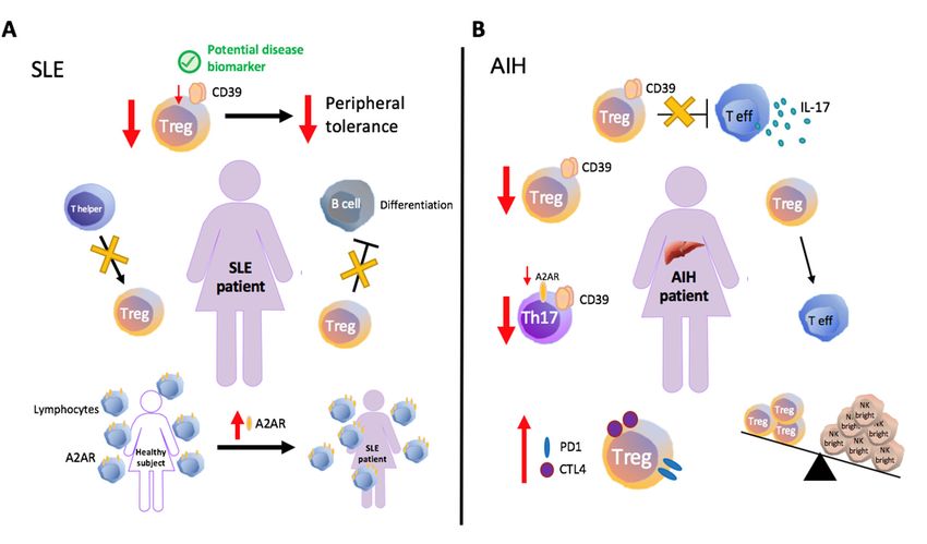
Int. J. Mol. Sci. 2019, 20, 528                                                                 2 of 15

      Ectonucleotidases are expressed on the plasma membrane of immune cells and belong to several
enzymatic families, which have been structurally and functionally characterized [5,6]. ENTPD1/CD39,
the prototype member of the NTPDase family, is a rate-limiting ectoenzyme that hydrolyzes ATP
into AMP, which is then further degraded into adenosine by the ecto-50 -nucleotidase/CD73 [5]. Once
generated, adenosine activates P1 receptors on target cells; it can undergo catalysis into inosine by
adenosine deaminase; or, alternatively, this nucleoside can be recaptured via cellular re-uptake and
used for purine salvage pathways [5]. ENTPD1/CD39 and CD73 are constitutively expressed in
human innate and adaptive immune cells such as monocytes, granulocytes, B-cells and T-cell subsets,
including regulatory and memory subpopulations. Pathological conditions, inflammatory stress and
exposure to specific compounds (e.g., aryl-hydrocarbon-receptor [AhR] ligands) markedly impact
ectonucleotidase expression and functionality [6–9].
      Both ENTPD1/CD39 and CD73 have been identified as major contributors to murine and human
regulatory T-cell (Treg) function [10–13]. Adenosine signaling stabilizes Foxp3 expression [14] while
the combined CD39/CD73 activity protects from the inhibitory and pro-apoptotic effects of P2X7R
signaling through eATP scavenging [15]. Notably, adenosine not only inhibits effector T-cells via
A2AR activation [16], but also increases ENTPD1/CD39 levels in an autocrine manner by acting on
this same A2AR on regulatory cells [17,18]. A2AR signaling on Foxp3+ Tregs induces CTLA-4 and
PD-1 expression, enhances cell proliferation and promotes Treg/dendritic cell (DC) interactions via
Epac1-Rap1-dependent pathways [19].
      Furthermore, recent investigations have revealed a competitive effect of A2AR expressed
by γδ T-cells on adenosine-mediated Treg functions.                 Once activated, γδT-cells markedly
upregulate adenosine receptors, depriving Foxp3+ T-cells of local adenosine, thus inhibiting their
expansion [20,21]. In a comparable fashion, A2BR mRNA is upregulated on activated regulatory cells
and endotoxin-induced Treg accumulation is impaired in A2BR deficient mice [22].
      ENTPD1/CD39 activity also supports the differentiation and function of IL-27-induced Tr-1
cells and ENTPD1/CD39 genetic deletion results in impaired differentiation, due to increased P2X7R
response that limits AhR signaling [23]. Interestingly, Tr-1 cells do not express CD73 and, in this
setting, adenosine generation relies on the combined activity of ENTPD1/CD39 on Tr-1 cells and
on CD73 present on adjacent DCs or effector cells [23]. IL-6 and TGF-β induce ENTPD1/CD39 and
CD73 expression in pro-inflammatory Th17-cells, conferring a non-pathogenic immunoregulatory
phenotype [24]. These so-called suppressor Th17 (SupTh17) cells further upregulate Foxp3 and
ENTPD1/CD39 expression in response to AhR agonists [7]. The adenosine generated stabilizes
SupTh17-cell differentiation likely by promoting the expression of stem cell-related transcription
factors (tcf-7 and lef-1) that limit differentiation into Th1-like phenotypes [25].
      ENTPD1/CD39 has been also detected in long-lived memory T-cells and exhausted effector
T-cells [26,27] where it impacts survival through the regulation of the mammalian-target-of-rapamycin
(mTOR) [28].
      In human peripheral blood mononuclear cells (PBMCs), exposure to TNF-α increases the
percentage of CD73+ CD4+ T-cells [29]. CD73 expression on conventional CD8+ and CD4+
T-lymphocytes can be also induced by the active form of vitamin D, retinoic acid and TFG-β [30,31].
On the other hand, terminally differentiated CD8+ T-cells potently decrease levels of CD73
expression [25]. A2AR activation in naïve/memory CD8+ T-cells regulates the transition to effectors
by inhibiting Wnt signaling [25,26]; whereas, in activated effector T-cells, A2AR stimulation induces
cAMP and decreases pro-inflammatory cytokine (e.g., TNF-α, IL-6) release [32]. Accordingly, global
A2AR deletion or blockade results in uncontrolled inflammatory responses with serious tissue damage
resulting from the dysregulated immune response [32].
      T-cell responses to adenosine can be inhibited by G protein-coupled receptor kinase
(GRKs)-mediated phosphorylation of P1 receptors or by PKA-mediated desensitization, consequent
to exposure to adenosine. Interference with P1 receptor response signaling can also derive from
upregulation of immune cell phosphodiesterases (PDE) or proteins that limit the A2A C-terminal-cAMP
Int. J. Mol. Sci. 2019, 20, 528 3 of 15
signaling [33–35]; and may also depend on the presence of A2AR splice variants that abrogate the
Ac-dependent cAMP responses [36].
2. Systemic Autoimmune Disorders
2.1. Systemic Lupus Erythematosus
Systemic lupus erythematosus (SLE) is a chronic systemic autoimmune disorder leading to
multi-organ inflammation. SLE may present with a wide spectrum of clinical manifestations and is
frequently characterized by recurring episodes of relapse and remission.
There is increasing evidence supporting the role of adenosine as a protective mediator, in SLE;
adenosine is operational via adenosine receptors (Figure 1A). In a murine model of lupus nephritis,
treatment with A2AR agonists results in significant improvement of renal histopathology, this being
associated with decreased blood urea, creatinine and proteinuria [37]. ENTPD1/CD39−/− and
CD73−/− mice show more pronounced endothelial cell dysfunction and exaggerated neutrophil
extracellular “trap” release in response to intraperitoneal administration of pristane, in a model of
Int. J. Mol.
lupus, whenSci. compared
2018, 19, x FOR
to PEER
wildREVIEW
type (WT) controls. 4 of 16
Figure 1. Immune and purinergic dysregulation in systemic lupus erythematosus (SLE) and
Figure 1. Immune and purinergic dysregulation in systemic lupus erythematosus (SLE) and
autoimmune hepatitis (AIH). (A) In SLE, defects in ENTPD1/CD39 expression and impaired Treg
autoimmune hepatitis (AIH). (A) In SLE, defects in ENTPD1/CD39 expression and impaired Treg
function have been associated with loss of peripheral tolerance. Studies conducted in active SLE
function have been associated with loss of peripheral tolerance. Studies conducted in active SLE
patients have indicated impaired suppression of B-cell differentiation and identified the abnormal
patients have indicated impaired suppression of B-cell differentiation and identified the abnormal
generation of regulatory T-cells (Treg) as result of limited transition from inducer/helper to suppressor
generation of regulatory T-cells (Treg) as result of limited transition from inducer/helper to
phenotype. Upregulation of A2AR is detectable in SLE patients, likely being linked to activation
suppressor phenotype. Upregulation of A2AR is detectable in SLE patients, likely being linked to
of compensatory pathways. (B) In type 1 autoimmune hepatitis (AIH-1), CD39+ Treg cells display
activation
impaired of compensatory
suppression of IL-17pathways.
production (B)byInCD4
type+ 1effectors.
autoimmuneAcutehepatitis (AIH-1),
AIH patients CD39
present
+ Treg cells
a low ratio
display Tregs
between impaired suppression
and NK of IL-17
bright cells, production
a specific NK subset by with + effectors.effector
CD4 activated Acute AIH patientsActivated
phenotype. present a
low ratio
memory between Tregs
phenotype and NK
and signs bright cells, including
of exhaustion, a specific NK subsetCTLA-4
increased with activated effector
and PD-1 phenotype.
levels are also
Activated memory phenotype and signs of exhaustion, including increased CTLA-4
typical of AIH Tregs. Reduced CD39+ Treg and CD39+ Th17-cell frequencies positively correlate and PD-1 with
levels
aredisease
the also typical of AIH
progression andTregs.
mightReduced
result fromCD39 + Treg andupon
cell instability CD39pro-inflammatory
+ Th17-cell frequencies positively
challenge, with
correlate with the disease progression and might result from cell instability upon
+
increased rate of conversion into effector lymphocytes. The reduction in CD39 Th17-cell numbers, pro-inflammatory
challenge,
also with
associates increased
with rate ofexpression
lower A2AR conversion(seeintotext
effector
below).lymphocytes. The reduction in CD39+ Th17-
cell numbers, also associates with lower A2AR expression (see text below).
2.2. Rheumatoid Arthritis
Rheumatoid arthritis (RA), as in the case of SLE, predominantly impacts females and is
characterized by joint inflammation and synovial tissue hyperplasia. These lesions eventually result
in cartilage and bone damage with increasing deformity and disability. Recent discoveries have
resulted in novel and improved therapies that, however, remain non curative [43].Int. J. Mol. Sci. 2019, 20, 528 4 of 15
Further studies have shown that CD73−/− mice have more activated B-cells in the spleen and
higher levels of plasma cell-free DNA, whereas ENTPD1/CD39 deficiency results in greater Th17-cell
expansion [38].
Accordingly, in SLE patients, loss of immunotolerance has been linked to defects in
ENTPD1/CD39 expression and impaired Treg function, suggesting that ENTPD1/CD39 deficient
Tregs could be associated with disease or might serve as disease biomarkers [39]. A study conducted
in active SLE patients has revealed abnormal generation of suppressor T-cells as result of the limited
transition from inducer/helper to suppressor phenotype. The dysfunction was observed in both
spontaneous and adenosine-inducible suppressor cells and, interestingly, this abnormality corrected
upon disease remission [40]. Another line of investigation has correlated immune dysregulation in
SLE with resistance of T-cells to adenosine-mediated effects [40].
To this end, SLE-derived T-cells lack adenosine receptor-coupled adenylate cyclase activity,
possibly contributing to impaired immunoregulation. T-lymphocytes from both SLE patients and
healthy subjects express A2R, but not A1R [41]. Although no differences have initially been observed
in A2R density and responses in SLE, a recent study has shown upregulation of A2AR in these
patients [41,42]. This finding might be linked with the activation of compensatory pathways, given
that A2AR activation is a strong immunoregulatory signal. Indeed, A2AR levels inversely correlate
with disease activity and the use of A2AR agonists might represent a potential therapeutic approach to
correct immunoregulation in SLE [42].
2.2. Rheumatoid Arthritis
Rheumatoid arthritis (RA), as in the case of SLE, predominantly impacts females and is
characterized by joint inflammation and synovial tissue hyperplasia. These lesions eventually result in
cartilage and bone damage with increasing deformity and disability. Recent discoveries have resulted
in novel and improved therapies that, however, remain non curative [43].
One first-line treatment choice for RA involves the use of methotrexate (MTX), the
anti-inflammatory action of which has been linked to increased levels of adenosine. This increased
engagement of adenosine via P1 receptors activates intracellular cascades, thereby promoting an
overall anti-inflammatory state.
Interestingly, studies conducted in murine models of arthritis have shown that CD39 blockade
and decreased adenosine generation reverse the therapeutic effect of MTX, while non-responder
patients express lower pre-treatment levels of CD39/ENTPD1 [44]. Low CD39 density on Treg and
MTX resistance have been both associated with alterations in TGFβRII and CREB1, which are TGF-β
signaling factors, in turn leading to CD39/ENTPD1 expression. In this regard, lower expression
of TGFβRII and CREB1 or decreased levels of p-SMAD2 and p-CREB might both result in MTX
resistance [45].
Notably, aspects of adenosine signaling protect from MTX toxicity. Five SNPs within the A2AR
gene have been linked to increased MTX gastrointestinal toxicity, serving as useful markers for high
risk patients prior to treatment [46].
Adenosine signaling is also involved in the protective effects of fructose 1,6 bisphosphate (FBP).
FBP administration attenuates experimental arthritis promoting immunoregulatory pathways mediated
by CD39/CD73 and A2AR signaling [47]. In collagen induced arthritis, IL-6 release by pro-inflammatory
cells negatively impacts the frequency of CD39+ Tregs in lymph nodes and spleen, this effect being
abrogated by antibody-mediated IL-6 neutralization [48]. Similarly, TNF-α accumulation causes
de-phosphorylation of Foxp3 leading to Treg functional impairment [49]. As a compensatory response,
RA patients present higher Treg frequencies in the joints, associated with increased CD39 function and
lower adenosine deaminase activity [50]. These synovial Foxp3+ CD39+ CD25+ T-cells, however, while
effectively suppressing IFNγ, TNF-α and IL-17F, fail to control IL-17A secretion by effector T-cells [51].
In addition to CD39, CD73 also plays a protective role in RA. In this context, patient-derived
Foxp3+ cells obtained from the synovium display low levels of CD73 [51]. Further, CD73 deletionInt. J. Mol. Sci. 2019, 20, 528 5 of 15
in non-hematopoietic cells results in higher Th1 cell responses and marked joint damage in a mouse
model of collagen induced arthritis [52].
Recent investigations have also reported a correlation between CD39/ENTPD1 and CD73
expression on CD4+ T-cell-derived microparticles (MPs) and disease activity. High levels of
CD4+ CD161+ CD39+ MPs positively correlate, while CD4+ CD39+ CD73+ MPs have a negative
correlation with RA activity. It is feasible that MPs with differential phenotypes might serve as
biomarkers for disease monitoring [53].
In PBMCs from RA patients, the disease-related high concentration of pro-inflammatory cytokines
induces expression of NF-κB and CREB as well as activation of the PI3K/Akt pathway, leading to
A2R and A3R upregulation [54–56]. Adenosine receptors upregulation inversely correlates with the
disease activity score and is associated with decreases in TNF-α, IL-1β and IL-6 levels [56]. Further,
agonist-induced A2AR and A3R activation in RA-derived lymphocytes results in inhibition of NF-κB
signaling and reduction in metalloproteinases [57].
2.3. Type 1 Diabetes
Type 1 diabetes is considered an autoimmune disorder targeting the insulin-producing pancreatic
β-cells. Genetic predisposition along with environmental factors have been proposed as disease
triggers. No cure is currently available, and the goal of current treatments is to control blood sugar
levels by insulin replacement therapies with the view of the prevention of vascular, neurological and
other complications.
Experimental evidence provided by murine models suggests that high levels of CD39,
in association with high A2AR and A2BR expression in T-helper cells confer protection
from streptozotocin-induced diabetes [58]. Further studies have revealed that suppression of
pro-inflammatory cytokine release is predominantly mediated upon A2BR engagement [59]. Positive
correlations between low CD39/ENTPD1 levels and disease activity has been observed in type 1
diabetic children, suggesting a potential compromise in Treg function [60].
Toll-like-receptor-9 (TLR9) deficiency has been associated with CD73-mediated beneficial effects.
TLR9−/− non-obese diabetic mice display higher levels of CD73 on CD4+ T-cells, lower levels of
pro-inflammatory cytokines and increased anti-inflammatory cytokine production; these all being
linked to protection against diabetes [61].
2.4. Autoimmune Hepatitis and Cholestatic Liver Disorders
There is growing evidence that defects in ectonucleotidase activity and impaired P1 receptor
levels contribute to loss of immunotolerance in autoimmune liver disorders (Figure 1B). In type-1
autoimmune hepatitis (AIH-1), CD39+ Treg cells are decreased in number and display impaired ability
to suppress IL-17 production by CD4+ effectors. Reduced CD39+ Treg frequencies might result from
cell instability upon pro-inflammatory challenge, with consequent increased rate of conversion into
effector lymphocytes [62]. In both AIH and autoimmune sclerosing cholangitis, there is a decrease in
CD39+ Th17-cell numbers, associated with impaired overall cell-associated ADPase activity and lower
A2AR expression [63]. Further, in acutely presenting, untreated AIH patients there is low ratio between
Tregs and a specific NK subset with activated effector phenotype [64]. In this same study, Tregs were
found to display an activated memory phenotype and exhibit signs of exhaustion, including increased
CTLA-4 and PD-1 receptor levels, as well as decreased ability to limit pro-inflammatory responses [64].
Murine studies conducted in the context of experimental cholestasis have proposed a pathogenic
role for A1R signaling in mediating liver injury, as lack of A1R limits the efflux of toxic biliary
constituents through the biliary excretory route [65]. On the other hand, A2BR activation in mouse
cholangiocytes, promotes IL-6 expression via cAMP and Ca2+ signaling, favoring cholangiocyte
survival during biliary cirrhosis [66]. Patients affected by primary biliary cholangitis exhibit
dramatic phenotypic alterations in CD8+ Tregs, as reflected by increased levels of CD127 and lower
CD39/ENTPD1 expression that correlate with lower responsiveness to IL-10 [67].Int. J. Mol. Sci. 2019, 20, 528 6 of 15
There is evidence that mice deficient in the ABCB4/multi-drug-resistant-protein2 (MDR2)
transporter protein—an experimental model for human primary sclerosing cholangitis (PSC)—show
expanded intrahepatic CD8+ lymphocytes that positively correlate with biliary injury and fibrosis [68].
Depletion of CD8+ cells in MDR2−/− /CD39−/− mice attenuates hepatobiliary injury and fibrosis;
while administration of αβ-ATP into Mdr2−/− /CD39 WT mice mirrors the phenotype of
MDR2−/− /CD39−/− mice [69].
2.5. Inflammatory Bowel Disease
A number of studies have demonstrated close links between ectonucleotidases or P1 receptor
signaling and the immunopathogenesis of inflammatory bowel disease (IBD); and, specifically, in
Crohn’s disease and ulcerative colitis. Th17-cells are pivotal players in IBD pathogenesis and
require eATP for complete differentiation [70]. High levels of CD39/ENTPD1 expression endow
Th17-cells with suppressor phenotypes and immunoregulatory function. Crohn’s patients present
lower frequencies of these SupTh17 lymphocytes, when compared to healthy subjects [71]. In mice,
CD39/ENTPD1 deletion exacerbates dextran-sulfate-sodium (DSS)-induced experimental colitis [72].
Furthermore, the presence of genetic polymorphisms of ENTPD1 and also levels of expression of CD39
on Tregs, have been associated with increased susceptibility to Crohn’s disease in humans and in
predicting the response to immunomodulatory therapy, respectively [72,73].
Importantly, in IBD patients during clinical and endoscopic remission, peripheral blood-derived
Tregs express higher CD39 levels [73]; further, therapeutic drug levels in responders are associated
with higher CD39 expression in FOXP3+ Tregs [73].
Regulatory effects of unconjugated bilirubin (UCB) are detectable in healthy human and
WT murine Th17-cells, significantly ameliorating DSS-colitis in vivo [7]. SupTh17-cells can be
induced upon exposure of conventional Th17-cells to certain metabolites e.g., UCB that boost
CD39/ENTPD1 expression via AhR engagement [7,8]. In Crohn’s disease, however, Th17-cells display
lower AhR expression and higher levels of hypoxia-inducible-factor-1alpha (HIF-1α), known to
inhibit AhR levels [23] and signaling [8]. Increases in HIF-1α results in heightened expression of
ATP-binding-cassette transporters that also induce extracellular efflux of immunometabolites like
UCB [8], therefore dampening immunosuppressive potential.
AhR activation, by the relatively non-toxic agonist 2-(10 H-indole-30 -carbonyl)-thiazole-4-carboxylic
acid methyl ester (ITE), increases CD39, IL-10 as well as granzyme B expression in Tregs [9]. High
CD39 levels in Foxp3+ Tregs have been associated with Crohn’s remission, also in response to
anti-TNF-α treatment [73]. Conversely, co-expression of CD39/ENTPD1 and CD161 in Th17-cells
correlates with a pro-inflammatory cellular phenotype that is upregulated in Crohn’s patients [74].
Exposure to CD3/CD28 stimulation induces CD39/ENTPD1 also in CD8+ T-cells, an effector subset
involved in IBD pathogenesis. CD39+ CD8+ T-cells thwart IFNγ production by CD39− CD8+ T-cells,
this effect being mediated by A2AR in a paracrine manner [75].
Other studies have indicated a protective role for CD73, the lack of which leads to
heightened susceptibility to DSS colitis, marked weight loss, gut permeability and accumulation
of pro-inflammatory cytokines [76] in mice. However, CD73 expression on effector CD4+ cells has
been also associated with a pro-inflammatory phenotype. Pro-inflammatory CD73+ CD4+ T-cells with
a Th17 signature are enriched in peripheral blood and lamina propria of IBD patients [77], suggesting
compensatory mechanisms that are activated during active inflammation.
Studies on P1 signaling have demonstrated that A2AR activation reduces intestinal inflammation,
TNF, IFNγ and IL-4 levels as well as colonic inflammatory cell infiltration in vivo [78]. Conversely,
global A2BR deletion protects from inflammatory damage induced by DSS, 2,4,6-trinitrobenzene
sulfonic acid (TNBS), Salmonella typhimurium and IL-8-induced colitis [79]. However, recent
investigations on mice with A2BR conditional deletion on vascular endothelial or intestinal epithelial
cells attributed the ability to reduce colonic inflammation only to A2BR expressed on epithelial
cells. Adenosine can also promote intestinal epithelial barrier restoration, especially during diseaseInt. J. Mol. Sci. 2019, 20, 528 7 of 15
remission. The effect is mediated by the nucleoside transporters 1 and 2 that remove adenosine from
the extracellular space upon A2BR activation [80].
2.6. Multiple Sclerosis
Ectonucleotidase activity and adenosine signaling exhibit protective properties also in multiple
sclerosis (MS), a neuroinflammatory autoimmune disorder driven by pathogenic T-cells specific for
myelin antigens in the central nervous system (CNS) [81,82].
In mice with experimental autoimmune encephalomyelitis (EAE), the murine model for MS,
administration of capsular polysaccharide A (PSA), the symbiosis factor for human intestinal
commensal Bacteroides fragilis, elicits immunotolerance by promoting expansion and accumulation of
CD39+ CD4+ cells in CNS lymphoid-draining sites [83]. PSA-mediated CD39+ CD4+ T-cell expansion
is driven by TLR2 signaling and the protective effect is completely abrogated in the absence of
ENTPD1/CD39 [84].
ENTPD1/CD39+ expression in human regulatory cells has been closely associated to MS different
stages. CD25+ Foxp3+ CD39+ Treg cells are impaired in peripheral blood of MS patients and cell
frequency is further reduced in the remitting/relapsing form of the disease [82,85]. Around 40% of
relapsing-remitting MS cases present with apparent Th17-cell expansion with a positive correlation
between Th17-cell numbers and ENTPD1/CD39+ Treg frequencies during remission but not during
relapse. These studies suggest that dysregulation of the Th17/CD39+ Treg functional balance may
contribute to exacerbation of the disease [86].
A1R regulates IL-6 and TNF-α expression. Interestingly, reduced A1R levels are detectable in the
microglia and peripheral blood of MS patients [87–89]; while A1R null mice develop a more severe
form of EAE, characterized by increased pro-inflammatory gene expression, microglial activation
and demyelination as compared to WT controls [87]. Accordingly, caffeine administration increases
A1R expression and improves animal clinical condition further supporting the receptor protective
effect [87].
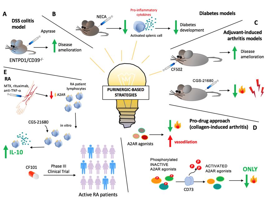
3. Therapeutic Implications
Modulation of adenosine signaling represents a promising therapeutic tool for several
autoimmune diseases (Figure 2). In previous studies, administration of apyrase, which has
ectoenzymatic activity comparable to CD39, strongly ameliorated DSS colitis in ENTPD1/CD39−/−
mice [72]. Protective effects of apyrase were also confirmed in subsequent studies conducted in the
context of already established DSS colitis [90].
Therapeutic strategies modulating the purinergic signaling involve direct targeting of adenosine
receptors, either by administration of receptors agonists, like adenosine, or by pharmacological
antagonization (Figure 2). Although most of these approaches are still under evaluation, some have
been already applied to the clinical setting, like the pharmacological preconditioning of explanted livers
with adenosine solution, which prevents ischemic damage consequent to organ reperfusion [91–93].
In two different experimental murine models, administration of the nonselective adenosine receptor
agonist 50 -N-ethylcarboxamidoadenosine (NECA), significantly prevented diabetes development
by suppressing expression of pro-inflammatory cytokines by activated splenic cells, including Th1
cells [59]. Experiments conducted in primary murine myeloid cells showed that A2AR activation
regulates bone turnover inhibiting osteoclast differentiation. The effect is mediated by PKA-dependent
inhibition of NF-κB nuclear translocation. This supports the use of A2AR agonists for targeting
inflammatory conditions affecting the bones, including RA [94]. Administration of the A2AR
agonist CGS 21680 results in anti-inflammatory as well as analgesic properties in a rat model of
adjuvant-induced arthritis. A2AR expression is upregulated in circulating lymphocytes of RA and
MS patients, probably as a compensatory response to counteract inflammation [95–97]. The increased
A2AR expression in RA lymphocytes is gradually reduced by anti-TNF-α agents like rituximab or
MTX; however, in vitro stimulation with the receptor agonist CGS 21680 significantly increases IL-10Int. J. Mol. Sci. 2019, 20, 528 8 of 15
production [97].
Int. J. Mol. Sci. 2018,Likewise, A2AR
19, x FOR PEER activation inhibits cell proliferation and pro-inflammatory cytokine
REVIEW 9 of 16
production in lymphocytes from MS patients [95].
Figure 2. Purinergic-based therapeutic strategies. (A) Administration of exogenous apyrase
Figure 2.ameliorates
strongly Purinergic-based therapeutic strategies.
dextran-sulfate-sodium (A) Administration
(DSS) of exogenous
colitis in ENTPD1/CD39 −/apyrase
− mice.strongly
(B) In
ameliorates dextran-sulfate-sodium (DSS) colitis in ENTPD1/CD39 −/− mice. (B) In experimental
experimental murine diabetes models, treatment with the nonselective adenosine receptor agonist
50murine diabetes models, treatment
-N-ethylcarboxamidoadenosine (NECA) with the nonselective
prevents adenosinebyreceptor
diabetes development agonist
suppressing 5′-N-
expression
ethylcarboxamidoadenosine (NECA) prevents diabetes development by suppressing expression of
of pro-inflammatory cytokines by activated splenic cells. (C) In rat models of adjuvant-induced
pro-inflammatory cytokines by activated splenic cells. (C) In rat models of adjuvant-induced arthritis,
arthritis, administration of the A2AR agonist CGS 21680 shows anti-inflammatory and analgesic
administration of the A2AR agonist CGS 21680 shows anti-inflammatory and analgesic properties.
properties. Similarly, orally administrated, low doses of the A3R agonist CF502 significantly
Similarly, orally administrated, low doses of the A3R agonist CF502 significantly ameliorate clinical
ameliorate clinical condition. (D) On the other hand, A2AR agonists have also been described
condition. (D) On the other hand, A2AR agonists have also been described as highly effective
as highly effective vasodilators, having, as a major side effect, hypotension. An improved and
vasodilators, having, as a major side effect, hypotension. An improved and promising therapeutic
promising therapeutic approach might be the use of phosphorylated A2AR agonists (prodrugs) that
approach might be the use of phosphorylated A2AR agonists (prodrugs) that need the ecto-5′-
need the ecto-50 -nucleotidase(CD73)-mediated de-phosphorylation in order to be activated. As an
nucleotidase(CD73)-mediated de-phosphorylation in order to be activated. As an example, in a
example, in a murine model of collagen-induced arthritis, the prodrug 2-(cyclohexylethylthio)adenosine
0murine model of collagen-induced arthritis, the prodrug 2-(cyclohexylethylthio)adenosine 5′-
5 -monophosphate (chet-AMP), showed potent immunosuppressive properties, with negligible
monophosphate (chet-AMP), showed potent immunosuppressive properties, with negligible
vasodilatory side effects. (E) In rheumatoid arthritis (RA) patients, anti-TNF-α agents, rituximab or
vasodilatory side effects. (E) In rheumatoid arthritis (RA) patients, anti-TNF-α agents, rituximab or
methotrexate (MTX) reduce the compensatory and protective increase in A2AR expression in peripheral
methotrexate (MTX) reduce the compensatory and protective increase in A2AR expression in
blood lymphocytes. However, studies reveal that in vitro stimulation with the receptor agonist CGS
peripheral blood lymphocytes. However, studies reveal that in vitro stimulation with the receptor
21680 significantly promotes immunoregulatory responses, increasing IL-10 production. Administration
agonist CGS 21680 significantly promotes immunoregulatory responses, increasing IL-10 production.
of CF101, an A3R specific agonist, is currently being tested in clinical trials.
Administration of CF101, an A3R specific agonist, is currently being tested in clinical trials.
A2AR plays a pivotal role also in modulating the inflammatory response in hypoxic conditions,
4. Concluding Remarks
especially in the acute setting. It is well established that lack of oxygen induces adenosine release in
severalThe
bodyJanus-like nature[98]
compartments of and
the there
purinergic
is now signaling involves
evidence that, release
in murine of eATP
models that boosts
of T-cell-mediated
inflammation;
acute and also
hepatitis, A2AR results in
mediates theeATP hydrolysis, which
hypoxia-induced leads to
protection the liver
from generation of adenosine
damage. that
A2AR deletion
suppresses the immune response. Dysregulation of the ATP/adenosine balance
and pharmacological antagonization significantly abrogate the hypoxia-mediated anti-inflammatory occurs in
autoimmune conditions and positively
effects in acute liver tissue injury [99]. correlates with disease severity and progression. Due to the
high levels of immunological heterogeneity of these conditions, current treatments directly targeting
the immune response are often poorly effective and are associated with significant side effects.Int. J. Mol. Sci. 2019, 20, 528 9 of 15
Additional studies have reported that synthesized phosphorylated A2AR agonists (prodrugs)
that need the ecto-50 -nucleotidase (CD73)-mediated de-phosphorylation in order to be activated,
were tested in a murine model of collagen-induced arthritis. The prodrug effect was evaluated also
upon inhibition of CD73 and A2AR. Among the tested compounds, 2-(cyclohexylethylthio)adenosine
50 -monophosphate (chet-AMP) showed potent immunosuppressive properties, with negligible
vasodilatory side effects, supporting the use of phosphorylated A2AR agonists for the specific treatment
of inflammation [100]. Encouraging results were also obtained with a non-absorbable, locally active
A2AR agonist, named as 4-(2-ethyl)-benzenesulfonic acid (7, PSB-0777) that was recently proposed
as novel treatment for inflammatory bowel syndrome. Ex vivo treatment of rat ileum/jejunum
preparations with PSB-0777 alone or in combination with A2BR antagonists, significantly ameliorated
the impaired acetylcholine-induced contractions induced by TNBS [101].
Further, in vitro treatment of fibroblast-like synoviocytes with CF502, a selective A3R agonist
with high affinity for the human subtype, markedly inhibited cell proliferation. Moreover, in a
rat experimental model of adjuvant-induced arthritis, oral administration of low doses of CF502
significantly ameliorated the clinical phenotype [102]. Previous studies have shown that in a phase II
clinical trial in patients with active RA, administration of CF101—a specific A3R agonist—resulted
in amelioration of disease that, however, did not reach statistical significance [103,104]. In the same
study, levels of A3R on patients’ PMBCs at baseline correlated directly with clinical response to CF101,
suggesting A3R as a predictive therapeutic biomarker [103,104]. In subsequent investigations, CF101
was reported to reduce pannus formation and lymphocyte infiltration in rats with osteoarthritis by
deregulating NF-κB [105]. CF101 is currently being tested in a phase III clinical trial in patients with
active RA (NCT02647762).
In experimental colitis, treatment with the A3R agonist N6 -(3-iodobenzyl)-adenosine-5-
N-methyluronamide (IB-MECA) significantly prevented colitis-induced gene dysregulation, weight
loss and gut injury [106].
4. Concluding Remarks
The Janus-like nature of the purinergic signaling involves release of eATP that boosts
inflammation; and also results in eATP hydrolysis, which leads to the generation of adenosine that
suppresses the immune response. Dysregulation of the ATP/adenosine balance occurs in autoimmune
conditions and positively correlates with disease severity and progression. Due to the high levels of
immunological heterogeneity of these conditions, current treatments directly targeting the immune
response are often poorly effective and are associated with significant side effects.
Modulation of the purinergic response could therefore be a novel approach to improve current
therapeutics. Promising molecular candidates have been already identified in ectonucleotidases
(especially ENTPD1/CD39 and CD73) and P1 receptor agonists. Pre-clinical data support the
pharmacological induction of ENTPD1/CD39 expression as well as the stimulation of A2AR and A3R
as potential ways of treatment. We propose that purinergic-based strategies, alone or in combination
with current treatments, might represent strong adjunctive therapeutics to help dampen inflammation
and interfere with disease progression without untoward toxicity.
Author Contributions: M.V. and R.P.H. wrote the manuscript; S.C.R. and M.S.L. reviewed and edited
the manuscript.
Funding: This work has been supported by the National Institute of Health (R01 DK108894 to M.S.L.; P01
HL107152 and R21 CA164970 to S.C.R.); AASLD Pilot Research Award (to M.S.L.); Pfizer research support to
S.C.R.; the Helmsley Charitable Trust (grant 281574.5069091.0010 to S.C.R.); and by the Department of Defense
Award W81XWH-16-0464 (to S.C.R.).
Conflicts of Interest: The authors declare no conflict of interest.Int. J. Mol. Sci. 2019, 20, 528 10 of 15
References
1. Burnstock, G. Purinergic signalling: From discovery to current developments. Exp. Physiol. 2014, 99, 16–34.
[CrossRef] [PubMed]
2. Di Virgilio, F.; Vuerich, M. Purinergic signaling in the immune system. Auton. Neurosci. 2015, 191, 117–123.
[CrossRef] [PubMed]
3. Eltzschig, H.K.; Sitkovsky, M.V.; Robson, S.C. Purinergic signaling during inflammation. N. Engl. J. Med.
2012, 367, 2322–2333. [CrossRef] [PubMed]
4. Longhi, M.S.; Moss, A.; Jiang, Z.G.; Robson, S.C. Purinergic signaling during intestinal inflammation.
J. Mol. Med. 2017, 95, 915–925. [CrossRef] [PubMed]
5. Yegutkin, G.G. Enzymes involved in metabolism of extracellular nucleotides and nucleosides: Functional
implications and measurement of activities. Crit. Rev. Biochem. Mol. Biol. 2014, 49, 473–497. [CrossRef]
[PubMed]
6. Allard, B.; Longhi, M.S.; Robson, S.C.; Stagg, J. The ectonucleotidases CD39 and CD73: Novel checkpoint
inhibitor targets. Immunol. Rev. 2017, 276, 121–144. [CrossRef]
7. Longhi, M.S.; Vuerich, M.; Kalbasi, A.; Kenison, J.E.; Yeste, A.; Csizmadia, E.; Vaughn, B.; Feldbrugge, L.;
Mitsuhashi, S.; Wegiel, B.; et al. Bilirubin suppresses Th17 immunity in colitis by upregulating CD39.
JCI Insight 2017, 2. [CrossRef]
8. Xie, A.; Robles, R.J.; Mukherjee, S.; Zhang, H.; Feldbrugge, L.; Csizmadia, E.; Wu, Y.; Enjyoji, K.; Moss, A.C.;
Otterbein, L.E.; et al. HIF-1alpha-induced xenobiotic transporters promote Th17 responses in Crohn’s disease.
J. Autoimmun. 2018, 94, 122–133. [CrossRef]
9. Goettel, J.A.; Gandhi, R.; Kenison, J.E.; Yeste, A.; Murugaiyan, G.; Sambanthamoorthy, S.; Griffith, A.E.;
Patel, B.; Shouval, D.S.; Weiner, H.L.; et al. AHR Activation Is Protective against Colitis Driven by T Cells in
Humanized Mice. Cell Rep. 2016, 17, 1318–1329. [CrossRef]
10. Deaglio, S.; Dwyer, K.M.; Gao, W.; Friedman, D.; Usheva, A.; Erat, A.; Chen, J.F.; Enjyoji, K.; Linden, J.;
Oukka, M.; et al. Adenosine generation catalyzed by CD39 and CD73 expressed on regulatory T cells
mediates immune suppression. J. Exp. Med. 2007, 204, 1257–1265. [CrossRef]
11. Dwyer, K.M.; Hanidziar, D.; Putheti, P.; Hill, P.A.; Pommey, S.; McRae, J.L.; Winterhalter, A.; Doherty, G.;
Deaglio, S.; Koulmanda, M.; et al. Expression of CD39 by human peripheral blood CD4+ CD25+ T cells
denotes a regulatory memory phenotype. Am. J. Transplant. 2010, 10, 2410–2420. [CrossRef] [PubMed]
12. Alam, M.S.; Kurtz, C.C.; Rowlett, R.M.; Reuter, B.K.; Wiznerowicz, E.; Das, S.; Linden, J.; Crowe, S.E.; Ernst, P.B.
CD73 is expressed by human regulatory T helper cells and suppresses proinflammatory cytokine production
and Helicobacter felis-induced gastritis in mice. J. Infect. Dis. 2009, 199, 494–504. [CrossRef] [PubMed]
13. Kobie, J.J.; Shah, P.R.; Yang, L.; Rebhahn, J.A.; Fowell, D.J.; Mosmann, T.R. T regulatory and primed
uncommitted CD4 T cells express CD73, which suppresses effector CD4 T cells by converting 5’-adenosine
monophosphate to adenosine. J. Immunol. 2006, 177, 6780–6786. [CrossRef]
14. Bao, R.; Hou, J.; Li, Y.; Bian, J.; Deng, X.; Zhu, X.; Yang, T. Adenosine promotes Foxp3 expression in Treg cells
in sepsis model by activating JNK/AP-1 pathway. Am. J. Transl. Res. 2016, 8, 2284–2292. [PubMed]
15. Schenk, U.; Frascoli, M.; Proietti, M.; Geffers, R.; Traggiai, E.; Buer, J.; Ricordi, C.; Westendorf, A.M.; Grassi, F.
ATP inhibits the generation and function of regulatory T cells through the activation of purinergic P2X
receptors. Sci. Signal. 2011, 4, ra12. [CrossRef] [PubMed]
16. Huang, S.; Apasov, S.; Koshiba, M.; Sitkovsky, M. Role of A2a extracellular adenosine receptor-mediated
signaling in adenosine-mediated inhibition of T-cell activation and expansion. Blood 1997, 90, 1600–1610.
[PubMed]
17. Liao, H.; Hyman, M.C.; Baek, A.E.; Fukase, K.; Pinsky, D.J. cAMP/CREB-mediated transcriptional regulation
of ectonucleoside triphosphate diphosphohydrolase 1 (CD39) expression. J. Biol. Chem. 2010, 285,
14791–14805. [CrossRef] [PubMed]
18. Kinsey, G.R.; Huang, L.; Jaworska, K.; Khutsishvili, K.; Becker, D.A.; Ye, H.; Lobo, P.I.; Okusa, M.D. Autocrine
adenosine signaling promotes regulatory T cell-mediated renal protection. J. Am. Soc. Nephrol. 2012, 23,
1528–1537. [CrossRef]
19. Ohta, A.; Kini, R.; Ohta, A.; Subramanian, M.; Madasu, M.; Sitkovsky, M. The development and
immunosuppressive functions of CD4+ CD25+ FoxP3+ regulatory T cells are under influence of the
adenosine-A2A adenosine receptor pathway. Front. Immunol. 2012, 3, 190. [CrossRef]Int. J. Mol. Sci. 2019, 20, 528 11 of 15
20. Liang, D.; Woo, J.I.; Shao, H.; Born, W.K.; O’Brien, R.L.; Kaplan, H.J.; Sun, D. Ability of gammadelta T cells to
modulate the Foxp3 T cell response is dependent on adenosine. PLoS ONE 2018, 13, e0197189. [CrossRef]
21. Ring, S.; Pushkarevskaya, A.; Schild, H.; Probst, H.C.; Jendrossek, V.; Wirsdorfer, F.; Ledent, C.; Robson, S.C.;
Enk, A.H.; Mahnke, K. Regulatory T cell-derived adenosine induces dendritic cell migration through the
Epac-Rap1 pathway. J. Immunol. 2015, 194, 3735–3744. [CrossRef] [PubMed]
22. Ehrentraut, H.; Westrich, J.A.; Eltzschig, H.K.; Clambey, E.T. Adora2b adenosine receptor engagement
enhances regulatory T cell abundance during endotoxin-induced pulmonary inflammation. PLoS ONE 2012,
7, e32416. [CrossRef] [PubMed]
23. Mascanfroni, I.D.; Takenaka, M.C.; Yeste, A.; Patel, B.; Wu, Y.; Kenison, J.E.; Siddiqui, S.; Basso, A.S.;
Otterbein, L.E.; Pardoll, D.M.; et al. Metabolic control of type 1 regulatory T cell differentiation by AHR and
HIF1-alpha. Nat. Med. 2015, 21, 638–646. [CrossRef] [PubMed]
24. McGeachy, M.J.; Bak-Jensen, K.S.; Chen, Y.; Tato, C.M.; Blumenschein, W.; McClanahan, T.; Cua, D.J. TGF-beta
and IL-6 drive the production of IL-17 and IL-10 by T cells and restrain T(H)-17 cell-mediated pathology.
Nat. Immunol. 2007, 8, 1390–1397. [CrossRef] [PubMed]
25. Flores-Santibanez, F.; Fernandez, D.; Meza, D.; Tejon, G.; Vargas, L.; Varela-Nallar, L.; Arredondo, S.; Guixe, V.;
Rosemblatt, M.; Bono, M.R.; et al. CD73-mediated adenosine production promotes stem cell-like properties
in mouse Tc17 cells. Immunology 2015, 146, 582–594. [CrossRef] [PubMed]
26. Bono, M.R.; Fernandez, D.; Flores-Santibanez, F.; Rosemblatt, M.; Sauma, D. CD73 and CD39
ectonucleotidases in T cell differentiation: Beyond immunosuppression. FEBS Lett. 2015, 589, 3454–3460.
[CrossRef] [PubMed]
27. Gupta, P.K.; Godec, J.; Wolski, D.; Adland, E.; Yates, K.; Pauken, K.E.; Cosgrove, C.; Ledderose, C.;
Junger, W.G.; Robson, S.C.; et al. CD39 Expression Identifies Terminally Exhausted CD8+ T Cells. PLoS Pathog.
2015, 11, e1005177. [CrossRef]
28. Hindupur, S.K.; Gonzalez, A.; Hall, M.N. The opposing actions of target of rapamycin and AMP-activated
protein kinase in cell growth control. Cold Spring Harb. Perspect. Biol. 2015, 7, a019141. [CrossRef]
29. Farez, M.F.; Mascanfroni, I.D.; Mendez-Huergo, S.P.; Yeste, A.; Murugaiyan, G.; Garo, L.P.; Balbuena
Aguirre, M.E.; Patel, B.; Ysrraelit, M.C.; Zhu, C.; et al. Melatonin Contributes to the Seasonality of Multiple
Sclerosis Relapses. Cell 2015, 162, 1338–1352. [CrossRef]
30. Mann, E.H.; Chambers, E.S.; Chen, Y.H.; Richards, D.F.; Hawrylowicz, C.M. 1alpha,25-dihydroxyvitamin D3
acts via transforming growth factor-beta to up-regulate expression of immunosuppressive CD73 on human
CD4+ Foxp3− T cells. Immunology 2015, 146, 423–431. [CrossRef]
31. Regateiro, F.S.; Howie, D.; Nolan, K.F.; Agorogiannis, E.I.; Greaves, D.R.; Cobbold, S.P.; Waldmann, H.
Generation of anti-inflammatory adenosine by leukocytes is regulated by TGF-beta. Eur. J. Immunol. 2011,
41, 2955–2965. [CrossRef] [PubMed]
32. Antonioli, L.; Blandizzi, C.; Pacher, P.; Hasko, G. Immunity, inflammation and cancer: A leading role for
adenosine. Nat. Rev. Cancer 2013, 13, 842–857. [CrossRef]
33. Gsandtner, I.; Charalambous, C.; Stefan, E.; Ogris, E.; Freissmuth, M.; Zezula, J. Heterotrimeric G
protein-independent signaling of a G protein-coupled receptor. Direct binding of ARNO/cytohesin-2 to the
carboxyl terminus of the A2A adenosine receptor is necessary for sustained activation of the ERK/MAP
kinase pathway. J. Biol. Chem. 2005, 280, 31898–31905. [CrossRef] [PubMed]
34. Peter, D.; Jin, S.L.; Conti, M.; Hatzelmann, A.; Zitt, C. Differential expression and function of
phosphodiesterase 4 (PDE4) subtypes in human primary CD4+ T cells: Predominant role of PDE4D.
J. Immunol. 2007, 178, 4820–4831. [CrossRef]
35. Keuerleber, S.; Gsandtner, I.; Freissmuth, M. From cradle to twilight: The carboxyl terminus directs the fate
of the A(2A)-adenosine receptor. Biochim. Biophys. Acta 2011, 1808, 1350–1357. [CrossRef] [PubMed]
36. Kreth, S.; Ledderose, C.; Kaufmann, I.; Groeger, G.; Thiel, M. Differential expression of 50 -UTR splice variants
of the adenosine A2A receptor gene in human granulocytes: Identification, characterization, and functional
impact on activation. FASEB J. 2008, 22, 3276–3286. [CrossRef]
37. Zhang, L.; Yang, N.; Wang, S.; Huang, B.; Li, F.; Tan, H.; Liang, Y.; Chen, M.; Li, Y.; Yu, X. Adenosine 2A
receptor is protective against renal injury in MRL/lpr mice. Lupus 2011, 20, 667–677. [CrossRef] [PubMed]
38. Knight, J.S.; Mazza, L.F.; Yalavarthi, S.; Sule, G.; Ali, R.A.; Hodgin, J.B.; Kanthi, Y.; Pinsky, D.J.
Ectonucleotidase-Mediated Suppression of Lupus Autoimmunity and Vascular Dysfunction. Front. Immunol.
2018, 9, 1322. [CrossRef] [PubMed]Int. J. Mol. Sci. 2019, 20, 528 12 of 15
39. Loza, M.J.; Anderson, A.S.; O’Rourke, K.S.; Wood, J.; Khan, I.U. T-cell specific defect in expression of the
NTPDase CD39 as a biomarker for lupus. Cell Immunol. 2011, 271, 110–117. [CrossRef]
40. Kammer, G.M.; Birch, R.E.; Polmar, S.H. Impaired immunoregulation in systemic lupus erythematosus:
Defective adenosine-induced suppressor T lymphocyte generation. J. Immunol. 1983, 130, 1706–1712.
41. Schultz, L.A.; Kammer, G.M.; Rudolph, S.A. Characterization of the human T lymphocyte adenosine receptor:
Comparison of normal and systemic lupus erythematosus cells. FASEB J. 1988, 2, 244–250. [CrossRef]
[PubMed]
42. Bortoluzzi, A.; Vincenzi, F.; Govoni, M.; Padovan, M.; Ravani, A.; Borea, P.A.; Varani, K. A2A adenosine
receptor upregulation correlates with disease activity in patients with systemic lupus erythematosus.
Arthritis Res. Ther. 2016, 18, 192. [CrossRef] [PubMed]
43. McInnes, I.B.; Schett, G. The pathogenesis of rheumatoid arthritis. N. Engl. J. Med. 2011, 365, 2205–2219.
[CrossRef] [PubMed]
44. Peres, R.S.; Donate, P.B.; Talbot, J.; Cecilio, N.T.; Lobo, P.R.; Machado, C.C.; Lima, K.W.A.; Oliveira, R.D.;
Carregaro, V.; Nakaya, H.I.; et al. TGF-beta signalling defect is linked to low CD39 expression on regulatory T
cells and methotrexate resistance in rheumatoid arthritis. J. Autoimmun. 2018, 90, 49–58. [CrossRef] [PubMed]
45. Peres, R.S.; Liew, F.Y.; Talbot, J.; Carregaro, V.; Oliveira, R.D.; Almeida, S.L.; Franca, R.F.; Donate, P.B.;
Pinto, L.G.; Ferreira, F.I.; et al. Low expression of CD39 on regulatory T cells as a biomarker for resistance to
methotrexate therapy in rheumatoid arthritis. Proc. Natl. Acad. Sci. USA 2015, 112, 2509–2514. [CrossRef]
[PubMed]
46. Hider, S.L.; Thomson, W.; Mack, L.F.; Armstrong, D.J.; Shadforth, M.; Bruce, I.N. Polymorphisms within the
adenosine receptor 2a gene are associated with adverse events in RA patients treated with MTX. Rheumatology
2008, 47, 1156–1159. [CrossRef] [PubMed]
47. Veras, F.P.; Peres, R.S.; Saraiva, A.L.; Pinto, L.G.; Louzada-Junior, P.; Cunha, T.M.; Paschoal, J.A.; Cunha, F.Q.;
Alves-Filho, J.C. Fructose 1,6-bisphosphate, a high-energy intermediate of glycolysis, attenuates experimental
arthritis by activating anti-inflammatory adenosinergic pathway. Sci. Rep. 2015, 5, 15171. [CrossRef]
48. Thiolat, A.; Semerano, L.; Pers, Y.M.; Biton, J.; Lemeiter, D.; Portales, P.; Quentin, J.; Jorgensen, C.; Decker, P.;
Boissier, M.C.; et al. Interleukin-6 receptor blockade enhances CD39+ regulatory T cell development in
rheumatoid arthritis and in experimental arthritis. Arthritis Rheumatol. 2014, 66, 273–283. [CrossRef]
49. Nie, H.; Zheng, Y.; Li, R.; Guo, T.B.; He, D.; Fang, L.; Liu, X.; Xiao, L.; Chen, X.; Wan, B.; et al. Phosphorylation
of FOXP3 controls regulatory T cell function and is inhibited by TNF-alpha in rheumatoid arthritis. Nat. Med.
2013, 19, 322–328. [CrossRef]
50. Dos Santos Jaques, J.A.; Becker, L.V.; Souza Vdo, C.; Leal, C.A.; Bertoldo, T.M.; de Vargas Pinheiro, K.;
Morsch, V.M.; Schetinger, M.R.; Leal, D.B. Activities of enzymes that hydrolyze adenine nucleotides in
lymphocytes from patients with rheumatoid arthritis. Cell Biochem. Funct. 2013, 31, 395–399. [CrossRef]
51. Herrath, J.; Chemin, K.; Albrecht, I.; Catrina, A.I.; Malmstrom, V. Surface expression of CD39 identifies an
enriched Treg-cell subset in the rheumatic joint, which does not suppress IL-17A secretion. Eur. J. Immunol.
2014, 44, 2979–2989. [CrossRef] [PubMed]
52. Chrobak, P.; Charlebois, R.; Rejtar, P.; El Bikai, R.; Allard, B.; Stagg, J. CD73 plays a protective role in
collagen-induced arthritis. J. Immunol. 2015, 194, 2487–2492. [CrossRef] [PubMed]
53. Fan, W.; Wang, W.; Wu, J.; Ma, L.; Guo, J. Identification of CD4+ T-cell-derived CD161+ CD39+ and
CD39+ CD73+ microparticles as new biomarkers for rheumatoid arthritis. Biomark. Med. 2017, 11, 107–116.
[CrossRef] [PubMed]
54. Madi, L.; Cohen, S.; Ochayin, A.; Bar-Yehuda, S.; Barer, F.; Fishman, P. Overexpression of A3 adenosine
receptor in peripheral blood mononuclear cells in rheumatoid arthritis: Involvement of nuclear factor-kappaB
in mediating receptor level. J. Rheumatol. 2007, 34, 20–26. [PubMed]
55. Ochaion, A.; Bar-Yehuda, S.; Cohen, S.; Barer, F.; Patoka, R.; Amital, H.; Reitblat, T.; Reitblat, A.; Ophir, J.;
Konfino, I.; et al. The anti-inflammatory target A(3) adenosine receptor is over-expressed in rheumatoid
arthritis, psoriasis and Crohn’s disease. Cell Immunol. 2009, 258, 115–122. [CrossRef] [PubMed]
56. Varani, K.; Padovan, M.; Vincenzi, F.; Targa, M.; Trotta, F.; Govoni, M.; Borea, P.A. A2A and A3 adenosine
receptor expression in rheumatoid arthritis: Upregulation, inverse correlation with disease activity score
and suppression of inflammatory cytokine and metalloproteinase release. Arthritis Res. Ther. 2011, 13, R197.
[CrossRef] [PubMed]Int. J. Mol. Sci. 2019, 20, 528 13 of 15
57. Ravani, A.; Vincenzi, F.; Bortoluzzi, A.; Padovan, M.; Pasquini, S.; Gessi, S.; Merighi, S.; Borea, P.A.;
Govoni, M.; Varani, K. Role and Function of A2A and A(3) Adenosine Receptors in Patients with Ankylosing
Spondylitis, Psoriatic Arthritis and Rheumatoid Arthritis. Int. J. Mol. Sci. 2017, 18, 697. [CrossRef]
58. Chia, J.S.; McRae, J.L.; Thomas, H.E.; Fynch, S.; Elkerbout, L.; Hill, P.; Murray-Segal, L.; Robson, S.C.;
Chen, J.F.; d’Apice, A.J.; et al. The protective effects of CD39 overexpression in multiple low-dose
streptozotocin-induced diabetes in mice. Diabetes 2013, 62, 2026–2035. [CrossRef]
59. Nemeth, Z.H.; Bleich, D.; Csoka, B.; Pacher, P.; Mabley, J.G.; Himer, L.; Vizi, E.S.; Deitch, E.A.; Szabo, C.;
Cronstein, B.N.; et al. Adenosine receptor activation ameliorates type 1 diabetes. FASEB J. 2007, 21, 2379–2388.
[CrossRef]
60. Akesson, K.; Tompa, A.; Ryden, A.; Faresjo, M. Low expression of CD39+ /CD45RA+ on regulatory T cells
(Treg ) cells in type 1 diabetic children in contrast to high expression of CD101+ /CD129+ on Treg cells in
children with coeliac disease. Clin. Exp. Immunol. 2015, 180, 70–82. [CrossRef]
61. Tai, N.; Wong, F.S.; Wen, L. TLR9 deficiency promotes CD73 expression in T cells and diabetes protection in
nonobese diabetic mice. J. Immunol. 2013, 191, 2926–2937. [CrossRef] [PubMed]
62. Grant, C.R.; Liberal, R.; Holder, B.S.; Cardone, J.; Ma, Y.; Robson, S.C.; Mieli-Vergani, G.; Vergani, D.;
Longhi, M.S. Dysfunctional CD39(POS) regulatory T cells and aberrant control of T-helper type 17 cells in
autoimmune hepatitis. Hepatology 2014, 59, 1007–1015. [CrossRef] [PubMed]
63. Liberal, R.; Grant, C.R.; Ma, Y.; Csizmadia, E.; Jiang, Z.G.; Heneghan, M.A.; Yee, E.U.; Mieli-Vergani, G.;
Vergani, D.; Robson, S.C.; et al. CD39 mediated regulation of Th17-cell effector function is impaired in
juvenile autoimmune liver disease. J. Autoimmun. 2016, 72, 102–112. [CrossRef] [PubMed]
64. Jeffery, H.C.; Braitch, M.K.; Bagnall, C.; Hodson, J.; Jeffery, L.E.; Wawman, R.E.; Wong, L.L.; Birtwistle, J.;
Bartlett, H.; Lohse, A.W.; et al. Changes in natural killer cells and exhausted memory regulatory T Cells
with corticosteroid therapy in acute autoimmune hepatitis. Hepatol. Commun. 2018, 2, 421–436. [CrossRef]
[PubMed]
65. Yang, P.; Chen, P.; Wang, T.; Zhan, Y.; Zhou, M.; Xia, L.; Cheng, R.; Guo, Y.; Zhu, L.; Zhang, J. Loss of
A(1) adenosine receptor attenuates alpha-naphthylisothiocyanate-induced cholestatic liver injury in mice.
Toxicol. Sci. 2013, 131, 128–138. [CrossRef] [PubMed]
66. Lavoie, E.G.; Fausther, M.; Goree, J.R.; Dranoff, J.A. The Cholangiocyte Adenosine-IL-6 Axis Regulates
Survival During Biliary Cirrhosis. Gene Expr. 2017, 17, 327–340. [CrossRef]
67. Bernuzzi, F.; Fenoglio, D.; Battaglia, F.; Fravega, M.; Gershwin, M.E.; Indiveri, F.; Ansari, A.A.; Podda, M.;
Invernizzi, P.; Filaci, G. Phenotypical and functional alterations of CD8 regulatory T cells in primary biliary
cirrhosis. J. Autoimmun. 2010, 35, 176–180. [CrossRef]
68. Taylor, A.E.; Carey, A.N.; Kudira, R.; Lages, C.S.; Shi, T.; Lam, S.; Karns, R.; Simmons, J.; Shanmukhappa, K.;
Almanan, M.; et al. Interleukin 2 Promotes Hepatic Regulatory T Cell Responses and Protects From Biliary
Fibrosis in Murine Sclerosing Cholangitis. Hepatology 2018, 68, 1905–1921. [CrossRef] [PubMed]
69. Peng, Z.W.; Rothweiler, S.; Wei, G.; Ikenaga, N.; Liu, S.B.; Sverdlov, D.Y.; Vaid, K.A.; Longhi, M.S.; Kuang, M.;
Robson, S.C.; et al. The ectonucleotidase ENTPD1/CD39 limits biliary injury and fibrosis in mouse models
of sclerosing cholangitis. Hepatol. Commun. 2017, 1, 957–972. [CrossRef] [PubMed]
70. Fujino, S.; Andoh, A.; Bamba, S.; Ogawa, A.; Hata, K.; Araki, Y.; Bamba, T.; Fujiyama, Y. Increased expression
of interleukin 17 in inflammatory bowel disease. Gut 2003, 52, 65–70. [CrossRef] [PubMed]
71. Longhi, M.S.; Moss, A.; Bai, A.; Wu, Y.; Huang, H.; Cheifetz, A.; Quintana, F.J.; Robson, S.C. Characterization
of human CD39+ Th17 cells with suppressor activity and modulation in inflammatory bowel disease.
PLoS ONE 2014, 9, e87956. [CrossRef] [PubMed]
72. Friedman, D.J.; Kunzli, B.M.; YI, A.R.; Sevigny, J.; Berberat, P.O.; Enjyoji, K.; Csizmadia, E.; Friess, H.;
Robson, S.C. From the Cover: CD39 deletion exacerbates experimental murine colitis and human
polymorphisms increase susceptibility to inflammatory bowel disease. Proc. Natl. Acad. Sci. USA 2009, 106,
16788–16793. [CrossRef] [PubMed]
73. Gibson, D.J.; Elliott, L.; McDermott, E.; Tosetto, M.; Keegan, D.; Byrne, K.; Martin, S.T.; Rispens, T.; Cullen, G.;
Mulcahy, H.E.; et al. Heightened Expression of CD39 by Regulatory T Lymphocytes Is Associated with
Therapeutic Remission in Inflammatory Bowel Disease. Inflamm. Bowel Dis. 2015, 21, 2806–2814. [CrossRef]
[PubMed]You can also read