Advances and Challenges of Biodegradable Implant Materials with a Focus on Magnesium-Alloys and Bacterial Infections - MDPI
←
→
Page content transcription
If your browser does not render page correctly, please read the page content below
metals
Review
Advances and Challenges of Biodegradable Implant
Materials with a Focus on Magnesium-Alloys and
Bacterial Infections
Muhammad Imran Rahim 1 , Sami Ullah 2 and Peter P. Mueller 3, *           ID

 1   Department of Prosthetic Dentistry and Biomedical Materials Science, Lower Saxony Centre for Biomedical
     Engineering, Implant Research and Development, Hannover Medical School, Carl-Neuberg-Straße 1,
     30625 Hannover, Germany; M.Imran.Rahim@outlook.com
 2   Department of MSYS, Helmholtz Center for Infection Research, Inhoffenstrasse 7,
     38124 Braunschweig, Germany; Sami.Ullah@helmholtz-hzi.de
 3   Department of Chemical Biology, Helmholtz Center for Infection Research, Inhoffenstrasse 7,
     38124 Braunschweig, Germany
 *   Correspondence: pmu@gbf.de; Tel.: +49-531-6181-5070

 Received: 12 June 2018; Accepted: 4 July 2018; Published: 10 July 2018

 Abstract: Medical implants made of biodegradable materials could be advantageous for temporary
 applications, such as mechanical support during bone-healing or as vascular stents to keep blood
 vessels open. After completion of the healing process, the implant would disappear, avoiding
 long-term side effects or the need for surgical removal. Various corrodible metal alloys based
 on magnesium, iron or zinc have been proposed as sturdier and potentially less inflammatory
 alternatives to degradable organic polymers, in particular for load-bearing applications. Despite the
 recent introduction of magnesium-based screws, the remaining hurdles to routine clinical applications
 are still challenging. These include limitations such as mechanical material characteristics or
 unsuitable corrosion characteristics. In this article, the salient features and clinical prospects of
 currently-investigated biodegradable implant materials are summarized, with a main focus on
 magnesium alloys. A mechanism of action for the stimulation of bone growth due to the exertion
 of mechanical force by magnesium corrosion products is discussed. To explain divergent in vitro
 and in vivo effects of magnesium, a novel model for bacterial biofilm infections is proposed which
 predicts crucial consequences for antibacterial implant strategies.

 Keywords: bioresorbable implants; corrosion layer; vascular stents; orthopedic implants;
 microbial infections

1. Introduction
      For metallic implants, industrially-developed, inert and long-lasting materials, such as titanium
(Ti) alloys, stainless steel (SS) and cobalt–chromium (CoCr) alloys, are most frequently used [1–4].
The duration of the healing process is highly variable depending on the extent of injury, disease
state, age and treatment. In general, healing time may range from a brief one month period, up to
a six month period in more complex cases. Permanent implants are frequently removed after the
completion of the healing process to avoid diverse side effects. Long-term disadvantages of this practice
include the failure to adapt to rapid growth in young children, bone degradation by stress shielding,
microbial implant infections, excessive fibrosis or persistent inflammation. Novel bioresorbable
metal implants could provide support during the healing process, and then disappear to avoid
long-term side effects without requiring surgical removal [5,6]. Conventionally, initial material tests
are done most economically in vitro under precisely defined technical conditions. Subsequent assays

Metals 2018, 8, 532; doi:10.3390/met8070532                                    www.mdpi.com/journal/metals
Metals 2018, 8, 532 2 of 14
are performed under increasingly complex and more costly cell culture conditions, followed by
small animal experiments and eventually tested in large animals or in clinical trials. However,
corrosion results obtained under simple technical conditions cannot be extrapolated to clinical
circumstances. In one study, the corrosion rate for magnesium alloys was reported to differ four orders
of magnitude between in vitro and in vivo conditions [7]. Due to inherent limitations in reproducing
the complexities of living tissue in vitro, this review preferentially refers to animal models or clinical
trials if available [8,9]. For molecular genetic and economic reasons, small animal studies are most
popular. However, for load bearing applications in particular, size is an important parameter that must
be kept in mind, and eventually large animal experiments and clinical data are essential. Even though
intense research efforts recently culminated in clinical reports, degradable metallic implants are not
yet routinely applied (see Section 7 for details). Compared to organic polymers, biodegradable metals
can achieve higher strengths and ductility and would therefore would be preferential for load bearing
applications such as bone plates, screws or coronary stents [10]. Three types of metal alloys have been
commonly-investigated as degradable implants for biomedical applications based on magnesium, iron
or zinc. The main purpose of this article is a brief and easily understandable overview of virtues and
clinical hurdles of self-degrading implants as screws, plates or intramedullary rods for load-bearing
orthopedic (musculoskeletal) applications or as vascular stents. In addition, a novel model for implant
infections is proposed to explain divergent effects of magnesium on bacteria in vitro and in vivo.
2. Material Requirements for Fully-Bioresorbable Vascular Stents
Clinical requirements provide the basis for the required implant material characteristics.
Age-related vascular malfunctions such as vessels clogged by a blood clot are of growing
importance [11,12]. One of the earliest effective treatments was antithrombotic therapy, but this
required some time until the clot was dissolved. Then, vascular stents, used to keep blood vessels
open, were shown to be superior, despite the fact that treatment had to be delayed to prevent patient
deaths. Balloon angioplasty is routinely applied and requires minimal invasive surgery. A small
folded stent on a balloon at the tip of a catheter is maneuvered through blood vessels until it is
located at the site of the restriction. The position within the body can be monitored with the help of
an X-ray camera. For this reason, an X-ray dense stent material is an advantage. Once positioned,
the balloon is inflated to unfold the stent. Stents are overextended to a limited degree to allow a
firm anchoring in the vessel wall to prevent migration, and to compensate for the inherent elastic
recoil of the stent after the balloon had deflated. The stent material must be sturdy enough to allow
for thin struts, minimize the recoil, and withstand the pressure of the tissue and the forces during
movements of the body [13]. It is clinically well established that thin, yet robust and highly ductile,
stainless steel or shape memory alloy stents fulfill these requirements. Nevertheless, initial stent
overextension and subsequent persistent mechanical stress, due to interactions with pulsing blood
vessel walls, stimulates smooth muscle cell proliferation in the vessel walls. In a process, termed
restenosis, a growing mass of proliferating vascular smooth muscle-related cells eventually obstruct
the stented vessel again. The blood flow may be reestablished by inserting a second stent or through
surgical bypass, leading to additional patient discomfort, risks and costs [14,15]. In clinical applications
restenosis has been successfully curbed by drug-eluting stents [16]. Thereby, unwanted cell growth is
suppressed locally by clinically well-established drug-loaded polymer coated stents that gradually
release immune suppressive agents like sirolimus or the antiproliferative-acting drug paclitaxel.
Even though these drugs could reduce the incidence of restenosis, serious side effects include a
delayed healing response, inflammation and persistent thrombosis risks [17–20]. This results in the
requirement of costly regular and prolonged antiplatelet treatments, and non-complying patients
drastically increase the thrombosis hazard [21]. Therefore, the application of drug-eluting stents
must be carefully considered for each patient individually depending on the restenosis risks and the
treatment-associated bleeding vulnerability. As an alternative, long-term side effects could be avoided
by using fully biodegradable stents which provide the essential support for a few weeks during theMetals 2018, 8, 532 3 of 14
healing process, and then completely disappear [22–25]. Key degradable stent material requirements
are appropriate corrosion characteristics, biocompatibility, high elasticity to allow for small folded
stents and sufficient strength to resist collapsing.
3. Material Requirements for Degradable Orthopedic Implants
To allow healing, broken bones must be firmly stabilized to avoid even micro-movements under
the influence of considerable forces. Since inflammation may antagonize bone repair, the implant
must be highly biocompatible. Clinically, all requirements are met by sturdy plates, screws or
intramedullary nails made of titanium alloys or stainless steel. Nevertheless, after completion of
the healing process stress shielding implants are mostly removed since their prolonged presence
can lead to bone degradation [26]. Strong, tissue friendly self-degrading implants with bone-like
mechanical parameters, to minimize stress-shielding, and suitable degradation characteristics could
reduce such side effects. Furthermore, they allow patients to avoid a second surgery for implant
removal, making them a highly attractive option. Whereas conventional permanent implant materials
are sturdy and biologically inert, resorbable polymeric materials, as well as corrodible metals, have
distinct biological characteristics (Table 1). In the following table, the cardinal properties of the most
intensively investigated prospective biodegradable implant materials for load-bearing applications
are described.
Table 1. Basic properties of degradable implant materials.
Implant Physical and Corrosion
Degradation Speed Biological Effects References
Material Characteristics
Potentially flexible but mostly
too weak for load-bearing
Organic Inflammatory acidic
Adjustable applications; Implant swelling [27,28]
polymers hydrolysis products
in moist environments; X-ray
transparent
Accumulation of
Very slow, complete
Sturdy but irregular corrosion inflammatory iron
Iron degradation may require [29–31]
characteristics hydroxide particles in
several years
various tissues
Slow, life-time by far
Zinc-based exceeds expected Suboptimal strength Non-inflammatory [32,33]
healing periods
Alloys with sufficient strength
Non-inflammatory; gas
available; compliance can be
accumulation in the
Rapid, danger of adjusted; irregular pitting
tissue; accumulating
mechanical implant corrosion; corrosion coat
Magnesium-based solid corrosion products [34–36]
failure before the healing formation due to slowly
or gaseous hydrogen
process is completed dissolving solid precipitates
may exert pressure on
resulting in reduction of initial
non-yielding bony tissue
corrosion rates
Sturdy, suitable for
load-bearing applications,
Surgical steel inert Non-inflammatory, inert [1]
allows for ductile thin
vascular stent struts
Non-inflammatory,
Sturdy, highly suitable for
Titanium inert bone-friendly surface [1]
load-bearing applications
oxide layer
4. Polymeric Vascular Stents
Even though they may act somewhat inflammatory compared to metals, biodegradable polymeric
implants have been routinely employed as suture material and to temporarily fix tendons to bones until
they eventually adhere by themselves [37,38]. Popular hydrolysable polymers used for bioresorbable
scaffolds are poly(lactic-co-glycolic) acid (PLGA), polylactic acid (PLA) or polyglycolic acid (PGA) [39].
A main research focus has been polymeric stents, resulting in data that revealed several features thatMetals 2018, 8, 532 4 of 14
had to be optimized. Since polymeric materials are generally less sturdy than metals, thicker struts
are required. This results in stents that are more difficult to direct through small vessels. Moreover,
they are X-ray transparent and therefore harder to localize in the patient. Polymers also tend to swell
in aqueous environments and acidic hydrolysis products can act inflammatory [27]. In experimental
animal models degrading polymer stents resulted in increased restenosis rates [40,41]. One of the first
commercially available fully absorbable polylactic acid stent (Absorb, Abbot) that was FDA approved
in 2016 dissolved in two to three years, but despite promising short-term results long-term side
effects were negative and sales were terminated by 2017 [42,43]. In clinical trials these polymer stents
were more difficult to insert due to the increased efforts required for imaging, and over a two-year
period induced higher rates of in-stent thrombosis than drug eluting metal stents [44]. In summary,
the presently investigated resorbable polymer stents were deemed inferior to established metal stents.
5. Iron as a Prospective Stent Material
Pure iron and iron alloys were proposed in 2001 for corrodible stent materials [45]. Despite
appropriate mechanical properties, iron implants take years to disappear. The corrosion rate is an
order of magnitude too small for the implant to disappear without long-term side effects [30,46,47].
The immediate oxidation products (Fe2+ ) and ferrous (Fe3+ ) ions are essential for life and presumably
non-toxic at the expected concentrations [48–52]. In pioneering animal experiments iron implants
analysis revealed insoluble iron hydroxide precipitates that accumulated mainly at the site of
implantation [45,53]. Further analyses, in a mouse model, revealed iron precipitates engulfed by
local cells. After a few weeks these iron laden cells could be detected in various organs throughout
the body [54]. In war veterans, corroding iron fragments from grenade splinters have been shown to
migrate in the body and to cause chronic inflammation [55–57]. Overall, the slow degradation rate
prolonged possible side effects after completion of the healing process, and inflammatory precipitates
impede clinical applications of iron implants.
6. Zinc Alloy Stents
Corrodible zinc-based implants have been introduced relatively recently in 2013 (reviewed
in [58]). Even though the mechanical properties can be adjusted according to the requirements,
zinc alloys, with a reported yield strength up to 300 MPa, are not as strong as titanium or stainless
steel [59]. When tested, zinc alloys corroded with favorable kinetics, faster than iron, but less
rapidly than magnesium alloys. Zinc alloy degradation products were considered sufficiently
biocompatible [60]. In a rat model, zinc stents were still structurally intact after four months in
the abdominal aorta. The implant and the relevant degradation product Zn2+ appeared non-toxic
and even anti-inflammatory [61]. One year after the implantation of a pure zinc stent in a rabbit
aorta, an examination revealed artery remodeling and tissue healing without signs of inflammation,
platelet aggregation or thrombosis [33]. It was therefore concluded that selected zinc alloys had
promising strength and excellent biocompatibility for prospective bio-corrodible stent applications [62].
Nevertheless, it remains to be demonstrated in clinical trials that zinc alloys provide advantages over
clinically established permanent metal alloys.
7. Characteristics of Magnesium-Based Implants
The first reported medical application of degradable magnesium alloys in humans, as ligature
wire, was investigated in 1878 [63]. Side effects included the occurrence of gas pockets in the tissue,
and rapid, irregular pitting corrosion leading to premature implant failure. In part, pure magnesium
has been experimentally used to simplify the interpretation of biological responses. In general, alloy
metals such as aluminum, calcium, lithium, zirconium and rare earth elements have been used to
adjust mechanical properties such as the same stiffness as bony tissue or to reduce the degradation rate.
In addition, grain refinement, protective surface coatings, and metallic glasses obtained by ultrafastMetals 2018, 8, 532 5 of 14
cooling techniques resulted in improved degradation characteristics, increased material strength and
bone-compatible elastic moduli [64–75].
In biological environments magnesium reacts with water molecules in a pitting type corrosion
with kinetics that depend on the surrounding tissue [76–78]. In addition, irregular corrosion could lead
to premature mechanical implant failure [79,80]. The primary magnesium corrosion products—soluble
magnesium ions (Mg2+ ), hydroxide ions (OH- ), and hydrogen gas (H2 )—are well tolerated by the
body. Mg2+ ions are essential for living cells, by complexing with the energy carrier adenosine
triphosphate and numerous enzymatic processes, and excess Mg2+ can be excreted in the urine [81,82].
Soluble hydroxide ions could in principle lead to toxic pH increases [76]. However, in biological
environments magnesium implants appear highly biocompatible presumably due to an adequate
buffering capacity of the tissue. In addition, magnesium and hydroxide ions combine in a pH neutral
way, and, together with carbonic acid, phosphates and other components present in surrounding
body fluids, precipitate to form a corrosion-retarding and highly biocompatible implant-tissue
interface [83,84]. However, perhaps initially surprisingly, during corrosion these precipitates can
transiently lead to increases of the overall implant mass and volume. This is particularly critical
for implants in non-yielding bony tissue. Stimulation of new bone growth and calcium phosphate
deposition has also been observed. This may be due to magnesium hydroxide deposition, calcium
phosphate precipitation at the tissue interface and the exertion of mechanical stress by the resulting
volume increase [85–87]. One gram of Mg can generate around one liter of hydrogen gas. Hydrogen
gas is non-toxic and easily diffusible, but excessive corrosion can nevertheless lead to formation
of undesirable gas bubbles (emphysema) in surrounding soft tissue. Excessive corrosion may also
lead to a buildup of pressure in bone enclosed cavities and may, therefore, stimulate bone growth in
appropriate setups [88,89].
In orthopedic applications selected magnesium alloys could achieve mechanical properties
more similar to human bone than titanium or steel. This could be favorable as it would reduce
implant-associated stress shielding and bone degradation [90,91]. Magnesium-based screws have
been used in bone healing clinical trials without notable side effects reported by patients [92,93].
The first commercial magnesium screws (Magnezix, Syntellix, Hannover, Germany) were available
in 2013, and completely disappeared one to two years after implantation [94]. Furthermore, recently
an additional interference screw, made of an MgYREZr-alloy, has been introduced to the market
(Milagro; DePuy Mitek, Leeds, United Kingdom) [95]. A transient appearance of radio translucent
areas around magnesium implants was reported [96]. In fact, such a phenomenon would be expected
from the above proposed mechanism; an initial magnesium implant size expansion by the deposition
and the subsequent resorption of solid corrosion products, leaving a temporary void space to be filled
by bony tissue.
Vascular magnesium alloy stents with reduced corrosion rates have been shown to be mechanically
stable for up to 6 months in animal experiments and were eventually evaluated in clinical trials [97–103].
Polymer-coated drug-eluting magnesium stents (Magmaris and DREAMS; Biotronik AG, 231, Bülach,
Switzerland) were commercially offered and claimed to be resorbed to 95% within a year in clinical
trials. Thus, they may thereby overcome long-term side effects [104–106]. Both orthopedic and vascular
magnesium implants appear promising but, with the exception of small orthopedic implants like pins
or screws, the development of these options is still in its infancy, and a broader clinical applicability
needs to be demonstrated [107].
8. Magnesium Implant Infection Susceptibility Mechanism: Race for the Surface versus
Susceptible Tissue Surface Model
Bacterial implant infections are difficult problem to treat in orthopedics, particularly in non-sterile
environments like the oral cavity [108]. Bacteria can form recalcitrant biofilms on implant surfaces that
are resistant to conventional antibiotic treatments. As a last resort, the entire implant may have to be
removed to allow an efficacious antibiotic treatment before the implant can be replaced. CorrodingMetals 2018, 8, 532 6 of 14
Metals 2018, 8, x 6 of 14
magnesium has been shown to act as an antibacterial in vitro due to the generation of hydroxide
ions and pH increases
susceptibility [109–112].
to bacterial In animal
infections has been studies,
observedan enhanced
[113,114]. susceptibility
The reasons that to bacterial infections
could enhance the
has been observed [113,114]. The reasons that could enhance the susceptibility
susceptibility to infection in vivo are not understood, and difficult to explain. Any model must take to infection in vivo
are
intonot understood,
account that theand difficulteffects
corrosion to explain.
are noAny modelinmust
different take
vitro, into there
where account is nothatsuch
the corrosion
enhanced
effects are no different in vitro, where there is no such enhanced susceptibility.
susceptibility. The proposed model is an attempt to explain this observation. Conventionally, The proposed model is
an attempt
exposed to explain
implant this observation.
surfaces are thought to Conventionally,
be susceptible exposed
to bacterialimplant surfaces
adherence in are thought to
competition be
with
susceptible to bacterial adherence in competition with host tissue adhesion
host tissue adhesion [115]. To allow bacterial adhesion and survival on the freshly implanted [115]. To allow bacterial
adhesion
magnesium, and toxic
survivalpHonincreases
the freshly implanted
directly magnesium,
at the interface toxic
would pH have
increases
to bedirectly at the interface
prevented in vivo.
would
Unfortunately, experimental observation of the initial steps of bacterial invasion initial
have to be prevented in vivo. Unfortunately, experimental observation of the has not steps of
been
bacterial invasion has not been accomplished so far. However, this scenario
accomplished so far. However, this scenario appears unlikely if a freshly implanted magnesium appears unlikely if a
freshly
surface implanted magnesium
does act bactericidal. surface does
Importantly, act bactericidal.
despite Importantly,
systemic antibiotic despitebacterial
treatment, systemicbiofilms
antibiotic
on
treatment,
magnesium were observed. Not only were they observed on the implant surface but, also on
bacterial biofilms on magnesium were observed. Not only were they observed in the
the
implant
adjacent surface but, also1),insuggesting
tissue (Figure the adjacent tissue
that (Figure
bacterial 1), suggesting
adhesion to the that bacterial
implant may adhesion
actually not to the
be
implant may actually not be essential
essential for biofilm formation [113]. for biofilm formation [113].
Alternatively,
Alternatively, similar
similartotoburn
burnwound
wound infections
infectionsor keratitis, initial
or keratitis, bacterial
initial invasion
bacterial could could
invasion occur
via thevia
occur wound liquid toliquid
the wound susceptible wounded
to susceptible tissue surfaces
wounded tissue (Figure
surfaces2)(Figure
[116,117].
2) If true for implanted
[116,117]. If true for
materials other than magnesium, this scenario would predict dire consequences
implanted materials other than magnesium, this scenario would predict dire consequences for implant infection for
prevention strategies.
implant infection prevention strategies.
Figure 1.
Figure 1. Bacterial
Bacterialbiofilm
biofilminintissue pockets
tissue at aatdistance
pockets from
a distance the the
from implant surface.
implant Magnesium
surface. discs
Magnesium
subcutaneously
discs implanted
subcutaneously into into
implanted standard
standard BALB/c
BALB/c micemicewere
wereimmediately
immediately infected with
infected with
Pseudomonas aeruginosa.
Pseudomonas aeruginosa. After
After one week, tissue
one week, tissue adjacent
adjacent to
to the
the implants
implants was
was subjected
subjected to
to scanning
scanning
transmission electron
transmission electron microscopic
microscopic analysis
analysis (for
(for aa more
more detailed
detailed description
description see
see Reference
Reference [113]).
[113]).
Bacteria (upper arrow) surrounded by clear areas (lower arrow), indicating
Bacteria (upper arrow) surrounded by clear areas (lower arrow), indicating the presence of the presence of
component. Reproduced with permission from
exopolysaccharide matrix material, a typical biofilm component.
Wiley 2016.
J. Biomed. Mater. Res.; Published by Wiley 2016.
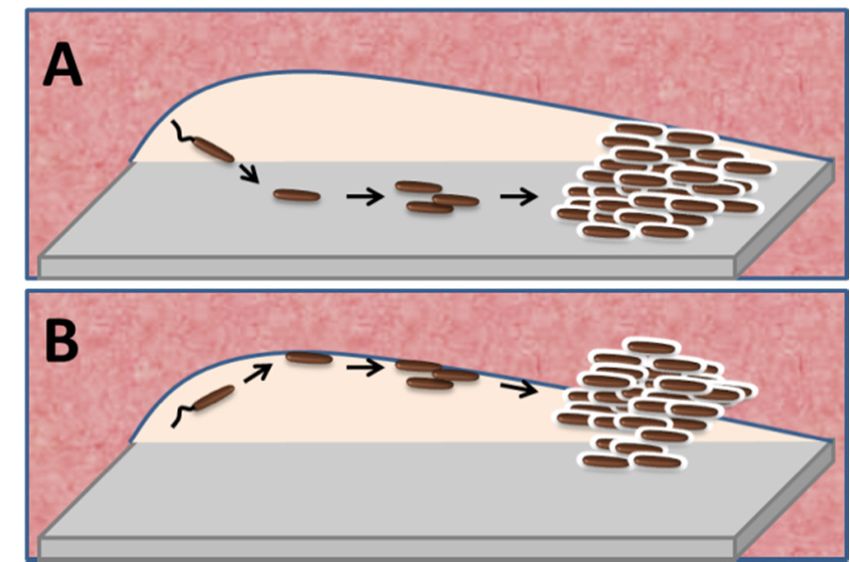
Figure 2. Model proposing tissue infection as initial key step of bacterial implant infections. (A)
Conventional model. Consecutive steps of biofilm infections are shown from left to right. Planktonic
bacteria (brown) enter the wound-liquid-filled interspace (colorless) between implant (grey) and
tissue (pink). As a crucial step towards biofilm formation, bacteria first adhere to the implant surfacesubcutaneously implanted into standard BALB/c mice were immediately infected with
Pseudomonas aeruginosa. After one week, tissue adjacent to the implants was subjected to scanning
transmission electron microscopic analysis (for a more detailed description see Reference [113]).
Bacteria (upper arrow) surrounded by clear areas (lower arrow), indicating the presence of
Metalsexopolysaccharide
2018, 8, 532 matrix material, a typical biofilm component. Reproduced with permission from 7 of 14
J. Biomed. Mater. Res.; Published by Wiley 2016.
Figure 2. Model proposing tissue infection as initial key step of bacterial implant infections. (A)
Figure 2. Model proposing tissue infection as initial key step of bacterial implant infections.
Conventional model. Consecutive steps of biofilm infections are shown from left to right. Planktonic
(A) Conventional model. Consecutive steps of biofilm infections are shown from left to right. Planktonic
bacteria (brown) enter the wound-liquid-filled interspace (colorless) between implant (grey) and
bacteria (brown) enter the wound-liquid-filled interspace (colorless) between implant (grey) and tissue
tissue (pink). As a crucial step towards biofilm formation, bacteria first adhere to the implant surface
(pink). As a crucial step towards biofilm formation, bacteria first adhere to the implant surface and
form micro-colonies. After reaching a critical density, bacteria switch to the biofilm mode and secrete
extracellular matrix compounds. Biofilm features, such as the encapsulation in the matrix, nutrient
restriction and slow growth, render the associated bacteria highly resistant to the host immune defenses
and to antibiotics. Subsequently, secreted exotoxins and proteases allow bacteria to invade the adjacent
host tissue. Alternatively, adhesion of host tissue to the implant acts to protect the implant surface
from bacterial attachment and subsequent biofilm formation. Based on the in vitro results, in this
scenario magnesium implants would be expected to act bactericidal. (B) Tissue infection model. Under
normal circumstances contiguous epithelial cell layers protect living tissue, whereas wounding renders
tissue highly susceptible to bacterial infections. After implant insertion the essential initial bacterial
attachment occurs primarily at the susceptible injured tissue surface. Bacterial colonies growing on the
tissue surface eventually switch to the biofilm mode with analogous outcomes as in the conventional
model. While bacterial adhesion to the implant may occur, it plays no essential role for the course of the
infection. Adhesion of host tissue to the implant would still be important to antagonize infections but
predominantly to protect the wound tissue surface, and not the implant, from bacterial colonization.
Despite acting bactericidal upon close contact, the observed enhanced infection susceptibility of
magnesium implants is explained by interference of corroding magnesium with host tissue adhesion.
Factors that prolong the wound surface exposure to bacteria could be alkaline pH immediately after
implantation, and hydrogen gas evolution or eroding solid corrosion layers thereafter.
9. Implications for the Design of Antibacterial Implants
A wide variety of anti-infective implant strategies have been investigated, mostly in vitro [118].
In the light of the proposed tissue invasion model, in order to be efficacious, antibacterial substances
would need to be diffusible to reach bacteria in the vicinity of the implant. Therefore, implant
nanostructures that act antiadhesive, or passive coatings that act bactericidal upon contact, would not
be expected to curb infections in patients. In addition, implant features that affect tissue adhesion play
an important, through different, role than previously thought. That is, to primarily prevent bacterial
adhesion to the injured tissue rather than to the implant (Table 2). Even though magnesium implants
could not curb bacterial infections in mice, clinical data is needed before a final conclusion can be
drawn. In addition, several alternative strategies are presently investigated, such as antibiotic-releasing
coatings for magnesium-based implants or the addition of antibacterial acting alloy metals like silver,
copper, or zinc that release cytotoxic ions [119–125]. The major challenge for such an approach is to
maintain the balance of achieving efficacious bactericidal ion concentrations in vivo without damaging
the host tissue.Metals 2018, 8, 532 8 of 14
Table 2. Implant features predicted by the tissue infection model to influence the susceptibility to
infections in vivo.
Ineffective Coatings Infection Risks Favorable Measures
Factors that hinder host tissue Surfaces favoring tissue
Surfaces that antagonize
adhesion; convex or integration; smooth, flat or
bacterial adhesion
microporous surfaces concave forms;
Contact-dependent Relative movement of Antibacterial
bactericidal surfaces implants versus tissue substance-releasing coatings
10. Conclusions
In long-term clinical trials biodegradable polymeric stents were inferior to conventional
drug-eluting metal stents, while recently introduced biocorrodible magnesium-based bone screws
were without noticeable side effects. However, since in vitro tests and even small animal studies
cannot predict the outcome in human patients, long-term clinical confirmation of the expected benefits,
with regard to potential risks, are needed. In addition, a novel model for implant infections suggests
that host cell adhesion to implants is important to prevent bacterial invasion of the exposed host tissue
surface, and not, as previously thought, to prevent bacterial adhesion to the implant. The model
predicts that passive antibacterial implant coating strategies would not be efficacious in vivo.
Funding: We acknowledge the financial support by a joint grant to M.I.R. and to S.U. of the German Academic
Exchange Service (DAAD), Germany, and the Higher Education Commission of Pakistan (HEC).
Conflicts of Interest: The authors declare that there is no conflict of interest regarding the publication of this paper.
References
1. Perren, S.M.; Regazzoni, P.; Fernandez, A.A. How to choose between the implant iaterials steel and titanium
in orthopedic trauma surgery: Part 2—Biological aspects. Acta Chir. Orthop. Traumatol. Cech. 2017, 84, 85–90.
[PubMed]
2. Osman, R.B.; Swain, M.V. A Critical Review of Dental Implant Materials with an Emphasis on Titanium
versus Zirconia. Materials (Basel) 2015, 8, 932–958. [CrossRef] [PubMed]
3. Williams, D.F. On the nature of biomaterials. Biomaterials 2009, 30, 5897–5909. [CrossRef] [PubMed]
4. Elias, C.N.; Lima, J.H.C.; Valiev, R.; Meyers, M.A. Biomedical applications of titanium and its alloys. JOM
2008, 60, 46–49. [CrossRef]
5. Sheikh, Z.; Najeeb, S.; Khurshid, Z.; Verma, V.; Rashid, H.; Glogauer, M. Biodegradable Materials for Bone
Repair and Tissue Engineering Applications. Materials (Basel) 2015, 8, 5744–5794. [CrossRef] [PubMed]
6. Middleton, J.C.; Tipton, A.J. Synthetic biodegradable polymers as orthopedic devices. Biomaterials 2000, 21,
2335–2346. [CrossRef]
7. Witte, F.; Fischer, J.; Nellesen, J.; Crostack, H.A.; Kaese, V.; Pisch, A.; Beckmann, F.; Windhagen, H. In vitro
and in vivo corrosion measurements of magnesium alloys. Biomaterials 2006, 27, 1013–1018. [CrossRef]
[PubMed]
8. Martinez Sanchez, A.H.; Luthringer, B.J.; Feyerabend, F.; Willumeit, R. Mg and Mg alloys: How comparable
are in vitro and in vivo corrosion rates? A review. Acta Biomater. 2015, 13, 16–31. [CrossRef] [PubMed]
9. Luthringer, B.J.; Feyerabend, F.; Willumeit-Romer, R. Magnesium-based implants: A mini-review.
Magnes. Res. 2014, 27, 142–154. [PubMed]
10. Li, H.; Zheng, Y.; Qin, L. Progress of biodegradable metals. Prog. Nat. Sci. 2014, 24, 414–422. [CrossRef]
11. Miller, A.P.; Huff, C.M.; Roubin, G.S. Vascular disease in the older adult. J. Geriatr. Cardiol. JGC 2016, 13,
727–732. [PubMed]
12. Martens, A.; Beckmann, E.; Kaufeld, T.; Umminger, J.; Fleissner, F.; Koigeldiyev, N.; Krueger, H.; Puntigam, J.;
Haverich, A.; Shrestha, M. Total aortic arch repair: Risk factor analysis and follow-up in 199 patients. Eur. J.
Cardiothorac. Surg. 2016, 50, 940–948. [CrossRef] [PubMed]Metals 2018, 8, 532 9 of 14
13. Rittersma, S.Z.; de Winter, R.J.; Koch, K.T.; Bax, M.; Schotborgh, C.E.; Mulder, K.J.; Tijssen, J.G.; Piek, J.J.
Impact of strut thickness on late luminal loss after coronary artery stent placement. Am. J. Cardiol. 2004, 93,
477–480. [CrossRef] [PubMed]
14. Onche, I.I.; Osagie, O.E.; INuhu, S. Removal of orthopaedic implants: Indications, outcome and economic
implications. J. West Afr. Coll. Surg. 2011, 1, 101–112. [PubMed]
15. Levy, J.A.; Podeszwa, D.A.; Lebus, G.; Ho, C.A.; Wimberly, R.L. Acute complications associated with removal
of flexible intramedullary femoral rods placed for pediatric femoral shaft fractures. J. Pediatr. Orthop. 2013,
33, 43–47. [CrossRef] [PubMed]
16. Buchanan, K.; Steinvil, A.; Waksman, R. Does the new generation of drug-eluting stents render bare metal
stents obsolete? Cardiovasc. Revasc. Med. 2017, 18, 456–461. [CrossRef] [PubMed]
17. Waksman, R. A new generation of drug-eluting stents: Indications and outcomes of bioresorbable vascular
scaffolds. Cleve. Clin. J. Med. 2017, 84 (Suppl. S4), e20–e24. [CrossRef] [PubMed]
18. Pleva, L.; Kukla, P.; Hlinomaz, O. Treatment of coronary in-stent restenosis: A systematic review.
J. Geriatr. Cardiol. 2018, 15, 173–184. [PubMed]
19. Artang, R.; Dieter, R.S. Analysis of 36 reported cases of late thrombosis in drug-eluting stents placed in
coronary arteries. Am. J. Cardiol. 2007, 99, 1039–1043. [CrossRef] [PubMed]
20. Mauri, L.; Hsieh, W.H.; Massaro, J.M.; Ho, K.K.; D’Agostino, R.; Cutlip, D.E. Stent thrombosis in randomized
clinical trials of drug-eluting stents. N. Engl. J. Med. 2007, 356, 1020–1029. [CrossRef] [PubMed]
21. Filion, K.B.; Roy, A.M.; Baboushkin, T.; Rinfret, S.; Eisenberg, M.J. Cost-effectiveness of drug-eluting stents
including the economic impact of late stent thrombosis. Am. J. Cardiol. 2009, 103, 338–344. [CrossRef]
[PubMed]
22. Erne, P.; Schier, M.; Resink, T.J. The road to bioabsorbable stents: Reaching clinical reality? Cardiovasc.
Intervent. Radiol. 2006, 29, 11–16. [CrossRef] [PubMed]
23. Tan, L.; Yu, X.; Wan, P.; Yang, K. Biodegradable Materials for Bone Repairs: A Review. J. Mater. Sci. Technol.
2013, 29, 503–513. [CrossRef]
24. Zheng, Y.F.; Gu, X.N.; Witte, F. Biodegradable metals. Mater. Sci. Eng. R Rep. 2014, 77, 1–34. [CrossRef]
25. Waksman, R.; Pakala, R. Biodegradable and bioabsorbable stents. Curr. Pharm. Des. 2010, 16, 4041–4051.
[CrossRef] [PubMed]
26. Sumner, D.R. Long-term implant fixation and stress-shielding in total hip replacement. J. Biomech. 2015, 48,
797–800. [CrossRef] [PubMed]
27. Ceonzo, K.; Gaynor, A.; Shaffer, L.; Kojima, K.; Vacanti, C.A.; Stahl, G.L. Polyglycolic acid-induced
inflammation: Role of hydrolysis and resulting complement activation. Tissue Eng. 2006, 12, 301–308.
[CrossRef] [PubMed]
28. Athanasiou, K.A.; Niederauer, G.G.; Agrawal, C.M. Sterilization, toxicity, biocompatibility and clinical
applications of polylactic acid/polyglycolic acid copolymers. Biomaterials 1996, 17, 93–102. [CrossRef]
29. Pierson, D.; Edick, J.; Tauscher, A.; Pokorney, E.; Bowen, P.; Gelbaugh, J.; Stinson, J.; Getty, H.; Lee, C.H.;
Drelich, J.; et al. A simplified in vivo approach for evaluating the bioabsorbable behavior of candidate stent
materials. J. Biomed. Mater. Res. B Appl. Biomater. 2012, 100, 58–67. [CrossRef] [PubMed]
30. Bowen, P.K.; Drelich, J.; Buxbaum, R.E.; Rajachar, R.M.; Goldman, J. New approaches in evaluating metallic
candidates for bioabsorbable stents. Emerg. Mater. Res. 2012, 1, 237–255. [CrossRef]
31. Hermawan, H.; Purnama, A.; Dube, D.; Couet, J.; Mantovani, D. Fe-Mn alloys for metallic biodegradable
stents: Degradation and cell viability studies. Acta Biomater. 2010, 6, 1852–1860. [CrossRef] [PubMed]
32. Vojtech, D.; Kubasek, J.; Serak, J.; Novak, P. Mechanical and corrosion properties of newly developed
biodegradable Zn-based alloys for bone fixation. Acta Biomater. 2011, 7, 3515–3522. [CrossRef] [PubMed]
33. Yang, H.; Wang, C.; Liu, C.; Chen, H.; Wu, Y.; Han, J.; Jia, Z.; Lin, W.; Zhang, D.; Li, W.; et al. Evolution
of the degradation mechanism of pure zinc stent in the one-year study of rabbit abdominal aorta model.
Biomaterials 2017, 145, 92–105. [CrossRef] [PubMed]
34. Zhao, D.; Wang, T.; Nahan, K.; Guo, X.; Zhang, Z.; Dong, Z.; Chen, S.; Chou, D.T.; Hong, D.; Kumta, P.N.; et al.
In vivo characterization of magnesium alloy biodegradation using electrochemical H2 monitoring, ICP-MS,
and XPS. Acta Biomater. 2017, 50, 556–565. [CrossRef] [PubMed]
35. Zhao, D.; Witte, F.; Lu, F.; Wang, J.; Li, J.; Qin, L. Current status on clinical applications of magnesium-based
orthopaedic implants: A review from clinical translational perspective. Biomaterials 2017, 112, 287–302.
[CrossRef] [PubMed]Metals 2018, 8, 532 10 of 14
36. Staiger, M.P.; Pietak, A.M.; Huadmai, J.; Dias, G. Magnesium and its alloys as orthopedic biomaterials:
A review. Biomaterials 2006, 27, 1728–1734. [CrossRef] [PubMed]
37. Debieux, P.; Franciozi, C.E.; Lenza, M.; Tamaoki, M.J.; Magnussen, R.A.; Faloppa, F.; Belloti, J.C. Bioabsorbable
versus metallic interference screws for graft fixation in anterior cruciate ligament reconstruction.
Cochrane Database Syst. Rev. 2016, 7, CD009772. [CrossRef] [PubMed]
38. Seitz, J.M.; Durisin, M.; Goldman, J.; Drelich, J.W. Recent Advances in Biodegradable Metals for Medical
Sutures: A Critical Review. Adv. Healthc. Mater. 2015, 4, 1915–1936. [CrossRef] [PubMed]
39. Doppalapudi, S.; Jain, A.; Khan, W.; Domb, A.J. Biodegradable polymers—An overview. Polym. Adv. Technol.
2014, 25, 427–435. [CrossRef]
40. Van der Giessen, W.J.; Lincoff, A.M.; Schwartz, R.S.; van Beusekom, H.M.; Serruys, P.W.; Holmes, D.R., Jr.;
Ellis, S.G.; Topol, E.J. Marked inflammatory sequelae to implantation of biodegradable and nonbiodegradable
polymers in porcine coronary arteries. Circulation 1996, 94, 1690–1697. [CrossRef] [PubMed]
41. Bunger, C.M.; Grabow, N.; Sternberg, K.; Kroger, C.; Ketner, L.; Schmitz, K.P.; Kreutzer, H.J.; Ince, H.;
Nienaber, C.A.; Klar, E.; et al. Sirolimus-eluting biodegradable poly-L-lactide stent for peripheral vascular
application: A preliminary study in porcine carotid arteries. J. Surg. Res. 2007, 139, 77–82. [CrossRef]
[PubMed]
42. Rizik, D.G.; Hermiller, J.B.; Kereiakes, D.J. Bioresorbable vascular scaffolds for the treatment of coronary
artery disease: Clinical outcomes from randomized controlled trials. Catheter. Cardiovasc. Interv. 2016, 88
(Suppl. S1), 21–30. [CrossRef] [PubMed]
43. Ali, Z.A.; Serruys, P.W.; Kimura, T.; Gao, R.; Ellis, S.G.; Kereiakes, D.J.; Onuma, Y.; Simonton, C.; Zhang, Z.;
Stone, G.W. 2-year outcomes with the Absorb bioresorbable scaffold for treatment of coronary artery disease:
A systematic review and meta-analysis of seven randomised trials with an individual patient data substudy.
Lancet 2017, 390, 760–772. [CrossRef]
44. Ali, Z.A.; Gao, R.; Kimura, T.; Onuma, Y.; Kereiakes, D.J.; Ellis, S.G.; Chevalier, B.; Vu, M.T.; Zhang, Z.;
Simonton, C.A.; et al. Three-Year Outcomes with the Absorb Bioresorbable Scaffold: Individual-Patient-Data
Meta-Analysis from the ABSORB Randomized Trials. Circulation 2018, 137, 464–479. [CrossRef] [PubMed]
45. Peuster, M.; Wohlsein, P.; Brugmann, M.; Ehlerding, M.; Seidler, K.; Fink, C.; Brauer, H.; Fischer, A.;
Hausdorf, G. A novel approach to temporary stenting: Degradable cardiovascular stents produced from
corrodible metal-results 6–18 months after implantation into New Zealand white rabbits. Heart 2001, 86,
563–569. [CrossRef] [PubMed]
46. Francis, A.; Yang, Y.; Virtanen, S.; Boccaccini, A.R. Iron and iron-based alloys for temporary cardiovascular
applications. J. Mater. Sci. Mater. Med. 2015, 26, 138. [CrossRef] [PubMed]
47. Peuster, M.; Hesse, C.; Schloo, T.; Fink, C.; Beerbaum, P.; von Schnakenburg, C. Long-term biocompatibility
of a corrodible peripheral iron stent in the porcine descending aorta. Biomaterials 2006, 27, 4955–4962.
[CrossRef] [PubMed]
48. Fagali, N.S.; Grillo, C.A.; Puntarulo, S.; Fernandez Lorenzo de Mele, M.A. Cytotoxicity of corrosion products
of degradable Fe-based stents: Relevance of pH and insoluble products. Colloids Surf. B Biointerfaces 2015,
128, 480–488. [CrossRef] [PubMed]
49. Moravej, M.; Purnama, A.; Fiset, M.; Couet, J.; Mantovani, D. Electroformed pure iron as a new biomaterial
for degradable stents: In vitro degradation and preliminary cell viability studies. Acta Biomater. 2010, 6,
1843–1851. [CrossRef] [PubMed]
50. Drynda, A.; Hoehn, R.; Peuster, M. Influence of Fe(II) and Fe(III) on the expression of genes related to
cholesterol- and fatty acid metabolism in human vascular smooth muscle cells. J. Mater. Sci. Mater. Med.
2010, 21, 1655–1663. [CrossRef] [PubMed]
51. Zhu, S.; Huang, N.; Xu, L.; Zhang, Y.; Liu, H.; Sun, H.; Leng, Y. Biocompatibility of pure iron: In vitro
assessment of degradation kinetics and cytotoxicity on endothelial cells. Mater. Sci. Eng. C 2009, 29,
1589–1592. [CrossRef]
52. Mueller, P.P.; May, T.; Perz, A.; Hauser, H.; Peuster, M. Control of smooth muscle cell proliferation by ferrous
iron. Biomaterials 2006, 27, 2193–2200. [CrossRef] [PubMed]
53. Kraus, T.; Moszner, F.; Fischerauer, S.; Fiedler, M.; Martinelli, E.; Eichler, J.; Witte, F.; Willbold, E.;
Schinhammer, M.; Meischel, M.; et al. Biodegradable Fe-based alloys for use in osteosynthesis: Outcome of
an in vivo study after 52 weeks. Acta Biomater. 2014, 10, 3346–3353. [CrossRef] [PubMed]Metals 2018, 8, 532 11 of 14
54. Mueller, P.P.; Arnold, S.; Badar, M.; Bormann, D.; Bach, F.W.; Drynda, A.; Meyer-Lindenberg, A.; Hauser, H.;
Peuster, M. Histological and molecular evaluation of iron as degradable medical implant material in a
murine animal model. J. Biomed. Mater. Res. A 2012, 100, 2881–2889. [CrossRef] [PubMed]
55. Voigtlaender, H. [Wandering of foreign bodies. (Grenate fragment in the common bile duct causing jaundice)].
Chirurg 1975, 46, 467–469. [PubMed]
56. Ghislain, P.D. Spontaneous extrusion of hand grenade fragments from the face 60 years after injury. JAMA
2003, 290, 1317–1318. [CrossRef] [PubMed]
57. Gaber, Y. [Grenade splinter injury simulating thrombophlebitis]. Vasa 2003, 32, 40–42. [CrossRef] [PubMed]
58. Mostaed, E.; Sikora-Jasinska, M.; Drelich, J.W.; Vedani, M. Zinc-based alloys for degradable vascular stent
applications. Acta Biomater. 2018, 71, 1–23. [CrossRef] [PubMed]
59. Jin, H.; Zhao, S.; Guillory, R.; Bowen, P.K.; Yin, Z.; Griebel, A.; Schaffer, J.; Earley, E.J.; Goldman, J.; Drelich, J.W.
Novel high-strength, low-alloys Zn-Mg (Metals 2018, 8, 532 12 of 14
76. Kirkland, N.T.; Birbilis, N.; Staiger, M.P. Assessing the corrosion of biodegradable magnesium implants:
A critical review of current methodologies and their limitations. Acta Biomater. 2012, 8, 925–936. [CrossRef]
[PubMed]
77. Huehnerschulte, T.A.; Angrisani, N.; Rittershaus, D.; Bormann, D.; Windhagen, H.; Meyer-Lindenberg, A.
In Vivo Corrosion of Two Novel Magnesium Alloys ZEK100 and AX30 and Their Mechanical Suitability as
Biodegradable Implants. Materials (Basel) 2011, 4, 1144–1167. [CrossRef] [PubMed]
78. Krause, A.; von der Höh, N.; Bormann, D.; Krause, C.; Bach, F.-W.; Windhagen, H.; Meyer-Lindenberg, A.
Degradation behaviour and mechanical properties of magnesium implants in rabbit tibiae. J. Mater. Sci.
2009, 45, 624. [CrossRef]
79. Zeng, R.; Dietzel, W.; Witte, F.; Hort, N.; Blawert, C. Progress and Challenge for Magnesium Alloys as
Biomaterials. Adv. Eng. Mater. 2008, 10, B3–B14. [CrossRef]
80. Kirkland, N.T.; Lespagnol, J.; Birbilis, N.; Staiger, M.P. A survey of bio-corrosion rates of magnesium alloys.
Corros. Sci. 2010, 52, 287–291. [CrossRef]
81. Jahnen-Dechent, W.; Ketteler, M. Magnesium basics. Clin. Kidney J. 2012, 5 (Suppl. S1), i3–i14. [CrossRef]
[PubMed]
82. De Baaij, J.H.; Hoenderop, J.G.; Bindels, R.J. Magnesium in man: Implications for health and disease.
Physiol. Rev. 2015, 95, 1–46. [CrossRef] [PubMed]
83. Han, P.; Cheng, P.; Zhang, S.; Zhao, C.; Ni, J.; Zhang, Y.; Zhong, W.; Hou, P.; Zhang, X.; Zheng, Y.; et al. In vitro
and in vivo studies on the degradation of high-purity Mg (99.99 wt.%) screw with femoral intracondylar
fractured rabbit model. Biomaterials 2015, 64, 57–69. [CrossRef] [PubMed]
84. Badar, M.; Lunsdorf, H.; Evertz, F.; Rahim, M.I.; Glasmacher, B.; Hauser, H.; Mueller, P.P. The formation of an
organic coat and the release of corrosion microparticles from metallic magnesium implants. Acta Biomater.
2013, 9, 7580–7589. [CrossRef] [PubMed]
85. Rahim, M.I.; Weizbauer, A.; Evertz, F.; Hoffmann, A.; Rohde, M.; Glasmacher, B.; Windhagen, H.; Gross, G.;
Seitz, J.M.; Mueller, P.P. Differential magnesium implant corrosion coat formation and contribution to bone
bonding. J. Biomed. Mater. Res. A 2017, 105, 697–709. [CrossRef] [PubMed]
86. Zhang, Y.; Xu, J.; Ruan, Y.C.; Yu, M.K.; O’Laughlin, M.; Wise, H.; Chen, D.; Tian, L.; Shi, D.; Wang, J.; et al.
Implant-derived magnesium induces local neuronal production of CGRP to improve bone-fracture healing
in rats. Nat. Med. 2016, 22, 1160–1169. [CrossRef] [PubMed]
87. Witte, F.; Kaese, V.; Haferkamp, H.; Switzer, E.; Meyer-Lindenberg, A.; Wirth, C.J.; Windhagen, H. In vivo
corrosion of four magnesium alloys and the associated bone response. Biomaterials 2005, 26, 3557–3563.
[CrossRef] [PubMed]
88. Kuhlmann, J.; Bartsch, I.; Willbold, E.; Schuchardt, S.; Holz, O.; Hort, N.; Hoche, D.; Heineman, W.R.; Witte, F.
Fast escape of hydrogen from gas cavities around corroding magnesium implants. Acta Biomater. 2013, 9,
8714–8721. [CrossRef] [PubMed]
89. Noviana, D.; Paramitha, D.; Ulum, M.F.; Hermawan, H. The effect of hydrogen gas evolution of magnesium
implant on the postimplantation mortality of rats. J. Orthop. Transl. 2016, 5, 9–15. [CrossRef]
90. Chen, Y.; Xu, Z.; Smith, C.; Sankar, J. Recent advances on the development of magnesium alloys for
biodegradable implants. Acta Biomater. 2014, 10, 4561–4573. [CrossRef] [PubMed]
91. Grünewald, T.A.; Rennhofer, H.; Hesse, B.; Burghammer, M.; Stanzl-Tschegg, S.E.; Cotte, M.; Löffler, J.F.;
Weinberg, A.M.; Lichtenegger, H.C. Magnesium from bioresorbable implants: Distribution and impact on
the nano- and mineral structure of bone. Biomaterials 2016, 76, 250–260. [CrossRef] [PubMed]
92. Seitz, J.-M.; Lucas, A.; Kirschner, M. Magnesium-Based Compression Screws: A Novelty in the Clinical Use
of Implants. JOM 2016, 68, 1177–1182. [CrossRef]
93. Plaass, C.; von Falck, C.; Ettinger, S.; Sonnow, L.; Calderone, F.; Weizbauer, A.; Reifenrath, J.; Claassen, L.;
Waizy, H.; Daniilidis, K.; et al. Bioabsorbable magnesium versus standard titanium compression screws for
fixation of distal metatarsal osteotomies—3 year results of a randomized clinical trial. J. Orthop. Sci. 2018, 23,
321–327. [CrossRef] [PubMed]
94. Windhagen, H.; Radtke, K.; Weizbauer, A.; Diekmann, J.; Noll, Y.; Kreimeyer, U.; Schavan, R.;
Stukenborg-Colsman, C.; Waizy, H. Biodegradable magnesium-based screw clinically equivalent to titanium
screw in hallux valgus surgery: Short term results of the first prospective, randomized, controlled clinical
pilot study. Biomed. Eng. Online 2013, 12, 62. [CrossRef] [PubMed]Metals 2018, 8, 532 13 of 14
95. Ezechieli, M.; Meyer, H.; Lucas, A.; Helmecke, P.; Becher, C.; Calliess, T.; Windhagen, H.; Ettinger, M.
Biomechanical Properties of a Novel Biodegradable Magnesium-Based Interference Screw. Orthop. Rev.
(Pavia) 2016, 8, 6445. [CrossRef] [PubMed]
96. Biber, R.; Pauser, J.; Brem, M.; Bail, H.J. Bioabsorbable metal screws in traumatology: A promising innovation.
Trauma Case Rep. 2017, 8, 11–15. [CrossRef] [PubMed]
97. Ang, H.Y.; Huang, Y.Y.; Lim, S.T.; Wong, P.; Joner, M.; Foin, N. Mechanical behavior of polymer-based vs.
metallic-based bioresorbable stents. J. Thorac. Dis. 2017, 9 (Suppl. S9), S923–S934. [CrossRef] [PubMed]
98. Zhao, N.; Watson, N.; Xu, Z.G.; Chen, Y.J.; Waterman, J.; Sankar, J.; Zhu, D.H. In Vitro Biocompatibility and
Endothelialization of Novel Magnesium-Rare Earth Alloys for Improved Stent Applications. PLoS ONE
2014, 9, e98674. [CrossRef] [PubMed]
99. Zhu, S.J.; Liu, Q.; Qian, Y.F.; Sun, B.; Wang, L.G.; Wu, J.M.; Guan, S.K. Effect of different processings on
mechanical property and corrosion behavior in simulated body fluid of Mg-Zn-Y-Nd alloy for cardiovascular
stent application. Front. Mater. Sci. 2014, 8, 256–263. [CrossRef]
100. Mao, L.; Shen, L.; Chen, J.; Zhang, X.; Kwak, M.; Wu, Y.; Fan, R.; Zhang, L.; Pei, J.; Yuan, G.; et al. A promising
biodegradable magnesium alloy suitable for clinical vascular stent application. Sci. Rep. 2017, 7, 46343.
[CrossRef] [PubMed]
101. Mao, L.; Shen, L.; Chen, J.; Wu, Y.; Kwak, M.; Lu, Y.; Xue, Q.; Pei, J.; Zhang, L.; Yuan, G.; et al. Enhanced
bioactivity of Mg-Nd-Zn-Zr alloy achieved with nanoscale MgF2 surface for vascular stent application.
ACS Appl. Mater. Interfaces 2015, 7, 5320–5330. [CrossRef] [PubMed]
102. Waksman, R.; Erbel, R.; Di Mario, C.; Bartunek, J.; de Bruyne, B.; Eberli, F.R.; Erne, P.; Haude, M.; Horrigan, M.;
Ilsley, C.; et al. Early- and long-term intravascular ultrasound and angiographic findings after bioabsorbable
magnesium stent implantation in human coronary arteries. JACC Cardiovasc. Interv. 2009, 2, 312–320.
[CrossRef] [PubMed]
103. Erbel, R.; Di Mario, C.; Bartunek, J.; Bonnier, J.; de Bruyne, B.; Eberli, F.R.; Erne, P.; Haude, M.; Heublein, B.;
Horrigan, M.; et al. Temporary scaffolding of coronary arteries with bioabsorbable magnesium stents:
A prospective, non-randomised multicentre trial. Lancet 2007, 369, 1869–1875. [CrossRef]
104. Rapetto, C.; Leoncini, M. Magmaris: A new generation metallic sirolimus-eluting fully bioresorbable scaffold:
Present status and future perspectives. J. Thorac. Dis. 2017, 9 (Suppl. S9), S903–S913. [CrossRef] [PubMed]
105. Haude, M.; Ince, H.; Kische, S.; Abizaid, A.; Tolg, R.; Alves Lemos, P.; Van Mieghem, N.M.; Verheye, S.;
von Birgelen, C.; Christiansen, E.H.; et al. Sustained safety and clinical performance of a drug-eluting
absorbable metal scaffold up to 24 months: Pooled outcomes of BIOSOLVE-II and BIOSOLVE-III.
EuroIntervention 2017, 13, 432–439. [PubMed]
106. Haude, M.; Erbel, R.; Erne, P.; Verheye, S.; Degen, H.; Bose, D.; Vermeersch, P.; Wijnbergen, I.; Weissman, N.;
Prati, F.; et al. Safety and performance of the drug-eluting absorbable metal scaffold (DREAMS) in patients
with de-novo coronary lesions: 12 month results of the prospective, multicentre, first-in-man BIOSOLVE-I
trial. Lancet 2013, 381, 836–844. [CrossRef]
107. Willbold, E.; Weizbauer, A.; Loos, A.; Seitz, J.M.; Angrisani, N.; Windhagen, H.; Reifenrath, J. Magnesium
alloys: A stony pathway from intensive research to clinical reality. Different test methods and
approval-related considerations. J. Biomed. Mater. Res. A 2017, 105, 329–347. [CrossRef] [PubMed]
108. Arciola, C.R.; Campoccia, D.; Speziale, P.; Montanaro, L.; Costerton, J.W. Biofilm formation in Staphylococcus
implant infections. A review of molecular mechanisms and implications for biofilm-resistant materials.
Biomaterials 2012, 33, 5967–5982. [CrossRef] [PubMed]
109. Rahim, M.I.; Eifler, R.; Rais, B.; Mueller, P.P. Alkalization is responsible for antibacterial effects of corroding
magnesium. J. Biomed. Mater. Res. A 2015, 103, 3526–3532. [CrossRef] [PubMed]
110. Feng, H.; Wang, G.; Jin, W.; Zhang, X.; Huang, Y.; Gao, A.; Wu, H.; Wu, G.; Chu, P.K. Systematic Study of
Inherent Antibacterial Properties of Magnesium-based Biomaterials. ACS Appl. Mater. Interfaces 2016, 8,
9662–9673. [CrossRef] [PubMed]
111. Lock, J.Y.; Wyatt, E.; Upadhyayula, S.; Whall, A.; Nunez, V.; Vullev, V.I.; Liu, H. Degradation and antibacterial
properties of magnesium alloys in artificial urine for potential resorbable ureteral stent applications. J. Biomed.
Mater. Res. A 2014, 102, 781–792. [CrossRef] [PubMed]
112. Robinson, D.A.; Griffith, R.W.; Shechtman, D.; Evans, R.B.; Conzemius, M.G. In vitro antibacterial
properties of magnesium metal against Escherichia coli, Pseudomonas aeruginosa and Staphylococcus aureus.
Acta Biomater. 2010, 6, 1869–1877. [CrossRef] [PubMed]Metals 2018, 8, 532 14 of 14
113. Rahim, M.I.; Rohde, M.; Rais, B.; Seitz, J.-M.; Mueller, P.P. Susceptibility of metallic magnesium implants to
bacterial biofilm infections. J. Biomed. Mater. Res. A 2016, 104, 1489–1499. [CrossRef] [PubMed]
114. Hou, P.; Zhao, C.; Cheng, P.; Wu, H.; Ni, J.; Zhang, S.; Lou, T.; Wang, C.; Han, P.; Zhang, X.; et al. Reduced
antibacterial property of metallic magnesium in vivo. Biomed. Mater. 2016, 12, 015010. [CrossRef] [PubMed]
115. Gristina, A.G.; Naylor, P.; Myrvik, Q. Infections from biomaterials and implants: A race for the surface.
Med. Prog. Technol. 1988, 14, 205–224. [PubMed]
116. Church, D.; Elsayed, S.; Reid, O.; Winston, B.; Lindsay, R. Burn wound infections. Clin. Microbiol. Rev. 2006,
19, 403–434. [CrossRef] [PubMed]
117. Stern, G.A. Pseudomonas keratitis and contact lens wear: The lens/eye is at fault. Cornea 1990, 9 (Suppl. S1),
S39–S40. [CrossRef] [PubMed]
118. Qin, S.; Xu, K.; Nie, B.; Ji, F.; Zhang, H. Approaches based on passive and active antibacterial coating on
titanium to achieve antibacterial activity. J. Biomed. Mater. Res. A 2018. [CrossRef] [PubMed]
119. Liu, Z.D.; Schade, R.; Luthringer, B.; Hort, N.; Rothe, H.; Muller, S.; Liefeith, K.; Willumeit-Romer, R.;
Feyerabend, F. Influence of the Microstructure and Silver Content on Degradation, Cytocompatibility, and
Antibacterial Properties of Magnesium-Silver Alloys In Vitro. Oxid. Med. Cell. Longev. 2017, 2017, 8091265.
[CrossRef] [PubMed]
120. Zhang, X.D.; Yi, J.H.; Zhao, G.W.; Huang, L.L.; Yan, G.J.; Chen, Y.S.; Liu, P. Layer-by-layer assembly of
silver nanoparticles embedded polyelectrolyte multilayer on magnesium alloy with enhanced antibacterial
property. Surf. Coat. Technol. 2016, 286, 103–112. [CrossRef]
121. Yu, W.L.; Chen, D.Y.; Ding, Z.Y.; Qiu, M.L.; Zhang, Z.W.; Shen, J.; Zhang, X.N.; Zhang, S.X.; He, Y.H.; Shi, Z.M.
Synergistic effect of a biodegradable Mg-Zn alloy on osteogenic activity and anti-biofilm ability: An in vitro
and in vivo study. RSC Adv. 2016, 6, 45219–45230. [CrossRef]
122. Zhao, C.; Hou, P.; Ni, J.; Han, P.; Chai, Y.; Zhang, X. Ag-Incorporated FHA Coating on Pure Mg: Degradation
and in Vitro Antibacterial Properties. ACS Appl. Mater. Interfaces 2016, 8, 5093–5103. [CrossRef] [PubMed]
123. Qin, H.; Zhao, Y.; An, Z.; Cheng, M.; Wang, Q.; Cheng, T.; Wang, Q.; Wang, J.; Jiang, Y.; Zhang, X.; et al.
Enhanced antibacterial properties, biocompatibility, and corrosion resistance of degradable Mg-Nd-Zn-Zr
alloy. Biomaterials 2015, 53, 211–220. [CrossRef] [PubMed]
124. Tie, D.; Feyerabend, F.; Hort, N.; Hoeche, D.; Kainer, K.U.; Willumeit, R.; Mueller, W.D. In vitro mechanical
and corrosion properties of biodegradable Mg-Ag alloys. Mater. Corros. 2014, 65, 569–576. [CrossRef]
125. Zhang, X.B.; Ba, Z.X.; Wang, Z.Z.; He, X.C.; Shen, C.; Wang, Q. Influence of silver addition on microstructure
and corrosion behavior of Mg-Nd-Zn-Zr alloys for biomedical application. Mater. Lett. 2013, 100, 188–191.
[CrossRef]
© 2018 by the authors. Licensee MDPI, Basel, Switzerland. This article is an open access
article distributed under the terms and conditions of the Creative Commons Attribution
(CC BY) license (http://creativecommons.org/licenses/by/4.0/).You can also read