Bacteria in the amniotic fluid without inflammation: early colonization vs. contamination
←
→
Page content transcription
If your browser does not render page correctly, please read the page content below
J. Perinat. Med. 2021; aop

Eunjung Jung, Roberto Romero*, Bo Hyun Yoon, Kevin R. Theis, Dereje W. Gudicha,
Adi L. Tarca, Ramiro Diaz-Primera, Andrew D. Winters, Nardhy Gomez-Lopez, Lami Yeo
and Chaur-Dong Hsu

Bacteria in the amniotic fluid without
inflammation: early colonization vs.
contamination
https://doi.org/10.1515/jpm-2021-0191                                   Methods: A retrospective cross-sectional study included
Received April 19, 2021; accepted May 19, 2021;                         360 patients with preterm labor and intact membranes who
published online July 6, 2021
                                                                        underwent transabdominal amniocentesis for evaluation
                                                                        of the microbial state of the amniotic cavity as well as intra-
Abstract
                                                                        amniotic inflammation. Cultivation techniques were used
Objectives: Intra-amniotic infection, defined by the pres-              to isolate microorganisms, and broad-range polymerase
ence of microorganisms in the amniotic cavity, is often                 chain reaction coupled with electrospray ionization mass
accompanied by intra-amniotic inflammation. Occasion-                   spectrometry (PCR/ESI-MS) was utilized to detect the
ally, laboratories report the growth of bacteria or the                 nucleic acids of bacteria, viruses, and fungi.
presence of microbial nucleic acids in amniotic fluid in the            Results: Patients whose amniotic fluid samples evinced
absence of intra-amniotic inflammation. This study was                  microorganisms but did not indicate inflammation had a
conducted to determine the clinical significance of the                 similar perinatal outcome to those without microorgan-
presence of bacteria in amniotic fluid samples in the                   isms or inflammation [amniocentesis-to-delivery interval
absence of intra-amniotic inflammation.
                                                                        (p=0.31), spontaneous preterm birth before 34 weeks

Chaur-Dong Hsu, Present affiliation: Department of Obstetrics and Gynecology, University of Arizona College of Medicine–Tucson, Tucson, AZ,
USA

*Corresponding author: Roberto Romero, MD, DMedSci, Perinatology Research Branch, Eunice Kennedy Shriver National Institute of Child
Health and Human Development, National Institutes of Health, U. S. Department of Health and Human Services, Hutzel Women’s Hospital, 3990
John R Street, 4 Brush, Detroit, MI 48201 USA; Department of Obstetrics and Gynecology, University of Michigan, Ann Arbor, MI, USA;
Department of Epidemiology and Biostatistics, Michigan State University, East Lansing, MI, USA; Center for Molecular Medicine and Genetics,
Wayne State University, Detroit, MI, USA; Detroit Medical Center, Detroit, MI, USA; and Department of Obstetrics and Gynecology, Florida
International University, Miami, FL, USA, Phone: +313 993 2700, E-mail: prbchiefstaff@med.wayne.edu
Eunjung Jung, Dereje W. Gudicha, Ramiro Diaz-Primera and Lami Yeo, Perinatology Research Branch, Eunice Kennedy Shriver National Institute
of Child Health and Human Development, National Institutes of Health, U.S. Department of Health and Human Services, Bethesda, MD, and
Detroit, MI, USA; and Department of Obstetrics and Gynecology, Wayne State University School of Medicine, Detroit, MI, USA
Bo Hyun Yoon, BioMedical Research Institute, Seoul National University Hospital, Seoul, Republic of Korea
Kevin R. Theis and Andrew D. Winters, Perinatology Research Branch, Eunice Kennedy Shriver National Institute of Child Health and Human
Development, National Institutes of Health, U.S. Department of Health and Human Services, Bethesda, MD, and Detroit, MI, USA; and
Department of Biochemistry, Microbiology and Immunology, Wayne State University School of Medicine, Detroit, MI, USA
Adi L. Tarca, Perinatology Research Branch, Eunice Kennedy Shriver National Institute of Child Health and Human Development, National
Institutes of Health, U.S. Department of Health and Human Services, Bethesda, MD, and Detroit, MI, USA; Department of Obstetrics and
Gynecology, Wayne State University School of Medicine, Detroit, MI, USA; and Department of Computer Science, College of Engineering, Wayne
State University, Detroit, MI, USA
Nardhy Gomez-Lopez, Perinatology Research Branch, Eunice Kennedy Shriver National Institute of Child Health and Human Development,
National Institutes of Health, U.S. Department of Health and Human Services, Bethesda, MD, and Detroit, MI, USA; Department of Obstetrics and
Gynecology, Wayne State University School of Medicine, Detroit, MI, USA; and Department of Biochemistry, Microbiology and Immunology,
Wayne State University School of Medicine, Detroit, MI, USA
Chaur-Dong Hsu, Perinatology Research Branch, Eunice Kennedy Shriver National Institute of Child Health and Human Development, National
Institutes of Health, U.S. Department of Health and Human Services, Bethesda, MD, and Detroit, MI, USA; Department of Obstetrics and
Gynecology, Wayne State University School of Medicine, Detroit, MI, USA; and Department of Physiology, Wayne State University School of
Medicine, Detroit, MI, USA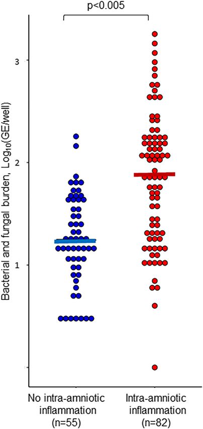
2        Jung et al.: Bacteria in amniotic fluid without inflammation

(p=0.83), acute placental inflammatory lesions (p=1), and                which the laboratory reports bacterial growth or the pres-
composite neonatal morbidity (p=0.8)].                                  ence of microbial nucleic acids in amniotic fluid in the
Conclusions: The isolation of microorganisms from a                     absence of intra-amniotic inflammation [6, 13, 34, 92,
sample of amniotic fluid in the absence of intra-amniotic               112–118]. The interpretation of this finding is a challenge.
inflammation is indicative of a benign condition, which                 Are bacteria the result of contamination of amniotic fluid at
most likely represents contamination of the specimen                    the time of specimen collection/laboratory processing, or
during the collection procedure or laboratory processing                the result of early colonization by pathogenic bacteria
rather than early colonization or infection.                            before an inflammatory process is established? This
                                                                        conundrum haunts clinicians in virtually every specialty of
Keywords: Acinetobacter; acute funisitis; acute histologic
                                                                        medicine dealing with a presumably sterile specimen, such
chorioamnionitis; amniocentesis; interleukin-6; intra-am-
                                                                        as cerebrospinal, pleural-pericardial, or amniotic fluid, or
niotic infection; intra-amniotic inflammation; microbial
                                                                        blood. Erroneous interpretation of a laboratory report can
burden; preterm labor with intact membranes; Propioni-
                                                                        lead to devastating consequences in obstetrics, including
bacterium acnes.
                                                                        iatrogenic preterm delivery. This study was conducted to
                                                                        determine the clinical significance of the presence of bac-
                                                                        teria in amniotic fluid in the absence of intra-amniotic
Introduction                                                            inflammation.

Infection is a major cause of morbidity and mortality for
mothers and newborns [1–3] who represent relatively                     Materials and methods
vulnerable hosts to the effects of microorganisms or mi-
crobial products (i.e. bacterial endotoxin). Therefore, the             Study population
accurate identification of intra-amniotic infection has
become important in patients at risk for adverse pregnancy              This retrospective cohort study was conducted by searching the clin-
outcome (preterm labor with intact membranes [4–13],                    ical database and bank of biological samples of Wayne State Univer-
preterm prelabor rupture of the membranes [14–25], clin-                sity, the Detroit Medical Center, and the Perinatology Research
                                                                        Branch. Patients diagnosed with preterm labor and intact membranes
ical chorioamnionitis [26–29], idiopathic vaginal bleeding
                                                                        at Hutzel Women’s Hospital were included in the study when they met
in the second or third trimester [30, 31], sonographic short            the following criteria: (1) a singleton gestation; (2) a transabdominal
cervix [32–34], cervical insufficiency [35]), and in patients            amniocentesis to assess the microbial state of the amniotic cavity
who conceive despite having an intrauterine contraceptive               between 20 and 35 weeks of gestation; (3) availability of amniotic fluid
device [36].                                                            for the performance of molecular microbiologic studies; and (4)
     Amniotic fluid does not contain bacteria under normal              known pregnancy and neonatal outcomes. Patients were excluded
                                                                        from the study when they presented with (1) a rupture of the cho-
circumstances [37–44]. However, microorganisms may
                                                                        rioamniotic membranes before amniotic fluid collection or (2) a
gain access to the amniotic cavity through different path-              chromosomal or structural fetal anomaly.
ways, of which the most frequent is an ascending route                        A transabdominal amniocentesis was offered to patients with the
from the lower genital tract [1, 45–48]. Bacteria in the                diagnosis of preterm labor and intact membranes at the discretion of
amniotic cavity, detectable by cultivation or molecular                 the attending physician to identify the microbial status of the amniotic
                                                                        cavity. Women who agreed to undergo this procedure were asked to
microbiologic techniques, may elicit a local inflammatory
                                                                        donate additional amniotic fluid and to allow the collection of clinical
response [49–63], and this state is referred to as intra-               information for research purposes. The administration of antibiotics
amniotic infection. Persuasive evidence indicates that                  was left to the discretion of the physician.
intra-amniotic infection is causally linked to spontaneous                    All patients provided written informed consent prior to the pro-
preterm labor and delivery [64–67], the development of                  cedure and the collection of samples. The use of biological specimens
acute histologic chorioamnionitis [65, 68–73] and funisitis             as well as clinical and ultrasound data for research purposes was
                                                                        approved by the Human Investigation Committee of Wayne State
[74, 75], a fetal inflammatory response [73, 76–85], and
                                                                        University.
adverse perinatal outcomes [6, 65, 86–106]. Recent evi-
dence suggests that maternal administration of antibiotics
                                                                        Clinical definitions
can eradicate intra-amniotic infection [32, 107–111].
     The definition of intra-amniotic infection requires the
                                                                        Preterm labor was defined by the presence of at least two regular
presence of both microorganisms and an intra-amniotic                   uterine contractions every 10 min associated with cervical changes in
inflammatory response [1, 45]. Occasionally, during clin-               patients with a gestational age between 20 and 36 6/7 weeks. Neonatal
ical management, patients present with a situation for                  death was defined as an infant death before 28 days of age [119].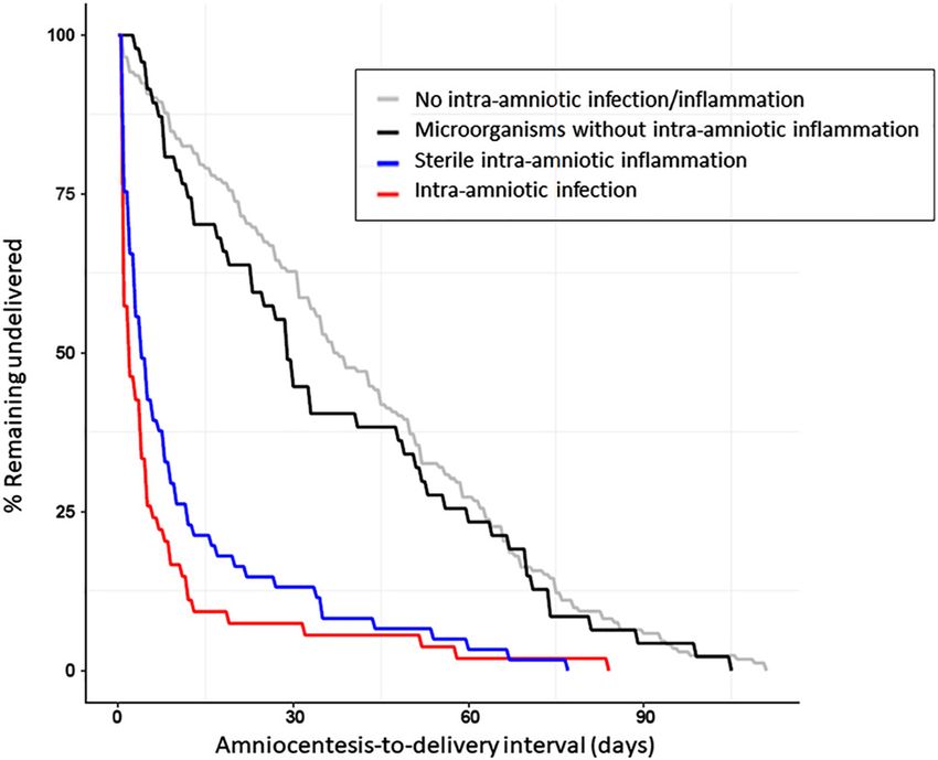
Jung et al.: Bacteria in amniotic fluid without inflammation           3

Composite neonatal morbidity included two or more of the following: a      assays have similar detection limits (100 CFU/mL) [11]. The LOD for the
5-min Apgar score
4         Jung et al.: Bacteria in amniotic fluid without inflammation

as a function of microbial burden. A p-value
Table : Microbiology of amniotic fluid in samples with positive cultures and/or PCR/ESI-MS according to the presence or absence of intra-amniotic inflammation.
Microorganisms without intra-amniotic inflammation Microorganisms with intra-amniotic inflammation (intra-amniotic infection)
Name of microorganisms No. of No. of No. of Microbial Name of microorganisms No. of No. of No. of Microbial burden
culture-positive PCR-positive both-positive burden culture-positive PCR-positive both-positive
samples samples samples samples samples samples
(GE/well)a (GE/well)a
Bacteria and fungi (–) Bacteria and fungi (–)
Propionibacterium acnes (–) Ureaplasma parvum (–,)
Acinetobacter junii (–) Haemophilus influenzae ;
Staphylococcus aureus (–) Mycoplasma hominis (–)
Streptococcus viridans ; Streptococcus pneumonia
Acidovorax temperans ; Ureaplasma urealyticum ;
Actinomyces odontolyticus Staphylococcus aureus ;
Aeromonas caviae Gardnerella vaginalis ;
Moraxella osloensis Sneathia spp. (–)
Pantoea dispersa Streptococcus agalactiae ; ;
Streptococcus spp. Escherichia coli
Lactobacillus acidophilus Candida albicans (–)
Streptococcus infantis Fusobacterium nucleatum (–)
Haemophilus influenzae Rhodococcus jostii
Clostridium septicum Pseudomonas mendocina
Corynebacterium Peptostreptococcus anaerobius
tuberculostearicum
Candida tropicalis Kytococcus schroeteri
Acinetobacter baumannii Acinetobacter junii (–)
Staphylococcus arlettae Streptococcus spp.
Stenotrophomonas maltophilia Streptobacillus moniliformis
Pseudomonas fluorescens Bacteroides fragilis
Streptococcus agalactiae Corynebacterium accolens
Corynebacterium spp. – Propionibacterium acnes (–)
Ureaplasma urealyticum – Acinetobacter lwoffii
Viruses (–) Pseudomonas entomophila
Human parvovirus B (–) Bacteroides ureolyticus
Human enterovirus Mobiluncus spp. –
Human herpesvirus (HHV-) ; Clostridium sporogenes –
Human herpesvirus (HHV-) (–) Staphylococcus capitis –
Streptococcus anginosus –
Viruses (–)
Human enterovirus ; ,
Jung et al.: Bacteria in amniotic fluid without inflammation
Human herpesvirus (HHV-) ; ;
Human herpesvirus (HHV-) (–)
Human parvovirus B (–)
Roseolovirus
5
PCR/ESI-MS, polymerase chain reaction with electrospray ionization mass spectrometry. aData are presented as median (interquartile range) or each value.6 Jung et al.: Bacteria in amniotic fluid without inflammation
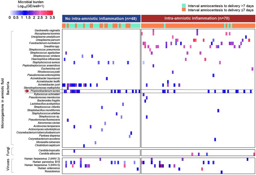
no intra-amniotic inflammation: 35.4% (17/48) had two or
more microorganisms and, overall, a total of 79 microor-
ganisms (bacteria, n=59; fungus, n=1; and viruses, n=19)
were identified (Table 3). Propionibacterium acnes (n=17)
was the most frequent microorganism reported, followed
by Acinetobacter junii (n=14). P. acnes was isolated by
culture in only one patient [6% (1/17)] and, in the rest of the
patients, positive results were attributable to PCR analysis
[94% (16/17)]. All samples positive for A. junii were detected
by PCR/ESI-MS (i.e. this organism was not detected by
culture).
Among the 70 patients with intra-amniotic infection
(positive for microorganisms and intra-amniotic inflam-
mation), 47.1% (33/70) had two or more microorganisms
and, overall, a total of 120 microorganisms (bacteria, n=97;
fungus, n=4; and viruses, n=19) were identified (Table 3).
Amniotic fluid samples from patients with intra-amniotic
infection were dominated by Sneathia spp., Fusobacterium
nucleatum, Ureaplasma parvum, Mycoplasma hominis,
Gardnerella vaginalis, and Candida albicans, which were
not found in patients without intra-amniotic inflammation.
Microbial burden was low in amniotic fluid
samples with microorganisms in the
absence of intra-amniotic inflammation
The microbial burden was defined as the number of mi-
croorganisms in amniotic fluid and estimated by the
number of gene copies per PCR well reaction (GE/well).
Bacterial and fungal burdens were lower in amniotic fluid
samples in the absence of intra-amniotic inflammation
than in those with intra-amniotic inflammation [median
(interquartile range (IQR), 17 (10–38) vs. 80 (20–178) GE/
Figure 1: Microbial burden in amniotic fluid between patients with
intra-amniotic inflammation and those without intra-amniotic well, pJung et al.: Bacteria in amniotic fluid without inflammation 7 Figure 2: Heatmap showing microbial taxa and microbial load in the amniotic fluid, [log10 (GE/well + 1)], according to PCR-based methods. High microbial burden is represented with color gradation which increases toward red, while low microbial burden tends toward blue, as shown in the color-key. The arrow indicates Propionibacterium acnes. M. hominis: 100% (6/6), Sneathia spp.: 100% (12/12), inflammation [spontaneous preterm birth
8
Table : Perinatal outcomes among the four subgroups of patients according to results of amniotic fluid culture, PCR/ESI-MS, and amniotic fluid IL- concentrations in patients with preterm labor
and intact membranes.
No intra-amniotic Microorganisms without Sterile intra-amniotic Intra-amniotic p-Value
inflammation/infection intra-amniotic inflammation inflammation infection
(n=) (n=) (n=) (n=)
Pregnancy outcomes
Gestational age at delivery, weeks . (.–.) . (.–.) . (.–.)c,e . (.–.)b,dJung et al.: Bacteria in amniotic fluid without inflammation 9
Patients with bacteria in amniotic fluid but culture techniques, 12.5% (45/360) by PCR/ESI-MS, and
without intra-amniotic inflammation have a 13.3% (48/360) by the combination of cultivation and PCR/
ESI-MS; (2) the most frequently identified microorganisms
similar interval-to-delivery to those without
in the 48 amniotic fluid specimens were P. acnes, followed
either bacteria or intra-amniotic by A. junii; 3) patients whose amniotic fluid samples had
inflammation microorganisms but no indication of inflammation had a
similar perinatal outcome to those in whom microorgan-
Figure 3 displays the amniocentesis-to-delivery interval isms were not found in the amniotic fluid [amniocentesis-
according to the presence or absence of microorganisms in to-delivery interval (p=0.31), frequency of spontaneous
amniotic fluid and intra-amniotic inflammation. Patients preterm birth before 34 weeks of gestation (p=0.83), acute
with microorganisms without intra-amniotic inflammation placental inflammatory lesions (p=1), and composite
had a significantly longer amniocentesis-to-delivery in- neonatal morbidity (p=0.8)]; and 4) the microbial load of
terval than those with intra-amniotic inflammation samples with bacteria reported by the laboratory in the
regardless of the presence of microorganisms [median absence of intra-amniotic inflammation was low. There-
(IQR), 29.4 (12–57) vs. 1.4 (0.7–5) days, p10 Jung et al.: Bacteria in amniotic fluid without inflammation
Moreover, clinical manifestations of microbial invasion of 189], nebulizers [192], water dispensers [193], hemodialysis
the amniotic cavity vary according to the virulence of the fluids [194], and intravenous fluids [194]. Therefore, in
microorganisms [21, 65, 89, 116, 151–157], microbial burden amniotic fluid, these microorganisms are likely to be
[11, 20, 26, 64, 121, 158–160], and time frame of the acute contaminants.
inflammatory response [66, 146, 161]. A. junii was the second most common species of
Contamination occurs when microorganisms from an microorganism isolated from samples with the absence of
outside source are introduced into a sample [162]. For intra-amniotic inflammation. Acinetobacter spp. are found
example, microorganisms normally present on the skin can in water and soil environments [195] and have previously
gain access to amniotic fluid during amniocentesis or been identified as contaminants [189, 196] in biology grade
during the procedures required to prepare a specimen [20, water [170], PCR reagents [170, 190], DNA extraction kits
163, 164]. Indeed, contamination can originate from many [170, 190], and air samples collected from a patient’s room
sources including the laboratory environments [165, 166], [197]. In the current study, any time Acinetobacter spp.
plastic consumables [167], nucleic acid extraction kits (Acinetobacter baumannii, A. junii, and Acinetobacter
[168–173], laboratory reagents [174–180], and cross- lwoffii) were found, the microbial burden was low, which is
contamination from other samples [181, 182]. It is now also a characteristic of contamination. Although Acineto-
well accepted that laboratory reagents and nucleic acid bacter spp. have been reported as potential pathogens
extraction kits harbor low levels of bacterial DNA [170, 183] causing nosocomial sepsis [198–200], preterm delivery
similar to that found in soil or water samples [170, 172]. [201, 202], acute chorioamnionitis, and a fetal inflamma-
DNA contamination of reagents is unavoidable, given the tory response [201, 202], questions always arise about
ubiquity of microorganisms and the fact that many re- whether these organisms are contaminants and whether
agents are products of microbial processes and engineer- the condition occurs as a sterile intra-amniotic inflamma-
ing [184]. tory process or as organisms that escape detection by
conventional methods. It is important to remember that,
even in recent times, some bacteria have been difficult to
Which bacteria are typical contaminants in identify for decades, including Borrelia burgdorferi, the
amniotic fluid? organism responsible for Lyme disease [203], and Heli-
cobacter pylori [204].
P. acnes was the most common microorganism isolated
from amniotic fluid in the absence of intra-amniotic
inflammation, and its presence should be considered Bacteria likely to be pathogens in amniotic
suggestive of contamination rather than true infection. fluid
Several arguments support this view: (1) P. acnes, a
commensal bacterium in the human skin microbiome [185, Microorganisms implicated as “true pathogens” in intra-
186], is a common contaminant detected in cultures of amniotic infection include Ureaplasma parvum, Mycoplasma
blood and cerebrospinal fluid [187, 188]. Such skin bacteria hominis, Sneathia spp., Candida albicans, Fusobacterium
can gain access to the amniotic fluid during an amnio- nucleatum, Staphylococcus aureus, Gardnerella vaginalis,
centesis or through the procedures required to prepare the Haemophilus influenzae, and Streptococcus agalactiae
specimen [20, 163, 164]; (2) Propionibacterium spp. are re- [20, 89, 116, 121, 151, 152, 158, 163, 205–213]. In the current
ported as common contaminants present in the DNA study, these taxa were abundant in the amniotic fluid
extraction kits and other laboratory reagents [170, 171, samples with intra-amniotic inflammation, as demon-
189–191]; in the current study, nearly all detection of strated in Figure 2, and their presence was associated with
P. acnes (97%; 31/32) came through the PCR method; and adverse pregnancy outcomes, including spontaneous
(3) all amniotic fluid samples positive with P. acnes yielded preterm delivery, severe acute chorioamnionitis or funisi-
a consistently low bacterial burden regardless of the tis, and a short interval-to-delivery.
presence or absence of intra-amniotic inflammation.
Acidovorax temperans, Pantoea dispersa, Staphy-
lococcus arlettae, and Stenotrophomonas maltophilia Abundance of microorganisms to
were detected in amniotic fluid samples with the absence of differentiate infection from contamination
intra-amniotic inflammation. These microorganisms have
rarely been reported to cause human infection and are Quantification of bacterial growth has been used to
found in laboratory environments [170, 189], reagents [170, distinguish between contamination and true infection inJung et al.: Bacteria in amniotic fluid without inflammation 11
the clinical setting. For example, the difference between
asymptomatic bacteriuria and contamination of a urine
specimen is based on the number of colony-forming
units in a urine specimen obtained via clean-catch. The
presence of bacteria is considered to be clinically signif-
icant if there are more than 105 colony-forming units
(CFU)/mL, and a lower number is thought to reflect
contamination when the urine travels from the bladder to
the container through the urethra, which normally con-
tains bacteria [214].
In the past, quantitative cultivation-based microbi-
ology methods have been used to assess the microbial
burden in amniotic fluid. We have observed that patients
with a higher microbial burden are more likely to have a
positive Gram stain in amniotic fluid [4, 126, 215] or to
present with preterm labor leading to preterm delivery [14].
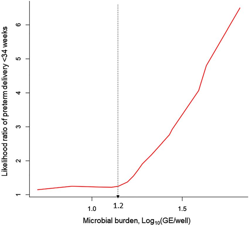
Given that bacteria grow exponentially in amniotic
Figure 4: Positive likelihood ratio for preterm birth before 34 weeks
fluid over time [66, 144–146], amniotic fluid samples with a of gestation as a function of microbial burden [log10 (GE/well)]. The
true infection will have a much higher microbial burden in risk of preterm birth before 34 weeks begins to increase
exponentially when the microbial burden (bacterial and fungal)
later stages of gestation than those that have been
exceeds 1.2 log10 (GE/well). Viral invasion of the amniotic cavity was
contaminated during collection and/or processing. excluded from this analysis. In patients with several
A low microbial burden in amniotic fluid assessed by microorganisms, the calculation was based on the microorganism
PCR methods has been attributed to background DNA with the highest microbial burden.
contamination in the extraction kit [42, 160], whereas a
high microbial burden has been observed in patients who Clinical significance of microorganisms in
have intra-amniotic infection [160] with a strong intra- amniotic fluid without intra-amniotic
amniotic inflammatory response [11, 20, 26, 121, 160]. We inflammation: contamination or early
reported that patients exhibiting a microbial burden higher infection?
than 17 GE/well had a higher frequency of intra-amniotic
inflammation, acute histologic chorioamnionitis, and Patients with microorganisms in amniotic fluid specimens
perinatal morbidity than those with a lower microbial (detected by either culture or molecular microbiologic
burden, assessed by PCR [11]. techniques) without intra-amniotic inflammation have
In the current study, pathogenic bacteria in the am- similar pregnancy outcomes to patients who did not have
niotic fluid exhibited a high microbial burden [i.e. median bacteria or inflammation. Therefore, we propose that
(IQR), GE/well; U. parvum: 573 (114–1,245), M. hominis: 446 finding bacteria in amniotic fluid in samples without intra-
(247–451), Sneathia spp.: 168 (117–180), C. albicans: 136 amniotic inflammation represents contamination. Conse-
quently, clinical decisions, such as inducing labor or
(45–337), and F. nucleatum: 108 (32–120)], whereas bacte-
withholding treatment, given the suspicion of intra-
ria considered as contaminants in amniotic fluid showed a
amniotic infection does not seem to be justifiable.
lower microbial load [i.e. median (IQR), GE/well; P. acne: 17
(13–24) GE/well] regardless of the presence or absence of
intra-amniotic inflammation. Moreover, the risk of preterm
Strengths and limitations
birth begins to increase exponentially when the microbial
burden in amniotic fluid exceeds 17 GE/well (Figure 4).
The major strengths of this study are emphasized as fol-
Therefore, we believe that a microbial burden, assessed lows: (1) both cultivation and molecular microbiologic
with molecular microbiologic techniques, can assist in techniques were used to identify microorganisms in the
determining whether a positive result reflects contamina- samples of amniotic fluid collected by transabdominal
tion rather than a true infection. amniocentesis from the amniotic cavity; therefore, the12 Jung et al.: Bacteria in amniotic fluid without inflammation
diagnosis of microbial invasion was based on the use of Ethical approval: The use of biological specimens as well as
state-of-the-art methodologies; (2) the assessment of intra- clinical and ultrasound data for research purposes was
amniotic inflammation using the concentration of IL-6 in approved by the Human Investigation Committee of Wayne
amniotic fluid; (3) the blinding of pathologists to obstet- State University.
rical diagnoses and perinatal outcomes; and (4) the use of
standardized protocols for placental examination.
The study also comprises the following limitations: (1) References
the duration of storage of the samples may have led to a
degradation of the IL-6 concentration in amniotic fluid, 1. Romero R, Mazor M. Infection and preterm labor. Clin Obstet
Gynecol 1988;31:553–84.
which, in turn, may have yielded a lower concentration of
2. Goldenberg RL, Hauth JC, Andrews WW. Intrauterine infection and
the analytes as compared to the use of freshly collected and preterm delivery. N Engl J Med 2000;342:1500–7.
processed samples of amniotic fluid [216, 217]; (2) the lack 3. McClure EM, Goldenberg RL. Infection and stillbirth. Semin Fetal
of use of molecular markers to identify the presence of Neonatal Med 2009;14:182–9.
microorganisms in the extra-chorionic membranes, chori- 4. Romero R, Sirtori M, Oyarzun E, Avila C, Mazor M, Callahan R,
et al. Infection and labor. V. Prevalence, microbiology, and
onic plate, and umbilical cord resulted in a lack of
clinical significance of intra-amniotic infection in women with
morphologic evidence of the location of microorganisms at preterm labor and intact membranes. Am J Obstet Gynecol 1989;
different sites in the samples; and (3) the lack of use of 161:817–24.
metagenomics, strain culture, and/or strain-directed 5. Skoll MA, Moretti ML, Sibai BM. The incidence of positive
sequencing, which may be utilized to make the distinc- amniotic fluid cultures in patients preterm labor with intact
membranes. Am J Obstet Gynecol 1989;161:813–6.
tion between contamination and colonization.
6. Yoon BH, Romero R, Moon JB, Shim SS, Kim M, Kim G, et al.
Clinical significance of intra-amniotic inflammation in patients
with preterm labor and intact membranes. Am J Obstet Gynecol
2001;185:1130–6.
Conclusions 7. Shim S, Yoon BH, Romero R, Shim J, Kim G, Jung H, et al. The
clinical significance of detecting Ureaplasma Urealyticum by PCR
in the amniotic fluid of patients with preterm labor and intact
The isolation of microorganisms or the detection of mi-
membranes. Am J Obstet Gynecol 2003;187:S129.
crobial nucleic acids from a sample of amniotic fluid, by 8. Gomez R, Romero R, Nien JK, Chaiworapongsa T, Medina L, Kim
cultivation and/or microbiologic molecular techniques in a YM, et al. A short cervix in women with preterm labor and intact
clinical laboratory setting in the absence of intra-amniotic membranes: a risk factor for microbial invasion of the amniotic
inflammation is a benign condition. Such a result most cavity. Am J Obstet Gynecol 2005;192:678–89.
9. Espinoza J, Goncalves LF, Romero R, Nien JK, Stites S, Kim YM, et al.
likely represents contamination of the specimen during the
The prevalence and clinical significance of amniotic fluid ’sludge’
collection procedure or laboratory processing rather than in patients with preterm labor and intact membranes. Ultrasound
early colonization or infection. Obstet Gynecol 2005;25:346–52.
10. Kim BJ, Romero R, Mi Lee S, Park CW, Shin Park J, Jun JK, et al.
Research funding: This research was supported, in part, by Clinical significance of oligohydramnios in patients with preterm
labor and intact membranes. J Perinat Med 2011;39:131–6.
the Perinatology Research Branch, Division of Obstetrics and
11. Romero R, Miranda J, Chaiworapongsa T, Chaemsaithong P,
Maternal-Fetal Medicine, Division of Intramural Research, Gotsch F, Dong Z, et al. A novel molecular microbiologic
Eunice Kennedy Shriver National Institute of Child Health and technique for the rapid diagnosis of microbial invasion of the
Human Development, National Institutes of Health, U.S. amniotic cavity and intra-amniotic infection in preterm labor
Department of Health and Human Services (NICHD/NIH/ with intact membranes. Am J Reprod Immunol 2014;71:
330–58.
DHHS); and, in part, with Federal funds from NICHD/NIH/
12. Romero R, Miranda J, Chaiworapongsa T, Korzeniewski SJ,
DHHS under Contract No. HHSN275201300006C. Chaemsaithong P, Gotsch F, et al. Prevalence and clinical
Author contributions: All authors have accepted significance of sterile intra-amniotic inflammation in patients
responsibility for the entire content of this manuscript with preterm labor and intact membranes. Am J Reprod Immunol
and approved its submission. Dr. Romero has contributed 2014;72:458–74.
to this work as part of his official duties as an employee of 13. Combs CA, Gravett M, Garite TJ, Hickok DE, Lapidus J, Porreco R,
et al. Amniotic fluid infection, inflammation, and colonization in
the United States Federal Government.
preterm labor with intact membranes. Am J Obstet Gynecol 2014;
Competing interests: Authors state no conflict of interest. 210:125.e1–15.
Informed consent: Informed consent was obtained from all 14. Romero R, Quintero R, Oyarzun E, Wu YK, Sabo V, Mazor M, et al.
individuals included in this study. Intraamniotic infection and the onset of labor in pretermJung et al.: Bacteria in amniotic fluid without inflammation 13
premature rupture of the membranes. Am J Obstet Gynecol 1988; 28. Martinez-Varea A, Romero R, Xu Y, Miller D, Ahmed AI,
159:661–6. Chaemsaithong P, et al. Clinical chorioamnionitis at term VII: the
15. Romero R, Ghidini A, Mazor M, Behnke E. Microbial invasion of amniotic fluid cellular immune response. J Perinat Med 2017;45:
the amniotic cavity in premature rupture of membranes. Clin 523–38.
Obstet Gynecol 1991;34:769–78. 29. Romero R, Pacora P, Kusanovic JP, Jung E, Panaitescu B,
16. Averbuch B, Mazor M, Shoham-Vardi I, Chaim W, Vardi H, Maymon E, et al. Clinical chorioamnionitis at term X:
Horowitz S, et al. Intra-uterine infection in women with preterm microbiology, clinical signs, placental pathology, and neonatal
premature rupture of membranes: maternal and neonatal bacteremia-implications for clinical care. J Perinat Med 2021;49:
characteristics. Eur J Obstet Gynecol Reprod Biol 1995;62:25–9. 275–98.
17. Jacobsson B, Mattsby-Baltzer I, Andersch B, Bokstrom H, Holst 30. Gomez R, Romero R, Nien JK, Medina L, Carstens M, Kim YM, et al.
RM, Nikolaitchouk N, et al. Microbial invasion and cytokine Idiopathic vaginal bleeding during pregnancy as the only clinical
response in amniotic fluid in a Swedish population of women manifestation of intrauterine infection. J Matern Fetal Neonat
with preterm prelabor rupture of membranes. Acta Obstet Med 2005;18:31–7.
Gynecol Scand 2003;82:423–31. 31. Madan I, Romero R, Kusanovic JP, Mittal P, Chaiworapongsa T,
18. Shim SS, Romero R, Hong JS, Park CW, Jun JK, Kim BI, et al. Clinical Dong Z, et al. The frequency and clinical significance of intra-
significance of intra-amniotic inflammation in patients with amniotic infection and/or inflammation in women with placenta
preterm premature rupture of membranes. Am J Obstet Gynecol previa and vaginal bleeding: an unexpected observation. J
2004;191:1339–45. Perinat Med 2010;38:275–9.
19. Witt A, Berger A, Gruber CJ, Petricevic L, Apfalter P, Worda C, et al. 32. Hassan S, Romero R, Hendler I, Gomez R, Khalek N, Espinoza J,
Increased intrauterine frequency of Ureaplasma urealyticum in et al. A sonographic short cervix as the only clinical
women with preterm labor and preterm premature rupture of the manifestation of intra-amniotic infection. J Perinat Med 2006;
membranes and subsequent cesarean delivery. Am J Obstet 34:13–9.
Gynecol 2005;193:1663–9. 33. Vaisbuch E, Hassan SS, Mazaki-Tovi S, Nhan-Chang CL,
20. DiGiulio DB, Romero R, Kusanovic JP, Gomez R, Kim CJ, Seok KS, Kusanovic JP, Chaiworapongsa T, et al. Patients with an
et al. Prevalence and diversity of microbes in the amniotic fluid, asymptomatic short cervix (14 Jung et al.: Bacteria in amniotic fluid without inflammation
pregnancies using molecular microbiology. Am J Obstet Gynecol 57. Gotsch F, Romero R, Kusanovic JP, Erez O, Espinoza J, Kim CJ, et al.
2017;217:71.e1–5. The anti-inflammatory limb of the immune response in preterm
42. Lim ES, Rodriguez C, Holtz LR. Amniotic fluid from healthy term labor, intra-amniotic infection/inflammation, and spontaneous
pregnancies does not harbor a detectable microbial community. parturition at term: a role for interleukin-10. J Matern Fetal
Microbiome 2018;6:87. Neonatal Med 2008;21:529–47.
43. Rehbinder EM, Lodrup Carlsen KC, Staff AC, Angell IL, Landro L, 58. Mittal P, Romero R, Kusanovic JP, Edwin SS, Gotsch F, Mazaki‐Tovi
Hilde K, et al. Is amniotic fluid of women with uncomplicated term S, et al. CXCL6 (granulocyte chemotactic protein‐2): a novel
pregnancies free of bacteria? Am J Obstet Gynecol 2018;219: chemokine involved in the innate immune response of the
289.e1–12. amniotic cavity. Am J Reprod Immunol 2008;60:246–57.
44. Liu Y, Li X, Zhu B, Zhao H, Ai Q, Tong Y, et al. Midtrimester amniotic 59. Nhan-Chang CL, Romero R, Kusanovic JP, Gotsch F, Edwin SS, Erez
fluid from healthy pregnancies has no microorganisms using O, et al. A role for CXCL13 (BCA-1) in pregnancy and intra-amniotic
multiple methods of microbiologic inquiry. Am J Obstet Gynecol infection/inflammation. J Matern Fetal Neonatal Med 2008;21:
2020;223:248.e1-248.e21. 763–75.
45. Romero R, Mazor M, Wu YK, Sirtori M, Oyarzun E, Mitchell MD, 60. Marconi C, de Andrade Ramos BR, Peracoli JC, Donders GG,
et al. Infection in the pathogenesis of preterm labor. Semin da Silva MG. Amniotic fluid interleukin-1 beta and interleukin-6,
Perinatol 1988;12:262–79. but not interleukin-8 correlate with microbial invasion of the
46. Goldenberg RL, Andrews WW, Hauth JC. Choriodecidual infection amniotic cavity in preterm labor. Am J Reprod Immunol 2011;65:
and preterm birth. Nutr Rev 2002;60:S19–25. 549–56.
47. Suff N, Karda R, Diaz JA, Ng J, Baruteau J, Perocheau D, et al. 61. Gervasi MT, Romero R, Bracalente G, Erez O, Dong Z, Hassan SS,
Ascending vaginal infection using bioluminescent bacteria et al. Midtrimester amniotic fluid concentrations of interleukin-6
evokes intrauterine inflammation, preterm birth, and neonatal and interferon-gamma-inducible protein-10: evidence for
brain injury in pregnant mice. Am J Pathol 2018;188: heterogeneity of intra-amniotic inflammation and associations
2164–76. with spontaneous early (32 weeks) preterm
48. Romero R, Gomez-Lopez N, Winters AD, Jung E, Shaman M, Bieda delivery. J Perinat Med 2012;40:329–43.
J, et al. Evidence that intra-amniotic infections are often the result 62. Gomez-Lopez N, Romero R, Leng Y, Xu Y, Slutsky R, Levenson D,
of an ascending invasion: a molecular microbiological study. J et al. The origin of amniotic fluid monocytes/macrophages in
Perinat Med 2019;47:915–31. women with intra-amniotic inflammation or infection. J Perinat
49. Romero R, Avila C, Santhanam U, Sehgal PB. Amniotic fluid Med 2019;47:822–40.
interleukin 6 in preterm labor. Association with infection. J Clin 63. Bhatti G, Romero R, Rice GE, Fitzgerald W, Pacora P, Gomez-Lopez
Invest 1990;85:1392–400. N, et al. Compartmentalized profiling of amniotic fluid cytokines
50. Romero R, Gomez R, Galasso M, Munoz H, Acosta L, Yoon BH, in women with preterm labor. PLoS One 2020;15:e0227881.
et al. Macrophage inflammatory protein‐1α in term and preterm 64. Thiersch JB. Effect of lipopolysaccharides of Gram negative bacilli
parturtition: effect of microbial invasion of the amniotic cavity. on the rat litter in utero. Proc Soc Exp Biol Med 1962;109:429–37.
Am J Reprod Immunol 1994;32:108–13. 65. Dombroski RA, Woodard DS, Harper MJ, Gibbs RS. A rabbit model
51. Fidel PL, Jr, Romero R, Wolf N, Cutright J, Ramirez M, Araneda H, for bacteria-induced preterm pregnancy loss. Am J Obstet
et al. Systemic and local cytokine profiles in endotoxin-induced Gynecol 1990;163:1938–43.
preterm parturition in mice. Am J Obstet Gynecol 1994;170: 66. Gravett MG, Witkin SS, Haluska GJ, Edwards JL, Cook MJ, Novy MJ.
1467–75. An experimental model for intraamniotic infection and preterm
52. Dudley DJ, Hunter C, Varner MW, Mitchell MD. Elevation of labor in rhesus monkeys. Am J Obstet Gynecol 1994;171:1660–7.
amniotic fluid interleukin-4 concentrations in women with 67. Gomez-Lopez N, Romero R, Arenas-Hernandez M, Panaitescu B,
preterm labor and chorioamnionitis. Am J Perinatol 1996;13: Garcia-Flores V, Mial TN, et al. Intra-amniotic administration of
443–7. lipopolysaccharide induces spontaneous preterm labor and birth
53. Maymon E, Ghezzi F, Edwin SS, Mazor M, Yoon BH, Gomez R, et al. in the absence of a body temperature change. J Matern Fetal
The tumor necrosis factor alpha and its soluble receptor profile in Neonat Med 2018;31:439–46.
term and preterm parturition. Am J Obstet Gynecol 1999;181: 68. Kallapur SG, Willet KE, Jobe AH, Ikegami M, Bachurski CJ. Intra-
1142–8. amniotic endotoxin: chorioamnionitis precedes lung maturation
54. Chaiworapongsa T, Romero R, Espinoza J, Kim YM, Edwin S, in preterm lambs. Am J Physiol Lung Cell Mol Physiol 2001;280:
Bujold E, et al. Macrophage migration inhibitory factor in patients L527–36.
with preterm parturition and microbial invasion of the amniotic 69. Kramer BW, Moss TJ, Willet KE, Newnham JP, Sly PD, Kallapur SG,
cavity. J Matern Fetal Neonatal Med 2005;18:405–16. et al. Dose and time response after intraamniotic endotoxin in
55. Esplin MS, Romero R, Chaiworapongsa T, Kim YM, Edwin S, preterm lambs. Am J Respir Crit Care Med 2001;164:982–8.
Gomez R, et al. Monocyte chemotactic protein-1 is increased in 70. Moss TJ, Nitsos I, Newnham JP, Ikegami M, Jobe AH.
the amniotic fluid of women who deliver preterm in the presence Chorioamnionitis induced by subchorionic endotoxin infusion in
or absence of intra-amniotic infection. J Matern Fetal Neonatal sheep. Am J Obstet Gynecol 2003;189:1771–6.
Med 2005;17:365–73. 71. Kramer BW, Ikegami M, Moss TJ, Nitsos I, Newnham JP, Jobe AH.
56. Jacobsson B, Holst RM, Andersson B, Hagberg H. Monocyte Endotoxin-induced chorioamnionitis modulates innate immunity
chemotactic protein-2 and -3 in amniotic fluid: relationship to of monocytes in preterm sheep. Am J Respir Crit Care Med 2005;
microbial invasion of the amniotic cavity, intra-amniotic 171:73–7.
inflammation and preterm delivery. Acta Obstet Gynecol Scand 72. Berry CA, Nitsos I, Hillman NH, Pillow JJ, Polglase GR, Kramer BW,
2005;84:566–71. et al. Interleukin-1 in lipopolysaccharide inducedJung et al.: Bacteria in amniotic fluid without inflammation 15
chorioamnionitis in the fetal sheep. Reprod Sci 2011;18: rhesus monkeys. Am J Obstet Gynecol 1996;174:1725–31.
1092–102. discussion 31–3.
73. Maxwell JR, Denson JL, Joste NE, Robinson S, Jantzie LL. 88. Yoon BH, Jun JK, Romero R, Park KH, Gomez R, Choi JH, et al.
Combined in utero hypoxia-ischemia and lipopolysaccharide Amniotic fluid inflammatory cytokines (interleukin-6,
administration in rats induces chorioamnionitis and a fetal interleukin-1beta, and tumor necrosis factor-alpha), neonatal
inflammatory response syndrome. Placenta 2015;36:1378–84. brain white matter lesions, and cerebral palsy. Am J Obstet
74. Yoon BH, Romero R, Park JS, Kim M, Oh SY, Kim CJ, et al. The Gynecol 1997;177:19–26.
relationship among inflammatory lesions of the umbilical cord 89. Yoon BH, Chang JW, Romero R. Isolation of Ureaplasma
(funisitis), umbilical cord plasma interleukin 6 concentration, urealyticum from the amniotic cavity and adverse outcome in
amniotic fluid infection, and neonatal sepsis. Am J Obstet preterm labor. Obstet Gynecol 1998;92:77–82.
Gynecol 2000;183:1124–9. 90. Yoon BH, Romero R, Park JS, Kim CJ, Kim SH, Choi JH, et al. Fetal
75. Watanabe T, Matsuda T, Hanita T, Okuyama K, Cho K, Kobayashi exposure to an intra-amniotic inflammation and the
K, et al. Induction of necrotizing funisitis by fetal administration development of cerebral palsy at the age of three years. Am J
of intravenous granulocyte-colony stimulating factor and intra- Obstet Gynecol 2000;182:675–81.
amniotic endotoxin in premature fetal sheep. Pediatr Res 2007; 91. Nelson KB, Willoughby RE. Infection, inflammation and the risk
62:670–3. of cerebral palsy. Curr Opin Neurol 2000;13:133–9.
76. Rounioja S, Rasanen J, Glumoff V, Ojaniemi M, Makikallio K, 92. Hitti J, Tarczy-Hornoch P, Murphy J, Hillier SL, Aura J, Eschenbach
Hallman M. Intra-amniotic lipopolysaccharide leads to fetal DA. Amniotic fluid infection, cytokines, and adverse outcome
cardiac dysfunction. A mouse model for fetal inflammatory among infants at 34 weeks’ gestation or less. Obstet Gynecol
response. Cardiovasc Res 2003;60:156–64. 2001;98:1080–8.
77. Cheah FC, Pillow JJ, Kramer BW, Polglase GR, Nitsos I, Newnham 93. Romero R, Gomez R, Chaiworapongsa T, Conoscenti G, Kim JC,
JP, et al. Airway inflammatory cell responses to intra-amniotic Kim YM. The role of infection in preterm labour and delivery.
lipopolysaccharide in a sheep model of chorioamnionitis. Am J Paediatr Perinat Epidemiol 2001;15 (2 Suppl):41–56.
Physiol Lung Cell Mol Physiol 2009;296:L384–93. 94. Moon JB, Kim JC, Yoon BH, Romero R, Kim G, Oh SY, et al.
78. Gavilanes AW, Strackx E, Kramer BW, Gantert M, Van den Hove D, Amniotic fluid matrix metalloproteinase-8 and the development
Steinbusch H, et al. Chorioamnionitis induced by intraamniotic of cerebral palsy. J Perinat Med 2002;30:301–6.
lipopolysaccharide resulted in an interval-dependent increase in 95. Yoon BH, Park CW, Chaiworapongsa T. Intrauterine infection and
central nervous system injury in the fetal sheep. Am J Obstet the development of cerebral palsy. BJOG 2003;110:124 (20
Gynecol 2009;200:437.e1–8. Suppl)–7.
79. Kramer BW, Kallapur SG, Moss TJ, Nitsos I, Polglase GP, 96. Neufeld MD, Frigon C, Graham AS, Mueller BA. Maternal
Newnham JP, et al. Modulation of fetal inflammatory response on infection and risk of cerebral palsy in term and preterm infants. J
exposure to lipopolysaccharide by chorioamnion, lung, or gut in Perinatol 2005;25:108–13.
sheep. Am J Obstet Gynecol 2010;202:77.e1–9. 97. Shatrov JG, Birch SC, Lam LT, Quinlivan JA, McIntyre S, Mendz
80. Kunzmann S, Glogger K, Been JV, Kallapur SG, Nitsos I, Moss TJ, GL. Chorioamnionitis and cerebral palsy: a meta-analysis.
et al. Thymic changes after chorioamnionitis induced by Obstet Gynecol 2010;116:387–92.
intraamniotic lipopolysaccharide in fetal sheep. Am J Obstet 98. Kasper DC, Mechtler TP, Bohm J, Petricevic L, Gleiss A, Spergser
Gynecol 2010;202:476.e1–9. J, et al. In utero exposure to Ureaplasma spp. is associated with
81. Bieghs V, Vlassaks E, Custers A, van Gorp PJ, Gijbels MJ, Bast A, increased rate of bronchopulmonary dysplasia and
et al. Chorioamnionitis induced hepatic inflammation and intraventricular hemorrhage in preterm infants. J Perinat Med
disturbed lipid metabolism in fetal sheep. Pediatr Res 2010;68: 2011;39:331–6.
466–72. 99. Bhandari V, Buhimschi CS, Han CS, Lee SY, Pettker CM,
82. Strackx E, Sparnaaij MA, Vlassaks E, Jellema R, Kuypers E, Vles JS, Campbell KH, et al. Cord blood erythropoietin and interleukin-6
et al. Lipopolysaccharide-induced chorioamnionitis causes acute for prediction of intraventricular hemorrhage in the preterm
inflammatory changes in the ovine central nervous system. CNS neonate. J Matern Fetal Neonatal Med 2011;24:673–9.
Neurol Disord Drug Targets 2015;14:77–84. 100. Korzeniewski SJ, Romero R, Cortez J, Pappas A, Schwartz AG,
83. Schmidt AF, Kannan PS, Chougnet CA, Danzer SC, Miller LA, Jobe Kim CJ, et al. A “multi-hit” model of neonatal white matter injury:
AH, et al. Intra-amniotic LPS causes acute neuroinflammation in cumulative contributions of chronic placental inflammation,
preterm rhesus macaques. J Neuroinflamm 2016;13:238. acute fetal inflammation and postnatal inflammatory events. J
84. Nguyen DN, Thymann T, Goericke-Pesch SK, Ren S, Wei W, Perinat Med 2014;42:731–43.
Skovgaard K, et al. Prenatal intra-amniotic endotoxin induces 101. Kuban KC, O’Shea TM, Allred EN, Paneth N, Hirtz D, Fichorova RN,
fetal gut and lung immune responses and postnatal systemic et al. Systemic inflammation and cerebral palsy risk in extremely
inflammation in preterm pigs. Am J Pathol 2018;188:2629–43. preterm infants. J Child Neurol 2014;29:1692–8.
85. Jung E, Romero R, Yeo L, Diaz-Primera R, Marin-Concha J, Para R, 102. Catov JM, Scifres CM, Caritis SN, Bertolet M, Larkin J, Parks WT.
et al. The fetal inflammatory response syndrome: the origins of a Neonatal outcomes following preterm birth classified according
concept, pathophysiology, diagnosis, and obstetrical to placental features. Am J Obstet Gynecol 2017;216:411. e1–4.
implications. Semin Fetal Neonatal Med 2020;25:101146. 103. Oh KJ, Park JY, Lee J, Hong JS, Romero R, Yoon BH. The combined
86. Leviton A. Preterm birth and cerebral palsy: is tumor necrosis exposure to intra-amniotic inflammation and neonatal
factor the missing link? Dev Med Child Neurol 1993;35:553–8. respiratory distress syndrome increases the risk of
87. Gravett MG, Haluska GJ, Cook MJ, Novy MJ. Fetal and maternal intraventricular hemorrhage in preterm neonates. J Perinat Med
endocrine responses to experimental intrauterine infection in 2018;46:9–20.16 Jung et al.: Bacteria in amniotic fluid without inflammation
104. Latino MA, Botta G, Badino C, Maria D, Petrozziello A, Sensini A, response in amniotic fluid in a Swedish population of women in
et al. Association between genital mycoplasmas, acute preterm labor. Acta Obstet Gynecol Scand 2003;82:120–8.
chorioamnionitis and fetal pneumonia in spontaneous 118. Jacobsson B, Mattsby-Baltzer I, Hagberg H. Interleukin-6 and
abortions. J Perinat Med 2018;46:503–8. interleukin-8 in cervical and amniotic fluid: relationship to
105. Faro J, Romero R, Schwenkel G, Garcia-Flores V, Arenas- microbial invasion of the chorioamniotic membranes. BJOG
Hernandez M, Leng Y, et al. Intra-amniotic inflammation induces 2005;112:719–24.
preterm birth by activating the NLRP3 inflammasomedagger. 119. WHO. Neonatal and perinatal mortality: country, regional and
Biol Reprod 2019;100:1290–305. global estimates. Geneva: World Health Organization; 2006.
106. Peiris HN, Romero R, Vaswani K, Reed S, Gomez-Lopez N, 120. Kim KW, Romero R, Park HS, Park CW, Shim SS, Jun JK, et al. A
Tarca AL, et al. Preterm labor is characterized by a high rapid matrix metalloproteinase-8 bedside test for the detection
abundance of amniotic fluid prostaglandins in patients with of intraamniotic inflammation in women with preterm
intra-amniotic infection or sterile intra-amniotic inflammation. J premature rupture of membranes. Am J Obstet Gynecol 2007;
Matern Fetal Neonatal Med 2019:1–16. https://doi.org/10. 197:292.e1–5.
1080/14767058.2019.1702953. 121. DiGiulio DB, Romero R, Amogan HP, Kusanovic JP, Bik EM,
107. Lee J, Romero R, Kim SM, Chaemsaithong P, Yoon BH. A new Gotsch F, et al. Microbial prevalence, diversity and abundance
antibiotic regimen treats and prevents intra-amniotic in amniotic fluid during preterm labor: a molecular and culture-
inflammation/infection in patients with preterm PROM. J Matern based investigation. PLoS One 2008;3:e3056.
Fetal Neonatal Med 2016;29:2727–37. 122. DiGiulio DB, Gervasi M, Romero R, Mazaki-Tovi S, Vaisbuch E,
108. Lee J, Romero R, Kim SM, Chaemsaithong P, Park CW, Park JS, Kusanovic JP, et al. Microbial invasion of the amniotic cavity in
et al. A new anti-microbial combination prolongs the latency preeclampsia as assessed by cultivation and sequence-based
period, reduces acute histologic chorioamnionitis as well as methods. J Perinat Med 2010;38:503–13.
funisitis, and improves neonatal outcomes in preterm PROM. J 123. DiGiulio DB, Gervasi MT, Romero R, Vaisbuch E, Mazaki-Tovi S,
Matern Fetal Neonatal Med 2016;29:707–20. Kusanovic JP, et al. Microbial invasion of the amniotic cavity in
109. Yoon BH, Romero R, Park JY, Oh KJ, Lee J, Conde-Agudelo A, et al. pregnancies with small-for-gestational-age fetuses. J Perinat
Antibiotic administration can eradicate intra-amniotic infection Med 2010;38:495–502.
or intra-amniotic inflammation in a subset of patients with 124. Romero R, Quintero R, Nores J, Avila C, Mazor M, Hanaoka S,
preterm labor and intact membranes. Am J Obstet Gynecol 2019; et al. Amniotic fluid white blood cell count: a rapid and simple
221:142.e1–22. test to diagnose microbial invasion of the amniotic cavity and
110. Oh KJ, Romero R, Park JY, Lee J, Conde-Agudelo A, Hong JS, et al. predict preterm delivery. Am J Obstet Gynecol 1991;165:821–30.
Evidence that antibiotic administration is effective in the 125. Romero R, Jimenez C, Lohda AK, Nores J, Hanaoka S, Avila C,
treatment of a subset of patients with intra-amniotic infection/ et al. Amniotic fluid glucose concentration: a rapid and simple
inflammation presenting with cervical insufficiency. Am J Obstet method for the detection of intraamniotic infection in preterm
Gynecol 2019;221:140.e1–18. labor. Am J Obstet Gynecol 1990;163:968–74.
111. Kacerovsky M, Romero R, Stepan M, Stranik J, Maly J, Pliskova L, 126. Romero R, Emamian M, Quintero R, Wan M, Hobbins JC, Mazor
et al. Antibiotic administration reduces the rate of intraamniotic M, et al. The value and limitations of the Gram stain examination
inflammation in preterm prelabor rupture of the membranes. Am in the diagnosis of intraamniotic infection. Am J Obstet Gynecol
J Obstet Gynecol 2020;223:114.e1–20. 1988;159:114–9.
112. Coultrip LL, Lien JM, Gomez R, Kapernick P, Khoury A, Grossman 127. Romero R, Miranda J, Chaemsaithong P, Chaiworapongsa T,
JH. The value of amniotic fluid interleukin-6 determination in Kusanovic JP, Dong Z, et al. Sterile and microbial-associated
patients with preterm labor and intact membranes in the intra-amniotic inflammation in preterm prelabor rupture of
detection of microbial invasion of the amniotic cavity. Am J membranes. J Matern Fetal Neonatal Med 2015;28:1394–409.
Obstet Gynecol 1994;171:901–11. 128. Eckert DJ, Sampath R, Li H, Massire C, Matthews HE, Toleno D,
113. Hitti J, Riley DE, Krohn MA, Hillier SL, Agnew KJ, Krieger JN, et al. et al. New technology for rapid molecular diagnosis of
Broad-spectrum bacterial rDNA polymerase chain reaction bloodstream infections. Expert Rev Mol Diagn 2010;10:
assay for detecting amniotic fluid infection among women in 399–415.
premature labor. Clin Infect Dis 1997;24:1228–32. 129. Metzgar D, Frinder M, Lovari R, Toleno D, Massire C, Blyn LB,
114. Greci LS, Gilson GJ, Nevils B, Izquierdo LA, Qualls CR, Curet LB. Is et al. Broad-spectrum biosensor capable of detecting and
amniotic fluid analysis the key to preterm labor? A model using identifying diverse bacterial and Candida species in blood. J Clin
interleukin-6 for predicting rapid delivery. Am J Obstet Gynecol Microbiol 2013;51:2670–8.
1998;179:172–8. 130. A broad range of tests to meet your needs. Available from: www.
115. Angus SR, Segel SY, Hsu CD, Locksmith GJ, Clark P, Sammel MD, athogen.com/consulting-services/microbial-tests.html.
et al. Amniotic fluid matrix metalloproteinase-8 indicates 131. Legoff J, Feghoul L, Mercier-Delarue S, Dalle JH, Scieux C, Cherot
intra-amniotic infection. Am J Obstet Gynecol 2001;185: J, et al. Broad-range PCR-electrospray ionization mass
1232–8. spectrometry for detection and typing of adenovirus and other
116. Yoon BH, Romero R, Lim JH, Shim SS, Hong JS, Shim JY, et al. The opportunistic viruses in stem cell transplant patients. J Clin
clinical significance of detecting Ureaplasma urealyticum by the Microbiol 2013;51:4186–92.
polymerase chain reaction in the amniotic fluid of patients with 132. Romero R, Kadar N, Miranda J, Korzeniewski SJ, Schwartz AG,
preterm labor. Am J Obstet Gynecol 2003;189:919–24. Chaemsaithong P, et al. The diagnostic performance of the Mass
117. Jacobsson B, Mattsby-Baltzer I, Andersch B, Bokstrom H, Holst Restricted (MR) score in the identification of microbial invasion
RM, Wennerholm UB, et al. Microbial invasion and cytokine of the amniotic cavity or intra-amniotic inflammation is notJung et al.: Bacteria in amniotic fluid without inflammation 17
superior to amniotic fluid interleukin-6. J Matern Fetal Neonatal 149. Baschat AA, Towbin J, Bowles NE, Harman CR, Weiner CP.
Med 2014;27:757–69. Prevalence of viral DNA in amniotic fluid of low-risk pregnancies
133. Redline RW, Faye-Petersen O, Heller D, Qureshi F, Savell V, in the second trimester. J Matern Fetal Neonatal Med 2003;13:
Vogler C, et al. Amniotic infection syndrome: nosology and 381–4.
reproducibility of placental reaction patterns. Pediatr Dev 150. Miller JL, Harman C, Weiner C, Baschat AA. Perinatal outcomes
Pathol 2003;6:435–48. after second trimester detection of amniotic fluid viral genome
134. Redline RW, Heller D, Keating S, Kingdom J. Placental diagnostic in asymptomatic patients. J Perinat Med 2009;37:140–3.
criteria and clinical correlation–a workshop report. Placenta 151. Yoon BH, Romero R, Park JS, Chang JW, Kim YA, Kim JC, et al.
2005;26 (A Suppl):S114–7. Microbial invasion of the amniotic cavity with Ureaplasma
135. Redline RW. Placental pathology: a systematic approach with urealyticum is associated with a robust host response in fetal,
clinical correlations. Placenta 2008;29 (A Suppl):S86–91. amniotic, and maternal compartments. Am J Obstet Gynecol
136. Kim CJ, Romero R, Chaemsaithong P, Chaiyasit N, Yoon BH, Kim 1998;179:1254–60.
YM. Acute chorioamnionitis and funisitis: definition, pathologic 152. Yoon BH, Romero R, Kim M, Kim EC, Kim T, Park JS, et al. Clinical
features, and clinical significance. Am J Obstet Gynecol 2015; implications of detection of Ureaplasma urealyticum in the
213 (4 Suppl):S29–52. amniotic cavity with the polymerase chain reaction. Am J Obstet
137. Khong TY, Mooney EE, Ariel I, Balmus NC, Boyd TK, Brundler MA, Gynecol 2000;183:1130–7.
et al. Sampling and definitions of placental lesions: amsterdam 153. Payne MS, Kemp MW, Kallapur SG, Kannan PS, Saito M, Miura Y,
placental workshop Group Consensus Statement. Arch Pathol et al. Intrauterine Candida albicans infection elicits severe
Lab Med 2016;140:698–713. inflammation in fetal sheep. Pediatr Res 2014;75:716–22.
138. Romero R, Kim YM, Pacora P, Kim CJ, Benshalom-Tirosh N, 154. Yoneda N, Yoneda S, Niimi H, Ueno T, Hayashi S, Ito M, et al.
Jaiman S, et al. The frequency and type of placental histologic Polymicrobial amniotic fluid infection with mycoplasma/
lesions in term pregnancies with normal outcome. J Perinat Med ureaplasma and other bacteria induces severe intra-amniotic
2018;46:613–30. inflammation associated with poor perinatal prognosis in
139. Kim CJ, Romero R, Kusanovic JP, Yoo W, Dong Z, Topping V, et al. preterm labor. Am J Reprod Immunol 2016;75:112–25.
The frequency, clinical significance, and pathological features 155. Sweeney EL, Kallapur SG, Meawad S, Gisslen T, Stephenson SA,
of chronic chorioamnionitis: a lesion associated with Jobe AH, et al. Ureaplasma species multiple banded Antigen
spontaneous preterm birth. Mod Pathol 2010;23:1000–11. (MBA) variation is associated with the Severity of inflammation
140. Ford-Martin P. Gale encyclopedia of medicine. Detroit: The Gale in vivo and in vitro in human placentae. Front Cell Infect
Group Inc.; 2004. Microbiol 2017;7:123.
141. Dani A. Colonization and infection. Cent European J Urol 2014; 156. Revello R, Alcaide MJ, Abehsera D, Martin-Camean M, Sousa
67:86–7. EFGM, Alonso-Luque B, et al. Prediction of chorioamnionitis in
142. Rehbinder EM, Lodrup Carlsen KC, Staff AC, Angell IL, Landro L, cases of intraamniotic infection by Ureaplasma urealyticum in
Hilde K, et al. Is amniotic fluid of women with uncomplicated women with very preterm premature rupture of membranes or
term pregnancies free of bacteria? Am J Obstet Gynecol 2018; preterm labour. J Matern Fetal Neonatal Med 2018;31:
219:289.e1-289.e12. 1839–44.
143. Burnham P, Gomez-Lopez N, Heyang M, Cheng AP, Lenz JS, 157. Oh KJ, Romero R, Park JY, Hong JS, Yoon BH. The earlier the
Dadhania DM, et al. Separating the signal from the noise in gestational age, the greater the intensity of the intra-amniotic
metagenomic cell-free DNA sequencing. Microbiome 2020;8:18. inflammatory response in women with preterm premature
144. Gravett MG, Adams KM, Sadowsky DW, Grosvenor AR, Witkin SS, rupture of membranes and amniotic fluid infection by
Axthelm MK, et al. Immunomodulators plus antibiotics delay Ureaplasma species. J Perinat Med 2019;47:516–27.
preterm delivery after experimental intraamniotic infection in a 158. Jacobsson B, Aaltonen R, Rantakokko-Jalava K, Morken NH,
nonhuman primate model. Am J Obstet Gynecol 2007;197: Alanen A. Quantification of Ureaplasma urealyticum DNA in the
518.e1–8. amniotic fluid from patients in PTL and pPROM and its relation to
145. Novy MJ, Duffy L, Axthelm MK, Sadowsky DW, Witkin SS, Gravett inflammatory cytokine levels. Acta Obstet Gynecol Scand 2009;
MG, et al. Ureaplasma parvum or Mycoplasma hominis as sole 88:63–70.
pathogens cause chorioamnionitis, preterm delivery, and fetal 159. Allen-Daniels MJ, Serrano MG, Pflugner LP, Fettweis JM,
pneumonia in rhesus macaques. Reprod Sci 2009;16:56–70. Prestosa MA, Koparde VN, et al. Identification of a gene in
146. Grigsby PL, Novy MJ, Sadowsky DW, Morgan TK, Long M, Acosta Mycoplasma hominis associated with preterm birth and
E, et al. Maternal azithromycin therapy for Ureaplasma microbial burden in intraamniotic infection. Am J Obstet Gynecol
intraamniotic infection delays preterm delivery and reduces 2015;212:779.e1–13.
fetal lung injury in a primate model. Am J Obstet Gynecol 2012; 160. Theis KR, Romero R, Motomura K, Galaz J, Winters AD, Pacora P,
207:475.e1–14. et al. Microbial burden and inflammasome activation in
147. Mazor M, Chaim W, Shinwell ES, Glezerman M. Asymptomatic amniotic fluid of patients with preterm prelabor rupture of
amniotic fluid invasion with Candida albicans in preterm membranes. J Perinat Med 2020;48:115–31.
premature rupture of membranes. Implications for obstetric and 161. McCall CE, Yoza B, Liu T, El Gazzar M. Gene-specific epigenetic
neonatal management. Acta Obstet Gynecol Scand 1993;72: regulation in serious infections with systemic inflammation. J
52–4. Innate Immun 2010;2:395–405.
148. Mandar R, Li K, Ehrenberg A, Smidt I, Raukas E, Kask V, et al. 162. Dargere S, Cormier H, Verdon R. Contaminants in blood cultures:
Amniotic fluid microflora in asymptomatic women at mid- importance, implications, interpretation and prevention. Clin
gestation. Scand J Infect Dis 2001;33:60–2. Microbiol Infect 2018;24:964–9.You can also read