Ferritin H deficiency deteriorates cellular iron handling and worsens Salmonella typhimurium infection by triggering hyperinflammation
←
→
Page content transcription
If your browser does not render page correctly, please read the page content below
Ferritin H deficiency deteriorates cellular iron handling and
worsens Salmonella typhimurium infection by triggering
hyperinflammation
David Haschka, … , Igor Theurl, Guenter Weiss
JCI Insight. 2021;6(13):e141760. https://doi.org/10.1172/jci.insight.141760.

 Research Article   Immunology      Infectious disease

Iron is an essential nutrient for mammals as well as for pathogens. Inflammation-driven changes in systemic and cellular
iron homeostasis are central for host-mediated antimicrobial strategies. Here, we studied the role of the iron storage
protein ferritin H (FTH) for the control of infections with the intracellular pathogen Salmonella enterica serovar
Typhimurium by macrophages. Mice lacking FTH in the myeloid lineage (LysM-Cre +/+Fthfl/fl mice) displayed impaired iron
storage capacities in the tissue leukocyte compartment, increased levels of labile iron in macrophages, and an
accelerated macrophage-mediated iron turnover. While under steady-state conditions, LysM-Cre +/+Fth+/+ and LysM-
Cre+/+Fthfl/fl animals showed comparable susceptibility to Salmonella infection, i.v. iron supplementation drastically
shortened survival of LysM-Cre +/+Fthfl/fl mice. Mechanistically, these animals displayed increased bacterial burden, which
contributed to uncontrolled triggering of NF-κB and inflammasome signaling and development of cytokine storm and
death. Importantly, pharmacologic inhibition of the inflammasome and IL-1β pathways reduced cytokine levels and
mortality and partly restored infection control in iron-treated ferritin-deficient mice. These findings uncover incompletely
characterized roles of ferritin and cellular iron turnover in myeloid cells in controlling bacterial spread and for modulating
NF-κB and inflammasome-mediated cytokine activation, which may be of vital importance in iron-overloaded individuals
suffering from severe infections and sepsis.

Find the latest version:
https://jci.me/141760/pdf
RESEARCH ARTICLE

                                         Ferritin H deficiency deteriorates cellular
                                         iron handling and worsens Salmonella
                                         typhimurium infection by triggering
                                         hyperinflammation
                                         David Haschka,1 Piotr Tymoszuk,1 Verena Petzer,1 Richard Hilbe,1 Simon Heeke,1 Stefanie Dichtl,1
                                         Sergej Skvortsov,2 Egon Demetz,1 Sylvia Berger,1 Markus Seifert,1 Anna-Maria Mitterstiller,1
                                         Patrizia Moser,3 Dirk Bumann,4 Manfred Nairz,1 Igor Theurl,1 and Guenter Weiss1
                                         Department of Internal Medicine II, Medical University of Innsbruck, Innsbruck, Austria. 2Department of Therapeutic
                                         1

                                         Radiology and Oncology, Laboratory for Experimental and Translational Research on Radiation Oncology, Tyrolean Cancer
                                         Research Institute, Medical University of Innsbruck, Innsbruck, Austria. 3Institute of Pathology, INNPATH, Innsbruck,
                                         Austria. 4Biozentrum, University of Basel, Klingelbergstrasse, Basel, Switzerland.

                                                 Iron is an essential nutrient for mammals as well as for pathogens. Inflammation-driven changes in
                                                 systemic and cellular iron homeostasis are central for host-mediated antimicrobial strategies. Here,
                                                 we studied the role of the iron storage protein ferritin H (FTH) for the control of infections with the
                                                 intracellular pathogen Salmonella enterica serovar Typhimurium by macrophages. Mice lacking FTH
                                                 in the myeloid lineage (LysM-Cre+/+Fthfl/fl mice) displayed impaired iron storage capacities in the
                                                 tissue leukocyte compartment, increased levels of labile iron in macrophages, and an accelerated
                                                 macrophage-mediated iron turnover. While under steady-state conditions, LysM-Cre+/+Fth+/+ and
                                                 LysM-Cre+/+Fthfl/fl animals showed comparable susceptibility to Salmonella infection, i.v. iron
                                                 supplementation drastically shortened survival of LysM-Cre+/+Fthfl/fl mice. Mechanistically, these
                                                 animals displayed increased bacterial burden, which contributed to uncontrolled triggering of
                                                 NF-κB and inflammasome signaling and development of cytokine storm and death. Importantly,
                                                 pharmacologic inhibition of the inflammasome and IL-1β pathways reduced cytokine levels and
                                                 mortality and partly restored infection control in iron-treated ferritin-deficient mice. These findings
                                                 uncover incompletely characterized roles of ferritin and cellular iron turnover in myeloid cells in
                                                 controlling bacterial spread and for modulating NF-κB and inflammasome-mediated cytokine
                                                 activation, which may be of vital importance in iron-overloaded individuals suffering from severe
                                                 infections and sepsis.

Authorship note: DH, PT, IT, and GW
contributed equally to this work.        Introduction
Conflict of interest: The authors have   Iron is essential for numerous metabolic processes in living cells, including heme production, oxidative phos-
declared that no conflict of interest    phorylation, and DNA synthesis, and it also acts as a cofactor of multiple metalloenzymes (1). Nevertheless,
exists.                                  a surplus of iron can be potentially harmful, for instance, as a catalyst of nonenzymatic generation of toxic
                                         oxygen radicals via Fenton/Haber–Weiss chemistry (2). In addition, iron is a critical nutrient for growth and
Copyright: © 2021, Haschka et
al. This is an open access article       virulence of most bacterial pathogens (3). Thus, during infection, the availability of iron is limited by the host
published under the terms of the         and, as a result, the replication of invading pathogens is restricted — a process known as “nutritional immu-
Creative Commons Attribution 4.0         nity” (4). Knowledge about the mechanisms of iron withholding has emerged in the last decades, pointing to
International License.                   a dependency on the localization and nature of a pathogen (5–7). Infections with intracellular bacteria such
Submitted: June 26, 2020                 as Salmonella or Listeria lead to increased iron efflux from bacteria-hosting macrophages to limit intracellular
Accepted: May 26, 2021                   iron availability (8, 9). In contrast, infection with extracellular bacteria promotes scavenging of extracellular
Published: July 8, 2021                  iron by macrophages, resulting in systemic hypoferremia and macrophage iron accumulation (10).
                                              Salmonella is a Gram-negative pathogen, which is mainly localized in a modified late phagosome known
Reference information: JCI Insight.
2021;6(13):e141760.                      as the Salmonella-containing vacuole (SCV) of macrophages (11). Salmonella pathogenicity relies on its ability
https://doi.org/10.1172/jci.             to invade macrophages and to use those cells as a habitat for multiplication and spreading within the host (12).
insight.141760.                          The intracellular replication of this pathogen is highly dependent on iron availability in the SCV (13–15).

                                                                                                                                                                 1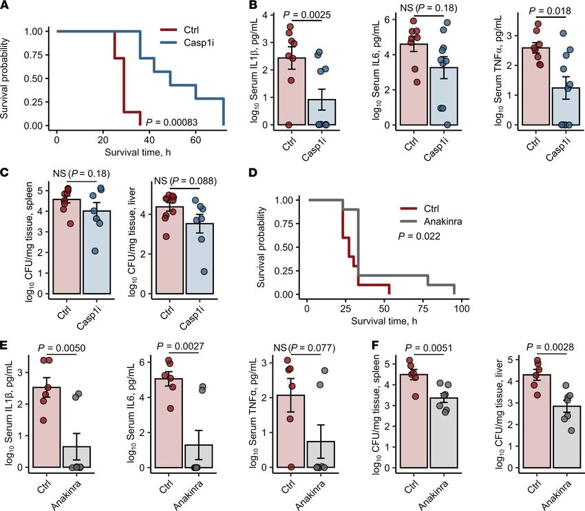
RESEARCH ARTICLE

                                       Iron homeostasis within macrophages is regulated at multiple levels. Under steady state, macrophages
                                  take part in systemic iron homeostasis by taking up, degrading, and recycling old or damaged erythrocytes
                                  (16). As the degradation of heme produces significant amounts of iron, macrophages are perfectly equipped
                                  to handle this potentially toxic trace metal. One such mechanism is increased iron efflux, which is mediated
                                  by the sole known iron exporter (FPN1) (17). The other option is the storage of intracellular iron within fer-
                                  ritin. Ferritin is a protein polymer consisting of a total of 24 ferritin H (FTH) and ferritin L (FTL) subunits
                                  forming a round structure that can store up to 4500 Fe3+ ions (18). The stoichiometric proportion of the L and
                                  the H subunits differs, depending on the tissue. However, only the H subunit possesses ferroxidase activity
                                  that is necessary for oxidation of cytoplasmic ferrous iron and its incorporation into the ferritin oligomer but
                                  also affects dietary iron absorption (19). Moreover, macrophages produce FTH-rich molecules, as they are
                                  exposed to significant variation of labile iron levels (20), and can release ferritin by specific pathways (21).
                                       Upon infection with intracellular bacteria, the cellular iron turnover in the macrophage is substantially
                                  altered to deprive invading pathogens of this essential nutrient (5, 6). In the case of Salmonella infection, mac-
                                  rophages upregulate FPN1 expression and, therefore, reduce the cytoplasmic labile iron pool (22). However,
                                  ferritin protein levels are also upregulated, although incorporation of iron is reduced (9). This is biologically rea-
                                  sonable, as Salmonella may be able to access ferritin-associated iron (23). Cellular regulation of ferritin and FPN1
                                  translation is mediated by iron regulatory proteins (IRPs) 1 and 2, which respond to changes in cellular iron
                                  levels. IRP deletion in macrophages leads to hyperferritinemia, and this in turn seems to increase iron availability
                                  for invading intracellular pathogens like Salmonella, resulting in shortened survival of the KO animals (23).
                                       Notably, protective effects of ferritin were described in diverse infection models, including infection with the
                                  intracellular, macrophage-colonizing pathogen Mycobacterium tuberculosis (24) and polymicrobial sepsis (25). In
                                  this setting, FTH expression and FTH-mediated control of cellular iron availability has been linked to disease
                                  tolerance and metabolic adaption (24). Recently, we demonstrated that iron loading of macrophages induces a
                                  pathogen-friendly metabolic environment by suppressing glycolysis and boosting Krebs cycle activity (26).
                                       In light of these contrasting findings, we sought to clarify the effect of macrophage-expressed ferritin
                                  on immune response against the model intracellular pathogen Salmonella enterica serovar Typhimurium. To
                                  this end, we characterized the baseline iron turnover in LysM-Cre+/+Fthfl/fl mice lacking FTH in myeloid cells
                                  and delved into their susceptibility to Salmonella infection. Our results functionally link the increased labile
                                  cellular iron levels in FTH-deficient macrophages to rapidly expanding pathogen burdens, which contribute
                                  to uncontrolled activation of the NF-κB and inflammasome pathways, detrimental cytokine formation, and
                                  dramatically shortened survival in course of infection with the intramacrophage pathogen S. Typhimurium.

                                  Results
                                  FTH depletion increases cellular labile iron and iron efflux in macrophages. FTH is the ferroxidase subunit of the
                                  ferritin complex, the activity of which is necessary for iron incorporation. Therefore, it is expected that Fth
                                  gene disruption severely impairs the iron storing capacity of cells and increases cellular levels of labile iron.
                                  To corroborate this assumption for FTH-deficient macrophages, we characterized alterations in iron turnover
                                  in mice lacking the Fth gene in the myeloid lineage (LysM-Cre+/+Fthfl/fl mice, herein referred to as FthΔ/Δ mice).
                                       First, we could prove efficient deletion of the Fth gene in control and iron-treated bone marrow–derived
                                  macrophages (BMDMs) from FthΔ/Δ mice by immunoblotting. Notably, a compensatory upregulation of the
                                  other ferritin subunit, FTL, could be observed in FthΔ/Δ BMDMs as compared with LysM-Cre+/+Fth+/+-derived
                                  macrophages (herein referred to as Fth+/+ macrophages) (Supplemental Figure 1; supplemental material avail-
                                  able online with this article; https://doi.org/10.1172/jci.insight.141760DS1).
                                       Deletion of the Fth gene in the myeloid compartment led to a substantial decrease of splenic nonheme iron
                                  content and markedly reduced numbers of iron-storing Pearl’s Prussian Blue–positive resident splenic macro-
                                  phages compared with those in Fth+/+ spleens. These differences preexisted in steady state and persisted after
                                  challenge with i.v. iron isomaltoside, a clinically approved preparation that delivers iron-containing nanoparti-
                                  cles directly to macrophages (27, 28). However, no significant difference was observed in the liver iron content
                                  between FthΔ/Δ and Fth+/+ mice (Figure 1, A and B). Under steady-state conditions, plasma iron levels were not
                                  affected by myeloid FTH deficiency, but these dramatically increased in FthΔ/Δ mice after iron challenge (Figure
                                  1C). Interestingly, in this setting, serum hepcidin levels rose upon stimulation with iron, but were unaffected
                                  by lack of myeloid ferritin (Supplemental Figure 3A). Leukocyte composition in the liver and the spleen was
                                  also unaltered in iron-loaded FthΔ/Δ mice in comparison with that in Fth+/+ mice, except for Ly6Chi monocytes
                                  in the spleen, which were significantly increased in iron-loaded FthΔ/Δ mice (Supplemental Figure 3, B and C).

JCI Insight 2021;6(13):e141760 https://doi.org/10.1172/jci.insight.141760                                                                             2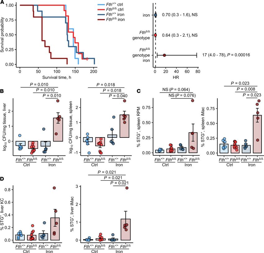
RESEARCH ARTICLE

                                  Cumulatively, myeloid FTH deficiency seems to impair the iron storage capacity of the spleen myeloid cells and
                                  increases circulating iron levels.
                                       To verify this on a cellular level, we investigated iron turnover rates in FTH-proficient and -deficient
                                  BMDMs. Interestingly, the iron uptake rate in FthΔ/Δ macrophages was initially slightly increased but leveled
                                  off 2 hours after iron challenge (Figure 1D). However, the iron efflux rates of FTH-deleted macrophages were
                                  significantly elevated as compared with WT cells (Figure 1D), suggesting reduced storage capacities in these
                                  cells. In line with the augmented export, total (Supplemental Figure 1) and surface levels (Figure 1E) of the
                                  sole iron exporter FPN1 were substantially increased in FTH-deficient cells at baseline and further boosted by
                                  iron challenge. Additionally, reduced iron storage capacities of FTH-deficient macrophages led to accumula-
                                  tion of cellular labile iron even without iron challenge, as measured by calcein quenching (Figure 1F). Expect-
                                  edly, expression of the central iron uptake receptor TFR1 was reduced in FTH-deficient cells, a finding which
                                  is in line with the known negative effects of cellular labile iron accumulation on Tfr1 mRNA stability (1).
                                       Taken together, myeloid-specific Fth-KO mice have a defective cellular iron storage, which leads to an
                                  expansion of the cellular labile iron pool, FPN1 induction, and increased export exacerbating upon iron loading.
                                       Myeloid FTH deficiency causes hyperresponsiveness to Salmonella infection. Next, we determined how myeloid
                                  FTH KO affects susceptibility to systemic infection with the intracellular bacterium, S. Typhimurium.
                                  Upon i.p. infection, we could not find a survival difference between control-treated Fth+/+ and FthΔ/Δ mice.
                                  In turn, i.v. iron loading prior to infection dramatically shortened the survival of FthΔ/Δ mice as compared
                                  with Fth+/+ mice (median survival 60 hours vs. 128.5 hours for iron-loaded Fth+/+, Figure 2A). When the
                                  animals were analyzed at the time points when all FthΔ/Δ mice were still alive, we could observe markedly
                                  elevated bacterial burdens in the spleen and liver as early as 12 hours after infection (Supplemental Figure
                                  6C). Similarly, pathogen counts were highly significantly increased by more than one order of magnitude
                                  20 hours after challenge (Figure 2B) in the iron-treated FthΔ/Δ as compared with the iron-administered WT
                                  cohort. Analogically, substantially higher bacterial colonization of inflammatory (iMac, CD11b+F4/80+)
                                  and resident macrophages (red-pulp macrophages and Kupffer cells, CD11b–F4/80+) of these organs could
                                  be ascertained in iron-treated KO mice (Figure 2, C and D).
                                       Notably, S. Typhimurium primarily infects and is taken up by macrophages (29). Yet, the higher
                                  bacterial burden in FthΔ/Δ animals could not be explained by elevated phagocytic capabilities since in vivo
                                  uptake of latex beads by liver and spleen macrophages was not positively affected by the Fth genotype nor
                                  iron loading. Uptake of latex beads was significantly impaired in iron-loaded FthΔ/Δ mice in comparison
                                  with that in FthΔ/Δ mice (Supplemental Figure 4A). Importantly, we could exclude any differences in first
                                  line defense and uptake of Salmonella, as iron-loaded FthΔ/Δ mice infected i.v. with bacteria demonstrated
                                  comparable spleen and liver bacterial burden (Supplemental Figure 4B) and macrophage colonization
                                  (Supplemental Figure 4C) 3 hours after infection.
                                       Taken together, deletion of FTH in myeloid leukocytes of iron-loaded FthΔ/Δ mice drastically reduced
                                  their survival. Notably, the unaltered bacterial burden and macrophage colonization 3 hours after infection let
                                  us speculate that primary defense mechanisms such as phagocytic exclusion of Salmonella or oxidative burst
                                  were not impaired in FthΔ/Δ animals and may not be the primary cause for this conspicuous phenotype.
                                       Myeloid FTH deficiency triggers uncontrolled cytokine response to Salmonella. To investigate dysregulated sig-
                                  naling circuits during infection in iron-loaded myeloid-specific Fth-KO animals, we analyzed whole-genome
                                  gene expression in the spleens of Fth+/+ and FthΔ/Δ animals challenged with/without i.v. iron and infected
                                  with 500 CFU Salmonella i.p. for 12 hours. Using 2-way ANOVA, we assessed the effect of iron and Fth
                                  genotype and, most importantly, the interaction of these 2 factors on expression of a particular gene (Sup-
                                  plemental Figure 5). Since the combination of the iron loading with the lacking myeloid Fth led to the dras-
                                  tically shortened survival, we assumed that the gene set regulated by the interaction of iron challenge and
                                  Fth genotype was most critical for this phenotype. By analyzing this iron/genotype interaction, we identified
                                  271 genes significantly upregulated and 893 genes significantly downregulated in iron-loaded FthΔ/Δ mice as
                                  compared with iron-treated WT animals (Supplemental Table 1).
                                       Functional analysis of the gene set downregulated by the iron/genotype interaction using Gene
                                  Ontology (GO) term enrichment (Figure 3A and Supplemental Tables 2 and 3) revealed a substantial
                                  enrichment in genes involved in regulation of adaptive immune response such as Cd4, Cd8a, Lat, and
                                  Zap70 (Figure 3C). In turn, a highly significant enrichment in genes linked to innate immune activation
                                  (Tnfaip3, Itgam, C5ar1, Tlr2, and Tnf), cytokine (Il1b, Il6, Il10, Il18, and Tnf), chemokine production
                                  (Ccl2, Ccl3, Ccl4, Cxcl1, Cxcl2, and Cxcl5), and TLR (Tlr2 and Cd14), NF-κB (Tnip, Tnfaip3, and Irak3),

JCI Insight 2021;6(13):e141760 https://doi.org/10.1172/jci.insight.141760                                                                          3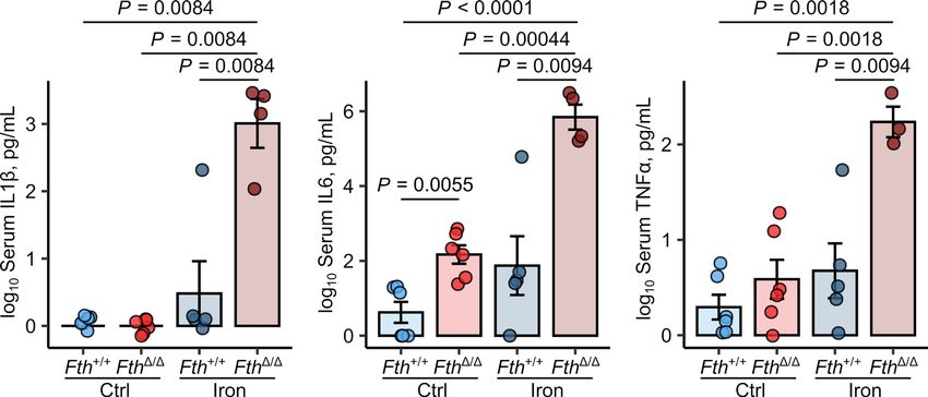
RESEARCH ARTICLE

JCI Insight 2021;6(13):e141760 https://doi.org/10.1172/jci.insight.141760                  4
RESEARCH ARTICLE

Figure 1. Fth deletion in myeloid cells increases cellular labile iron pool and iron turnover in macrophages. (A–C) Fthfl/fl (Fth+/+) and LysM-Cre Fthfl/fl (FthΔ/Δ)
mice were injected i.v. with iron isomaltoside (2 mg elementary Fe/mouse) or left untreated and analyzed 3 days later. (A) Nonheme iron content of the spleen
and liver (n = 3 per group). (B) Counts of splenic and hepatic PPB-positive macrophages. Each point denotes mean from at least 3 high-power fields (HPFs) per
animal (spleen: Fth+/+ ctrl, Fth+/+ iron, FthΔ/Δ ctrl — n = 4, FthΔ/Δ iron —n = 3; liver: n = 7 per group). (C) Nonheme iron concentrations in serum (n = 7 per group). (D)
Iron uptake and release from Fth+/+ and FthΔ/Δ bone marrow–derived macrophages (BMDMs). Iron uptake was measured in cultures stimulated with 5 μM 59Fe3+
(in form of FeCl3) for the indicated time points. Iron release from BMDMs loaded with 5 μM 59Fe3+ into culture supernatant was determined at the indicated time
points (uptake and release: n = 6 per group). (E) Surface expression of FPN1 and TFR1 in Fth+/+ and FthΔ/Δ BMDMs cultivated with/without 10 μM Fe3+ (FeCl3) for
12 hours was measured by flow cytometry (n = 5 per group). (F) Calcein-stained Fth+/+ and FthΔ/Δ BMDMs were stimulated with 50 μM Fe3+ (FeCl3) for the indicat-
ed time points. Calcein fluorescence expressed as ΔMFI was determined by flow cytometry. Each point denotes single observation (A and C–F) or a mean from
at least 3 HPFs per animal (B). Bars with whiskers represent mean ± SEM. Statistical significance was assessed with 2-way ANOVA (A–C and E), Kruskal-Wallis
test (E, FPN1), or repeated-measures 2-way ANOVA (E, TFR1 data, and F) with Benjamini-Hochberg-corrected 2-tailed post hoc t tests (A–F) or Mann-Whitney U
tests (E, FPN1). In the plots, post-hoc test P values are indicated.

                                       and inflammasome signaling (Nlrp3, Il1b, and Il18) could be observed within the gene set upregulated in
                                       iron-treated FTH-deficient animals (Figure 3, B and D).
                                            Next, we hypothesized that distinct transcription factors (TFs) may mediate the differences in gene
                                       expression as a function of the iron/genotype interaction. Therefore, we investigated possible common TF
                                       orchestrating the exacerbated transcriptional response to Salmonella in iron-loaded FthΔ/Δ animals (Figure 3,
                                       E and F, and Supplemental Tables 4 and 5). To this end, we compared the frequency of a particular TF
                                       binding site in the gene sets downregulated and upregulated by the iron/Fth interaction with the frequency in
                                       the whole genome. This analysis revealed a prominent overrepresentation of binding sites of various NF-κB
                                       isoforms (NF-κB, NFKB1, REL, and RELA) both in promoters of the downregulated and upregulated gene
                                       set, indicating that the aberrant iron turnover in iron-loaded FthΔ/Δ animals upon infection massively interferes
                                       with the NF-κB signaling pathway (Figure 3, E and F). Taken together, the combination of the increased iron
                                       loading with myeloid FTH deficiency triggers an excessive proinflammatory response to Salmonella manifest-
                                       ed by, among others, an unharnessed NF-κB activation, cytokine, and chemokine production.
                                            To further verify the previous results, we took a closer look at serum cytokine levels and infiltration of
                                       proinflammatory myeloid cells in the spleen and liver of iron-treated FthΔ/Δ mice infected with Salmonella for 12
                                       and 20 hours. Twelve hours after infection, the levels of circulating inflammatory cytokines (IL-1β, IL-6, IL-18,
                                       and TNF-α) were still hardly detectable in most of the animals. Independent of iron status and genotype, we
                                       identified elevated Il1b and Il6 transcript levels in the spleens of the iron-loaded FthΔ/Δ animals as compared
                                       with the remaining study groups (Supplemental Figure 6, A and B). In turn, a dramatic rise in IL-1β, IL-6, and
                                       TNF-α plasma concentrations was evident 20 hours after infection in i.v. iron-treated Fth-KO animals as com-
                                       pared with control and iron-administered WT animals and control-treated FthΔ/Δ mice (Figure 4).
                                            Importantly, a quantitatively and qualitatively similar cytokine response to Salmonella in myeloid
                                       FTH-deficient mice could be elicited by iron loading with another clinically applicable i.v. iron preparation,
                                       iron gluconate, at the identical elementary iron dose (Supplemental Figure 7).
                                            Pharmacologic interference with the inflammasome/IL-1β pathway prevents the Salmonella-elicited cytokine storm in
                                       iron-loaded myeloid FTH-deficient mice. Our microarray analysis pointed toward possible involvement of NF-κB
                                       and inflammasome signaling as well as myeloid cell recruitment in development of the uncontrolled inflam-
                                       matory response in iron-treated and Salmonella-infected myeloid FTH-deficient mice (Figure 3, B, D, and F).
                                       To identify the functionally most relevant pathway, we first tested for NF-κB hyperactivity and treated control
                                       or iron-loaded Fth+/+ and FthΔ/Δ animals with the canonical TLR/NF-κB signaling inducer LPS at a sublethal
                                       dose of 20 μg/kg (30). Animals were sacrificed 9 hours after the treatment, i.e., shortly after passing the peak of
                                       systemic inflammatory response defined by a body temperature drop (Supplemental Figure 7A). In this setting,
                                       iron-loaded FthΔ/Δ mice showed significantly decreased nadir body temperature (Supplemental Figure 8A) and
                                       substantially risen serum levels of IL-6 and TNF-α (Supplemental Figure 8B) compared with other experimen-
                                       tal groups. However, the magnitude of the cytokine response to LPS did not reach the levels elicited by living
                                       Salmonella in the iron-challenged myeloid FthΔ/Δ (Supplemental Figure 7B and Figure 4) (Salmonella: 1.4 ± 0.7
                                       × 106 pg/mL IL-6 and 200 ± 80 pg/mL TNF-α; LPS: 4.2 ± 0.7 × 10⁴ pg/mL IL-6 and 70 ± 6 pg/mL TNF-α).
                                       Importantly, serum IL-1β, which was dramatically increased in iron-loaded FthΔ/Δ mice in Salmonella infection,
                                       was barely altered in the setting of LPS challenge.
                                            However, even though some level of hypersensitivity of the NF-κB signaling pathway upon iron load-
                                       ing and myeloid FTH deficiency could be observed, it is unlikely that this mechanism solely underlies the
                                       dramatically increased cytokine response and mortality in iron-loaded FthΔ/Δ mice upon bacterial infection.

JCI Insight 2021;6(13):e141760 https://doi.org/10.1172/jci.insight.141760                                                                                                 5
RESEARCH ARTICLE
Figure 2. Loss of FTH in myeloid cells increases susceptibility of
iron-loaded mice to Salmonella infection. Fthfl/fl (Fth+/+) and LysM-Cre
Fthfl/fl (FthΔ/Δ) mice were i.v. administered PBS or iron isomaltoside
(2 mg elementary Fe per animal) and infected 3 days later with 500
CFU GFP-expressing S. Typhimurium (STG). (A) Surviving fractions of
control and iron-loaded Fth+/+ and FthΔ/Δ mice as a function of time
after infection (Fth+/+ ctrl, Fth+/+ iron, FthΔ/Δ iron: n = 11, FthΔ/Δ ctrl: n =
10). The forest plots show results of Cox proportional hazard modeling
of the data (HR, hazard ratio). (B) The number of GFP-expressing
bacteria in the spleen and liver determined by flow cytometry of organ
lysates 20 hours after infection (Fth+/+ ctrl: n = 6, Fth+/+ iron: n = 5,
FthΔ/Δ ctrl: n = 9, FthΔ/Δ iron: n = 5). (C and D) Bacterial colonization of
spleen red-pulp (RPM) and inflammatory macrophages (iMacs) (C) and
of liver Kupffer cells (KCs) and iMac (D) 20 hours after was measured
by flow cytometry and expressed as percent of STG-positive cells
within the parent population (Fth+/+ ctrl: n = 6, Fth+/+ iron: n = 5, FthΔ/Δ
ctrl: n = 9, FthΔ/Δ iron: n = 5). In A, data are presented as Kaplan-Meier
plot and forest plot with points representing Cox regression estimates
and whiskers depicting 95% CI for the estimates. In the other panels,
each point denotes a single animal; bars with whiskers represent
mean ± SEM. In A, statistical significance was assessed with Cox
proportional hazard modeling for genotype, iron, and genotype: iron
interaction terms, points in the forest plot are labeled with estimate
values, 95% CI and P values. In other panels, statistical significance
was assessed with Kruskal-Wallis test and with Benjamini-Hoch-
berg-corrected Mann-Whitney U tests. In the plots, post hoc test P
values are indicated.
This finding is further supported by the observation that the treatment of iron-loaded FthΔ/Δ mice, with a
pharmacological NF-κB blocker BAY11-7082 (31) concomitantly with Salmonella infection, could neither
reduce systemic cytokine production (Supplemental Figure 9A) nor improve infection control (Supplemen-
tal Figure 9B) and worsened the pathological phenotype.
JCI Insight 2021;6(13):e141760 https://doi.org/10.1172/jci.insight.141760 6RESEARCH ARTICLE JCI Insight 2021;6(13):e141760 https://doi.org/10.1172/jci.insight.141760 7
RESEARCH ARTICLE

Figure 3. S. Typhimurium triggers unrestrained expression of proinflammatory NF-κB targets in iron-loaded FthΔ/Δ mice. Fthfl/fl (Fth+/+) and LysM-Cre
Fthfl/fl (FthΔ/Δ) mice were i.v. administered PBS or iron isomaltoside (2 mg elementary Fe per animal) and infected 3 days later with 500 CFU GFP-ex-
pressing S. Typhimurium (STG) (n = 3 mice per group). Twelve hours after infection, total spleen RNA was isolated and subjected to a whole transcrip-
tome measurement with gene microarrays. Genes significantly downregulated (PANOVA iron/genotype < 0.05 and estimateiron/genotype < -1.5, n = 893 genes) and
upregulated (PANOVA iron/genotype < 0.05 and estimateiron/genotype > 1.5, n = 271 genes) were identified by 2-way ANOVA and linear regression as described in
Methods and Supplemental Figure 5. For a list of significant genes with ANOVA P values and regression estimates, see Supplemental Table 1. (A and B)
GO term enrichment analysis for genes significantly downregulated (A) and upregulated (B) by the iron/genotype interaction. Significant GO terms are
highlighted in blue and red, respectively (downregulated genes: n = 10 significant GO terms, upregulated genes: n = 31 significant GO terms), 10 most
significantly enriched GO terms are labeled with their names. For full results of GO term enrichment analysis, see Supplemental Tables 2 and 3. (C and D)
Heatmap representation of normalized gene expression values (z score) for genes assigned to selected significantly enriched GO terms. (C) Significantly
downregulated genes.(D) Significantly upregulated genes. Color scale corresponds to normalized expression. (E and F) Transcription factor (TF) binding
site enrichment analysis for genes significantly downregulated (E) and upregulated (F) by the iron/genotype interaction. For each TF-binding motif, the
Benjamini-Hochberg-corrected P value and fold enrichment are plotted. Significant TF-binding motifs are highlighted in blue and red (downregulated
genes: n = 35, upregulated genes: n = 5 significant TF binding motifs), 10 most significantly enriched TF-binding motifs are labeled with their names.

                                        To corroborate the prime effect of the inflammasome signaling on unharnessed cytokine response
                                   to Salmonella, we treated iron-loaded and Salmonella-infected FthΔ/Δ mice first with AC-YVAD-cmk, an
                                   oligopeptide inhibitor of the key common inflammasome component caspase-1. Administration of the
                                   inhibitor significantly prolonged survival of the animals and reduced the levels of circulating IL-1β
                                   and TNF-α and partly IL-6 at 20 hours after infection (Figure 5, A and B). However, this intervention
                                   only marginally improved infection control, as demonstrated by only slightly reduced bacterial loads
                                   of the spleen and liver (Figure 5C). To verify these promising results, we next administered the clini-
                                   cally applicable trap receptor for the key inflammasome-activated cytokine IL-1β, anakinra (25), to the
                                   infected iron-loaded myeloid FTH-deficient animals. By these means, we could significantly prolong
                                   survival (Figure 5D) and reduce not only the systemic levels of IL-1β but also of inflammasome-in-
                                   dependent cytokines IL-6 and TNF-α as compared with iron-loaded FthΔ/Δ animals injected with PBS
                                   (Figure 5E). Importantly, systemic IL-1β neutralization significantly reduced bacterial burden in the
                                   liver and spleen as well (Figure 5F).
                                        Taken together, the substantial reversal of the cytokine storm phenotype in iron-loaded FthΔ/Δ animals
                                   by interference with caspase-1 activity and the action of the inflammasome-dependent cytokine IL-1β
                                   activity strongly suggests that an exaggerated triggering of the inflammasome pathway represents the key
                                   effector mechanism driving lethal hyperresponsiveness to Salmonella.
                                        FTH deficiency in infected primary macrophages leads to overreactivity of the NF-κB and inflammasome signaling
                                   pathways and unharnessed cytokine secretion. Given the complexity of the myeloid leukocyte compartment and its
                                   differential roles in hosting and eliminating Salmonella as well as cell-type-specific triggering of inflammatory
                                   pathways (32), we sought to verify the contribution of the NF-κB and inflammasome signaling to the cytokine
                                   hyperresponsiveness upon FTH deficiency of macrophages, the prime host cells for Salmonella.
                                        To this end, we tested cytokine production capacities of primary peritoneal macrophages (PMs)
                                   derived from WT and FthΔ/Δ donors upon iron and Salmonella challenge in vitro. Notably, PMs were
                                   isolated from the mice without prior inflammatory stimulation in vivo, such as thioglycolate, to avoid
                                   any nonspecific priming of the cells. Surprisingly, FTH-deficient cells demonstrated high iron-indepen-
                                   dent production of IL-1β, IL-6, and TNF-α even without bacteria (Supplemental Figure 10A), indicat-
                                   ing steady activity of proinflammatory signaling, most likely as a result of distorted iron metabolism
                                   (Figure 1). The secretion of the strictly inflammasome-dependent IL-1β (see the results of costimula-
                                   tion with AC-YVAD-cmk, Supplemental Figure 10B) was further strongly augmented by a short-time
                                   3-hour Salmonella infection in those cells and was dramatically higher in the KO than in WT macro-
                                   phages (Supplemental Figure 10A). Interestingly, production of IL-6 and TNF-α orchestrated in PMs
                                   primarily by NF-κB (see the results of costimulation with BAY11-7082; Supplemental Figure 10B) was
                                   stimulated by bacteria to a much lower extent in the FthΔ/Δ cells than in IL-1β. However, the levels of
                                   those cytokines upon Salmonella were significantly higher than in the WT cells (Supplemental Figure
                                   10A). Although the fully fledged cytokine storm phenotype in FthΔ/Δ in vivo could only be observed in
                                   iron-loaded mice, production of the investigated cytokines was hardly boosted by iron; the significant
                                   but marginal effect of iron was found solely for IL-1β.
                                        Collectively, at the level of the isolated macrophage, FTH deficiency goes hand in hand with uncon-
                                   trolled spontaneous activation of the NF-κB and inflammasome signaling culminating in production of
                                   IL-1β, IL-6, and TNF-α. The latter signaling circuit is further augmented by Salmonella, leading to a dramatic

JCI Insight 2021;6(13):e141760 https://doi.org/10.1172/jci.insight.141760                                                                                   8
RESEARCH ARTICLE
Figure 4. S. Typhimurium elicits unbraked cytokine and cellular innate response in iron-loaded FthΔ/Δ mice. Fthfl/fl (Fth+/+) and LysM-Cre Fthfl/fl (FthΔ/Δ)
mice were i.v. administered PBS or iron isomaltoside (2 mg elementary Fe per animal) and infected 3 days later with 500 CFU GFP-expressing S. Typh-
imurium (STG). The animals were analyzed 20 hours after infection. Serum levels of IL-1β, IL-6, and TNF-α were measured by ELISA (Fth+/+ ctrl: n = 6,
Fth+/+ iron: n = 5, FthΔ/Δ ctrl: n = 6, FthΔ/Δ iron: n = 4). Each point denotes single animal; bars with whiskers represent mean ± SEM. Statistical significance
was assessed with 2-way ANOVA with Benjamini-Hochberg-corrected 2-tailed post hoc t tests. In the plots, post hoc test P values are indicated.
increase of IL-1β secretion. At the systemic level, the labile iron-rich environment in the iron-loaded FthΔ/Δ
mice further promotes pathogen growth, which in turn viciously amplifies the hyperreactive inflammatory
NF-κB and inflammasome pathways causing fatal cytokine storm.
Discussion
Ferritin is the most important intracellular iron storage protein. It is critically involved in maintaining iron
homeostasis under steady-state situations and, even more importantly, in the case of infection. Intracellular
free iron concentrations stimulate ferritin expression by stabilizing the mRNA via the iron response element/
IRP system (33). As shown previously, infection with Salmonella or Mycobacterium spp. induces ferritin expres-
sion in macrophages (9, 34). However, less iron is incorporated into ferritin in the case of infection with an
intracellular pathogen (9). Nevertheless, to date the molecular role of FTH in infection with intracellular patho-
gens has scarcely been investigated. Deletion of Fth and therefore, lack of ferroxidase activity, in the myeloid
cell line should result in reduced iron storage capacities. We could show that the iron content of the spleen
was decreased in myeloid FTH-deficient mice. This was in line with recently published results also showing
impaired iron storage capacities in the spleen (24). Additionally, the expression of the iron export protein FPN1
and the labile iron pool was increased, which has been reported in FTH-deficient enterocytes (19).
Surprisingly, infection of myeloid FTH-deficient mice with Salmonella without additional iron
loading resulted in unaltered survival or bacterial load as compared with WT animals. This is of
interest, as a recent report with another intracellular bacterium, Mycobacterium avium, showed highly
significantly increased susceptibility of myeloid FTH-deficient mice (24). Although both bacteria live
intracellularly, there is a decisive difference concerning their exact localization: whereas Mycobacterium
resides in the early phagosome, Salmonella primarily lives in the late phagosome (35). As we could
show recently, the subcellular exact localization of intracellular pathogens matters (8). This might be
one explanation for the pronounced phenotype in the Mycobacterium infection model.
However, by challenging the mice with an iron source used in clinics, we observed a significantly short-
ened survival and impaired infection control in myeloid FTH-deficient mice resulting in strongly elevated
bacterial burden of the spleen and liver. In addition, we found a sustained upregulation of multiple inflam-
matory transcripts in iron-loaded FthΔ/Δ mice as early as 12 hours after infection and the resultant massive
overproduction of the key proinflammatory cytokines IL-1β, IL-6, and TNF-α. Although, the cytokine
storm poses the prime cause of mortality of the iron-administered myeloid FTH-deficient animals, as it
could be inferred from the effects of AC-YVAD and anakinra treatments in vivo, we could not definitively
state whether the increased bacterial loads further trigger the pathology or rather the impaired pathogen
control results for a cytokine-mediated immunoparalysis. In light of published evidence and the analysis of
the mice 12 hours after pathogen challenge, the first possibility seems to be the case. Specifically, multiple
JCI Insight 2021;6(13):e141760 https://doi.org/10.1172/jci.insight.141760 9RESEARCH ARTICLE
Figure 5. Inhibition of the inflammasome/IL-1β pathway prevents the S. Typhimurium–induced cytokine storm in iron-loaded FthΔ/Δ mice. FthΔ/Δ mice were
i.v. administered iron isomaltoside (2 mg elementary Fe per animal) and infected 3 days later with 500 CFU GFP-expressing S. Typhimurium (STG). Mice were
i.p. injected with the caspase-1 inhibitor Ac-YVAD-cmk (Casp1i, 8 mg/kg, 1 hour after infection, A–C) or the IL-1β receptor antagonist anakinra (25 mg/kg, 3 hours
after infection, D–F). The treatment with Ac-YVAD-cmk or anakinra was repeated in 12-hour intervals. Control mice were administered PBS. (A) Surviving animal
fractions as a function of time (n = 7 mice per group). (B) Serum levels of IL-1β, IL-6, and TNF-α measured by ELISA 20 hours after infection (Ctrl: n = 10, Casp1i: n
= 7). (C) Bacterial burden of the spleen and liver determined by plating of organ lysates (Ctrl: n = 10, Casp1i: n = 7). (D) Surviving animal fractions as a function of
time (n = 10 mice per group). (E) Serum levels of IL-1β, IL-6, and TNF-α measured by ELISA 20 hours after infection (n = 7 per group). (F) Bacterial burden of the
spleen and liver was determined by plating of organ lysates 20 hours after infection (n = 7 per group). In A and D, data are presented as Kaplan-Meier plots. In
other panels, each point denotes single animal, and bars with whiskers represent mean ± SEM. In A and D, statistical significance was assessed with Wilcoxon
test. In the other panels, statistical significance was assessed with 2-tailed t test. In the plots, Wilcoxon and t test P value are indicated.
intracellular pathogens, including Salmonella, benefit from the elevated availability of the limiting micro-
element iron (23, 36) and such conditions are present in FTH-deficient macrophages. Therefore, these
increased bacteria counts are likely to drive classical inflammatory pathways activating NF-κB (37) and
inflammasome subtypes such as NLRC4 (38). However, we observed a strongly augmented inflammatory
signaling in FTH-deficient primary macrophages even without any bacterial or proinflammatory stimulus
and Salmonella infection could drastically boost secretion of the inflammasome-dependent IL-1β in these
cells. Notably, both the labile iron accumulating in the FthΔ/Δ macrophages and ferritin on its own were
postulated to affect NF-κB (39–42) and inflammasome signaling (43). Thus, under FTH deficiency and
iron administration, the coincident accelerated pathogen growth providing a trigger for the inflammatory
JCI Insight 2021;6(13):e141760 https://doi.org/10.1172/jci.insight.141760 10RESEARCH ARTICLE
pathways along with the pronounced activation of the NF-κB and inflammasome signaling pose a self-per-
petuating vicious cycle peaking at cytokine storm and death.
In line with the above hypothesis, administration of the caspase-1 inhibitor AC-YVAD-fmk (44)
and the IL-1β trap receptor anakinra (45) effectively reduced cytokine levels in the iron-treated FthΔ/Δ
animals and prolong their survival. In the case of anakinra, an improved pathogen control could be
observed as well. This later effect was less accentuated in the AC-YVAD-fmk treatment, supposedly
as a result of interference with caspase-1 — mediated pyroptosis being an important bacteria-clearing
mechanism (38). Analogically, a negative interference with NF-κB microbial defense pathways may be
the reason why the NF-κB inhibitor treatment was not able to reduce bacterial burden and to keep the
pathogen-induced cytokine storm at bay in the iron-loaded myeloid FTH-deficient animals.
Taken together, we put forward a potentially novel link between FTH as a scavenger of intracellular
labile iron, intracellular pathogen growth, and control of inflammatory response to infection. This might be
especially relevant in patients with hereditary or acquired iron overload, such as poly-transfused patients,
or patients with chemotherapy and severe infection or sepsis who have a higher risk for death in associa-
tion with uncontrolled systemic iron homeostasis (46). Disrupted iron homeostasis has also been linked to
adverse outcomes in patients with COVID-19 (47, 48), pointing to the general importance of iron homeo-
static control for immune control and the course of infection (49). Of interest, anakinra has been applied
for treatment of patients with severe COVID-19 who suffer from dysregulated hyperinflammatory immune
response but also altered systemic iron trafficking (50–52). Future studies will have to clarify if such treat-
ment will be of benefit for treatment of such infections with an exaggerated cytokine storm and whether
part of this effect can be traced back to anakinra-mediated effects on iron homeostasis and associated
induction of hyperinflammation as well as on iron delivery for microbes.
Methods
Bacteria and mice. The SifB-GFP–expressing Salmonella enterica serovar typhimurium was described elsewhere
(53) and used in all infection experiments.
FthΔ/Δ mice were generated by crossing the LysM-Cre+/+ (JAX mice line B6.129P2-Lyz2tm1(cre)Ifo/J) and
Fth (JAX mice line B6.129-Fth1tm1.1Lck/J) animals and were gifted by Lukas Kühn (Ecole Polytechnique
fl/fl
Fédérale de Lausanne, Lausanne, Switzerland). Fth+/+ mice bred from the FthΔ/Δ line served as WT con-
trols. Male mice (aged 8–12 weeks) were used in all experiments. Mice were kept in standard laboratory
conditions and with standard rodent diet containing 180 mg elementary Fe/kg (SNIFF).
Iron loading, Salmonella, and LPS treatment in vivo. Iron loading was accomplished by an i.v. admin-
istration of iron isomaltoside (Medice) or i.p. injection of iron gluconate (Sanofi). The dose of each
iron preparation was adjusted to 2 mg elementary Fe per animal. Salmonella infection or iron turnover
analyses were conducted 72 hours after iron loading.
In most infection experiments, mice were i.p. administered 500 CFU Salmonella. Bacterial load in the spleen
and liver was determined either by plating serial dilutions of organ homogenates on LB agar (Sigma-Aldrich)
or by flow cytometry. For the cytometric load measurement, the absolute count of GFP-positive bacteria in the
organ homogenate was determined with Precision Count Beads (BioLegend).
For the in vivo LPS challenge, mice were i.p. administered Salmonella LPS (Sigma-Aldrich) at a
dose of 20 μg/kg.
In each in vivo assay, surface body temperature was measured in at least 12-hour intervals. Loss of reflexes
(righting and grabbing reflex) and/or body temperature drop of the animal of more than 5°C compared with
the preinfection baseline were deemed a humane endpoint for infection and survival experiments.
In vivo blocking of NF-κB and IL-1β neutralization. For NF-κB blocking, mice were i.p. injected with
BAY11-7082 (10 mg/kg, Sigma-Aldrich) concomitantly with Salmonella infection.
For in vivo neutralization of IL-1, animals were i.p. treated with anakinra (25 mg/kg, Amgen) 3 hours
after Salmonella infection. The anakinra treatment, which is known to antagonize murine IL-1β in experimental
sepsis (54), was repeated in 12-hour intervals. Control animals were injected with PBS.
For in vivo selective and irreversible blocking of caspase-1, animals were infected with Salmonella as
described. One hour later, we treated the animals with Ac-YVAD-cmk (8 mg/kg, Sigma-Aldrich) i.p. and repeat-
ed this treatment in 12-hour intervals. Control animals were injected with a 1% DMSO solution in water.
Primary macrophage culture. Macrophages were cultured in DMEM with 10% FCS (PAN Biotech), 2 mM
L-glutamine (Lonza), and 1% penicillin-streptomycin (Lonza).
JCI Insight 2021;6(13):e141760 https://doi.org/10.1172/jci.insight.141760 11RESEARCH ARTICLE
For BMDM differentiation, bone marrow cells were obtained from tibias and femurs flushed with cold
PBS containing 1% penicillin-streptomycin. Bone marrow cells were cultured for 7 days in the presence of
50 ng/mL recombinant murine M-CSF (Preprotech).
For isolation of peritoneal exudate macrophages (PEM), peritoneal cavities of the mice without any
inflammatory pretreatment were washed 3 times with 10 mL prewarmed PBS. PMs were incubated in DMEM
with 10% FCS and antibiotics with/without 50 μM Fe3+ (iron III sulfate) for 3 hours and subsequently infected
with logarithmic phase GFP-expressing S. Typhimurium at multiplicity of infection (MOI) of 10 CFU bacteria
to 1 cell as described before (23). Gentamycin at 25 μg/mL (Thermo Fisher) was added 1 hour after infection to
eliminate extracellular bacteria and supernatants collected at 3 hours after bacteria challenge.
Flow cytometry, cellular labile iron pool measurement. Spleen cells were isolated by meshing the organ through a
100 μm cell strainer (Corning) with PBS. Liver leukocytes were obtained from mechanically dissociated, Liber-
ase TM/DNaseI digested organs (0.16 U/mL and 10 μg/mL, respectively, both from Sigma-Aldrich) essential-
ly as described previously (55). Erythrocyte lysis was done with ACK buffer (150 mM NH4Cl, 10 mM KHCO3,
and 0.1 mM Na2EDTA, all from Sigma-Aldrich). Flow cytometry staining was performed as described previ-
ously (56) with the following antibodies: anti-mouse CD45 (clone 3F11), CD11b (M1/70), Gr-1 (RB6-8C5),
Ly6C (HK1.4), and F4/80 (BM8). Flow cytometry antibodies were purchased from BioLegend and Thermo
Fisher. Organ-infiltrating leukocytes were identified as presented in Supplemental Figure 2. Total organ bacte-
rial load was determined as described previously. Leukocyte population-specific infection with GFP-expressing
Salmonella was measured as described previously (57) and presented in Supplemental Figure 2.
Measurement of cellular labile iron pool (LIP) was performed essentially as described previously (58, 59).
Briefly, adherent macrophages were treated with 50 μM Fe3+ for indicated time points, harvested by scraping
and stained with 1 μg/μL calcein AM (Thermo Fisher) for 5 minutes at 37°C. The difference between calcein
fluorescence between the iron-untreated sample and the given iron-treated sample (ΔMFI calcein) is assumed
to be proportional to cellular LIP size (59).
Flow cytometry measurement were conducted with the Gallios instrument (Beckman Coulter).
Data analysis was accomplished with FlowJo software (BD).
Determination of iron, hepcidin, nontransferrin-bound iron and cytokine concentration, lactate dehydrogenase
activity, and Prussian Blue staining. Liver and spleen tissues samples were dried, and their nonheme iron
content was measured using the bathophenanthroline method as described previously (60). The results
were calibrated to the protein content as determined with the BCA protein assay (Thermo Fisher). Plasma
iron was determined with a kit from BioAssay Systems. Plasma hepcidin, IL-6, TNF-α, and IL-1β were
determined with ELISA kits (hepcidin: Intrinsic Lifescience, all others: BD). Prussian Blue staining of
formalin-fixed paraffin-embedded spleen and liver sections was performed as described previously (14).
Quantitative real-time PCR and Western blotting. RNA was isolated after lysis by PeqGold TriFast (Peqlab)
followed by chloroform-phenol extraction. Reverse transcription was performed with M-MLV reverse tran-
scriptase (Thermo Fisher) and random hexamer primers (Carl Roth) as described previously (8). Quantitative
real-time PCR was performed with SoFast Eva Green master mix (BioRad) and primers, the sequences of
which are listed in Supplemental Table 6. Hprt (hypoxanthine guanine phosphoribosyl transferase, Entrez
Gene ID 15452) served as the house-keeping gene for expression normalization. Relative gene expression was
calculated with the ΔCt method and presented as log2 expression, where log2 expressiongene = Ctgene — CtHprt.
Protein extraction and Western blotting were performed exactly as described previously (61). We used
the following antibodies: rabbit anti-mouse FTH1 (1:1000; Cell Signaling, 3998), rabbit anti-mouse Fer-
ritin light chain (1:1000; Abcam, ab69090), mouse anti-human Transferrin receptor (1:1000; Invitrogen,
PA1-84854), rabbit anti-human FPN1 (1:2000; Eurogentec, NRU 451443), and rabbit anti-human β-actin
(1:500; Sigma-Aldrich, A2066).
Iron uptake and release assay. Iron uptake and release assays were essentially done as previously described (8).
For the macrophage iron import assay, cells were cultured with 5 μM 59Fe-citrate (Perkin Elmer). At indicated
time points, radioactivity of the culture supernatant was measured with a γ-counter (Perkin Elmer). For the
iron release assay, cells were first incubated for 2 hours with 5 μM 59Fe-citrate, washed extensively, and cultured
in non-radioactive medium. At indicated time points, radioactivity of the culture supernatant was determined
with a gamma counter (Wallac, Perkin Elmer). For both assays, supernatant radioactivity was expressed as
cpm normalized to whole-cell lysate protein concentration to account for differences in culture density.
Whole-transcriptome analysis with microarrays. Fthfl/fl (Fth+/+) and LysM-Cre Fthfl/fl (FthΔ/Δ) mice were i.v. admin-
istered PBS or iron isomaltoside (2 mg elementary Fe per animal) and infected 3 days later with 500 CFU
JCI Insight 2021;6(13):e141760 https://doi.org/10.1172/jci.insight.141760 12RESEARCH ARTICLE
GFP-expressing S. Typhimurium (n = 3 per group). Twelve hours after infection, animals were sacrificed and
whole-spleen RNA was isolated as described. RNA quality tested with RNA 6000 Nano Kit and Bioanalyzer
2100 (both from Agilent) and exceeded 8.2 RNA Integrity Number. Whole-genome expression was performed
with Mouse Gene 2.0 ST Array (Thermo Fisher) following the manufacturer’s protocol.
Whole-transcriptome analysis was performed with R programming suite, version 3.6.1. Probe signals were
normalized with the RMA algorithm (Robust Microarray Average), assigned to transcript identifiers and log2
expression levels calculated with package oligo (62). For identification of probes, the expression of which was
significantly differentially regulated by the Fth genotype, iron loading, and the iron/genotype interaction 2-way
ANOVA and factorial linear regression was applied. Genes significantly downregulated (PANOVA iron/genotype < 0.05
and regression estimateiron/genotype < –log2 1.5, n = 893 genes) and upregulated (PANOVA iron/genotype < 0.05 and regres-
sion estimateiron/genotype > 1.5, n = 271 genes) by were further investigated. For a list of significant genes with
ANOVA P values and regression estimates see Supplemental Table 1. Mouse microarray data can be accessed
from the Gene Expression Omnibus (GSE145114).
GO enrichment analysis for the significantly regulated genes was performed with DAVID version
6.8 (63). For full results of GO term enrichment analysis for the downregulated and upregulated genes,
see Supplemental Tables 2 and 3.
TF binding site enrichment analysis for the significantly regulated genes was performed with an
in-house written R script (https://github.com/PiotrTymoszuk/TF-enrichment). First, TF binding (for all
TF represented in the JASPAR and TRANSFAC databases) in the promoters of genes of interest and in the
whole mouse transcriptome was predicted with D-Light with the default program settings (64). Next, for a
particular TF, the total count of binding sites in the gene set of interest (containing n genes) was compared
with the total count of binding sites in 106 randomly generated gene sets (containing n random genes) from
the whole mouse genome. To obtain the P value for enrichment significance (Pbootstrap), the number of ran-
dom gene sets with the binding site count exceeding the binding site count in the gene set of interest was
determined and divided by the number of random gene sets. Pbootstrap was adjusted for multiple comparisons
with the Benjamini-Hochberg method. Adjusted P values of less than 0.05 were considered significant.
For full results of TF-binding site enrichment analysis for the downregulated and upregulated genes, see
Supplemental Tables 4 and 5.
Statistics. Statistical data analysis and result visualization was performed with R programming suite
(version 4.0.3, tidyverse package bundle). Details on microarray data analysis are mentioned above. All
other data were analyzed with 2-tailed t tests, 1- and 2-way ANOVA, and/or factorial linear regression,
as indicated in the figure legends. Post hoc testing was accomplished with Benjamini-Hochberg adjusted
2-tailed t tests. Strongly nonnormal distributed variables (Shapiro-Wilk test and visual inspection of qq
plots) were log10 transformed prior to analysis. Mouse survival data were analyzed with Cox regression and
Kaplan-Meier method using Wilcoxon’s test. If not indicated otherwise, data are presented as dot plots,
where each point represents a single observation. Data are shown as the mean ± SEM. A P value of less than
0.05 was considered significant.
Study approval. All animal experiments were performed in accordance with the Austrian Experimental
Animal Welfare Act 2012 (Tierversuchsgesetz 2012) and were approved by the Federal Ministry of Science
and Education (approval no. BMWF-66.011/0142-WF/V/3b/2014, Vienna, Austria).
Author contributions
DH and PT conceived the project, designed and performed experiments, analyzed and interpreted data,
and wrote the manuscript. VP, RH, SD, SS, ED, SB, MS, and AMM performed experiments. DB and
MN provided intellectual input. IT and GW conceived the project, designed experiments, analyzed and
interpreted data, and wrote the manuscript.
Acknowledgments
We would like to express our gratitude to Sabine Engl for excellent technical support and to Lukas C. Kühn
(Ecole Polytechnique Fédérale de Lausanne — EPFL, 1015 Lausanne, Switzerland) for providing the floxed
FTH mice. This study was supported by the Austrian Research Fund (FWF): project P 28302 to IT, P 33062
to MN, I 3321 Epicross to GW, W 1253 HOROS to GW and VP; by the DOC stipend of the Austrian Acad-
emy of Sciences to DH; by the Österreichische Krebshilfe Tirol to PT (project no. 15024); by the Christian
Doppler research Society (to GW); and by the Society for Funding Research and Education in Molecular
JCI Insight 2021;6(13):e141760 https://doi.org/10.1172/jci.insight.141760 13RESEARCH ARTICLE
Immunology and Infectiology (“Verein zur Förderung von Forschung und Weiterbildung in Molekularer
Immunologie und Infektiologie”).
Address correspondence to: Guenter Weiss or Igor Theurl, Medical University of Innsbruck, Depart-
ment of Internal Medicine II, Anichstrasse 35, A-6020 Innsbruck, Austria. Phone: 43.512.504.23251;
Email: guenter.weiss@i-med.ac.at (GW); Email: igor.theurl@i-med.ac.at (IT).
1. Muckenthaler MU, et al. A red carpet for iron metabolism. Cell. 2017;168(3):344–361.
2. Koskenkorva-Frank TS, et al. The complex interplay of iron metabolism, reactive oxygen species, and reactive nitrogen species:
insights into the potential of various iron therapies to induce oxidative and nitrosative stress. Free Radic Biol Med. 2013;65:1174–1194.
3. Nairz M, et al. Iron and innate antimicrobial immunity-Depriving the pathogen, defending the host. J Trace Elem Med Biol.
2018;48:118–133.
4. Weinberg ED. Nutritional immunity. Host’s attempt to withold iron from microbial invaders. JAMA. 1975;231(1):39–41.
5. Soares MP, Weiss G. The Iron age of host-microbe interactions. EMBO Rep. 2015;16(11):1482–1500.
6. Drakesmith H, Prentice AM. Hepcidin and the iron-infection axis. Science. 2012;338(6108):768–772.
7. Lopez CA, Skaar EP. The impact of dietary transition metals on host-bacterial interactions. Cell Host Microbe. 2018;23(6):737–748.
8. Haschka D, et al. Contrasting regulation of macrophage iron homeostasis in response to infection with Listeria monocytogenes
depending on localization of bacteria. Metallomics. 2015;7(6):1036–1045.
9. Nairz M, et al. The co-ordinated regulation of iron homeostasis in murine macrophages limits the availability of iron for intra-
cellular Salmonella typhimurium. Cell Microbiol. 2007;9(9):2126–2140.
10. Ganz T, Nemeth E. Iron homeostasis in host defence and inflammation. Nat Rev Immunol. 2015;15(8):500–510.
11. Schaible UE, Kaufmann SH. Iron and microbial infection. Nat Rev Microbiol. 2004;2(12):946–953.
12. Vazquez-Torres A, et al. Extraintestinal dissemination of Salmonella by CD18-expressing phagocytes. Nature.
1999;401(6755):804–808.
13. Andrews-Polymenis HL, et al. Taming the elephant: Salmonella biology, pathogenesis, and prevention. Infect Immun.
2010;78(6):2356–2369.
14. Nairz M, et al. Absence of functional Hfe protects mice from invasive Salmonella enterica serovar Typhimurium infection via
induction of lipocalin-2. Blood. 2009;114(17):3642–3651.
15. Behnsen J, et al. Exploiting host immunity: the Salmonella paradigm. Trends Immunol. 2015;36(2):112–120.
16. Pantopoulos K, et al. Mechanisms of mammalian iron homeostasis. Biochemistry. 2012;51(29):5705–5724.
17. Nemeth E, et al. Hepcidin regulates cellular iron efflux by binding to ferroportin and inducing its internalization. Science.
2004;306(5704):2090–2093.
18. Harrison PM, Arosio P. The ferritins: molecular properties, iron storage function and cellular regulation. Biochim Biophys Acta.
1996;1275(3):161–203.
19. Vanoaica L, et al. Intestinal ferritin H is required for an accurate control of iron absorption. Cell Metab. 2010;12(3):273–282.
20. Torti FM, Torti SV. Regulation of ferritin genes and protein. Blood. 2002;99(10):3505–3516.
21. Truman-Rosentsvit M, et al. Ferritin is secreted via 2 distinct nonclassical vesicular pathways. Blood. 2018;131(3):342–352.
22. Nairz M, et al. Nitric oxide-mediated regulation of ferroportin-1 controls macrophage iron homeostasis and immune function
in Salmonella infection. J Exp Med. 2013;210(5):855–873.
23. Nairz M, et al. Iron regulatory proteins mediate host resistance to Salmonella infection. Cell Host Microbe. 2015;18(2):254–261.
24. Reddy VP, et al. Ferritin H deficiency in myeloid compartments dysregulates host energy metabolism and increases susceptibili-
ty to mycobacterium tuberculosis infection. Front Immunol. 2018;9:860.
25. Weis S, et al. Metabolic adaptation establishes disease tolerance to sepsis. Cell. 2017;169(7):1263–1275.
26. Telser J, et al. Metabolic reprogramming of Salmonella infected macrophages and its modulation by iron availability and the
mTOR pathway. Microb Cell. 2019;6(12):531–543.
27. Petzer V, et al. Established and emerging concepts to treat imbalances of iron homeostasis in inflammatory diseases. Pharmaceuticals
(Basel). 2018;11(4):E135.
28. Bhandari S, et al. Intravenous irons: from basic science to clinical practice. Pharmaceuticals (Basel). 2018;11(3):82.
29. Vazquez-Torres A, et al. Toll-like receptor 4 dependence of innate and adaptive immunity to Salmonella: importance of the
Kupffer cell network. J Immunol. 2004;172(10):6202–6208.
30. Quatrini L, et al. Host resistance to endotoxic shock requires the neuroendocrine regulation of group 1 innate lymphoid cells.
J Exp Med. 2017;214(12):3531–3541.
31. Li J, et al. Sensitizing leukemia stem cells to NF-κB inhibitor treatment in vivo by inactivation of both TNF and IL-1 signaling.
Oncotarget. 2017;8(5):8420–8435.
32. Chen KW, et al. The neutrophil NLRC4 inflammasome selectively promotes IL-1β maturation without pyroptosis during
acute Salmonella challenge. Cell Rep. 2014;8(2):570–582.
33. Wilkinson N, Pantopoulos K. The IRP/IRE system in vivo: insights from mouse models. Front Pharmacol. 2014;5:176.
34. Silva-Gomes S, et al. Mycobacterium avium infection induces H-ferritin expression in mouse primary macrophages by activat-
ing Toll-like receptor 2. PLoS One. 2013;8(12):e82874.
35. Weiss G, Schaible UE. Macrophage defense mechanisms against intracellular bacteria. Immunol Rev. 2015;264(1):182–203.
36. Chlosta S, et al. The iron efflux protein ferroportin regulates the intracellular growth of Salmonella enterica. Infect Immun.
2006;74(5):3065–3067.
37. Talbot S, et al. Toll-like receptor 4 signalling through MyD88 is essential to control Salmonella enterica serovar typhimurium
infection, but not for the initiation of bacterial clearance. Immunology. 2009;128(4):472–483.
38. Miao EA, et al. Caspase-1-induced pyroptosis is an innate immune effector mechanism against intracellular bacteria. Nat Immunol.
JCI Insight 2021;6(13):e141760 https://doi.org/10.1172/jci.insight.141760 14You can also read