Nuclear lamina defects cause ATM-dependent NF-kB activation and link accelerated aging to a systemic inflammatory response
←
→
Page content transcription
If your browser does not render page correctly, please read the page content below
Downloaded from genesdev.cshlp.org on March 19, 2015 - Published by Cold Spring Harbor Laboratory Press

Nuclear lamina defects cause
ATM-dependent NF-kB activation
and link accelerated aging to
a systemic inflammatory response
Fernando G. Osorio,1 Clea Bárcena,1 Clara Soria-Valles,1 Andrew J. Ramsay,1 Félix de Carlos,2
Juan Cobo,2 Antonio Fueyo,3 José M.P. Freije,1,4 and Carlos López-Otı́n1,4
1
 Departamento de Bioquı́mica y Biologı́a Molecular, Facultad de Medicina, Instituto Universitario de Oncologı́a, Universidad de
Oviedo, 33006-Oviedo, Spain; 2Departamento de Cirugı́a y Especialidades Médico-Quirúrgicas, Instituto Asturiano de Odontologı́a,
Universidad de Oviedo, 33006-Oviedo, Spain; 3Área de Fisiologı́a, Departamento de Biologı́a Funcional, Facultad de Medicina,
Instituto Universitario de Oncologı́a, Universidad de Oviedo, 33006-Oviedo, Spain

Alterations in the architecture and dynamics of the nuclear lamina have a causal role in normal and accelerated
aging through both cell-autonomous and systemic mechanisms. However, the precise nature of the molecular cues
involved in this process remains incompletely defined. Here we report that the accumulation of prelamin A
isoforms at the nuclear lamina triggers an ATM- and NEMO-dependent signaling pathway that leads to NF-kB
activation and secretion of high levels of proinflammatory cytokines in two different mouse models of accelerated
aging (Zmpste24–/– and LmnaG609G/G609G mice). Causal involvement of NF-kB in accelerated aging was
demonstrated by the fact that both genetic and pharmacological inhibition of NF-kB signaling prevents age-
associated features in these animal models, significantly extending their longevity. Our findings provide in vivo
proof of principle for the feasibility of pharmacological modulation of the NF-kB pathway to slow down the
progression of physiological and pathological aging.
[Keywords: nuclear envelope; longevity; progeroid laminopathies]
Supplemental material is available for this article.
Received June 5, 2012; revised version accepted September 4, 2012.

Aging is associated with the progressive and irreversible                   the nuclear lamina forms a protein network that provides
loss of tissue homeostasis. Several stressors have been                     a scaffold for nuclear envelope proteins and chromatin
related to this process, including DNA damage, telomere                     (Gruenbaum et al. 2005; Mekhail and Moazed 2010).
attrition, or accumulation of damaged macromolecules                           Alterations in the nuclear lamina affect nuclear struc-
(Kirkwood 2005; Vijg and Campisi 2008). An exaggerated                      ture and DNA integrity, leading to defects in DNA repli-
accumulation of cellular damage or the inefficient ability                  cation and repair processes and inducing the accumulation
to respond to stress causes progeroid syndromes, charac-                    of different types of DNA damage (Worman and Foisner
terized by a precocious manifestation of several features                   2010). Thus, several mutations in lamins and lamin-binding
related to human aging (Burtner and Kennedy 2010). Most                     proteins have been associated with a subgroup of progeroid
progeroid syndromes are caused by defective DNA re-                         syndromes known collectively as progeroid laminopathies
pair mechanisms or by alterations in the nuclear lamina                     (Worman et al. 2010). Among them, Hutchinson-Gilford
(Ramirez et al. 2007; Hoeijmakers 2009). The nuclear                        progeria syndrome (HGPS) is one of the most compre-
lamina is a complex structure that surrounds and protects                   hensively studied. HGPS patients show growth impair-
the nuclear content, playing important roles in genome                      ment, lipodystrophy, dermal and bone abnormalities, and
regulation, organization, and maintenance (Dechat et al.                    cardiovascular alterations, leading to a dramatically short-
2008). Covering the inner face of the nuclear membrane,                     ened life span (Hennekam 2006; Merideth et al. 2008).
                                                                            HGPS is primarily caused by a de novo silent mutation
                                                                            within exon 11 of the LMNA gene encoding lamin A
4
  Corresponding authors                                                     (c.1824C>T; p.Gly608Gly) (De Sandre-Giovannoli et al.
E-mail clo@uniovi.es                                                        2003; Eriksson et al. 2003).
E-mail jmpf@uniovi.es
Article published online ahead of print. Article and publication date are      Lamin A, a core component of the nuclear lamina,
online at http://www.genesdev.org/cgi/doi/10.1101/gad.197954.112.           undergoes a complex maturation process, including

       GENES & DEVELOPMENT 26:2311–2324 Ó 2012 by Cold Spring Harbor Laboratory Press ISSN 0890-9369/12; www.genesdev.org            2311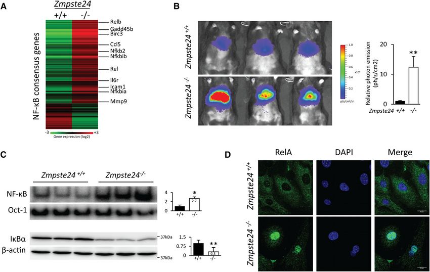
Downloaded from genesdev.cshlp.org on March 19, 2015 - Published by Cold Spring Harbor Laboratory Press

Osorio et al.

farnesylation, carboxyl methylation, and proteolytic               formulate potential therapeutic intervention strategies.
processing by the metalloproteinase ZMPSTE24/FACE-1.               Here we report that accumulation of prelamin A or
The HGPS mutation c.1824C>T activates a cryptic splice             progerin in the nuclear envelope of progeroid mice triggers
donor site, leading to a truncated form of prelamin A,             a signaling pathway involving ATM and NEMO proteins
called LAD50 or progerin, which lacks a 50-residue-long            to activate NF-kB. We also demonstrate that NF-kB-driven
fragment containing the cleavage site for ZMPSTE24.                inflammation is responsible for the development of sev-
Accordingly, Zmpste24 / mice show a premature aging                eral important features of progeroid phenotypes. Notably,
phenotype that phenocopies human HGPS (Bergo et al.                inhibition of the NF-kB pathway using genetic or pharma-
2002; Pendas et al. 2002; Osorio et al. 2011a). In all cases,      cological strategies was able to prevent these alterations,
the accumulation of farnesylated forms of prelamin A in            demonstrating the causal involvement of this inflamma-
the nuclear envelope leads to the nuclear abnormalities            tory pathway in the pathogenesis of accelerated aging.
and functional defects characteristic of cells from HGPS
patients and Zmpste24 / mice. Interestingly, progerin
accumulation is also detected during normal aging,                 Results
thereby adding a new level of interest to the study of the
                                                                   Zmpste24-deficient mice show NF-kB hyperactivation
mechanisms that underlie progerin formation and accu-
mulation (Scaffidi and Misteli 2006).                              Zmpste24-deficient mice accumulate farnesylated prel-
   NF-kB transcription factors form a cytoplasmic sensor           amin A at the nuclear envelope and develop a progeroid
system responding to not only pathogen attack, but also            syndrome that phenocopies most features of HGPS, in-
a variety of external and internal danger signals, such as         cluding growth impairment, lipodystrophy, dermal and
oxidative stress, hypoxia, and genotoxic stress (Perkins           bone abnormalities, and shortened life span (Bergo et al.
2007; Hayden and Ghosh 2008). NF-kB active transcrip-              2002; Pendas et al. 2002). Previous studies in Zmpste24 /
tion factors are composed of dimeric combinations of               mice have revealed that prelamin A accumulation in-
members of the Rel family. In mammals, five different              duces cellular senescence as well as important changes in
Rel family members have been identified: RelA (p65),               nuclear dynamics (Varela et al. 2005; Osorio et al. 2010).
RelB, c-Rel, NF-kB1 (p50 and its precursor, p105), and             On the other hand, NF-kB activation during aging has
NF-kB2 (p52 and its precursor, p100), which can form               been reported in human and mouse tissues as well as in
heterodimers or homodimers (Hayden and Ghosh 2012).                cells from HGPS patients (Adler et al. 2007; Kawahara
Many known stimuli that activate NF-kB require signal-             et al. 2009; Tilstra et al. 2011).
ing through the conserved IKK complex, consisting of at               To evaluate the activity of NF-kB in Zmpste24-
least two catalytic subunits, IKKa and IKKb, and a regu-           deficient mice, we first analyzed the transcriptional
latory subunit known as NEMO (also known as IKKg).                 profile of liver tissues from Zmpste24 / and wild-type
Activation of the NF-kB signaling pathway is one of the            littermates. By using the gene set enrichment analysis
cellular responses evoked to maintain homeostasis after            (GSEA) algorithm, we identified a strong positive corre-
DNA damage (Janssens and Tschopp 2006). As part of the             lation between the transcriptional changes in tissues from
DNA damage response (DDR), activation of the kinase                mutant mice and a gene set composed of genes whose
ataxia telangiectasia mutated (ATM) triggers a signaling           promoters contain the recognition sequence for NF-kB
pathway involving NEMO to directly link DNA damage                 transcription factors (Fig. 1A; Supplemental Fig. 1). Next,
events in the nucleus with the cytoplasmic activation of           we analyzed NF-kB activation in vivo in these progeroid
NF-kB (Miyamoto 2011). Thus, upon DNA damage, NEMO                 mice using a gene reporter-based assay (Fig. 1B; Supple-
undergoes a sequence of post-translational modifications           mental Fig. 2). To this end, we hydrodynamically de-
that includes sumoylation, ATM-mediated phosphorylation,           livered a plasmid vector encoding the firefly luciferase
and ubiquitylation (Huang et al. 2003; Wu et al. 2006).            gene under the control of an NF-kB response element in
NEMO trafficking between the cytoplasm and nucleus is              Zmpste24 / and wild-type animals. This approach allowed
a consequence of these modifications and finally results           us to quantify NF-kB activation in vivo by measuring the
in the activation of the IKK complex and the nuclear trans-        bioluminescence image obtained from these mice. Remark-
location of NF-kB active dimers (McCool and Miyamoto               ably, the bioluminescence signal of Zmpste24-deficient
2012).                                                             mice was >10-fold higher as compared with age-matched
   NF-kB hyperactivation has been related to the aging             control animals (Fig. 1B). NF-kB hyperactivation was
process (Adler et al. 2007). In addition, aberrant NF-kB           further confirmed by NF-kB electrophoretic mobility
activation is well documented in numerous age-associ-              shift assay (EMSA) using nuclear extracts from livers of
ated diseases, including neurodegeneration, osteoporosis,          Zmpste24 / and control mice (Fig. 1C). Accordingly, re-
diabetes, sarcopenia, immunosenescence, or atheroscle-             duced levels of the NF-kB inhibitor IkBa were also repro-
rosis (Tak and Firestein 2001; Le Saux et al. 2012). Given         ducibly observed in Zmpste24-deficient samples (Fig. 1C).
the links between accelerated aging, chronic DDRs, and                We next examined NF-kB activation in other tissues
NF-kB activation (Salminen et al. 2008; Ugalde et al.              previously reported to be affected during the development
2011a; Tilstra et al. 2012), we hypothesized that struc-           of progeria in Zmpste24 / mice (Pendas et al. 2002;
tural abnormalities in the nuclear envelope could activate         Varela et al. 2005). Thus, EMSA analysis of the thymus,
the NF-kB pathway. Accordingly, we used two different              spleen, kidney, and heart revealed an increase of >2.5-fold
progeroid mouse models to test this hypothesis and                 in NF-kB activity in Zmpste24 / tissues as compared

2312       GENES & DEVELOPMENT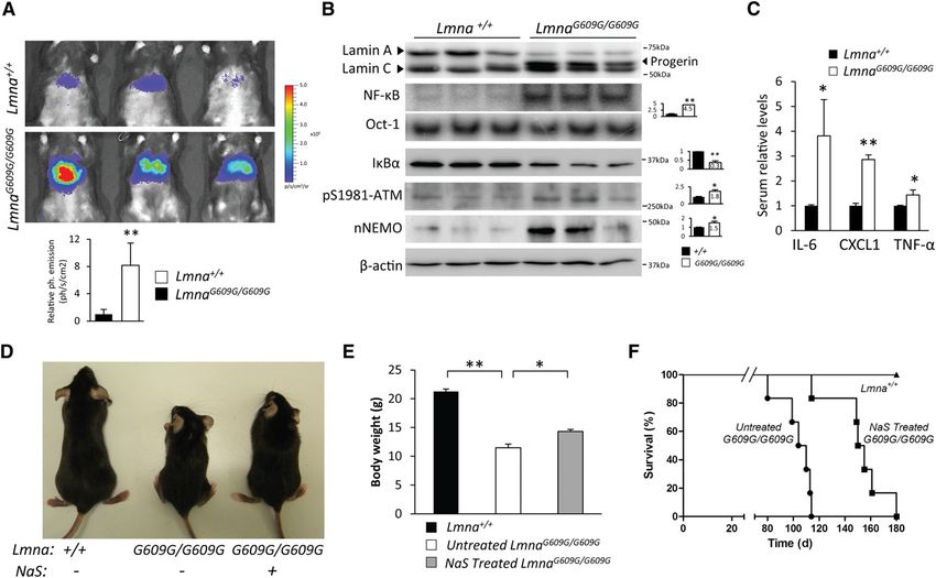
Downloaded from genesdev.cshlp.org on March 19, 2015 - Published by Cold Spring Harbor Laboratory Press
Causal role of NF-kB in progeria
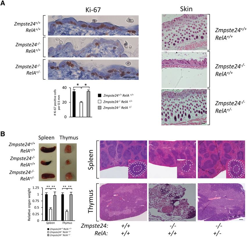
Figure 1. NF-kB hyperactivation in Zmpste24-deficient mice. (A) NF-kB target gene induction in Zmpste24-deficient mice. Heat map
represents gene expression analysis (in Zmpste24 / and wild-type livers) of genes that contain the consensus binding site for the NF-
kB transcription factor, displayed as Log2 transformed expression signals. (B) Hydrodynamic delivery of NF-kB–luciferase gene reporter
in the livers of 3-mo-old Zmpste24 / mice (n = 10) and age-matched control littermates (n = 10). The bioluminescence image was
recorded as the photon flux per second and square centimeter, and three representative mice from each genotype are shown. Relative
mean values are represented, and error bars indicate SEM. P < 0.01, two-tailed Student’s t-test. See also Supplemental Figure 2. (C) NF-
kB EMSA of livers from 3-mo-old Zmpste24 / mice (n = 3) and wild-type littermates (n = 3). Nuclear extracts were analyzed by using
32
P-labeled NF-kB and Oct-1 probes. Western blot of IkBa was performed using total liver extracts. b-Actin was used as loading control.
Signals were quantified, and plots represent relative mean values 6 SEM. (*) P < 0.05; (**) P < 0.01, two-tailed Student’s t-test. (D)
Representative image of immunofluorescence analysis using a RelA-specific antibody in fibroblasts from Zmpste24-deficient and wild-
type mice. Bar, 20 mm.
with wild-type tissues (Supplemental Fig. 3). In agreement pathway in the hyperactivation of NF-kB in tissues from
with this preferential nuclear localization of NF-kB in Zmpste24-deficient mice, we first analyzed the status
progeroid cells, immunofluorescence analysis revealed of ATM in these progeroid mice. As shown in Figure 2A,
nuclear accumulation of RelA in Zmpste24 / fibroblasts Western blot analysis of phospho-Ser1981-ATM re-
(Fig. 1D). Together, these data demonstrate that NF-kB is vealed a 1.5-fold increase in the levels of active ATM in
constitutively hyperactive in Zmpste24-deficient mouse Zmpste24 / mice as compared with wild-type animals.
tissues. As NEMO accumulation in the nucleus has been re-
ported during DDR activation, we next prepared whole-cell
and nuclear protein extracts from Zmpste24-deficient
ATM and NEMO cooperate to induce NF-kB activation
and control mice and analyzed NEMO levels by Western
upon prelamin A accumulation
blot. This approach revealed that the nuclear levels of
Prelamin A accumulation at the nuclear envelope causes NEMO were higher in Zmpste24 / mice (Fig. 2A). Immu-
genomic instability and activates several DNA repair nofluorescence analysis of NEMO in mouse fibroblasts
mechanisms that configure an integrated DDR (Liu et al. further confirmed this result (Fig. 2B). We next tested
2005; Ugalde et al. 2011b). ATM kinase has a central role whether NF-kB activation was dependent on ATM acti-
in coordinating the cellular response to DNA damage vation. To this end, we chemically inhibited ATM and
(Lavin 2008; Bensimon et al. 2011) and is essential for ablated ATM by using siRNAs (Supplemental Fig. 4) in
nuclear activation of NF-kB (Wu et al. 2006). To gain fibroblasts derived from Zmpste24-deficient mice. Both
insight into the putative relevance of this molecular ATM chemical inhibition and siRNA knockdown were
GENES & DEVELOPMENT 2313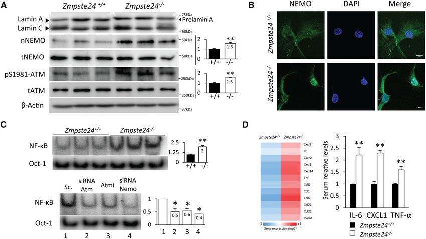
Downloaded from genesdev.cshlp.org on March 19, 2015 - Published by Cold Spring Harbor Laboratory Press

Osorio et al.

Figure 2. A signaling pathway involving ATM and NEMO activates NF-kB in Zmpste24-deficient mice. (A) ATM activation and
nuclear translocation of NEMO in Zmpste24 / cells. Western blot of NEMO protein was performed in nuclear extracts from livers of
3-mo-old Zmpste24 / mice (n = 3) and wild-type littermates (n = 3). Total NEMO, ATM, and pSer1981-ATM levels were analyzed in
total extracts of the same animals. b-Actin was used as loading control. Signals were quantified and are represented as relative mean
values 6 SEM. (*) P < 0.05; (**) P < 0.01, two-tailed Student’s t-test. (B) Nuclear staining of NEMO protein in fibroblasts from
Zmpste24-deficient mice supports nuclear translocation upon prelamin A-induced nuclear stress. Representative images are shown.
Bar, 10 mm. (C) ATM and NEMO siRNA transfection experiments demonstrate causal involvement of these proteins in NF-kB
activation. (Top panel) Nuclear extracts from Zmpste24 / and Zmpste24+/+ fibroblasts were analyzed by EMSA using 32P-labeled NF-
kB and Oct-1 probes. (Bottom panel) siRNA transfection analysis by EMSA as well as ATM inhibitor KU55933 incubations were
performed in three independent Zmpste24 / fibroblast cell lines, and the image shows a representative example. Signals were
quantified and are represented as relative mean values 6 SEM. (*) P < 0.05; (**) P < 0.01, two-tailed Student’s t-test. See also
Supplemental Figure 4. (D, left panel) Transcriptional analysis of SASP-related genes in Zmpste24 / and wild-type livers. The right
panel shows serum determinations of IL-6, CXCL1, and TNF-a in 3-mo-old Zmpste24 / (n = 5) and Zmpste24+/+ mice (n = 5). Plot
represents relative mean values 6 SEM. (*) P < 0.05; (**) P < 0.01, two-tailed Student’s t-test.

able to reduce the nuclear presence of NF-kB dimers to               sion molecules, such as Cxcr2 or Icam1, were also found
levels comparable with those of wild-type fibroblasts (Fig.          altered in these mutant animals. Moreover, serum levels
2C). Moreover, and consistent with the proposed role of              of IL-6, CXCL1, and TNF-a were increased in Zmpste24-
NEMO in nuclear NF-kB activation, NEMO siRNA also                    deficient mice (Fig. 2D). These three cytokines are impor-
abrogated NF-kB activation (Fig. 2C).                                tant mediators of inflammatory processes and have been
                                                                     related to aging and DDR (Kuilman and Peeper 2009; Biton
The secretory phenotype of senescent cells contributes               and Ashkenazi 2011).
to the establishment of chronic inflammation                           Together, these results provide experimental evidence
in Zmpste24-deficient mice                                           that prelamin A accumulation induces NF-kB activation
                                                                     by an ATM- and NEMO-dependent pathway. Nuclear
Senescent cells secrete a plethora of interleukins, in-
                                                                     activation of NF-kB contributes to the establishment of
flammatory cytokines, and growth factors that can affect
                                                                     a secretory phenotype that could give rise to a systemic
surrounding cells, ultimately developing a secretory phe-
                                                                     inflammatory situation.
notype of senescent cells (SASP) (Kuilman and Peeper
2009; Coppe et al. 2010; Freund et al. 2010). Accordingly,
                                                                     RelA heterozygosity extends longevity and prevents
we analyzed the transcriptional profile of SASP-associated
                                                                     the development of progeroid features
genes in livers from Zmpste24-deficient mice and found
                                                                     in Zmpste24 / mice
a >1.5-fold up-regulation of several cytokine-encoding
genes, such as Il6, Cxcl1, Cxcl2, Ccl8, and Tnf (Fig. 2D).           To test the specific contribution of NF-kB activation to
Likewise, genes encoding different receptors and adhe-               the progeroid phenotype shown by Zmpste24-deficient

2314       GENES & DEVELOPMENT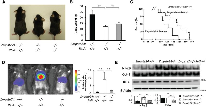
Downloaded from genesdev.cshlp.org on March 19, 2015 - Published by Cold Spring Harbor Laboratory Press

                                                                                                          Causal role of NF-kB in progeria

mice, we used a previously described method of NF-kB                   lated to a reduction in NF-kB activity in vivo, as assessed
genetic reduction (Kawahara et al. 2009; Tilstra et al.                by hydrodynamic delivery of the NF-kB luciferase reporter.
2012) based on the use of RelA-haploinsufficient mice                  Thus, Zmpste24 / RelA+/ mice have lower biolumi-
(RelA+/ ). RelA is a component of NF-kB pathway with                   nescence signals as compared with Zmpste24 / RelA+/
                                                                       +
essential developmental functions (Hayden and Ghosh                      , reaching levels similar to wild-type animals (Fig. 3D).
2004). Thus, RelA / mice present embryonic lethality,                  Furthermore, NF-kB EMSA and Western blot experi-
but RelA+/ mice are viable and apparently normal de-                   ments confirmed that RelA haploinsufficiency reduces
spite having reduced NF-kB activity (Beg et al. 1995). We              the amount of nuclear RelA dimers (Fig. 3E; Supplemental
crossed Zmpste24+/ mice with RelA-haploinsufficient                    Fig. 5) without affecting the levels of other NF-kB active
mice to create double-mutant Zmpste24+/ RelA+/ ani-                    members, such as p52 or RelB (Supplemental Fig. 5).
mals, which were then inbred to obtain Zmpste24 knock-                 Remarkably, no differences were found in nuclear levels
out mice with different dosages of RelA. As expected, we               of NEMO protein between Zmpste24 / RelA+/ and
did not obtain any RelA / mice, as they were embryonic-                Zmpste24 / RelA+/+ animals, supporting the fact that
lethal, and Zmpste24 / RelA+/+ animals resulting from                  NEMO translocation is an upstream event not affected
these crosses exhibited a phenotype identical to that                  by RelA haploinsufficiency.
of Zmpste24 / mice (Pendas et al. 2002). However,                          The accumulation of farnesylated forms of prelamin A
Zmpste24 / RelA+/ showed improved body weights                         has been associated with profound dermal alterations,
(Fig. 3A,B) and extended life spans as compared with                   including loss of hypodermal adipocytes, decreased pro-
Zmpste24 / RelA+/+ animals (Fig. 3C). Significantly, the               liferation of keratinocytes, structural aberrations in hair
mean survival of Zmpste24 / RelA+/ mice was extended                   follicles, and functional defects in stem cells present in
from 118 to 146 d, and the maximum survival was ex-                    this tissue (Espada et al. 2008; Sagelius et al. 2008; Wang
tended from 147 to 174 d (P < 0.05) (Fig. 3C). Notably,                et al. 2008). Notably, Zmpste24 / RelA+/ mice showed
increased life span in RelA-haploinsufficient mice corre-              a complete recovery in most of the skin phenotypes

Figure 3. RelA haploinsufficiency extends longevity of Zmpste24-deficient mice by reducing NF-kB activity. (A) Representative
photograph of 3-mo-old wild-type, Zmpste24 / RelA+/+, and Zmpste24 / RelA+/ littermates. (B) Zmpste24 / RelA+/ mice (n = 8)
showed improved body weight as compared with Zmpste24 / RelA+/+ mice (n = 7). P < 0.01, two-tailed Student’s t-test. Plot represents
mean values 6 SEM. (C) Kaplan-Meier survival plot showing the increase in life span of Zmpste24 / RelA+/ mice (n = 8) as compared
with Zmpste24 / RelA+/+ littermates (n = 7). P < 0.05, log-rank/Mantel-Cox test. (D) Reduced NF-kB activity in vivo in Zmpste24 / RelA+/
animals. Hydrodynamic delivery of the NF-kB–luciferase gene reporter in the livers of 3-mo-old wild-type (n = 5), Zmpste24 / RelA+/+
(n = 5) and Zmpste24 / RelA+/ (n = 5) mice. The bioluminescence image was recorded as the photon flux per second and square
centimeter, and representative mice of each genotype are shown. Relative mean values are shown, and error bars indicate SEM. P < 0.01
Zmpste24 / RelA+/+ as compared with wild-type or Zmpste24 / RelA+/ animals, two-tailed Student’s t-test). (E) NF-kB and Oct-1
EMSA of 3-mo-old wild-type (n = 3), Zmpste24 / RelA+/+ (n = 3), and Zmpste24 / RelA+/ (n = 3) livers. Western blot shows reduced
levels of RelA in haploinsufficient RelA+/ animals. b-Actin was used as loading control. Signals were quantified and are represented as
relative mean values 6 SEM. P < 0.01, two-tailed Student’s t-test.

                                                                                                     GENES & DEVELOPMENT             2315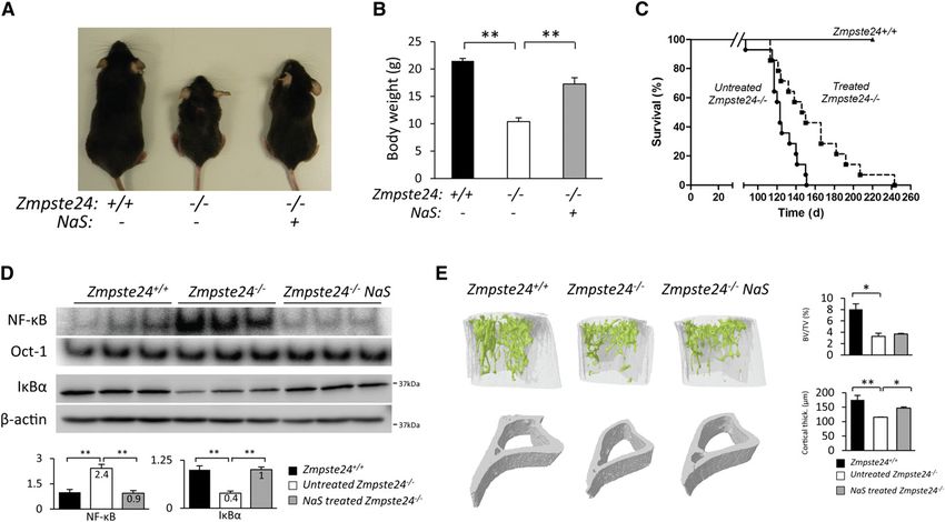
Downloaded from genesdev.cshlp.org on March 19, 2015 - Published by Cold Spring Harbor Laboratory Press

Osorio et al.

studied, which are also present in normal aging. Thus,               size and cellularity as well as abnormal lymphoid folli-
NF-kB blockade in the skin of Zmpste24-deficient mice                cles, which present an expansion of the mantle, disrupt-
increased cell proliferation, as indicated by increased              ing the germinal centers (Fig. 4B). Notably, the thymus
expression of the proliferation marker Ki-67 (Fig. 4A).              from Zmpste24 / mice shows an involution process char-
Additionally, Zmpste24 / RelA+/ showed a normal                      acterized by a reduction in tissue mass and thymic cellular-
subcutaneous fat layer and well-structured hair follicles            ity, loss of tissue structure, and abnormal architecture
(Fig. 4A).                                                           (Fig. 4B). Zmpste24 / RelA+/ mice display a notable im-
   NF-kB activity is also essential for a normal activity of         provement in both tissues. The spleen exhibits normal
the immune system, as demonstrated by the fact that                  lymphoid follicles, whereas the thymus has recovered nor-
mutations in several NF-kB genes, including NEMO,                    mal tissue mass, cellularity, and architecture, demonstrating
cause immunodeficiency syndromes (Li and Verma 2002).                the direct implication of NF-kB activation in the lymphoid
In this regard, we observed that Zmpste24-deficient mice             alterations characteristic of Zmpste24-deficient mice.
show marked histological alterations in the spleen and                  We also studied bone architecture in order to explore
thymus, two of the most important lymphoid organs.                   the potential involvement of NF-kB activation in the
Thus, Zmpste24-deficient mice exhibit reduced spleen                 bone abnormalities described in Zmpste24-deficient mice

Figure 4. RelA heterozygosis prevents important progeroid features of Zmpste24 / mice. (A) Reversal of skin alterations by NF-kB
blockade. (Left panel) Ki-67 immunohistochemistry of 3-mo-old wild-type (n = 3), Zmpste24 / RelA+/+ (n = 3), and Zmpste24 / RelA+/
(n = 3) mice. Representative photographs are shown. Plot represents average number of Ki-67-positive cells 6SEM. P < 0.01, two-tailed
Student’s t-test. Bar, 30 mm. The right panel shows full recovery of the subcutaneous fat layer in 3-mo-old Zmpste24 / RelA+/ mice
as compared with Zmpste24 / RelA+/+ littermates. Representative hematoxylin-eosin (H&E) staining micrographs are shown. Bar,
100 mm. (B) Thymus and spleen involution in Zmpste24-deficient mice is prevented in the RelA+/ background. (Top left panel)
Representative photographs of spleen and thymus from 3-mo-old wild-type (n = 3), Zmpste24 / RelA+/+ (n = 3), and Zmpste24 / RelA+/
(n = 3) mice are shown. (Bottom left panel) Relative weight values of thymus and spleen are represented 6SEM. P < 0.01, two-tailed
Student’s t-test. The right panel shows representative micrographs from spleen and thymus tissues of wild-type, Zmpste24 / RelA+/+,
and Zmpste24 / RelA+/ animals. Spleen details show representative lymphoid follicles; different regions are circled by dotted white
lines (germinal center and mantle and marginal zones). Note that the Zmpste24 / RelA+/+ thymus shows a notable tissue involution
where lymphoid tissue has been mostly replaced by adipose tissue. Bar, 100 mm.

2316       GENES & DEVELOPMENT
Downloaded from genesdev.cshlp.org on March 19, 2015 - Published by Cold Spring Harbor Laboratory Press

                                                                                                          Causal role of NF-kB in progeria

(de Carlos et al. 2008). To this end, microcomputed                    the same biological effect could be achieved by using
tomography (mCT) analyses were performed on tibias                     a pharmacological strategy. To this end, we chose sodium
from Zmpste24 / RelA+/ and Zmpste24 / RelA+/+ mice.                    salicylate, a nonsteroidal anti-inflammatory drug that at
No differences were found between the two groups in the                high doses is able to efficiently inhibit the IKK complex
trabecular region, and only a slight improvement was found             and has been satisfactorily used to reduce NF-kB activa-
in the cortical region of bones from Zmpste24 / RelA+/ as              tion in animal models of muscular dystrophy (Cai et al.
compared with Zmpste24 / RelA+/+ mice (Supplemental                    2004). Thus, we treated Zmpste-24-deficient mice with
Fig. 6). These experiments demonstrate that NF-kB hyper-               sodium salicylate (200 mg/kg per day), and both treated
activation contributes to reducing the life span of proge-             and control mice were weighted and observed during
roid mice and is responsible for some important features of            their lifetimes. Salicylate-treated Zmpste24 / mice
the progeroid phenotype. Moreover, the phenotypic im-                  showed improved body weights (Fig. 5A,B) and extended
provement of progeroid mice upon genetic reduction of                  life spans as compared with nontreated Zmpste24-
NF-kB supports the feasibility of developing pharmaco-                 deficient mice (Fig. 5C). Thus, the mean survival of
logical strategies aimed at inhibiting this pathway to treat           salicylate-treated Zmpste24 / mice was extended from
progeria.                                                              123 to 148 d, and the maximum survival was extended
                                                                       from 151 to 243 d (P < 0.01) (Fig. 5C).
                                                                          NF-kB EMSA analysis of livers from treated and un-
Sodium salicylate treatment efficiently prevents NF-kB
                                                                       treated mice demonstrated the targeted effect of salic-
activation and its associated alterations
                                                                       ylate on NF-kB activation (Fig. 5D), as treated mice
in Zmpste24-deficient mice
                                                                       showed decreased amounts of RelA dimers in the nucleus
Having shown that genetic inhibition of NF-kB extends                  as well as reduced degradation of the NF-kB inhibitor
longevity in Zmspte24-deficient mice, we asked whether                 IkBa (Fig. 5D). Salicylate-treated Zmpste24 / mice showed

Figure 5. Sodium salicylate treatment extends longevity and prevents progeroid features of Zmpste24-deficient mice. (A) Represen-
tative photographs of 3-mo-old Zmpste24+/+, untreated, and salicylate-treated Zmpste24-deficient mice. (B) Sodium salicylate-treated
Zmpste24 / mice (n = 12) showed improved body weight as compared with untreated Zmpste24 / mice (n = 12). Plot represents
weight mean values 6 SEM. P < 0.01, two-tailed Student’s t-test. (C) Kaplan-Meier survival plot showing the increase in life span of
salicylate-treated Zmpste24 / mice (n = 12) as compared with untreated Zmpste24 / littermates (n = 12). P < 0.01, log-rank/Mantel-
Cox test. (D) Liver NF-kB and Oct-1 EMSA analysis demonstrates effective NF-kB inhibition in salicylate-treated Zmpste24-deficient
mice. Western blot of IkBa was performed in total liver extracts. b-Actin was used as loading control. Signals were quantified and are
represented as relative mean values 6 SEM. P < 0.01, two-tailed Student’s t-test. (E, left panel) Representative three-dimensional image
generated from mCT analysis of tibias from 3-mo-old Zmpste24+/+ (n = 3), untreated (n = 3), and treated Zmpste24 / (n = 3) littermates.
The right panel contains a quantitative analysis of relative bone volumes (bone volume/tissue volume [BV/TV]) and cortical thickness.
Mean values are represented, and error bars indicate SEM. P < 0.05 as compared treated and untreated Zmpste24-deficient mice, two-
tailed Student’s t-test.

                                                                                                     GENES & DEVELOPMENT             2317
Downloaded from genesdev.cshlp.org on March 19, 2015 - Published by Cold Spring Harbor Laboratory Press

Osorio et al.

an increase in cell proliferation, subcutaneous fat layer          from the same mechanism as in the animal models de-
thickness, and normal hair follicles in the skin (Supple-          scribed above. To gain insight into the potential relevance
mental Fig. 7). Moreover, this treatment also prevented            of this phenomenon during normal human aging, we
thymic and spleen involution, with these organs having             analyzed NF-kB activation in fibroblasts from aged healthy
a size similar to wild-type organs (Supplemental Fig. 8).          donors. As shown in Supplemental Figure 11B, cells from
Notably, bone architecture analysis revealed no signifi-           advanced age donors showed a fivefold increase in NF-kB
cant changes in the trabecular region upon salicylate              activity as compared with fibroblasts obtained from
treatment. However, treated Zmpste24-deficient mice                young donors. In addition, aged fibroblasts showed in-
showed significantly improved cortical regions (Fig. 5E).          creased ATM activation and nuclear NEMO transloca-
This effect of salicylate treatment may derive from the            tion (Supplemental Fig. 11B), supporting that ATM- and
known inhibitory effect of this compound on the synthe-            NEMO-dependent NF-kB activation could be at least in
sis of prostaglandins involved in bone homeostasis (Ricciotti      part responsible for the increased inflammation observed
and FitzGerald 2011). In summary, these data demon-                during normal aging (Adler et al. 2007).
strate that salicylate treatment is effective at inhibiting
NF-kB activation in vivo and at significantly extending            Pharmacological inhibition of NF-kB extends longevity
the longevity of progeroid Zmpste24-deficient mice.                in the LmnaG609G/G609G model of HGPS
                                                                   Currently, LmnaG609G/G609G mice represent the best an-
NF-kB is hyperactivated in LmnaG609G/G609G mice                    imal model available for the preclinical testing of thera-
We recently generated a knock-in mouse strain carrying             peutic approaches for both HGPS and the age-associated
the most frequent HGPS mutation (Lmna c.1827C>T;                   pathologies derived from progerin accumulation in nor-
p.Gly609Gly) (Osorio et al. 2011a). LmnaG609G/G609G mice           mal aging. Thus, aiming to provide preclinical proof of
accumulate progerin and phenocopy the main clinical                concept of the feasibility of using nonsteroidal anti-
manifestations of HGPS, being a valuable model for the             inflammatory drugs to prevent the pathological conse-
study of progeroid syndromes as well as for the preclinical        quences of progerin accumulation, we decided to extend
evaluation of approaches aimed at preventing the patho-            the salicylate treatment results to the LmnaG609G/G609G
logical features of this condition. Comparison of transcrip-       mouse strain. To this end, we treated LmnaG609G/G609G
tional profiles from LmnaG609G/G609G and Lmna+/+ mouse             mice with sodium salicylate (200 mg/kg per day) and
livers using GSEA showed a significant correlation be-             found that salicylate-treated animals showed improved
tween transcriptional alterations detected and the gene            body weight as well an extended life span as compared
set that contains NF-kB-regulated genes (Supplemental              with untreated mice (Fig. 6D–F). Moreover, the mean
Fig. 9). Next, we analyzed the NF-kB activation status in          survival of salicylate-treated mice was extended from
LmnaG609G/G609G mice. Hydrodynamic delivery of the                 107 to 152 d, and the maximum survival was extended
NF-kB luciferase reporter showed that mutant mice have             from 114 to 180 d (P < 0.01).
an eightfold increase in bioluminescent signal (Fig. 6A) as           Together, these results demonstrate that hyperactiva-
compared with control littermates, indicating that these           tion of the NF-kB pathway plays an important role in the
mutant mice present an activation of the NF-kB pathway             development of aging-associated pathologies derived from
similar to that observed in Zmpste24-deficient animals.            the accumulation of abnormal lamin A precursors at the
These results were also confirmed by NF-kB EMSA exper-             nuclear lamina. Moreover, these data provide evidence of
iments and IkBa Western blot analysis using nuclear                the feasibility of developing anti-aging strategies based on
extracts from control and mutant liver samples (Fig. 6B).          the use of anti-inflammatory compounds.
   The nuclear accumulation of progerin activates ATM
kinase, as revealed by the increased amounts of phospho-
                                                                   Discussion
Ser1981-ATM present in LmnaG609G/G609G samples (Fig. 6B).
Consequently, NEMO accumulation in the nucleus sup-                Over recent years, mechanistic studies on human aging
ports the view that both prelamin A and progerin activate          have gained new insights from the elucidation of the
NF-kB through the same molecular mechanism. Serum                  molecular defects underlying the development of accel-
levels of IL-6, CXCL1, and TNF-a were also increased in            erated aging syndromes. These complex and dramatic
LmnaG609G/G609G mice (Fig. 6C), which is in agreement              diseases result from the combined action of both cell-
with the systemic inflammation condition described                 autonomous and systemic alterations (Marino et al. 2008;
above for Zmpste24-deficient mice and could be respon-             Osorio et al. 2011b). Thus, nuclear envelope defects caus-
sible for the similar immunological alterations found in           ative of progeroid laminopathies lead to perturbations in
LmnaG609G/G609G mice (Supplemental Fig. 10).                       cellular pathways, including p53-dependent cell senes-
   To explore the possible occurrence of these alterations         cence, deregulation of the somatotroph axis, and changes
in human progeria, we first performed an EMSA analysis             in metabolic master regulators (Lammerding et al. 2004;
on HGPS fibroblasts. This study revealed a twofold in-             Varela et al. 2005; Scaffidi and Misteli 2008; Marino et al.
crease in NF-kB activity in progeria cells (Supplemental           2010). Moreover, secretion of signaling molecules by
Fig. 11A). HGPS cells also showed increased levels of              affected cells could be a major contributor to progeria
phospho-Ser1981-ATM and nuclear NEMO, suggesting                   development, as secreted molecules can act on distant
that NF-kB activation in these human progeria cells results        organs, leading to an amplifying cascade of aged signals.

2318       GENES & DEVELOPMENT
Downloaded from genesdev.cshlp.org on March 19, 2015 - Published by Cold Spring Harbor Laboratory Press
Causal role of NF-kB in progeria
Figure 6. Salicylate treatment reduces NF-kB hyperactivation and extends longevity in LmnaG609G/G609G mice. (A) LmnaG609G/G609G
mice show an increased NF-kB activation, as demonstrated by hydrodynamic delivery of NF-kB-luciferase gene reporter in the livers of
Lmna+/+ (n = 5) and LmnaG609G/G609G (n = 5) mice. The bioluminescence image was recorded as the photon flux per second and square
centimeter, and three representative mice from each genotype are shown. Plot represents relative mean values, and error bars indicate
SEM. P < 0.01, two-tailed Student’s t-test. (B) NF-kB activation in LmnaG609G/G609G mouse livers. Western blot of lamin A/C isoforms
showing progerin accumulation in mutant mice. NF-kB and Oct-1 were analyzed by EMSA. Western blots of lamin A/C, IkBa, pS1981-
ATM, and b-actin were performed in whole-liver extracts. NEMO Western blot was performed in nuclear extracts. Signals were
quantified and are represented as relative mean values 6 SEM. (*) P < 0.05; (**) P < 0.01, two-tailed Student’s t-test. (C) Serum
determinations of IL-6, CXCL1, and TNF-a in 3-mo-old Lmna+/+ (n = 5) and LmnaG609G/G609G (n = 5) mice. Plot represents relative mean
values 6 SEM. (*) P < 0.05; (**) P < 0.01, two-tailed Student’s t-test. (D) Representative photographs of 3-mo-old Lmna+/+, untreated, and
sodium salicylate-treated LmnaG609G/G609G mice. (E) Sodium salicylate-treated LmnaG609G/G609G mice (n = 6) showed improved body
weight as compared with untreated LmnaG609G/G609G mice (n = 6). Plot represents weight mean values 6 SEM. P < 0.01, two-tailed
Student’s t-test. (F) Kaplan-Meier survival plot showing a significant increase in the life span of salicylate-treated LmnaG609G/G609G
mice (n = 6) as compared with LmnaG609G/G609G littermates (n = 6). P < 0.01, log-rank/Mantel-Cox test.
However, little is known about the nature of systemic this context, ATM and NEMO act coordinately to acti-
factors involved in aging or aging-like processes as well as vate NF-kB, as demonstrated by the finding that their
the mechanisms connecting nuclear defects to perturba- respective inhibition prevents prelamin A-induced NF-kB
tions in such systemic factors. Here, we present an ATM- activation. Consistent with the observed activation of
dependent NF-kB activation pathway that links nuclear NF-kB signaling in Zmpste24 / and LmnaG609G/G609G
lamina defects to systemic inflammation. We also dem- progeroid mice, several cytokines and adhesion mole-
onstrate in vivo the causal role for this pathway in the cules are strongly up-regulated in cells and tissues from
emergence of age-associated pathologies and illustrate these mice, likely contributing to the initiation and
the feasibility of targeting this signaling cascade for the maintenance of an inflammatory response. Among the
treatment of premature aging symptoms. plethora of proinflammatory cytokines secreted by se-
By using two different mouse models of progeroid nescent cells, we propose that IL-6, CXCL1, and TNF-a
laminopathy (Zmpste24-deficient and LmnaG609G mice), may have essential roles in progeria development by
we showed that accumulation of prelamin A/progerin at nonautonomous stimulation of surrounding cells through
the nuclear lamina activates the NF-kB pathway in an the activation of their cognate cell surface receptors and
ATM- and NEMO-dependent manner, illustrating that signal transduction pathways (Coppe et al. 2010; Freund
alterations in the nuclear architecture generate stress et al. 2010). Thus, systemically increased levels of cyto-
signals that activate important DNA damage sensors. In kines amplify the inflammatory stimuli and are implicated
GENES & DEVELOPMENT 2319Downloaded from genesdev.cshlp.org on March 19, 2015 - Published by Cold Spring Harbor Laboratory Press
Osorio et al.
in the establishment of a feed-forward signaling process the leading regulators of aging (Aw and Palmer 2011). The
(Biton and Ashkenazi 2011). Since senescent cells are reduction in tissue mass and cellularity and the loss of
potentially time-persisting, the continued production of tissue structure leads to a decline in naive T-cell output
cytokines and the subsequent NF-kB activation lead to an as well as to the occurrence of changes in the peripheral
increased inflammation in progeroid mice that contrib- T-cell compartment that contribute to the clinical signs
utes to age-related wasting and dramatically shortens of immunosenescence (Hale et al. 2010). Consistent with
organismal life span (Fig. 7). these observations, we demonstrated in this study that
According to our results, NF-kB could be regarded as NF-kB hyperactivation and the systemic inflammation
a major regulator of accelerated aging, as demonstrated by derived therefrom drive thymus and spleen involution
the fact that NF-kB blockade significantly increases life and that NF-kB inhibition is able to prevent these age-
span in both Zmpste24 / and LmnaG609G/G609G pro- related alterations.
geroid mice. Furthermore, the NF-kB blockade strategies Although the biological significance of NF-kB activa-
used in this study have allowed us to provide new molec- tion during aging is not completely clear, the findings
ular insights into the involvement of NF-kB in dermal and reported herein, together with the fact that other models
immunological homeostasis. The primary manifestations of normal and accelerated aging show increased levels of
of accelerated aging in skin affect cell proliferation, hair NF-kB activity (Kawahara et al. 2009; Rodier et al. 2009),
follicles, and the subcutaneous fat layer (Sur et al. 2008). support the idea that inflammation is a major regulator
NF-kB blockade was able to prevent these alterations, of the aging process. The mechanism by which NF-kB
demonstrating a causal role of NF-kB deregulation in age- signaling is activated with age also remains largely un-
associated defects in skin homeostasis. A similar situa- explored, but our data indicate that the nuclear envelope
tion has been shown in lymphoid organs, which is of abnormalities occurring in both normal and premature
special interest, as thymic involution is considered one of aging (Scaffidi and Misteli 2006) may contribute, at least
in part, to the activation of this inflammatory pathway.
The primary function of NF-kB activation in response to
nuclear envelope defects could be to protect damaged
cells against apoptosis (Wang et al. 1999; Salminen et al.
2011). Since a proper clearance of senescent cells by the
immune system seems to be crucial for homeostasis
maintenance in aging and cancer (Baker et al. 2011; Kang
et al. 2011), both NF-kB hyperactivation and the sub-
sequent age-related immunological decline could be com-
promising an appropriate response against age-accumulated
senescent cells.
The results of the present work also suggest that the
use of nonsteroidal anti-inflammatory drugs, alone or
in combination with statins and aminobisphophonates
(Varela et al. 2008), could be useful for the treatment of
accelerated aging-associated alterations occurring during
the course of progeroid laminopathies (Hennekam 2006;
Merideth et al. 2008; Puente et al. 2011). Moreover,
pharmacological modulation of the NF-kB pathway also
could be of interest for slowing down the progression of
physiological aging (Rando and Chang 2012). The identi-
fication of NF-kB signaling activation in mouse models
with accelerated aging also provides further in vivo
support for the provocative proposal that the mainte-
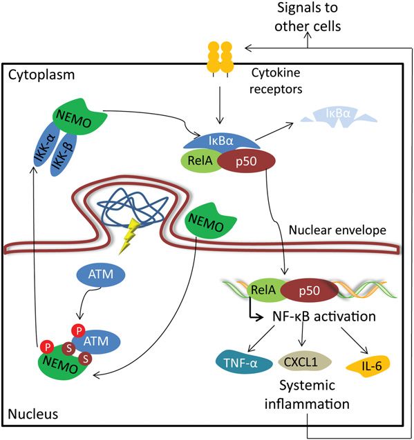
Figure 7. A model depicting ATM- and NEMO-mediated NF- nance of the aged state requires an active signaling
kB activation upon accumulation of prelamin A isoforms. program and that age-linked phenotypes can be substan-
Nuclear envelope alterations caused by the accumulation of tially reversed by intervention on the activity of individ-
prelamin A isoforms trigger a nuclear stress pathway involv- ual genes (Adler et al. 2007; Freije and Lopez-Otin 2012;
ing ATM and NEMO proteins. As a consequence of prelamin Rando and Chang 2012). Accordingly, we suggest that the
A/progerin accumulation, ATM is activated, and NEMO is trans- progeroid mice used in this study may represent a valu-
located into the nucleus. NEMO phosphorylation by ATM able tool for further exploration of this proposal due to
induces their translocation out of the nucleus to activate the their faithful recapitulation of the biological dysfunc-
cytoplasmic IKK complex. Subsequent NF-kB activation triggers
tions normally associated with advanced age. Likewise,
an inflammatory-associated transcriptional program that leads
to the secretion of IL-6, CXCL1, and TNF-a. Increased levels of
we propose that these progeroid mice are an essential tool
senescence-associated cytokines cause systemic inflammation for the development of putative rejuvenation strategies
by acting over distant cells and tissues. Systemic inflammation aimed at controlling the inflammatory responses driven
amplifies a cascade of aged signals, critically affecting tissue by NF-kB signaling that occur during both normal and
homeostasis and finally reducing the life span of progeroid mice. pathological aging.
2320 GENES & DEVELOPMENTDownloaded from genesdev.cshlp.org on March 19, 2015 - Published by Cold Spring Harbor Laboratory Press
Causal role of NF-kB in progeria
Materials and methods mycotic (Gibco). Studies with primary mouse fibroblasts were
performed at postnatal day 3 (P3). HGPS (AG01972) and control
Antibodies and reagents fibroblasts (AG10803) were obtained from Coriell Cell reposi-
tory. Dermal fibroblasts derived from young (GM05565, 3-yr-old;
Antibodies specific for lamin A/C (sc-6215, Santa Cruz Bio- and GM00038, 9-yr-old) and advanced age donors (AG04059, 96-
technology), IkBa (4812, Cell Signaling), RelA (8242, Cell Signal- yr-old) were obtained from Coriell Cell repository, except the
ing), NEMO (2685, Cell Signaling), ATM (2873, Cell Signaling), JM0097Y cell line that was obtained directly from a 97-yr-old
phospho-Ser1981-ATM (mouse: 200-301-400, Rockland Labora- donor. For transfection of siRNAs, we followed the manufac-
tories; human: 5883S, Cell Signaling), p52 (sc-7386, Santa Cruz turer’s instructions (Stealth siRNAs, Life Technologies). Briefly,
Biotechnology), RelB (sc-48366, Santa Cruz Biotechnology), Ki- cells were plated at low density (30%–50%), and up to 150 pg of
67 (KI681C01, DCS-Innovative Diagnostik-Systeme), a-tubulin siRNA was added with 7.5 mL of Lipofectamine RNAiMAX (Life
(T6074, Sigma), and b-actin (AC-40, Sigma) were used in this Technologies). The same procedure was repeated after 4 h, and
study for Western blot, immunofluorescence, and immunohisto- analyses were performed 48 h after the first transfection. For
chemistry experiments. The ATM inhibitor KU55933 was ob- pharmacological inhibition of ATM kinase activity, cells were
tained from Calbiochem. incubated for 12 h in the presence of 10 mM KU55933 ATM
inhibitor.
Mice
We generated and genotyped Zmpste24 / and LmnaG609G EMSA
mice as previously described (Pendas et al. 2002; Osorio et al. Nuclear extracts from cells were prepared as described pre-
2011a). RelA+/ mice were obtained from Jackson Laboratories. viously (Schreiber et al. 1989). For the preparation of nuclear
Zmpste24+/ and RelA+/ were crossed, and Zmpste24+/ RelA+/ extracts from tissues, a slightly modified protocol was used.
progeny were interbred to obtain Zmspte24 / RelA+/ mice. We Briefly, frozen tissues were homogenized in cold buffer A
administrated sodium salicylate (200 mg/kg per day; Sigma- (10 mM Tris-HCl at pH 8, 1.5 mM MgCl2, 10 mM KCl, 1 mM
Aldrich) in PBS intraperitoneally to mice every day. Neither DTT, 23 protease inhibitor cocktail) with a Potter homoge-
vehicle alone nor sodium salicylate treatment produced any nizer. After incubation on ice for 15 min, samples were pelleted
apparent damage or stress responses in mice. We performed mCT by centrifugation and resuspended in cold buffer A containing
analysis of tibias with a mCT SkyScan 1172 system (SkyScan). 0.1% Triton X-100. After incubation on ice, cells were pelleted,
For histology analysis, we fixed samples with 4% paraformalde- and nuclear pellets were resuspended in cold buffer C (20 mM
hyde in PBS, processed the resulting preparations into serial Tris-HCl at pH 8, 25% glycerol [v/v], 0.4 M NaCl, 1.5 mM
paraffin sections, and stained each with hematoxylin and eosin MgCl2, 0.2 mM EDTA, 0.5 mM DTT, 23 protease inhibitor
(H&E). All of the animal experiments were performed in accor- cocktail). Samples were vigorously rocked for 15 min at 4°C on
dance with the guidelines of the Committee for Animal Exper- a shaking platform. The nuclear extracts were finally centri-
imentation of the Universidad de Oviedo. fuged for 5 min in a microfuge at 4°C, and the supernatants
were frozen in aliquots at 70°C. NF-kB and Oct-1 consensus
Hydrodynamic delivery of an NF-kB–luciferase gene reporter oligonucleotides (Promega) were radiolabeled by T4 polynucle-
otide kinase (New England Biolabs) in the presence of g-32P-
A plasmid containing a firefly luciferase gene under the control ATP. Labeled oligonucleotides were incubated with 15–20 mg
of a minimal CMV promoter and tandem repeats of the NFkB of nuclear extracts in 13 binding buffer (43 glycerol, 20 mM
transcriptional response element was injected by using the Tris-HCl at pH 8, 60 mM NaCl, 5 mM MgCl2, 1 mM DTT, 13
hydrodynamic technique previously described (Herweijer and protease inhibitor cocktail) in the presence of poly-dI–dC.
Wolff 2007). In brief, 10 mg/mL solution of the plasmid was Complexes were run in nondenaturing, 6% acrylamide gels,
prepared in sterile Ringer’s buffer at room temperature. Mice and exposed to X-ray detector Fuji PhosphorImager (Fujifilm
were anaesthetized, and the lateral tail vein was accessed with Global).
a 21-gauge needle. Administration of the solution (1 mL/10 g)
was performed without extravasation.
Western blot analysis
Cultured cells were washed twice with 13 PBS and resus-
Bioluminescent imaging and analysis
pended in 100 mM Tris-HCl (pH 7.4), 2% SDS, and 50 mM
Mice were anaesthetized and injected intraperitoneally with EDTA. Tissues were snap-frozen in liquid nitrogen. Frozen
200 mL of D-luciferin solution (15 g/L in PBS; Melford Laboratories). tissues (;50 mg in each sample) were homogenized in 300 mL
Imaging was completed between 2 and 5 min after injection with of 100 mM Tris-HCl (pH 7.4), 2% SDS, and 50 mM EDTA with
a Xenogen IVIS system coupled to Living Image acquisition and a Polytron homogenizer. Protein concentration was evaluated
analysis software (Xenogen). Photon flux was calculated for each with the bicinchoninic acid technique (Pierce BCA protein
mouse by using a rectangular region of interest. This value was assay kit). Equal amounts of proteins were loaded onto SDS–
scaled for each mouse to a comparable background value. Rep- polyacrylamide gels. After electrophoresis, gels were electro-
resented values were recorded 7 d after injection to avoid any transferred onto nitrocellulose membranes or Immobilon-FL
potential interference caused by experimental procedure-induced polyvinylidene fluoride membranes (Millipore), blocked with
liver inflammation. 5% nonfat dry milk in TBS-T buffer (20 mM Tris at pH 7.4,
150 mM NaCl, 0.05% Tween 20), and incubated overnight at 4°C
with the different primary antibodies. Finally, blots were in-
Cell culture and siRNA delivery
cubated with 1:10,000 secondary antibody conjugated with
We extracted mouse fibroblasts from 15-wk-old ears as pre- horseradish peroxidase (HRP) (Jackson ImmunoResearch Labo-
viously described (Varela et al. 2005). We maintained cultures ratories) in 1.5% nonfat milk in TBS-T. Then, we washed and
in Dulbecco’s modified Eagle’s medium (Gibco) supplemented developed the immunoreactive bands with Immobilon Western
with 10% fetal bovine serum (Gibco) and 1% antibiotic anti- chemiluminescent HRP substrate (Millipore).
GENES & DEVELOPMENT 2321Downloaded from genesdev.cshlp.org on March 19, 2015 - Published by Cold Spring Harbor Laboratory Press Osorio et al. Immunohistochemistry and immunofluorescence analysis GSEA Formalin-fixed paraffin-embedded tissue sections were cut at GSEA was performed as described in the original citation 5 mm for immunohistochemical detection of Ki67 on a Discover (Subramanian et al. 2005). For data analysis, we used GSEA automated immunostainer (Ventana Medical Systems). Depar- release 2.06 and MSigDB release 2.5 (http://www.broadinstitute. affinization and heat-induced antigen retrieval were performed org/gsea/index.jsp). Weighted enrichment scores were calculated directly on the stainer. Antigen retrieval procedures were as with gene expression lists ranked by signal to noise ratio. The follows: Retrieval was performed with CC2 solution (Ventana maximum gene set size was set to 500 genes, the minimum gene Medical Systems) for 30 min at 95°C. Primary antibody in- set size was set to 20 genes, and the number of permutations was cubation was performed for 1 h at 37°C. Finally, HRP-conjugated set to 1000. Analyses were performed with a gene set composed antibody (OmniMap anti-Rb HRP, Ventana Medical Systems) of genes that contain, in the promoter regions [ 2 kb, 2 kb] was applied for 16 min at 37°C. Staining was visualized by using around the transcription start site, the motif GGGRATTTCC, ChromoMap DAB kit (Ventana Medical Systems). Cells were which matches the consensus binding site for the NF-kB tran- counterstained with hematoxylin and visualized by light mi- scription factor. Selected enriched pathways had a relaxed false croscopy. Quantitative analyses were performed according to the discovery rate of
Downloaded from genesdev.cshlp.org on March 19, 2015 - Published by Cold Spring Harbor Laboratory Press

                                                                                                      Causal role of NF-kB in progeria

   IKKb/NF-kB activation causes severe muscle wasting in              kB-dependent gene expression and organismal life span. Cell
   mice. Cell 119: 285–298.                                           136: 62–74.
Coppe JP, Desprez PY, Krtolica A, Campisi J. 2010. The             Kirkwood TB. 2005. Understanding the odd science of aging.
   senescence-associated secretory phenotype: The dark side           Cell 120: 437–447.
   of tumor suppression. Annu Rev Pathol 5: 99–118.                Kuilman T, Peeper DS. 2009. Senescence-messaging secretome:
de Carlos F, Varela I, Germana A, Montalbano G, Freije JM,            SMS-ing cellular stress. Nat Rev Cancer 9: 81–94.
   Vega JA, Lopez-Otin C, Cobo JM. 2008. Microcephalia with        Lammerding J, Schulze PC, Takahashi T, Kozlov S, Sullivan T,
   mandibular and dental dysplasia in adult Zmpste24-deficient        Kamm RD, Stewart CL, Lee RT. 2004. Lamin A/C deficiency
   mice. J Anat 213: 509–519.                                         causes defective nuclear mechanics and mechanotransduc-
Dechat T, Pfleghaar K, Sengupta K, Shimi T, Shumaker DK,              tion. J Clin Invest 113: 370–378.
   Solimando L, Goldman RD. 2008. Nuclear lamins: Major            Lavin MF. 2008. Ataxia-telangiectasia: From a rare disorder to
   factors in the structural organization and function of the         a paradigm for cell signalling and cancer. Nat Rev Mol Cell
   nucleus and chromatin. Genes Dev 22: 832–853.                      Biol 9: 759–769.
De Sandre-Giovannoli A, Bernard R, Cau P, Navarro C, Amiel J,      Le Saux S, Weyand CM, Goronzy JJ. 2012. Mechanisms of
   Boccaccio I, Lyonnet S, Stewart CL, Munnich A, Le Merrer           immunosenescence: Lessons from models of accelerated im-
   M, et al. 2003. Lamin a truncation in Hutchinson-Gilford           mune aging. Ann N Y Acad Sci 1247: 69–82.
   progeria. Science 300: 2055. doi: 10.1126/science.1084125.      Li Q, Verma IM. 2002. NF-kB regulation in the immune system.
Eriksson M, Brown WT, Gordon LB, Glynn MW, Singer J,                  Nat Rev Immunol 2: 725–734.
   Scott L, Erdos MR, Robbins CM, Moses TY, Berglund P,            Liu B, Wang J, Chan KM, Tjia WM, Deng W, Guan X, Huang JD,
   et al. 2003. Recurrent de novo point mutations in lamin            Li KM, Chau PY, Chen DJ, et al. 2005. Genomic instability in
   A cause Hutchinson-Gilford progeria syndrome. Nature 423:          laminopathy-based premature aging. Nat Med 11: 780–785.
   293–298.                                                        Marino G, Ugalde AP, Salvador-Montoliu N, Varela I, Quiros
Espada J, Varela I, Flores I, Ugalde AP, Cadinanos J, Pendas AM,      PM, Cadinanos J, van der Pluijm I, Freije JM, Lopez-Otin C.
   Stewart CL, Tryggvason K, Blasco MA, Freije JM, et al. 2008.       2008. Premature aging in mice activates a systemic meta-
   Nuclear envelope defects cause stem cell dysfunction in            bolic response involving autophagy induction. Hum Mol
   premature-aging mice. J Cell Biol 181: 27–35.                      Genet 17: 2196–2211.
Freije JM, Lopez-Otin C. 2012. Reprogramming aging and             Marino G, Ugalde AP, Fernandez AF, Osorio FG, Fueyo A, Freije
   progeria. Curr Opin Cell Biol doi: 10.1016/j.ceb.2012.08.009.      JM, Lopez-Otin C. 2010. Insulin-like growth factor 1 treat-
Freund A, Orjalo AV, Desprez PY, Campisi J. 2010. Inflammatory        ment extends longevity in a mouse model of human pre-
   networks during cellular senescence: Causes and conse-             mature aging by restoring somatotroph axis function. Proc
   quences. Trends Mol Med 16: 238–246.                               Natl Acad Sci 107: 16268–16273.
Gruenbaum Y, Margalit A, Goldman RD, Shumaker DK, Wilson           McCool KW, Miyamoto S. 2012. DNA damage-dependent NF-
   KL. 2005. The nuclear lamina comes of age. Nat Rev Mol             kB activation: NEMO turns nuclear signaling inside out.
   Cell Biol 6: 21–31.                                                Immunol Rev 246: 311–326.
Hale JS, Frock RL, Mamman SA, Fink PJ, Kennedy BK. 2010.           Mekhail K, Moazed D. 2010. The nuclear envelope in genome
   Cell-extrinsic defective lymphocyte development in Lmna /          organization, expression and stability. Nat Rev Mol Cell Biol
   mice. PLoS ONE 5: e10127. doi: 10.1371/journal.pone.               11: 317–328.
   0010127.                                                        Merideth MA, Gordon LB, Clauss S, Sachdev V, Smith AC, Perry
Hayden MS, Ghosh S. 2004. Signaling to NF-kB. Genes Dev 18:           MB, Brewer CC, Zalewski C, Kim HJ, Solomon B, et al. 2008.
   2195–2224.                                                         Phenotype and course of Hutchinson-Gilford progeria syn-
Hayden MS, Ghosh S. 2008. Shared principles in NF-kB signal-          drome. N Engl J Med 358: 592–604.
   ing. Cell 132: 344–362.                                         Miyamoto S. 2011. Nuclear initiated NF-kB signaling: NEMO
Hayden MS, Ghosh S. 2012. NF-kB, the first quarter-century:           and ATM take center stage. Cell Res 21: 116–130.
   Remarkable progress and outstanding questions. Genes Dev        Osorio FG, Varela I, Lara E, Puente XS, Espada J, Santoro R,
   26: 203–234.                                                       Freije JM, Fraga MF, Lopez-Otin C. 2010. Nuclear envelope
Hennekam RC. 2006. Hutchinson-Gilford progeria syndrome:              alterations generate an aging-like epigenetic pattern in mice
   Review of the phenotype. Am J Med Genet A 140: 2603–               deficient in Zmpste24 metalloprotease. Aging Cell 9: 947–
   2624.                                                              957.
Herweijer H, Wolff JA. 2007. Gene therapy progress and pros-       Osorio FG, Navarro CL, Cadinanos J, Lopez-Mejia IC, Quiros
   pects: Hydrodynamic gene delivery. Gene Ther 14: 99–107.           PM, Bartoli C, Rivera J, Tazi J, Guzman G, Varela I, et al.
Hoeijmakers JH. 2009. DNA damage, aging, and cancer. N Engl           2011a. Splicing-directed therapy in a new mouse model of
   J Med 361: 1475–1485.                                              human accelerated aging. Sci Transl Med 3: 106ra107. doi:
Huang TT, Wuerzberger-Davis SM, Wu ZH, Miyamoto S. 2003.              10.1126/scitranslmed.3002847.
   Sequential modification of NEMO/IKKg by SUMO-1 and              Osorio FG, Ugalde AP, Marino G, Puente XS, Freije JM, Lopez-
   ubiquitin mediates NF-kB activation by genotoxic stress.           Otin C. 2011b. Cell autonomous and systemic factors in
   Cell 115: 565–576.                                                 progeria development. Biochem Soc Trans 39: 1710–1714.
Janssens S, Tschopp J. 2006. Signals from within: The DNA-         Pendas AM, Zhou Z, Cadinanos J, Freije JM, Wang J, Hultenby
   damage-induced NF-kB response. Cell Death Differ 13: 773–          K, Astudillo A, Wernerson A, Rodriguez F, Tryggvason K,
   784.                                                               et al. 2002. Defective prelamin A processing and muscular
Kang TW, Yevsa T, Woller N, Hoenicke L, Wuestefeld T, Dauch           and adipocyte alterations in Zmpste24 metalloproteinase-
   D, Hohmeyer A, Gereke M, Rudalska R, Potapova A, et al.            deficient mice. Nat Genet 31: 94–99.
   2011. Senescence surveillance of pre-malignant hepatocytes      Perkins ND. 2007. Integrating cell-signalling pathways with NF-
   limits liver cancer development. Nature 479: 547–551.              kB and IKK function. Nat Rev Mol Cell Biol 8: 49–62.
Kawahara TL, Michishita E, Adler AS, Damian M, Berber E, Lin       Puente XS, Quesada V, Osorio FG, Cabanillas R, Cadinanos J,
   M, McCord RA, Ongaigui KC, Boxer LD, Chang HY, et al.              Fraile JM, Ordonez GR, Puente DA, Gutierrez-Fernandez A,
   2009. SIRT6 links histone H3 lysine 9 deacetylation to NF-         Fanjul-Fernandez M, et al. 2011. Exome sequencing and

                                                                                                GENES & DEVELOPMENT              2323
You can also read