Genome analysis of a halophilic bacterium Halomonas malpeensis YU PRIM 29T reveals its exopolysaccharide and pigment producing capabilities - Nature
←
→
Page content transcription
If your browser does not render page correctly, please read the page content below
www.nature.com/scientificreports
OPEN Genome analysis of a halophilic
bacterium Halomonas malpeensis
YU‑PRIM‑29T reveals its
exopolysaccharide and pigment
producing capabilities
Athmika1,2, Sudeep D. Ghate1,2, A. B. Arun1, Sneha S. Rao1, S. T. Arun Kumar1,
Mrudula Kinarulla Kandiyil1, Kanekar Saptami1 & P. D. Rekha1*
Halomonas malpeensis strain YU-PRIM-29T is a yellow pigmented, exopolysaccharide (EPS) producing
halophilic bacterium isolated from the coastal region. To understand the biosynthesis pathways
involved in the EPS and pigment production, whole genome analysis was performed. The complete
genome sequencing and the de novo assembly were carried out using Illumina sequencing and
SPAdes genome assembler (ver 3.11.1) respectively followed by detailed genome annotation. The
genome consists of 3,607,821 bp distributed in 18 contigs with 3337 protein coding genes and 53%
of the annotated CDS are having putative functions. Gene annotation disclosed the presence of
genes involved in ABC transporter-dependent pathway of EPS biosynthesis. As the ABC transporter-
dependent pathway is also implicated in the capsular polysaccharide (CPS) biosynthesis, we employed
extraction protocols for both EPS (from the culture supernatants) and CPS (from the cells) and found
that the secreted polysaccharide i.e., EPS was predominant. The EPS showed good emulsifying
activities against the petroleum hydrocarbons and its production was dependent on the carbon source
supplied. The genome analysis also revealed genes involved in industrially important metabolites
such as zeaxanthin pigment, ectoine and polyhydroxyalkanoate (PHA) biosynthesis. To confirm the
genome data, we extracted these metabolites from the cultures and successfully identified them. The
pigment extracted from the cells showed the distinct UV–Vis spectra having characteristic absorption
peak of zeaxanthin (λmax 448 nm) with potent antioxidant activities. The ability of H. malpeensis strain
YU-PRIM-29T to produce important biomolecules makes it an industrially important bacterium.
Halomonas malpeensis YU-PRIM-29T belongs to Halomonadaceae family within the Gammaproteobacteria. The
species of the genus Halomonas are Gram-negative, rod-shaped, aerobic and non-spore forming bacteria1. They
are highly halotolerant (up to 20% salinity) and are mostly associated with saline environments2. Many members
have been isolated from diverse saline or hyper-saline environments such as ocean water3 and hyper-saline lakes4
and also reported from varying pH and temperature conditions5.
Bacterial survival in the challenging extreme habitats is possible due to its unique capabilities in the biosyn-
thesis of metabolites that offer protection against such conditions. These molecules serve as osmolytes and protect
the cells from damage while allowing normal cellular functions. Many members of the genus Halomonas also
produce pigments and exopolysaccharides (EPS)5 having specific functional role in the adaptation and survival in
the extreme environmental conditions6. In addition to providing protection against the osmotic stress prevailing
in the marine environments, the EPS serves as a tactic for adhesion to solid surfaces and helps in the retention
of water and nutrients. It imparts stability to the structure of biofilms and forms a layer surrounding the cell to
provide an effective barrier against salinity, bacterial attacks and facilitate biochemical interactions among the
cells and the adjacent e nvironment7,8.
The important members of halophilic bacteria that produce commercially important EPS from Halo-
monadaceae family include Volcaniella eurihalina, Deleya marina, H. maura, H. anticariensis, H. ventosae, H.
1
Yenepoya Research Centre, Yenepoya (Deemed to be University), University Road, Deralakatte, Mangalore 575018,
India. 2These authors contributed equally: Athmika and Sudeep D. Ghate. *email: rekhapd@yenepoya.edu.in
Scientific Reports | (2021) 11:1749 | https://doi.org/10.1038/s41598-021-81395-1 1
Vol.:(0123456789)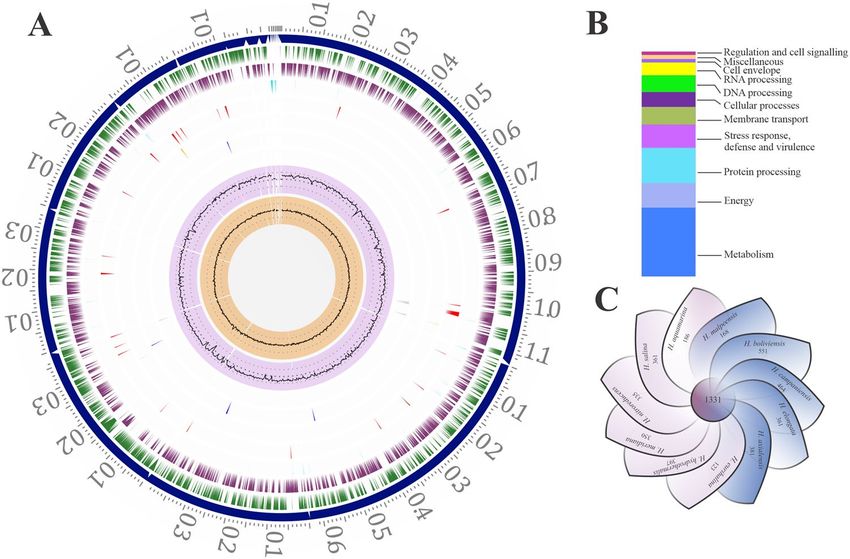
www.nature.com/scientificreports/
almeriensis, H. nitroreducens, H. cerina, H. fontilapidosi, H. rifensis, and H. stenophila9. The structural and func-
tional diversity among the EPS are seen depending on the species or the strain of the bacteria. Some of the EPS
produced by Halomonas spp. possess excellent emulsifying potential. Examples include H. ventosae strains A l12T
and Al16 and H. anticariensis strains F P35T and F P3610. The EPS of H. eurihalina H96 also exhibits high emul-
sifying activity and an ability to form a gel in acidic p H11. H. maura produce an EPS named mauran, containing
mannose, galactose, glucose, and glucuronic acid. It forms highly viscous solutions, similar in properties to that
of xanthan12. These EPS exhibit amphiphilic nature suitable for biodegradation of o ils13.
EPS biosynthesis is an energy exhaustive process involving three significant steps i.e., synthesis of the nucleo-
side diphosphate monosaccharides, polymerisation of the repeating unit, its transport and secretion. Intracellular
production of the EPS includes substrate uptake, metabolite pathway and the assembly. The internalized sugar
molecules are converted to specific monosaccharide by enzymatic chemical m odification14. Based on the pres-
ence of enzymes necessary for addition of chemical groups such as, acetyl, pyruvate, phosphate etc., conversion
of the monosaccharide occurs. These modified monosaccharides are then converted to nucleoside diphosphate
sugars and are assembled on undecaprenyl pyrophosphate that is found attached to the inner plasma membrane15.
Polymerisation occurs in the inner membrane by any of the two mechanisms: Wzx/Wzy-dependent pathway and
ABC transporter-dependent p athway16. The role of the ABC transporter-dependent pathway is well established in
the capsular polysaccharide (CPS) production; however, its involvement in the EPS production is also r eported17.
In addition to the production of osmolytes, H. malpeensis produces a yellow pigment that may be of industrial
importance. Microbially derived natural pigments have advantages as the production is not limited by season
coupled with lower costs for downstream processing18,19. The natural pigments have high industrial value due
to their enormous applications as antioxidants, functional foods, natural food colorants, antimicrobial agents,
etc. Among the characterised pigments of Halomonas species, H. elongata and H. aquamarina are known to
produce β-carotene and bacterioruberin respectively20–22. Among the many other metabolites of Halomonas
species ectoine has attracted a great interest as an osmolyte having application in cosmetic i ndustry23,24.
The whole genome analysis of a few members of Halomonas species has revealed the versatile functional
capabilities of the b acteria25–27. However, in-depth analysis of the EPS biosynthesis pathway in Halomonas spe-
cies is not yet elucidated. Hence, the genome analysis of H. malpeensis was performed to understand the EPS
biosynthesis and export pathways as well as to provide necessary information on other industrially exploitable
metabolites.
Results
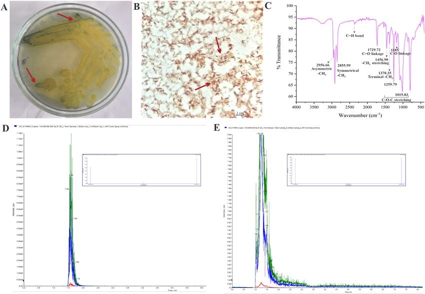
General features of the H. malpeensis genome. The complete genome sequencing of H. malpeensis
YU-PRIM-29T performed using Illumina sequencing produced 1,173,355 paired end raw reads. The genome
consists of 3,607,821 bp distributed in 18 contigs with G + C content of 63.8% (Fig. 1A). It contains 3337 protein
coding genes (CDS) and 53% of the annotated CDS having putative functions, while the remaining 47% genes
are annotated as hypothetical proteins (Fig. 1B). The salient features of the genome are summarized in Table 1. A
total of 251 pathways were identified and the important metabolic pathways in the genome are glycolysis, gluco-
neogenesis, glyoxylate bypass, TCA cycle and pentose phosphate pathway. The biosynthesis pathway for amino
acids includes serine, threonine, cysteine, methionine, histidine, arginine, proline, valine, leucine, isoleucine,
phenylalanine, tyrosine, tryptophan and lysine. The metabolic pathways for vitamins like biotin, thiamine and
riboflavin are also assigned.
The distribution of genes into clusters of orthologous groups (COGs) functional categories is listed in Table 2.
Using PATRIC Protein Family Sorter tool, a core genome containing 1,331 protein-coding genes that are shared
across 11 Halomonas strains (Supplementary Table S1) was identified as shown in Fig. 1C. This core genome is
made up of only 22–42% of the proteome of each strain, signifying a high amount of genomic diversity among
species of the Halomonas genus. The pan genome and core-genome contain 8,011 and 1,331 genes respectively.
EPS biosynthesis, transport of sugars and nucleotide sugar synthesis. The key EPS biosynthesis
pathway was identified by employing Kyoto Encyclopedia of Genes and Genomes (KEGG) and KEGG Auto-
matic Annotation Server (KAAS) functional annotation tools along with Prokka and Pathosystems Resource
Integration Center (PATRIC) annotations. The genes identified from the whole genome sequence were verified
by performing a translated Basic Local Alignment Search Tool (BLAST) against known sequences available
in National Center for Biotechnology Information (NCBI) Genbank database (https://www.ncbi.nlm.nih.gov/
genbank/). In general, based on the available literature, the bacterial EPS synthesis follows one of the three
mechanisms: Wzx/Wzy-dependent pathway, ABC transporter-dependent pathway and/or synthase depend-
ent pathway28. However, the ABC transporter-dependent pathway is mostly associated with CPS biosynthesis.
Annotation of the EPS biosynthesis pathway in H. malpeensis shows the presence of 184 ABC transporters, but
the genes involved in Wzx/Wzy-dependent (wzx, wzy and wzz genes) and synthase dependent pathways are
absent (Supplementary Table S2). The EPS synthesis machinery involves three significant steps i.e., synthesis of
the nucleoside diphosphate sugars (NDP sugars), polymerisation of the repeating units followed by transloca-
tion and secretion. Further, the EPS gene cluster was identified with antiSMASH (Node 477,025–155,069 bps)
and showed 10% similarity with Lactobacillus johnosonii EPS c luster29. However, the database is limited as only
a few biosynthetic gene clusters (BGC) of EPS are present in the MIBiG database with most of them belonging
to Gram-positive bacteria30.
In ABC transporter-dependent pathway, the polysaccharide is exported across the inner membrane through
various transporters. The transporters that are involved in the transport of molecules are listed in Table 3. A total
of 184 genes in the ATP binding cassette (ABC) transporters are identified for the transport of substrates like ions,
salts, sugars, vitamins, amino acids and purines across the cell membrane in an ATP-dependent manner. Apart
Scientific Reports | (2021) 11:1749 | https://doi.org/10.1038/s41598-021-81395-1 2
Vol:.(1234567890)
www.nature.com/scientificreports/

                                  Figure 1.  Genomic features and a comparative genomic analysis of H. malpeensis YU-PRIM-29T. (A) Circular
                                  plot representing the genome annotations of H. malpeensis YU-PRIM-29T. Circles are numbered from 1
                                  (outermost) to 8 (innermost). Circle 1 represents the contigs; Circles 2 and 3 show the locations of predicted
                                  coding sequences (CDSs) on the forward and reverse strands, respectively; Circle 4, RNA genes; Circle 5, CDS
                                  with homology to known antimicrobial resistance genes; Circle 6, CDS with homology to known virulence
                                  factors; Circle 7, % G + C; Circle 8, GC skew [(G − C)/(G + C)]. (B) Bar-chart representing PATRIC subsystems
                                  in the H. malpeensis YU-PRIM-29T genome. (C) Genomic diversity of 11 Halomonas strains. Each strain is
                                  represented by a petal. The central number represents the orthologous CDSs present in all strains. Overlapping
                                  regions show the number of CDSs conserved only within the specified genomes, while the number of CDSs
                                  unique to each strain is represented in non-overlapping portions. The total number of CDSs within each
                                  genome is enumerated beneath the strain name.

                                   Feature                                        H. malpeensis YU-PRIM-29T
                                   Domain                                         Bacteria
                                   Taxonomy                                       Proteobacteria; Gammaproteobacteria; Oceanospirillales; Halomonadaceae; Halomonas
                                   Genome size                                    3,607,821 bp
                                   G + C content                                  63.75%
                                   Completeness                                   99.00%
                                   Contamination                                  0.00%
                                   Number of coding sequences (CDSs) in PATRIC    3337
                                   Proteins with functional assignments           2653
                                   Hypothetical proteins                          684
                                   Proteins with EC number assignments            930
                                   Proteins with KEGG pathway assignments         700
                                   Genes assigned to COGs                         1817
                                   Number of tRNA                                 58
                                   Number of rRNA                                 12
                                   G + C content of tRNA                          58.51%
                                   G + C content of rRNA                          56.57%
                                   N50 value                                      410,886 bp
                                   L50 value                                      3

                                  Table 1.  General features of the H. malpeensis YU-PRIM-29T draft genome.

Scientific Reports |   (2021) 11:1749 |                     https://doi.org/10.1038/s41598-021-81395-1                                                            3

                                                                                                                                                            Vol.:(0123456789)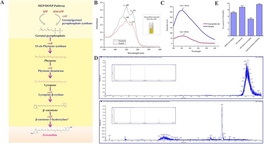
www.nature.com/scientificreports/

                                             COG code        Number of genes       Percentage     Description
                                             Cellular processes and signalling
                                             D                 30                   0.90          Cell cycle control, cell division, chromosome partitioning
                                             M               121                    3.63          Cell wall/membrane/envelope biogenesis
                                             N                 61                   1.83          Cell motility
                                             O                 81                   2.43          Post-translational modification, protein turnover, chaperones
                                             T                 67                   2.01          Signal transduction mechanisms
                                             U                 34                   1.02          Intracellular trafficking, secretion, and vesicular transport
                                             V                 29                   0.87          Defense mechanisms
                                             W                  0                   0.00          Extracellular structures
                                             Y                  0                   0.00          Nuclear structure
                                             Z                  0                   0.00          Cytoskeleton
                                             Information storage and processing
                                             A                  1                   0.03          RNA processing and modification
                                             B                  1                   0.03          Chromatin structure and dynamics
                                             J               193                    5.78          Translation, ribosomal structure and biogenesis
                                             K                 62                   1.86          Transcription
                                             L                 85                   2.55          Replication, recombination and repair
                                             X                 17                   0.51          Mobilome: prophages, transposons
                                             Metabolism
                                             C               127                    3.81          Energy production and conversion
                                             E               179                    5.36          Amino acid transport and metabolism
                                             F                 68                   2.04          Nucleotide transport and metabolism
                                             G                 89                   2.67          Carbohydrate transport and metabolism
                                             H               104                    3.12          Coenzyme transport and metabolism
                                             I                 64                   1.92          Lipid transport and metabolism
                                             P               107                    3.21          Inorganic ion transport and metabolism
                                             Q                 30                   0.90          Secondary metabolites biosynthesis, transport and catabolism
                                             Poorly characterised
                                             R               120                    3.60          General function prediction only
                                             S               147                    4.41          Function unknown
                                             –               1520                  45.55          Not in COGs

                                            Table 2.  Number of genes associated with the general cluster of orthologous group (COG) functional
                                            categories in H. malpeensis YU-PRIM-29T genome.

                                             Category               Molecules
                                             Metal ions             Na, K, Cu, Mg, Co, Ni, Pb, Cd, Zn, Hg, Fe, F, Ca, Mn
                                             Anions                 Phosphate, chromate, phosphite, nitrate, nitrite, sulphate, aminobenzoyl-glutamate
                                             Other cations          Ammonium
                                             Amino acids            Serine, threonine, proline, choline, histidine/lysine/arginine/ornithine, cysteine, methionine, glutamine, glutamate
                                             Carbohydrates          Ribose/xylose/arabinose/galactose, glucose, maltose, glyceraldehyde-3-phosphate, fructose
                                                                    Vitamin B12, dicarboxylate, tricarboxylate, glycine betaine, riboflavin, spermidine, putrescine, glycerol-3 phosphate,
                                             Other molecules
                                                                    ectoine, hydroxyectoine, queuosine precursor, 2-nitroimidazole, sialic acid

                                            Table 3.  Transporter systems for transfer of molecules identified in the H. malpeensis YU-PRIM-29T genome.

                                            from ABC transporters H. malpeensis also possesses genes for carbohydrate transport involved in the uptake of
                                            D-mannose, D-xylose, L-arabinose, D-fructose, sucrose, D-glucose and maltose (Supplementary Table S3). On
                                            its import into the cell’s interior, the sugars are converted to nucleotide sugar precursors. Based on the functional
                                            annotation, the enzymes involved in the synthesis of UDP-glucose and GDP-mannose from monosaccharides
                                            are shown in Fig. 2A. The gene galU coding for the enzyme UTP-glucose-1-phosphate uridylyl transferase (EC
                                            2.7.7.9), galE for UDP-glucose-4-epimerase (EC 5.1.3.2) involved in the production of UDP-galactose, ugd for
                                            UDP-glucose-6-dehydrogenase (EC 1.1.1.22) for the production of UDP-glucuronate from UDP-glucose and
                                            the gene manC for mannose-1-phosphate guanylyl transferases (EC 2.7.7.13) for GDP-mannose biosynthesis
                                            are identified (Supplementary Table S2). In the biosynthesis process, the sugar polymerisation is initiated by
                                            the action of glycosyltransferases by mediating glycosidic bond formation and these enzymes use sugar donors
                                            that contain nucleoside phosphate or lipid phosphate leaving groups. The presence of 29 glycosyltransferases

          Scientific Reports |   (2021) 11:1749 |                      https://doi.org/10.1038/s41598-021-81395-1                                                                            4

Vol:.(1234567890)
www.nature.com/scientificreports/
Figure 2. (A) Genes identified for the polysaccharide biosynthesis in H. malpeensis YU-PRIM-29T from the
genomic data. #Results obtained from tBLASTn analysis and the details are provided in the Supplementary
Tables S2 and S10. (B) ABC transporter-dependent assembly and transport of polysaccharides in H. malpeensis.
(C) Influence of carbon source on growth of H. malpeensis. Data points are OD600 values obtained from broth
cultures incubated with different carbon sources recorded at different time points. (D) EPS yield obtained
from different carbon sources at 48 h, *indicates p value < 0.001. Data points are mean ± SD and n = 3. (E)
FTIR spectra of EPS with peaks showing the important bands and (F) FESEM image of biofilm formed by H.
malpeensis on glass surface.
is identified using PATRIC, Rapid Annotations using Subsystems Technology (RAST), dbCAN2 and Prokka
annotation of which four are found in the EPS cluster hinting their role in transport of the produced EPS. The
detailed list of the glycosyltransferases is given in Table 4.
For the export of EPS outside the cell, H. malpeensis is proposed to follow an ABC transporter-dependent
pathway16. The genes coding for Kps proteins involved in ABC transporter-dependent pathway such as kpsD,
kpsM, kpsE, kpsT, kpsS and kpsC are identified in the genome. The kps gene cluster has been predicted using
antiSMASH. The structural details of these proteins were searched using InterPro database. The KpsS and KpsC
proteins (β-Kdo transferases) synthesise a capsular polysaccharide export system protein (oligo-Kdo linker);
KpsT is an ATP binding protein; KpsM is an ABC transporter permease protein that interacts with KpsT; KpsD
is an export system periplasmic protein and KpsE is an export system inner membrane protein (Fig. 2B).
The possible genes involved in EPS modifications were searched using Prokka annotation and tBLASTn. A
gene ugd coding for UDP-glucose-6-dehydrogenase (EC 1.1.1.22) involved in the conversion of the nucleotide
sugar UDP-glucose to UDP-glucuronic acid is found.
As the ABC transporter-dependent pathway is mainly associated with CPS, we followed extraction procedures
for both CPS and EPS and observed that EPS was predominantly extracted. Ability of the bacteria to produce
EPS was tested using different carbon source supplementation. Among the carbon sources tested based on the
genomic data, the growth was favoured by all the sugars (Fig. 2C), while, the EPS yield was higher in D-glucose,
D-xylose, D-fructose and L-arabinose supplemented media (Fig. 2D). The EPS yield in the media containing
D-mannose and maltose were significantly (p < 0.001) lower compared to the others. Biochemical analysis of the
Scientific Reports | (2021) 11:1749 | https://doi.org/10.1038/s41598-021-81395-1 5
Vol.:(0123456789)www.nature.com/scientificreports/
PATRIC ID Encoded protein Length (aa) Molecular weight (kDa) GT family
fig|2745.436.peg.139 Glycosyltransferase, group 1 373 41.13 GT4
fig|2745.436.peg.211 Glycosyltransferase, group 1 641 69.25 GT4
fig|2745.436.peg.583 Zeaxanthin glycosyltransferase 429 46.36
fig|2745.436.peg.676 Glycosyltransferase 1023 117.2 GT4
fig|2745.436.peg.886 Glycosyltransferase, group 1 351 38.37
fig|2745.436.peg.887 Glycosyltransferase, group 1 698 76.28 GT4
fig|2745.436.peg.888 Glycosyltransferase, group 1 291 32.79 GT4
fig|2745.436.peg.906 Glycosyltransferase 387 42.3 GT4
fig|2745.436.peg.1361 peptidoglycan transglycosylase 238 27.17 GT51
fig|2745.436.peg.1426 ADP-glucose transglucosylase 565 61.41 GT5
fig|2745.436.peg.1489 Multimodular transpeptidase-transglycosylase 842 92.15 GT51
fig|2745.436.peg.1748 Glycosyltransferase, group 2 family 316 36.12
fig|2745.436.peg.2024 Glycosyltransferase WecB/TagA/CpsF 225 25.94 GT26
fig|2745.436.peg.2035 Glucosyl-3-phosphoglycerate synthase 406 46.26 GT81
fig|2745.436.peg.2038 Uncharacterised glycosyltransferase YcjM 606 67.74 GH13
fig|2745.436.peg.2094 Glycosyltransferase, group 2 657 76.92
fig|2745.436.peg.2104 Glycosyltransferase, group 1 992 108.63
fig|2745.436.peg.2106 Glycosyltransferase, group 2 family 727 82.2 GT2
fig|2745.436.peg.2107 Glycosyltransferase, group 1 256 39.47
fig|2745.436.peg.2292 Peptidoglycan glycosyltransferase FtsW (EC 2.4.1.129) 393 42.95
fig|2745.436.peg.2293 N-acetylglucosaminyltransferase 365 38.94 GT28
fig|2745.436.peg.2551 Glycosyltransferase, group 1
fig|2745.436.peg.2881 Biofilm PGA synthesis N-glycosyltransferase 424 48.67 GT2
fig|2745.436.peg.3032 Lipid-A-disaccharide synthase 390 42.57 GT19
ADP-heptose–lipooligosaccharide heptosyltransferase
fig|2745.436.peg.3208 350 37.89 GT9
II
fig|2745.436.peg.3210 Lipopolysaccharide biosynthesis glycosyltransferase 355 40.53
fig|2745.436.peg.3212 glycosyltransferase, group 1 372 40.67 GT4
ADP-heptose–lipooligosaccharide heptosyltransferase
fig|2745.436.peg.3211 345 36.82 GT9
II
fig|2745.436.peg.3213 3-deoxy-D-manno-octulosonic acid kinase 238 26.14 GT9
Table 4. Glycosyltransferases present in H. malpeensis YU-PRIM-29T genome. The GT’s in bold are the
glycosyl transferases identified in the EPS gene cluster.
EPS showed 76% total sugar, 5% protein on w/w basis and among the sugars more than 50% were uronic acid
containing. The Fourier Transform Infrared Spectroscopy (FTIR) analysis showed the characteristic carbohydrate
peaks at 3600–2900 cm−1 (–OH groups), 2937 cm−1 (–CH2 stretching), 1735 cm−1 (uronic acid) and 1045 cm−1
(acetyl group) among others (Fig. 2E).
The extracted EPS showed emulsification activity against petroleum hydrocarbons. The highest emulsifica-
tion index ( EI24) was against toluene 64 ± 4%, followed by kerosene 63 ± 4%, xylene 62 ± 4%, hexane 60 ± 6%
and petrol 53 ± 2%. These values were significantly higher than Tween 20 (used as positive control) at the same
concentration.
Chemotaxis and biofilm formation. Chemotaxis is one of the mechanisms adapted by the bacteria to
sense the external environment in order to modify the mode of growth. The chemoattractant molecules influ-
ence the flagellar motor to direct the movement of the bacterial cell either towards or away from the chemical
signal. The genes coding for the proteins belonging to the chemosensory pathway such as cheR, cheB, cheA,
cheW, cheY, cheZ, fliG, fliM, fliN, motA and motB in the genome of H. malpeensis were identified using KEGG.
The membrane cofactor protein (MCP; CD46) can allow the attractant or repellent to be taken into the cell.
The function of MCP is regulated by cheR that codes for methyl transferase (EC 2.1.1.80) and cheB coding for
methyl esterase (EC 3.1.1.61) which in turn are regulated by cheA gene encoding for two component signalling
kinase (EC 2.7.13.3) and cheW for a coupling protein. The CheA and CheW proteins activate CheY to regulate
the function of MotA and MotB through activation of the fli genes (fliG, fliM and fliN). The genes for the biofilm
formation such as, wspA, wspE, wspF, wspR, sadC, tpbB, mucR and algA were identified by KEGG (Supplemen-
tary Table S4).
To test whether the bacteria form biofilm, the crystal violet staining and Field Emission Scanning Electron
Microscopy (FESEM) were used (Fig. 2F). The bacteria formed strongly adherent biofilms and the intensity
increased over the incubation period as recorded by the crystal violet staining method. The biofilm intensities
corresponded to an O D580 value of 1.48, 1.97 and 3.31 at 24 h, 48 h and 96 h of incubation respectively.
Scientific Reports | (2021) 11:1749 | https://doi.org/10.1038/s41598-021-81395-1 6
Vol:.(1234567890)www.nature.com/scientificreports/
Figure 3. Zeaxanthin biosynthesis in H. malpeensis YU-PRIM-29T. (A) Complete zeaxanthin biosynthesis
pathway as predicted from genomic sequence data. The biosynthesis pathway was constructed based on the
KEGG database, PATRIC genome annotation and Prokka. *Result obtained from tBLASTn analysis and details
are provided in the Supplementary Tables S5 and S10. (B) UV–Vis spectra of zeaxanthin extracted from H.
malpeensis. Standard zeaxanthin was used for comparison. Inset showing solvent extracted zeaxanthin from the
cells. (C) Fluorescence spectra of zeaxanthin extracted from H. malpeensis compared to standard zeaxanthin
showing maximum absorption at 520 nm. (D) Targeted LC–MS MRM spectra of extracted zeaxanthin with the
standard acquired in positive polarity. Two transitions were observed in the extract. (E) IC50 values (µg mL−1) for
zeaxanthin showing DPPH scavenging, NO reduction, OH radical scavenging and lipid peroxidation inhibition
activities. The bars represent mean ± SD of triplicate results.
Pigment biosynthesis. Microbial pigments occur in two forms; either secreted by the cell or cell bound.
In this bacterium the intense yellow pigment is cell bound and was water insoluble. Hence, it required extraction
using organic solvents. The genes crtB, crtI, crtY and crtZ responsible for the carotenoid biosynthesis were pre-
sent. These genes are responsible for the enzymes of 15-cis-phytoene synthase (EC 2.5.1.32) that converts gera-
nyl pyrophosphate to phytoene, phytoene desaturase (EC 1.3.99.31), converts phytoene to lycopene, lycopene
β-cyclase (EC 5.5.1.19) converts lycopene to β-carotene and β-carotene-3-hydroxylase (EC 1.14.15.24) converts
β-carotene to zeaxanthin. The detailed biosynthesis pathway identified for the zeaxanthin production is given
in Fig. 3A. Further, the yellow pigment produced by the bacterium was extracted from the cell pellets. It was
insoluble in water and was extractable in organic solvent methanol. The visible spectra of the extracted yellow
pigment showed characteristic absorption peaks with λmax 448 nm and a shoulder peak at 429 nm corresponding
to that of zeaxanthin used as a s tandard31,32 (Fig. 3B). The fluorescence spectral characteristics also corresponded
to the zeaxanthin pigment (Fig. 3C). The LC–MS/MS data obtained against the standard confirms the presence
of zeaxanthin (Fig. 3D). The isolated pigment showed potent antioxidant activities with IC50 values of 7.13 µg,
8.82 µg, 5.17 µg and 9.66 µg for DPPH scavenging, nitric oxide reduction, hydroxyl radical scavenging and lipid
peroxidation inhibition respectively (Fig. 3E).
KEGG annotation further revealed the enzymes involved in other pigment production pathways namely beta-
lain pathway and riboflavin pathway. The betalain biosynthesis pathway begins with L-tyrosine that is converted
to L-DOPA, which in the presence of 4,5-DOPA dioxygenase extradiol (EC 1.13.11.-) is converted to betalamic
acid. This conjugates with amino acids spontaneously and forms yellow betaxanthins. The 4,5-DOPA dioxygenase
extradiol gene is involved in this pathway. Riboflavin is a yellow coloured water-soluble pigment that is produced
by a wide variety of microorganisms and its biosynthesis in H. malpeensis was identified using KEGG. The impor-
tant proteins in the pathway such as RibA (EC 3.5.4.25), RibD1 (EC 3.5.4.26), RibD2 (EC 1.1.1.193), YigB (EC
3.1.3.104), RibH (EC 2.5.1.78), RibE (EC 2.5.1.9), RibF (EC 2.7.1.26), FAD1 (EC 2.7.7.2), RibB (EC 4.1.99.12)
and SsuE (EC 1.5.1.38) were annotated using the genome data. The pathway begins with purine metabolism with
seven enzymes namely; GTP cyclohydrolase II (RibA), pyrimidine deaminase (RibD1), pyrimidine reductase
(RibD2), uracil phosphatase (YigB), 3,4-DHBP synthase (RibH) and ribityllumazine synthase (RibE) for the
formation of the pigment (Supplementary Table S5). Apart from these enzymes, flavin mononucleotide (FMN)
hydrolase (EC 3.1.3.102) that converts FMN to riboflavin is present.
Scientific Reports | (2021) 11:1749 | https://doi.org/10.1038/s41598-021-81395-1 7
Vol.:(0123456789)www.nature.com/scientificreports/
Figure 4. (A) Demonstration of polyhydroxyalkanoate production by Sudan Black B staining of H.
malpeensis YU-PRIM-29T. Bluish black coloured colonies appearing on plate with 0.02% of dye indicated
by red arrow shows the presence of PHA. (B) Light microscopy images of Sudan Black B stained cells of H.
malpeensis observed at 100X. Red arrow indicates the PHA stained with Sudan Black B. (C) FTIR spectra of
polyhydroxyalkanoate extracted from H. malpeensis. (D) Targeted LC–MS MRM transition spectra of standard
Ectoine. (E) Targeted LC–MS MRM transition spectra of ectoine extracted from H. malpeensis with fragment
peaks shown as inset comparable to the standard acquired in positive polarity.
Polyhydroxyalkanoates and ectoine. Polyhydroxyalkanoates (PHAs) are important metabolites that
are involved in cellular energy storage. The annotation using PATRIC allowed the identification of acetoacetyl-
CoA reductase (PhbB; EC 1.1.1.36) and acetyl-CoA acetyltransferase (PhaA; EC 2.3.1.9) involved in PHA pro-
duction. Although the presence of PHA operon was not observed in the genome, but, the polyhydroxyalkanoate
synthase genes (phaC1 and phaC2), polyhydroxyalkanoate depolymerase (phaZ1, phaZ2, and phaZ3), polyhy-
droxyalkanoate synthesis repressor (phaR) and phasin (phaP) were identified using tBLASTn.
Ectoine is an industrially important osmolyte that is produced by many strains of Halomonas spp., to avoid
loss of turgor pressure in extreme environments with high osmotic stress conditions prevailing in marine habi-
tats. Using Prokka and PATRIC annotation the genes involved in the ectoine biosynthesis pathway were curated.
These included lysC, ectB, ectA, ectC and ectD. The genes coding for proteins involved in the degradation of the
synthesised ectoine present are doeA, doeB, doeD and doeC.
Experimentally the PHA production was tested by Sudan Black B staining of the colonies and the cells
(Fig. 4A,B). We could extract 11.6 mg PHA per gram dry weight of the cell. The IR spectra of the PHA extracted
from the cells showed important peaks corresponding to the PHA as follows; C=O groups (1729 cm−1), asym-
metric methyl group (2956 cm−1), symmetrical methyl group (2856 cm–1), stretching vibration (1456 cm−1) and
terminal methyl group vibration (1378 cm−1), C–O–C stretching vibration (1260 cm−1 and 1020 cm−1) and C–O
stretching (1185 cm−1) (Fig. 4C). Similarly, the ectoine produced by the bacteria was extracted from the cell pellet
(~ 0.20 g/L) and was identified using LC–MS/MS against the ectoine standard (Sigma Aldrich, USA) (Fig. 4D).
The genome of H. malpeensis YU-PRIM-29T also contains several genes that allow bacterial adaptation to
stressful environmental conditions. The important are the spermidine synthase gene (Srm) and genes for base
excision repair including many DNA glycosylases (Supplementary Table S6). Using antiSMASH 17 putative
BGC were identified and these included clusters for saccharide, fatty acids, terpenes, betalactone and ectoine
(Supplementary Table S7).
Scientific Reports | (2021) 11:1749 | https://doi.org/10.1038/s41598-021-81395-1 8
Vol:.(1234567890)www.nature.com/scientificreports/
Discussion
The genus Halomonas consists of more than 100 species described from marine and terrestrial high saline
environments. Genome annotation helps to identify the functional components in the genome sequence such
as genes involved in biosynthesis of important metabolites. From the draft genome of H. malpeensis YU-PRIM-
29T, we have identified the genes involved in the biosynthesis of EPS, PHA, carotenoid (zeaxanthin) and ectoine
pathways. The experimental evidences also suggest their production in the bacterial culture.
Bacterial cell surface glycoconjugates are found in various forms either attached to the cell or released into
the environment. Many polymers exist between these two states of capsular (cell-bound) or secreted (EPS) slimy
forms33. The polysaccharide biosynthesis and transport is a complex mechanism that follow one of the following
pathways namely; Wzx/Wzy-dependent pathway, ABC transporter-dependent pathway and synthase depend-
ent pathway34. While, literature shows that ABC transporter-dependent pathway is mostly associated with CPS
production, there are also evidences that implicate its role in EPS biosynthesis17. The polysaccharide is exported
out of the cell with the help of a translocation pathway which is formed by a polysaccharide copolymerase (PCP)
namely, KpsD protein and an outer membrane polysaccharide export protein (OPX) namely KpsE which together
form a channel. KpsE determines the length of the polymer chain while KpsD forms the export channel16,17. The
synthesis and translocation from the periplasm to the cell’s exterior needs the presence of KpsE (PCP-3 family)
and KpsD (OPX family) proteins similar to Wza-Wzc p roteins17,35,36. Four glycosyltransferases involved in the
EPS biosynthesis pathway cluster are identified from the draft genome. The presence of genes coding for ABC
transporter KpsMT; encoded by kpsM and kpsT genes37 and OPX (KpsD) and PCP (KpsE) proteins in the genome
suggests that H. malpeensis follows ABC transporter-dependent pathway for the export of polysaccharide and
its secretion to the environment as EPS. The genes kpsS and kpsC are seen in the kps cluster of H. malpeensis
which are known to code for Kdo linkers mostly associated with CPS38. However, there are many reports on EPS
production by bacteria with Kdo linkers as reported from Cobetia, Burkholderia and Pseudomonas genus39–43.
The growth phase dependent studies on the EPS production in H. malpeensis also shows a gradual increase
in EPS production during the incubation period peaking at late stationary phase (data not shown). This suggests
that, the polysaccharide exported out of the cell may gradually mature and be released to the environment. This
may be facilitated by inorganic/chemical mediators present in the environment. Among the reported EPS pro-
ducing Halomonas spp., H. stenophila HK30 produces haloglycan type of EPS and in aqueous medium it shows
moderate to high viscosity and pseudoplastic b ehaviour44. H. xianhensis SUR308 EPS exhibit high viscosity and
pseudoplasticity and stable over a wide pH r ange45. The EPS produced from Halomonas spp. has demonstrated
excellent emulsification activities suitable to remove the oil content in contaminated water and s ludge46. The
EPS extracted from H. malpeensis also showed emulsification of petroleum hydrocarbons. The genome based
metabolic systems engineering approach in H. smyrnensis AAD6T isolate from Camalti Saltern area in Turkey
resulted in an increased levan production when compared to the wild type strain47.
The biofilm formation capability of H. malpeensis was confirmed by the presence of genes involved and
experimental results. The biofilm mode of growth provides many advantages to the bacteria such as metabolite
exchange platform, better resource capturing, protection from desiccation, drugs, environmental stress and soci-
omicrobial interaction providing advantages compared to free living/planktonic counterparts48,49. The extracel-
lular polymeric substance forms an extracellular matrix for the microbial community in the biofilm and plays a
crucial role in binding the cells together50. H. malpeensis is capable of forming a biofilm on polystyrene material
surface as reported in other members of the genus50,51. Halomonas spp. are also reported as part of corrosive
biofilm community in the marine e nvironments52. Chemotaxis can also drive biofilm formation based on envi-
ronmental cues with the expression of wspA and wspE genes. These genes get activated when the bacterial cells
come in contact with the surface that in turn activates wspR, sadC, tpbB and mucR genes.
Bacteria produce many pigments through different biosynthesis pathways. For the biosynthesis of carotenoids,
lycopene β-cyclase is needed. Phytoene desaturase catalyzes the conversion of phytoene to lycopene by desatura-
tion at four sites53. Phytoene synthase catalyzes the condensation reaction of two molecules of geranylgeranyl
diphosphate to produce phytoene, a precursor of β-carotene. They produce triterpene and tetraterpene precur-
sors for many diverse sterol and carotenoid end products. H. malpeensis contains all the enzymes involved in
carotenoid pathway and precursor pathway. Zeaxanthin is an important antioxidant, a product of carotenoid
pathway and the enzymes for its synthesis are detected in the genome of H. malpeensis. However, H. elongata that
does not produce zeaxanthin was genetically engineered to produce β-carotene by expressing carotenoid pathway
genes crtE, crtY, crtI, and crtB derived from Pantoea agglomerans and IPP isomerase gene from Haematococcus
pluvialis20. Other pigments produced from the Halomonas spp. are, bacterioruberin, a carotenoid derivative from
H. aquamarina MB59821 and aminophenoxazinones from Halomonas sp. GWS-BW-H8hM54.
In addition to carotenoid pathway, the genes involved in the biosynthesis of betalain and riboflavin are
identified in H. malpeensis. Betalains, the yellow or violet pigments usually synthesised in plants and fungi are
reported to be synthesised in bacteria such as Gluconacetobacter diazotrophicus. However, it requires a growth
medium supplemented with L-DOPA55. The genome of H. malpeensis contained the gene coding for the enzyme,
4,5-DOPA dioxygenase extradiol.
Halomonas spp. are also significant for the production of ectoine, a well-known osmolyte which is produced
and released in response to the varying salinity stress. These molecules function as compatible solutes, have no
disturbance to the cell even at higher concentrations and reduce the detrimental effect of freezing, desiccation
and high t emperatures56. They do so by interacting with the cell’s protein and contributing to protein folding
and are responsible for increased protein s tability57. Ectoine is now one of the widely used compatible solute in
cosmetic industries as skin protectants and anti-ageing p roducts58, in healthcare products as anti-inflammatory
agents for treatment of allergies and for the treatment of epithelial derived inflammatory a ilments59. Currently,
H. elongata is the preferred strain for industrial production of e ctoine2. H. malpeensis possesses all the genes
Scientific Reports | (2021) 11:1749 | https://doi.org/10.1038/s41598-021-81395-1 9
Vol.:(0123456789)www.nature.com/scientificreports/
coding for the enzymes and transporters involved in ectoine biosynthesis pathway (Supplementary Table S8). The
bacterium was able to produce PHA that confirms the role of genes involved in the PHA biosynthesis pathway.
Studies on the distribution of PHA genes in Halomonas sp. SF2003 suggest that the genes are not clustered in
one operon but distant from each o ther60. A similar scattered occurrence of PHA relevant genes was reported
in the genome of Halomonas sp. TD0. The phaP and phaC1 are connected with a space of 92 bp similar to our
observation showing a gap of 90 bp between the two g enes61.
In summary, the draft whole genome of halophilic strain H. malpeensis YU-PRIM-29T was annotated using
bioinformatic tools to explore the production of commercially important metabolites. The EPS, pigment, PHA
and ectoine biosynthesis pathways described in this study may provide prospects to exploit this bacterium
industrially.
Methods
Bacterial strain and culture conditions. Halomonas malpeensis YU-PRIM-29T was originally isolated
from the rhizosphere soil in the coastal region of Malpe (13° 21′ 10.22″ N, 74° 42′ 29.99″ E). It was cultured in
Zobell marine agar 2216 (HiMedia, India) at 32 °C. For DNA isolation, the bacterium was cultured in Zobell
marine broth 2216 by incubating at 32 °C, under shaking (120 rpm) for 24 h. The cells were harvested by cen-
trifugation (7500 rpm), washed and lysed by lysis buffer followed by proteinase K treatment. The DNA was
extracted using Qiagen kit (Cat No./ID: 51304) following the manufacturer’s instruction for Gram-negative
bacteria. The DNA concentration and purity were assessed by the absorbance readings at 260 to 280 nm using
a Nanodrop spectrophotometer (Colibri, Titertek Berthold). Polymerase Chain Reaction (PCR) was performed
to amplify the 16S rRNA region, and the quality of the DNA sample was checked by gel electrophoresis prior to
genome sequencing.
Sequencing, assembly and annotation. Illumina sequencing was done on a MiSeq platform using
MiSeq Reagent Kit v3, 600 Cycles reagents (Catalog # MS-102-3003). It produced 1,173,355 raw reads after pre-
processing (adaptor trimming) for each of R1 and R2. This gave average genome coverage of 190X for a 5 Mb
genome size with a data yield of 1,598,696 reads. FASTQC was used to assess the raw reads quality and trimming
was performed by trimmomatic (Version 0.35), default settings, identifying a Phred cutoff of Q20. The sequence
was uploaded to the web annotation service RAST (http://rast.nmpdr.org/rast.cgi)62 as well as PATRIC (https://
www.patricbrc.org/) for automated annotation.
De novo assembly of the sequences were performed using SPAdes 3.11.1 and the resulting assembly with best
N50 value was taken into gene prediction using Prokka (kbase.us)63. The PATRIC gene features were considered
as a basis for annotation. PATRIC output was checked and corroborated by comparing to that from Prokka and
RAST. The construction of genomic and metabolic pathways was executed using all three. These data sources were
combined to affirm product description for predicted proteins. A total of 11 Halomonas genomes were used for
comparing protein-family across genomic groups (10 from PATRIC and the new genome from this study) using
Protein Family Sorter tool in PATRIC. The gene features of essential biosystems were further manually confirmed
using BLASTp (https://blast.ncbi.nlm.nih.gov/Blast.cgi) against non-redundant database of NCBI (details pro-
vided in Supplementary Table S9). The proteins involved in EPS and pigment production pathways were identi-
fied by the local tBLASTn of selected EPS/pigment production pathway proteins from known microbial genomes/
proteomes against whole genome sequence with alignment length of at least 80% and e-value cut-off of ≤ 10−5
(details provided in Supplementary Table S10). The best BLAST hit with the highest alignment length percentage
and identity match was assigned as the annotation of the predicted gene. Essential enzyme functional prediction
was obtained from KEGG (http://www.genome .jp/kegg/)64 using KAAS server65. This functional annotation was
used to reconstruct the metabolic pathways related to EPS biosynthesis and pigment production. The dbCAN2
meta server (http://cys.bios.niu.edu/dbCAN2/index.php) was used to identify the glycosyltransferases involved
in EPS biosynthesis. Similarity searches against Transporter Classification Database (TCDB) (www.tcdb.org)
was performed to confirm the genes coding for the ABC transporters involved in EPS export and annotations
of best-matching hits with an e-value cut-off of 1 0−9. WebMGA was used for the functional characterisation of
the protein coding genes66 and were mapped to the COG functional category assignment67. For identifying the
functional features of the proteins InterPro68 and UniProt BLAST69 were used. Biosynthetic gene clusters for
secondary metabolites were predicted using a ntiSMASH70 with default search parameters. Based on the genes
identified, visualisation of the biosynthesis pathways for EPS production and transport as well as for the pigment
were done using CorelDraw Technical Suite, 2019 and GraphPad Prism 5.03 software.
Isolation of EPS, pigment and other metabolites from H. malpeensis cultures. For extracting
the EPS, bacteria were cultured in Marine broth for 72 h under shaking at 32 °C. EPS was isolated from the cell-
free supernatant by cold ethanol precipitation. The harvested EPS was dialysed against MilliQ water, lyophilised
and the yield was recorded. The basic biochemical characterisation of the EPS was performed by estimating
the total sugar and total protein content by phenol sulphuric a cid71 and Bradford m
ethods72 respectively. Basic
structural characterisation of the EPS was performed by the FTIR spectroscopy.
For estimating the growth and EPS yield, bacteria were grown in MY media (g/L; sodium chloride 51.3, mag-
nesium sulphate heptahydrate 13.0, yeast extract 3.0, magnesium chloride 9.0, potassium chloride 1.3, sodium
bicarbonate 0.05, peptone 5.0, glucose 10, malt extract 3.0, calcium chloride 0.2, sodium bromide 0.15, and
ferrous chloride tetrahydrate 0.036) containing 7.5% salt supplemented with different carbon sources (glucose,
fructose, sucrose, maltose, arabinose, xylose and mannose). The culture conditions included temperature 32 ℃,
pH 7.2, aeration 1:5 and agitation 120 rpm. Growth was monitored based on the O D600 readings and EPS was
Scientific Reports | (2021) 11:1749 | https://doi.org/10.1038/s41598-021-81395-1 10
Vol:.(1234567890)www.nature.com/scientificreports/
harvested by chilled ethanol precipitation. EPS was purified by dialysis using MilliQ water and lyophilised to
estimate the dry weight.
For the extraction of the yellow pigment, the cell pellet collected by centrifugation was used. Pigment was
extracted several times with methanol till the cells were bleached completely. The extracts were pooled and con-
centrated using a vacuum evaporator. To this hexane and distilled water were added and mixed well to separate
the other organics from the pigment. The pigment extracted in the hexane layer was subjected to UV–Vis and
fluorescent spectrophotometry against standard zeaxanthin. Further, confirmation of zeaxanthin was made
based on the LC–MS/MS analysis (detailed methods are given in Supplementary_ Methods file). The isolated
pigment was tested for antioxidant activities by DPPH s cavenging73, nitric oxide s cavenging74, hydroxyl radical
scavenging75 and lipid peroxidation inhibition a ctivities76 for which the IC50 values were calculated.
ethod51. For this, bacteria
The biofilm forming ability of the bacteria was tested using crystal violet staining m
were inoculated to Zobell marine broth in polystyrene cuvettes and incubated for different time intervals up to
96 h. The planktonic cells were removed by carefully decanting the contents and the static biofilm was washed
with sterile PBS twice, fixed with methanol (10 min), washed again and dried. After drying, 0.1% crystal violet
was added and kept for staining (5 min). The stain was solubilised using acetic acid (33%) and the absorbance was
recorded at 580 nm. The biofilm adherence capacity was based on the O D580 readings compared with the blank
as OD ≤ ODc (non-adherent), OD < ODc ≤ 2 × ODc (weakly adherent), 2 × ODc < OD ≤ 4 × ODc assessed (moder-
ately adherent) and 4 × ODc < OD (strongly adherent)51. For FESEM, the biofilm was developed on a sterile glass
coupon (1 × 1 cm), fixed with methanol, dehydrated and subjected to sputter coating prior to FESEM analysis77.
The PHA producing ability of the bacterium was tested by Sudan Black B staining method. Here, the bacterial
colonies in the agar plates were stained with 0.02% of ethanolic solution of Sudan Black B dye for 1 h. The excess
stain was removed by 70% ethanol. The darkly stained culture plate was photographed. For microscopy, bacterial
smear was prepared by heat fixing, stained with Sudan Black B. Xylene was used for decolourizing and the cells
were counter stained using 0.05% safranin for 10 s. The stained cells were observed under 100X magnification78.
Extraction of PHA was performed from the bacterial cells after lyophilisation according to previously described
methods79. The PHA content was determined as the percent ratio of PHA to cell dry weight. FTIR spectrum of
PHA was recorded using Shimadzu FTIR spectrophotometer (4000–400 cm−1, spectral resolution of 4 cm−1 and
45 scans). The spectrum obtained was plotted using Origin 2017 SR2 software.
For extraction of ectoine a previously described method was used80. Briefly, ectoine was extracted from the
cell pellet with methanol/chloroform/water (10/5/4 v/v/v) by vigorous shaking for 90 min. Equal volume of
chloroform and water (130 µL/mL) was added, mixed well for 30 min and collected from the aqueous phase
by centrifugation (6500 rpm, 30 min). Ectoine was identified using LC–MS/MS against the standard (detailed
methodology in Supplementary document).
Bacterium strain and sequence. The sequencing data of the draft genome of halophilic H. malpeensis YU-PRIM-
29T is available online as BioProject PRJNA579246, NCBI taxonomy ID 1172368 from the NCBI database. The
genome description and the predictive annotation are available in PATRIC server with genome ID 2745.436
and RAST server with genome ID 2745.437. The Whole Genome Shotgun project of strain YU-PRIM-29T was
deposited at DDBJ/EMBL/GenBank under the accession number WHVL00000000. The version described in
this paper is version WHVL00000000. The 16S rRNA gene sequence is available in GenBank with the accession
ID JQ730736.
Data availability
The datasets generated during and/or analysed during this study are obtainable from the corresponding author
on reasonable request.
Received: 12 February 2020; Accepted: 4 January 2021
References
1. Kämpfer, P. et al. Halomonas malpeensis sp. nov., isolated from rhizosphere sand of a coastal sand dune plant. Int. J. Syst. Evol.
Microbiol. 68, 1037–1046 (2018).
2. Dauga, C. Balneatrix. In Bergey’s Manual of Systematics of Archaea and Bacteria (ed. Whitman, W. B.) (Wiley, Hoboken, 2015).
3. Gutierrez, T., Morris, G., Ellis, D., Mulloy, B. & Aitken, M. D. Production and characterisation of a marine Halomonas surface-
active exopolymer. Appl. Microbiol. Biotechnol. 104, 1063–1076 (2020).
4. Ollivier, B., Caumette, P., Garcia, J. L. & Mah, R. A. Anaerobic bacteria from hypersaline environments. Microbiol. Rev. 58, 27–38
(1994).
5. Kim, K. K., Lee, J.-S. & Stevens, D. A. Microbiology and epidemiology of Halomonas species. Future Microbiol. 8, 1559–1573 (2013).
6. Ventosa, A., de la Haba, R. R., Sanchez-Porro, C. & Papke, R. T. Microbial diversity of hypersaline environments: A metagenomic
approach. Curr. Opin. Microbiol. 25, 80–87 (2015).
7. Poli, A., Anzelmo, G. & Nicolaus, B. Bacterial exopolysaccharides from extreme marine habitats: Production, characterization and
biological activities. Mar. Drugs 8, 1779–1802 (2010).
8. Casillo, A., Lanzetta, R., Parrilli, M. & Corsaro, M. M. Exopolysaccharides from marine and marine extremophilic bacteria:
Structures, properties, ecological roles and applications. Mar. Drugs 16, 69 (2018).
9. de la Haba, R. R., Arahal, D. R., Marquez, M. C. & Ventosa, A. Phylogenetic relationships within the family Halomonadaceae based
on comparative 23S and 16S rRNA gene sequence analysis. Int. J. Syst. Evol. Microbiol. 60, 737–748 (2010).
10. Mata, J. A. et al. Exopolysaccharides produced by the recently described halophilic bacteria Halomonas ventosae and Halomonas
anticariensis. Res. Microbiol. 157, 827–835 (2006).
11. Calvo, C., Martinez-Checa, F., Mota, A., Bejar, V. & Quesada, E. Effect of cations, pH and sulfate content on the viscosity and
emulsifying activity of the Halomonas eurihalina exopolysaccharide. J. Ind. Microbiol. Biotechnol. 20, 205–209 (1998).
Scientific Reports | (2021) 11:1749 | https://doi.org/10.1038/s41598-021-81395-1 11
Vol.:(0123456789)www.nature.com/scientificreports/
12. Arias, S. et al. Mauran, an exopolysaccharide produced by the halophilic bacterium Halomonas maura, with a novel composition
and interesting properties for biotechnology. Extremophiles 7, 319–326 (2003).
13. Gutierrez, T., Biller, D. V., Shimmield, T. & Green, D. H. Metal binding properties of the EPS produced by Halomonas sp. TG39
and its potential in enhancing trace element bioavailability to eukaryotic phytoplankton. Biometals 25, 1185–1194 (2012).
14. Sutherland, I. W. Biotechnology of Microbial Exopolysaccharides. Cambridge Studies in Biotechnology (Cambridge University Press,
Cambridge, 1990).
15. Whitfield, G. B., Marmont, L. S. & Howell, P. L. Enzymatic modifications of exopolysaccharides enhance bacterial persistence.
Front. Microbiol. 6, 471 (2015).
16. Freitas, F., Alves, V. D. & Reis, M. A. M. Advances in bacterial exopolysaccharides: From production to biotechnological applica-
tions. Trends Biotechnol. 29, 388–398 (2011).
17. Cuthbertson, L., Mainprize, I. L., Naismith, J. H. & Whitfield, C. Pivotal roles of the outer membrane polysaccharide export and
polysaccharide copolymerase protein families in export of extracellular polysaccharides in gram-negative bacteria. Microbiol. Mol.
Biol. Rev. 73, 155–177 (2009).
18. Narsing Rao, M. P., Xiao, M. & Li, W. J. Fungal and bacterial pigments: Secondary metabolites with wide applications. Front.
Microbiol. 8, 1113 (2017).
19. Arulselvi, P., Umamaheswari, S., Sharma, G., Karthik, C. & Jayakrishna, C. Screening of yellow pigment producing bacterial isolates
from various eco-climatic areas and analysis of the carotenoid produced by the isolate. J. Food Process. Technol. 5, 1–8 (2014).
20. Rodríguez-sáiz, M., Sánchez-porro, C., Luis, J., Fuente, D. L. & Mellado, E. Engineering the halophilic bacterium Halomonas
elongata to produce β-carotene. Appl. Microbiol. Biotechnol. 77, 637–643 (2007).
21. Fariq, A., Yasmin, A. & Jamil, M. Production, characterization and antimicrobial activities of bio-pigments by Aquisalibacillus
elongatus MB592, Salinicoccus sesuvii MB597 and Halomonas aquamarina MB598 isolated from Khewra salt range, Pakistan.
Extremophiles 23, 435–449 (2019).
22. Ramesh, C., Vinithkumar, N. V. & Kirubagaran, R. Multifaceted applications of microbial pigments: Current knowledge, challenges
and future directions for public health implications. Microorganisms 7, 186 (2019).
23. Nakayama, H., Yoshida, K., Ono, H., Murooka, Y. & Shinmyo, A. Ectoine, the compatible solute of Halomonas elongata, confers
hyperosmotic tolerance in cultured tobacco cells. Plant Physiol. 122, 1239–1247 (2000).
24. Guzman, H., Van-Thuoc, D., Martin, J., Hatti-Kaul, R. & Quillaguaman, J. A process for the production of ectoine and poly(3-
hydroxybutyrate) by Halomonas boliviensis. Appl. Microbiol. Biotechnol. 84, 1069–1077 (2009).
25. Mormile, M. R. et al. Whole-genome analysis of Halomonas sp. Soap Lake# 7 reveals it possesses putative Mrp antiporter operon
groups 1 and 2. Genome Biol. Evol. 6, 1706–1709 (2019).
26. Sánchez-Porro, C. et al. Draft genome of the marine gammaproteobacterium Halomonas titanicae. Genome Announc. 1,
e00083-e113 (2013).
27. Kushwaha, B. et al. Whole-genome shotgun sequence of Halomonas sp. strain SBS 10, isolated from a hypersaline lake in India.
Microbiol. Resour. Announc. 9, e01270 (2020).
28. Schmid, J., Sieber, V. & Rehm, B. Bacterial exopolysaccharides: Biosynthesis pathways and engineering strategies. Front. Microbiol.
6, 496 (2015).
29. Berger, B. et al. Similarity and differences in the Lactobacillus acidophilus group identified by polyphasic analysis and comparative
genomics. J. Bacteriol. 189, 1311–1321 (2007).
30. Medema, M. H. et al. Minimum information about a biosynthetic gene cluster. Nat. Chem. Biol. 11, 625–631 (2015).
31. Prabhu, S., Rekha, P. D. & Arun, A. B. Zeaxanthin biosynthesis by members of the genus Muricauda. Polish J. Microbiol. 63, 115–119
(2014).
32. Zaghdoudi, K. et al. Extraction, identification and photo-physical characterization of persimmon (Diospyros kaki L.) carotenoids.
Foods (Basel, Switzerland) 6, 4 (2017).
33. Whitfield, C., Wear, S. S. & Sande, C. Assembly of bacterial capsular polysaccharides and exopolysaccharides. Annu. Rev. Microbiol.
74, 521–543 (2020).
34. Cuthbertson, L., Kos, V. & Whitfield, C. ABC transporters involved in export of cell surface glycoconjugates. Microbiol. Mol. Biol.
Rev. 74, 341–362 (2010).
35. Willis, L. M. & Whitfield, C. Structure, biosynthesis, and function of bacterial capsular polysaccharides synthesized by ABC
transporter-dependent pathways. Carbohydr. Res. 378, 35–44 (2013).
36. Liston, S. D. et al. Periplasmic depolymerase provides insight into ABC transporter-dependent secretion of bacterial capsular
polysaccharides. Proc. Natl. Acad. Sci. 115, E4870–E4879 (2018).
37. Chrismas, N. A. M., Barker, G., Anesio, A. M. & Sánchez-Baracaldo, P. Genomic mechanisms for cold tolerance and production
of exopolysaccharides in the Arctic cyanobacterium Phormidesmis priestleyi BC1401. BMC Genomics 17, 533 (2016).
38. Willis, L. M. & Whitfield, C. KpsC and KpsS are retaining 3-deoxy-oct-2-ulosonic acid (Kdo) transferases involved in synthesis
of bacterial capsules. Proc. Natl. Acad. Sci. 110, 20753–20758 (2013).
39. Lelchat, F. et al. The marine bacteria Cobetia marina DSMZ 4741 synthesizes an unexpected K-antigen-like exopolysaccharide.
Carbohydr. Polym. 124, 347–356 (2015).
40. Cescutti, P. et al. Exopolysaccharides produced by a clinical strain of Burkholderia cepacia isolated from a cystic fibrosis patient.
Carbohydr. Res. 338, 2687–2695 (2003).
41. Cuzzi, B. et al. Versatility of the Burkholderia cepacia complex for the biosynthesis of exopolysaccharides: A comparative structural
investigation. PLoS ONE 9, e94372 (2014).
42. Vanhaverbeke, C., Heyraud, A., Achouak, W. & Heulin, T. Structural analysis of the exopolysaccharide from Burkholderia caribensis
strain MWAP71. Carbohydr. Res. 334, 127–133 (2001).
43. Christensen, B. E., Kjosbakken, J. & Smidsrød, O. Partial chemical and physical characterization of two extracellular polysaccharides
produced by marine, periphytic Pseudomonas sp. strain NCMB 2021. Appl. Environ. Microbiol. 50, 837–845 (1985).
44. Amjres, H. et al. Characterization of haloglycan, an exopolysaccharide produced by Halomonas stenophila HK30. Int. J. Biol.
Macromol. 72, 117–124 (2015).
45. Biswas, J. & Paul, A. K. Optimization of factors influencing exopolysaccharide production by Halomonas xianhensis SUR308 under
batch culture. AIMS Microbiol. 3, 564–579 (2017).
46. Bouchotroch, S., Quesada, E., Izquierdo, I., Rodríguez, M. & Béjar, V. Bacterial exopolysaccharides produced by newly discovered
bacteria belonging to the genus Halomonas, isolated from hypersaline habitats in Morocco. J. Ind. Microbiol. Biotechnol. 24, 374–378
(2000).
47. Diken, E. et al. Genomic analysis reveals the biotechnological and industrial potential of levan producing halophilic extremophile,
Halomonas smyrnensis AAD6T. Springerplus 4, 393 (2015).
48. Santos, A. L. et al. What are the advantages of living in a community? A microbial biofilm perspective!. Mem. Inst. Oswaldo Cruz
113, e180212 (2018).
49. Jefferson, K. K. & Cerca, N. Bacterial-bacterial cell interactions in biofilms: Detection of polysaccharide intercellular adhesins by
blotting and confocal microscopy. Methods Mol. Biol. 341, 119–126 (2006).
50. Qurashi, A. W. & Sabri, A. N. Biofilm formation in moderately halophilic bacteria is influenced by varying salinity levels. J. Basic
Microbiol. 52, 1–7 (2011).
Scientific Reports | (2021) 11:1749 | https://doi.org/10.1038/s41598-021-81395-1 12
Vol:.(1234567890)You can also read