ELAV Proteins Bind and Stabilize C/EBP mRNA in the Induction of Long-Term Memory in Aplysia
←
→
Page content transcription
If your browser does not render page correctly, please read the page content below
The Journal of Neuroscience, February 3, 2021 • 41(5):947–959 • 947

Cellular/Molecular

ELAV Proteins Bind and Stabilize C/EBP mRNA in the
Induction of Long-Term Memory in Aplysia
    Anastasios A. Mirisis,1 Ashley M. Kopec,2 and Thomas J. Carew1
1
 Center for Neural Science, New York University, New York, New York 10003, and 2Department of Neuroscience and Experimental Therapeutics,
Albany Medical College, Albany, New York 12208

Long-term memory (LTM) formation is a critical survival process by which an animal retains information about prior experi-
ences to guide future behavior. In the experimentally advantageous marine mollusk Aplysia, LTM for sensitization can be
induced by the presentation of two aversive shocks to the animal’s tail. Each of these training trials recruits distinct growth
factor signaling systems that promote LTM formation. Specifically, whereas intact TrkB signaling during Trial 1 promotes an
initial and transient increase of the immediate early gene apc/ebp mRNA, a prolonged increase in apc/ebp gene expression
required for LTM formation requires the addition of TGFb signaling during Trial 2. Here we explored the molecular mecha-
nisms by which Trial 2 achieves the essential prolonged gene expression of apc/ebp. We find that this prolonged gene expres-
sion is not dependent on de novo transcription, but that apc/ebp mRNA synthesized by Trial 1 is post-transcriptionally
stabilized by interacting with the RNA-binding protein ApELAV. This interaction is promoted by p38 MAPK activation initi-
ated by TGFb. We further demonstrate that blocking the interaction of ApELAV with its target mRNA during Trial 2 blocks
both the prolonged increase in apc/ebp gene expression and the behavioral induction of LTM. Collectively, our findings eluci-
date both when and how ELAV proteins are recruited for the stabilization of mRNA in LTM formation. Stabilization of a
transiently expressed immediate early gene mRNA by a repeated training trial may therefore serve as a “filter” for learning,
permitting only specific events to cause lasting transcriptional changes and behavioral LTM.
Key words: Aplysia; c/ebp; ELAV; long-term memory; mRNA; post-transcriptional regulation

     Significance Statement:
     In the present paper, we significantly extend the general field of molecular processing in long-term memory (LTM) by describ-
     ing a novel form of pretranslational processing required for LTM, which relies on the stabilization of a newly synthesized
     mRNA by a class of RNA binding proteins (ELAVs). There are now compelling data showing that important processing can
     occur after transcription of a gene, but before translation of the message into protein. Although the potential importance of
     ELAV proteins in LTM formation has previously been reported, the specific actions of ELAV proteins during LTM formation
     remained to be understood. Our new findings thus complement and extend this literature by demonstrating when and how
     this post-transcriptional gene regulation is mediated in the induction of LTM.

                                                                                                                  Introduction
Received Aug. 31, 2020; revised Oct. 22, 2020; accepted Nov. 23, 2020.
                                                                                                                  Long-term memory (LTM) can be mechanistically distinguished
   Author contributions: A.A.M., A.M.K., and T.J.C. designed research; A.A.M. and A.M.K. performed research;      from short-term and intermediate-term memory by its depend-
A.A.M. and A.M.K. analyzed data; A.A.M., A.M.K., and T.J.C. wrote the first draft of the paper; A.A.M., A.M.K.,   ence on de novo transcription (Sutton et al., 2001; Alberini, 2009;
and T.J.C. edited the paper; A.A.M., A.M.K., and T.J.C. wrote the paper.                                          Kandel, 2012). LTM for sensitization of the defensive tail and
   The authors declare no competing financial interests.                                                          siphon withdrawal responses in Aplysia californica provides an
   This work was supported by National Institute of Mental Health R01 MH 094792 to T.J.C. and a Hellenic
                                                                                                                  experimentally advantageous system in which to study the mo-
Medical Society of New York Leonidas Lantzounis Research Grant to A.A.M. Figures were created with
BioRender.com. We thank A. Alexandrescu, P.E. Miranda, and N.V. Kukushkin for comments on the
                                                                                                                  lecular mechanisms underlying LTM formation. In this system,
manuscript; Bong-Kiun Kaang for sharing ApELAV plasmids; the Hochwagen Laboratory for use of the                  training trials (brief electrical shocks to the tail) lead to the
Deltavision Elite microscope for smFISH imaging; and the NYU Center for Genomics and Systems Biology              release of the neuromodulator serotonin (5-HT) in the CNS of
Genomics Core for use of the Roche 480 LightCycler for qPCR experiments.                                          Aplysia (Marinesco and Carew, 2002), which in turn enhances
   Correspondence should be addressed to Thomas J. Carew at tcarew@nyu.edu.                                       synaptic transmission in the reflex circuit (Brunelli et al., 1976).
   https://doi.org/10.1523/JNEUROSCI.2284-20.2020
                                                                                                                  Studies focusing on the sensory neurons (SNs) mediating these
Copyright © 2021 Mirisis et al.
    This is an open-access article distributed under the terms of the Creative Commons Attribution License        reflexes have elucidated critical molecular events underlying LTM
Creative Commons Attribution 4.0 International, which permits unrestricted use, distribution and reproduction     for sensitization. For example, initiation of CREB-mediated tran-
in any medium provided that the original work is properly attributed.                                             scription of several target genes, including the immediate early gene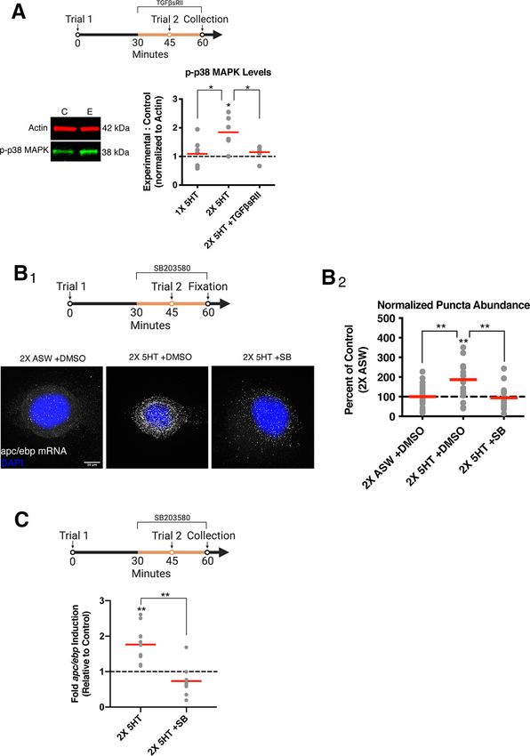
948 • J. Neurosci., February 3, 2021 • 41(5):947–959                                                    Mirisis et al. · mRNA Stabilization by ELAV Mediates LTM Formation

Aplysia c/ebp (apc/ebp), is required for LTM formation (Alberini et              Ganglia were perfused with ASW for at least 1.5 h to clear MgCl2 before
al., 1994; Alberini, 2009; Kandel, 2012; Mirisis et al., 2016).                  experimentation. Experimental and contralateral within-animal control
     As in many other species, the induction of LTM in Aplysia                   ganglia both received GF/drug/vehicle treatment, while only the experi-
critically depends on the number and pattern of training trials.                 mental ganglia received two-trial 5-HT training.
For example, two training trials separated by 45 min (but not by                      Training and drug incubation for gene expression analyses. After
                                                                                 MgCl2 washout, two-trial analog training was delivered by two pulses
15 or 60 min) induce LTM formation (Philips et al., 2013),
                                                                                 of 5-HT (5 min; 50 mM, Sigma Millipore, #H9523-25MG) in ASW
revealing a narrow time window during which a second trial                       (ITI = 45 min). As previously described (Kopec et al., 2015), ganglia in
interacts with a “molecular context” produced by the first trial to              GF treatment experiments were blocked with BSA for at least 5 min
give rise to LTM formation (Philips et al., 2013).                               pre-GF treatment, and GF/drug/vehicle was applied at the appropri-
     In this paper, we explore the role of mRNA stabilization                    ate experimental time point. Drugs used were as follows: rhTGF b 1
induced by growth factors (GFs) in LTM formation, with an em-                    (R&D Systems, #240-B-002), rhTGF b sRII (R&D Systems, #241-R2-
phasis on regulation that occurs between transcription and trans-                025), SB203580 (Tocris Bioscience, #1202), Actinomycin D (ActinoD,
lation within the molecular context established by Trial 1. Kopec                Sigma Millipore, #A1410-10MG), and CMLD-2 (Millipore, #538339).
et al. (2015) found that signaling through two distinct GF fami-                 Drug incubation was from 15 min before the training trial, during the
lies, TrkB and TGF b , is required during different training trials              training trial, and proceeded until SN cluster collection. Within-ani-
for the induction of the molecular events critical for LTM forma-                mal control ganglia concurrently received appropriate vehicle and
                                                                                 mock two-trial training (pulses of ASW without 5-HT). Control and
tion. Specifically, apc/ebp gene expression induced by Trial 1
                                                                                 experimental SN clusters were collected at the experimental time
peaked at 45 min, was dependent on TrkB signaling, and                           point and prepared for either Western blotting or qPCR analysis.
returned to baseline by 1 h (Philips et al., 2013; Kopec et al.,                      RNA isolation, cDNA synthesis, qPCR analysis. RNA was isolated
2015). However, if Trial 2 was delivered during peak apc/ebp                     and purified using Ambion RNAqueous Micro kits (Invitrogen), and
mRNA levels at 45 min after Trial 1, apc/ebp mRNA level                          total RNA for cDNA synthesis was normalized between experimental
remained elevated for .1 h, and this elevation critically required               and control ganglia (;100 ng RNA). cDNA was synthesized using
TGF b signaling during Trial 2. Further, the induction of LTM                    Superscript IV cDNA synthesis reagents (Invitrogen). qPCR was per-
required TrkB signaling during Trial 1 and TGF b signaling dur-                  formed using a Roche LightCycler 480, with 1 ml cDNA, 1 ml each of
ing Trial 2 (Kopec et al., 2015).                                                5 mM F9 and R9 primers, and 18 ml 1 SYBR Green (Roche Diagnostics).
     Trial 2-dependent apc/ebp gene expression was directly de-                  Primer sequences are as follows: apc/ebp-F: caccacctcactcccatctc; apc/
pendent on the TrkB-mediated events set in motion by Trial 1                     ebp-R: ctgacgtctgcgagactttg; apgapdh-F: ctctgagggtgctttgaagg; apgapdh-
(Kopec et al., 2015). This dependence could be explained in one of               R: gttgtcgttgagggcaattc. The amplification program was 95°C for 3 min,
                                                                                 30 cycles of 95°C for 10 s, 60°C for 20 s, and 72°C for 30 s, and last, a
two ways: (1) TGF b signaling in Trial 2 also induces apc/ebp gene
                                                                                 melting curve to confirm a single PCR product. Data were analyzed by
expression in Trial 2; or (2) TGFb signaling in Trial 2 engages                  normalizing the quantity of apc/ebp (measured by Ct) to apgapdh within
post-transcriptional mechanisms, which act to stabilize the apc/                 the same sample (DCt), then comparing normalized values between ex-
ebp mRNA that was generated by TrkB signaling in Trial 1.                        perimental and within-animal control groups (DDCt). Data are displayed
     In considering the second alternative, a candidate mechanism for            as fold induction relative to the control group.
the stabilization of the mRNA is the RNA-binding protein ELAV                         Western blotting. Samples were lysed and snap-frozen in RIPA buffer
(embryonic lethal abnormal vision), which confers stability to                   (Sigma Millipore) with 1 Halt protease and phosphatase inhibitor cock-
mRNAs containing AU-rich elements (AREs) in their 39 UTR                         tail (Invitrogen), loaded onto 4%-12% Bis-Tris gels (Novex; Invitrogen)
(Wang et al., 2000; Brennan and Steitz, 2001). Two ELAV family                   for electrophoresis, and then transferred to nitrocellulose membranes for
members, ApELAV1 and ApELAV2, have been identified and                           incubation with primary antibodies and secondary antibodies: anti-HuR
                                                                                 (1:1000; Santa Cruz Biotechnology, sc-5261), anti-b -actin (1:5000; Cell
cloned in Aplysia, and ApELAV1 has been shown to bind to apc/ebp
                                                                                 Signaling Technology, #3700), anti-p-p38 MAPK (1:1000; Cell Signaling
mRNA both in vitro and in vivo (Yim et al., 2006). Whereas ELAV                  Technology, #4511), anti-b -tubulin (1:5000; Cell Signaling Technology,
proteins have been implicated in memory formation (Quattrone et                  #2146), anti-mouse IgG IRDye 680LT (1:10,000; LICOR Biosciences, #926-
al., 2001; Yim et al., 2006; Bolognani et al., 2007), these studies lacked       68 020), and anti-rabbit IgG IRDye 800CW (1:10,000; LICOR Biosciences,
temporal resolution and did not elucidate the mechanisms recruiting              #926-32211). For ApELAV and p-p38 MAPK experiments, protein levels
ELAV-mediated mRNA stabilization and its role in LTM formation.                  within each sample were assessed using the LICOR imaging system. Each
     In the present paper, we examine mRNA stabilization                         protein of interest band was normalized to the tubulin or actin within the
induced by ApELAV during LTM formation and show that it is                       same sample, and normalized protein of interest in the experimental ganglia
specifically regulated by TGF b -induced p38 MAPK signaling                      was compared with within-animal control ganglia. Data are displayed as
initiated by Trial 2. Disrupting the ApELAV-apc/ebp interaction                  fold expression of normalized protein of interest in experimental sample rel-
                                                                                 ative to control sample.
blocks the prolonged expression of apc/ebp mRNA, and this
                                                                                      Single molecule FISH (smFISH). Aplysia SNs were individually iso-
interaction is required for the behavioral induction of LTM.                     lated with a glass microelectrode from the ventral SN cluster of the pleu-
Collectively, these data demonstrate both when and how mRNA                      ral ganglion of 80 g animals according to Zhao et al. (2009) in 50 mm
stabilization is achieved in LTM formation.                                      Mattek plates at 16 °C with culture medium consisting of salt-adjusted
                                                                                 L15 (Leibovitz) medium (Sigma; supplemented with 264 mM NaCl,
Materials and Methods                                                            26 mM MgSO4, 27 mM MgCl2, 5 mM KCl, 2 mM NaHCO3, 11 mM CaCl2,
Experimental model and subject details                                           15 mM HEPES, 35 mM glucose, 2 mM L-glutamine, 100 U/ml penicillin,
A. californica were acquired from South Coast Bio-Marine and                     0.1 mg/ml streptomycin) with 50% Aplysia hemolymph. SNs were dis-
University of Miami National Resource for Aplysia. All animals were              tributed very sparsely throughout the coverglass. After 4 DIV, 7 plates of
allowed to acclimate for at least 4 d in circulating tanks with artificial sea   10-20 SNs were chosen and separated into four different groups ran-
water (ASW) (Instant Ocean) at 15°C.                                             domly. Two plates each were designated as “2 5-HT 1 DMSO,” “2
                                                                                 5-HT 1 SB,” and “2 ASW 1 DMSO.” One plate received no treat-
Method details                                                                   ments but was used as a “no probe” control to determine background
   Ganglion preparation. Pleural-pedal ganglia were dissected from               fluorescence of SNs without DNA probes.
anesthetized A. californica (150-250 g). In a 1:1 solution of MgCl2 and               The smFISH protocol was adapted from Eliscovich et al. (2017).
ASW, the pleural SN cluster and SN-MN neuropil were exposed.                     Following treatment according to the experimental paradigm (5-HT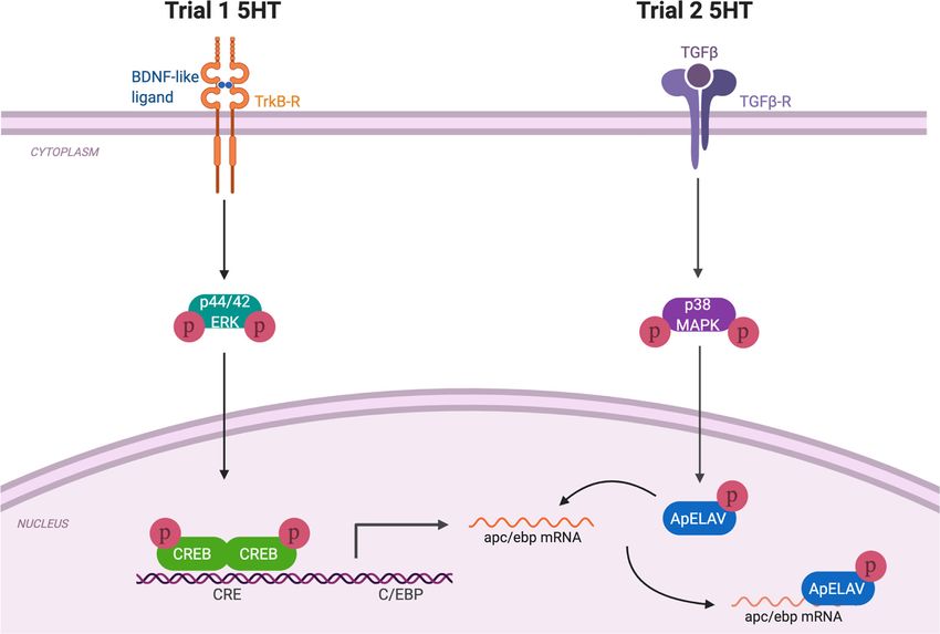
Mirisis et al. · mRNA Stabilization by ELAV Mediates LTM Formation                                              J. Neurosci., February 3, 2021 • 41(5):947–959 • 949

treatment was 50 mM in ASW, ASW treatment was an equivalent volume           drug-treated) and within-animal control (mock two-trial training and
of ASW), the cells were fixed for 30 min at room temperature in 4%           vehicle) groups.
formaldehyde in 30% sucrose/DEPC-treated PBS with supplemented                   ApELAV1 protein expression. The coding region for ApELAV1 was
1 mM magnesium chloride and 0.1 mM calcium chloride (DEPC-PBS-               isolated by PCR from a previously published plasmid kindly provided by
MC). Cells were washed quickly 2 with DEPC-PBS-MC and quenched              Bong-Kiun Kaang (Seoul National University) (Yim et al., 2006) and
with glycine in DEPC-PBS-MC. Quench buffer was removed, and cells            ligated into pGEX vector to make pGEX-ApELAV1-GST. Rosetta 2 DE3
were incubated in permeabilization-block buffer (0.2% Triton X-100,          cells (Millipore) were transformed with this plasmid, and cells were
0.5% Ultrapure BSA in DEPC-PBS-MC) for 30 min at room temperature            induced for 2 h at 37°C with 1 mM IPTG during log-phase growth. Cells
while shaking. Permeabilization-block buffer was removed, and cells          were subsequently lysed and subjected to Western blotting.
were incubated with prehybridization buffer (10% deionized formamide,            Semi-intact behavioral preparation. Preparations were completed as
2 SSC, 0.5% Ultrapure BSA in nuclease-free water) for 30 min at room        previously described (Sutton et al., 2001; Kopec et al., 2015). Briefly, the
temperature without shaking. Prehybridization buffer was removed, and        ring ganglia (cerebral and paired pleural-pedal ganglia) from anesthe-
cells were incubated with hybridization buffer (10% deionized formam-        tized Aplysia (250-400 g; South Coast Bio-Marine) were surgically iso-
ide, 2 SSC, 1 mg/ml Escherichia coli tRNA, 20 mg/ml BSA, 2 SSC,            lated, leaving p9 and pleural-abdominal innervation to the tail and
2 mM vanadyl ribonucleoside complex, 10% dextran sulfate, 125 nM of          abdominal ganglia, respectively, intact. The tail and mantle were surgi-
DNA probes in nuclease-free water) for 4 h at 37°C. Thirty-three DNA         cally removed, and the siphon artery was cannulated with Dow Corning
probes conjugated to Quasar 670 dye (LGC Biosearch Technology) were          SILASTIC tubing (0.025 in I.D.; Thermo Fisher Scientific) and perfused
designed to be antisense to all regions on apc/ebp mRNA (Accession           at ;5 ml/min, while the tail was perfused at ;0.5 ml/min via three 22-
#NP_001191392.1; NCBI) without significant complementarity to other          gauge needles inserted into the tail. The tail and mantle were pinned to
mRNAs in the Aplysia Refseq database (NCBI). Cells were washed 4             the chamber floor, while the ring ganglia with both pleural-pedal ganglia
times with 2 SSC and then once quickly with nuclease-free water.            desheathed were pinned in an isolated CNS chamber. Both the chamber
Coverslips were detached from the plates and mounted using Prolong           containing the peripheral tissues as well as the CNS chamber were con-
Gold Antifade Reagent with DAPI (Invitrogen).                                tinuously perfused with seawater (Instant Ocean, 15°C). The p9 and
    A Deltavision Elite imaging system (GE) equipped with Olympus            pleural-abdominal nerves exited the CNS chamber through small slits
60/1.42 NA oil immersion lens and an InsightSSI Solid State                 that were sealed with Vaseline. Preparations were allowed at least 2 h to
Illumination module were used to image samples. Individual full SN           recover before baseline measurements.
somata were imaged in a z stack with 500 nm spacing. Raw images were             Training and drug incubation for LTM analyses. An average of 3 or
loaded into FISH-QUANT software (Mueller et al., 2013) and batch             4 baseline siphon-withdrawal measurements were recorded by stimulat-
processed after detecting nucleus size (by DAPI) and tracing the outline     ing the medial posterior tip of the tail with a water jet (0.4 s, 45 psi; ITI
of each SN. Initial analyses determined that, in the cells that received     15 min; Teledyne Water Pik) and measuring the duration of time with
mock treatment (2 ASW), the number of puncta counted in each cell           which the siphon withdrew. No significant differences in the pretraining
was correlated strongly to the area of the cell nucleus, which corresponds   baseline duration of T-SWRs were observed in any experimental condi-
to previous literature noting that the absolute amount of RNA can be de-     tion. The CNS was incubated with drug/vehicle 15 min before Trial 2,
pendent on cell size (Marguerat and Bähler, 2012). Therefore, the num-       which was washed out 30 min after Trial 2. Two-trial LTM training
ber of puncta counted in each cell was normalized to the area of the         (100 mA, 1.5 s, ITI 45 min) was delivered medially to the anterior por-
nucleus in each cell, as this value was calculated automatically by FISH-    tion of the tail through a hand-held electrode. LTM for sensitization of
QUANT.                                                                       the T-SWR was assessed by 3 or 4 tests (ITI = 15 min) 15-22 h after train-
    Cross-linking immunoprecipitation (CLIP)-qPCR. The protocol was          ing by an experimenter blind to the experimental condition (drug vs ve-
adapted from Yoon and Gorospe (2016). Experimental and within-ani-           hicle). The data are displayed as duration of siphon withdrawal as a
mal contralateral control ganglia both received GF/drug/vehicle treat-       percent of pretraining baseline.
ment, while only the experimental ganglia received two-trial 5-HT
training. At 1 h following the onset of Trial 1, experimental and control    Quantification and statistical analysis
ganglia were exposed to two consecutive pulses of 400 mJ UV light            Data were analyzed with parametric statistics using GraphPad Prism.
(254 nm) in a Stratagene Gene Linker to crosslink ribonucleoprotein          Statistical details for all experiments can be found within the main text
complexes. Two pulses were used to ensure that UV light penetrated           and/or in the figure legends. Within-group analyses were performed
deep into the ventral SN cluster of the intact ganglion, as has been         using paired t test. LTM for sensitization of the T-SWR is compared rel-
described for other tissues, such as triturated mouse brain (Darnell,        ative to pretraining baseline T-SWR within the same preparation.
2012). SN clusters of 8 animals were collected and lysed in ice-cold NP-     Between-group analyses (drug vs vehicle) were performed using t tests.
40 lysis buffer (20 mM Tris-HCl, pH 7.5, 100 mM KCl, 5 mM MgCl2, 0.5%        When appropriate, ANOVAs were used to determine whether there was
NP-40, 1 mM DTT) supplemented with 1 Halt protease and phospha-             a difference among the groups and are indicated in the main text.
tase inhibitor cocktail and 40 U/ml RNaseOUT (Invitrogen), and pooled        Significant results were followed by planned parametric comparisons.
for each experimental and contralateral control sample. Lysates were         Outliers .2 SDs from the mean were excluded resulting in the removal
split into two fractions each and immunoprecipitated with either anti-       of 5 data points (,5% of all data points; reported in Results). Because of
bodies against HuR (Santa Cruz Biotechnology, sc-5261; see Fig. 3F) or       normal distribution of data, data in all figures are depicted as mean 6
normal mouse IgG1 (Santa Cruz Biotechnology, sc-3877) using Protein          SEM. Significant within-group comparisons are displayed as asterisks
G Dynabeads (Invitrogen). Beads were washed with ice-cold NT-2 buffer        above the summary data, and significant between-group comparisons
(50 mM Tris-HCl, pH 7.5, 150 mM NaCl, 1 mM MgCl2, 0.05% NP-40).              are displayed as asterisks above a bar drawn between the summary data.
Following consecutive digestions with DNase I and Proteinase K (New
England Biolabs), beads were magnetically separated from the superna-
tant. RNA was isolated from the supernatant with Trizol extraction           Results
(Invitrogen), and cDNA was synthesized using Superscript IV cDNA             The Trial 1-dependent increase in apc/ebp mRNA level is
synthesis reagents (Invitrogen). qPCR was performed as described             transcription-dependent
above. Importantly, primers were designed to lie within the 39 UTR of
                                                                             We have shown that apc/ebp gene expression is increased 45 min
apc/ebp mRNA, the binding region of ELAV. The primer sequences
were as follows: apc/ebpUTR-F: cccaagcatgttgtgatagttgt; apc/ebpUTR-R:
                                                                             following Trial 1 (Philips et al., 2013; Kopec et al., 2015). Thus,
tgggaggagatagaagcagtg. Data were analyzed by normalizing the differ-         we first asked whether Trial 1-dependent increase in apc/ebp
ence in apc/ebp between ELAV- and IgG-immunoprecipitated samples             gene expression was because of (1) increased gene transcription
to the difference in apgapdh between the same samples, then comparing        or (2) alternative mechanisms, such as an increase in transcript
these normalized values between experimental (two-trial training and         stability of baseline levels of apc/ebp mRNA.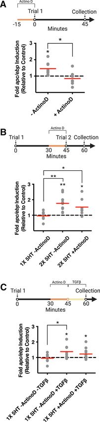
950 • J. Neurosci., February 3, 2021 • 41(5):947–959 Mirisis et al. · mRNA Stabilization by ELAV Mediates LTM Formation
In order to distinguish between these possibilities, we first
conducted experiments to determine whether transcription was
required for the Trial 1-dependent increase in apc/ebp gene
expression. Dissected pleural-pedal ganglia were incubated with
the irreversible transcription inhibitor Actinomycin D (40 mg/ml,
Sigma Millipore) for 15 min, and contralateral within-animal
control ganglia were incubated with vehicle (an equivalent vol-
ume of ddH2O). Following drug/vehicle washout, experimental
ganglia were administered Trial 1 (50 mM 5-HT in ASW, Sigma
Millipore) for 5 min (1 5-HT) to simulate a tail shock training
trial (Philips et al., 2013). Within-animal contralateral control
ganglia were administered mock Trial 1 (pulse of ASW without
5-HT). Five minutes following pulse onset, ganglia were washed
with fresh ASW, and pleural SN clusters were collected at 45 min
and analyzed for apc/ebp gene expression via qPCR. No signifi-
cant difference in apc/ebp gene expression was observed between
(1) experimental ganglia, which received Trial 1 in the presence
of ActinoD, and (2) within-animal control ganglia, which
received mock training in the presence of vehicle (Fig. 1A;
within-group paired t test: 1ActinoD: n = 7, 0.8505 6 0.1601,
p = 0.3865). In this and following experiments, the ratio of apc/
ebp gene expression in experimental ganglia to contralateral con-
trol ganglia was calculated for each pair of ganglia (data in histo-
grams are represented as a ratio of drug conditions to vehicle
conditions). Consistent with previous results (Kopec et al., 2015),
a within-group comparison revealed a significant increase in apc/
ebp gene expression in experimental SN clusters collected 45 min
after Trial 1 without drug treatment, compared with within-ani-
mal contralateral control ganglia, which received mock Trial 1
without drug treatment (Fig. 1A; within-group paired t test: –
ActinoD: n = 8, 1.456 6 0.140, p = 0.0139). A between-group com-
parison revealed a significant difference between apc/ebp gene
expression of SN clusters, which had received Trial 1 with
ActinoD compared with SN clusters, which had received Trial 1
without ActinoD (Fig. 1A; between-group unpaired t test: –
ActinoD vs 1ActinoD: t = 2.862, df = 13, p = 0.0134), demonstrat-
ing that Trial 1-dependent apc/ebp gene expression at 45 min after
Trial 1 is dependent on de novo gene transcription.
The Trial 2-dependent increase in apc/ebp mRNA level is
transcription-independent
When Trial 2 is delivered 45 min after Trial 1, apc/ebp gene
expression at 1 h after Trial 1 is dependent on (1) TrkB signaling
during Trial 1 and (2) TGF b signaling during Trial 2 (Kopec et
al., 2015). This suggests that either (1) Trial 2 TGF b -dependent
mechanisms cause a new wave of apc/ebp transcription or (2)
Trial 2 induces a form of transcription-independent stabilization
/
this and all subsequent figures, unless specified otherwise, data are displayed as aligned dot
plots. Red lines within each histogram indicate means. Within-group statistical significance is
displayed with an asterisk within histograms, and between-group statistical significance is
indicated above the histograms being compared. n = 7 or 8. pp , 0.05. For full statistical
details, see Results. B, Top, Experimental paradigm. SN somata are collected at 1 h following
the onset of Trial 1. Experimental ganglia are treated with ActinoD at 30-45 min (dark yellow
line). Bottom, A second training trial (Trial 2) at 45 min following the onset of Trial 1 results
Figure 1. Trial 1 initiates a transcription-dependent increase in apc/ebp mRNA levels, in a transcription-independent significant increase in apc/ebp gene expression at 1 h. n = 7-
whereas Trial 2 prolongs this gene expression through a transcription-independent mecha- 11. pp , 0.05. ppp , 0.01. C, Top, Experimental paradigm. SN somata are collected at 1 h
nism. A, Top, Experimental paradigm. Trial 1 is delivered at time = 0:00, and SN somata are following the onset of Trial 1. Experimental ganglia are treated with ActinoD at 30-45 min
collected at 45 min following the onset of Trial 1. Experimental ganglia are treated with (dark yellow line). TGF b 1 is applied at 45 min until collection (light yellow line). Bottom,
ActinoD 15 min before Trial 1 (dark yellow line). Bottom, Blocking transcription before Trial 1 Treatment with TGF b 1 alone at 45 min results in a transcription-independent increase in
significantly disrupts apc/ebp gene expression at 45 min. Histograms are labeled by their cor- apc/ebp mRNA level. 1 5-HT –ActinoD -TGF b group control data are from B. n = 7-12.
responding drug condition. Drug conditions are expressed as a ratio to vehicle conditions. In pp , 0.05.Mirisis et al. · mRNA Stabilization by ELAV Mediates LTM Formation J. Neurosci., February 3, 2021 • 41(5):947–959 • 951
of apc/ebp mRNA synthesized from Trial 1 (via TrkB-dependent asked whether the TGF b signaling-dependent effect of increased
mechanisms). Experimental ganglia were treated with ActinoD for apc/ebp mRNA levels at 1 h after Trial 1 required transcription.
15 min before Trial 2 (30 min following Trial 1). Following drug Ganglia were treated at 30 min after Trial 1 with ActinoD. At
washout, experimental ganglia were exposed to 5-HT for 5 min 45 min after Trial 1, ActinoD was washed out and ganglia were
(Trial 2), thus receiving two-trial training (2 5-HT). 5-HT was immediately treated with TGF b 1 until SN cluster collection and
washed out with fresh ASW, and pleural SN clusters were collected lysis at 1 h after Trial 1. Contralateral within-animal control gan-
at 1 h after Trial 1. In this and subsequent experiments where ex- glia received mock Trial 1 and vehicle (0.1% BSA in ASW and
perimental ganglia are trained with two-trial training, within-ani- ddH2O). TGF b 1 treatment concomitant with a transcriptional
mal contralateral control ganglia received mock two-trial training block still resulted in significantly increased apc/ebp mRNA level
(5 min pulses of ASW without 5-HT) and vehicle. A within-group compared with within-animal contralateral control ganglia (Fig.
analysis revealed a significant increase in apc/ebp mRNA levels 1C; within-group paired t test: 1 5-HT 1ActinoD 1TGF b :
in ganglia, which had received two-trial training, even in the 1.230 6 0.0954, n = 12, p = 0.0345). Moreover, there was no sig-
presence of ActinoD before Trial 2 (Fig. 1B; within-group nificant difference between groups that received TGF b treat-
paired t test: 2 5-HT –ActinoD: 1.765 6 0.1463, n = 11, p = ment alone and TGF b treatment with ActinoD (Fig. 1C;
0.0004; 2 5-HT 1ActinoD: 1.527 6 0.1830, n = 8, p = 0.0237). between-group unpaired t test: 1 5-HT –ActinoD 1TGF b vs
Consistent with previous findings (Kopec et al., 2015), Trial 1 1 5-HT 1ActinoD 1TGF b : t = 0.9160, df = 18, p = 0.3718).
alone was not sufficient for apc/ebp gene expression 1 h later Collectively, these data support the hypothesis that TGF b sig-
(Fig. 1B; within-group paired t test: 1 5-HT –ActinoD: 0.9600 6 naling during Trial 2 is responsible for the post-transcriptional
0.1118, n = 7, p = 0.7325). ANOVA revealed a significant difference stabilization effect on apc/ebp mRNA.
among the three groups (F = 6.786; p = 0.0048). Subsequent
planned t tests revealed a significant increase in apc/ebp mRNA p38 MAPK is activated by two-trial training and is blocked
levels in both the 2 5-HT –ActinoD and 2 5-HT 1ActinoD by blocking TGFb signaling
groups, compared with ganglia that had received only Trial 1 (1 Our findings raise the important question of how Trial 2 pro-
5-HT), and no significant difference was noted between the 2 longs apc/ebp gene expression through a transcription-inde-
5-HT –ActinoD and 2 5-HT 1ActinoD groups (Fig. 1B; be- pendent mechanism downstream of TGF b signaling. To
tween-group unpaired t test: 1 5-HT –ActinoD vs 2 5-HT examine this question, we investigated a particular kinase, p38
–ActinoD: t = 3.926, df = 16, p = 0.0012; 1 5-HT –ActinoD MAPK, which is both activated downstream of TGF b signal-
vs 2 5-HT 1ActinoD: t = 2.550, df = 13, p = 0.0242; 2 ing (Yamashita et al., 2008) and plays a cellular role in broker-
5-HT –ActinoD vs 2 5-HT 1ActinoD: t = 1.028, df = 17, p = ing to regulating mRNA stability (Varela-Rey et al., 2002;
0.3181). These data demonstrate that the Trial 2-dependent Dean et al., 2003; Soni et al., 2019). Thus, we assayed p38
increase in apc/ebp mRNA level at 1 h after Trial 1 is not de- MAPK activation in the pleural SNs following two-trial train-
pendent on de novo gene transcription and supports the hy- ing. Our experiments revealed a significant increase in p38
pothesis that the increased apc/ebp mRNA level may result MAPK activation in SNs following two-trial training in experi-
from TGF b -dependent mRNA stabilization. mental ganglia compared with within-animal contralateral
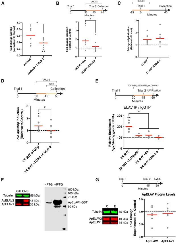
control ganglia (Fig. 2A; within-group paired t test: 2 5-HT:
TGFb1 treatment at 45 min is sufficient for Trial 2- 1.845 6 0.232, n = 6, p = 0.0148). This increase was blocked ei-
dependent increase in apc/ebp mRNA level ther (1) by eliminating Trial 2 or (2) by delivering Trial 2 in
By sequestering endogenously released TGF b -like ligands dur- the presence of TGF b sRII (5 mg/ml, R&D Systems), the solu-
ing Trial 2, we have previously shown that intact TGF b signal- ble extracellular portion of the human Type-II TGF b recep-
ing is required during Trial 2 for apc/ebp gene expression at 1 h tor, which acts as an inhibitor of TGF b signaling by
(Kopec et al., 2015). We here sought to determine whether treat- sequestering extracellular ligands, preventing their association
ment with recombinant human TGF b 1 alone could duplicate with the endogenous Aplysia TGFb receptor (Kopec et al., 2015)
these effects. (Fig. 2A; within-group paired t test: 1 5-HT: 1.089 6 0.184, n = 7,
To investigate this question, ganglia were treated with p = 0.9975; 2 5-HT 1TGFb sRII: 1.148 6 0.086, n = 7, p = 0.25;
TGF b 1 (100 ng/ml, R&D Systems) starting at 45 min after between-group unpaired t test: 1 5-HT vs 2 5-HT: t = 2.582,
Trial 1 until SN cluster collection and lysis at 1 h after Trial 1 df = 11, p = 0.0255; 2 5-HT vs 2 5-HT 1TGFb sRII: t = 2.996,
(Trial 2 was not administered). Treatment with TGF b 1 has df = 11, p = 0.0122). These data demonstrate that p38 MAPK acti-
previously been demonstrated to (1) recruit persistent ERK sig- vation at 1 h is dependent on TGF b signaling and may serve an
naling, a necessary step in LTM formation; and (2) induce intermediate role between TGF b signaling and apc/ebp mRNA
long-term facilitation in isolated Aplysia ganglia (Zhang et al., stabilization.
1997; Shobe et al., 2016). Contralateral control ganglia received
mock Trial 1 and were treated with an equivalent volume of ve- p38 MAPK activation is required for prolonged increase in
hicle (0.1% BSA in ASW). Treatment with TGF b 1 resulted in a apc/ebp mRNA levels
significant increase in apc/ebp mRNA levels compared with To further examine the role of p38 MAPK in the stabilization of
within-animal controls that had received only vehicle (Fig. 1C; apc/ebp mRNA, we directly measured apc/ebp mRNA levels, by
within-group paired t test: 1 5-HT –ActinoD 1TGF b : performing smFISH on isolated Aplysia SNs. This technique
1.384 6 0.1493, n = 8, p = 0.0369). Between-group comparisons allows for fluorescent visualization of individual apc/ebp RNA
further revealed a significant difference between the Trial 1 molecules as puncta resulting from the combined fluorescence of
only and Trial 1 1TGF b -treated groups (Fig. 1C; between- 33 antisense DNA probes (LGC Biosearch Technology) (Fig. 2B1).
group unpaired t test: 1 5-HT –ActinoD -TGF b vs 1 5-HT ANOVA revealed a significant difference in puncta abundance
–ActinoD 1TGF b : t = 2.219, df = 13, p = 0.0449). between: (1) SNs that received two-trial training in the presence of
Given that TGF b 1 treatment at 45 min after Trial 1 is suffi- vehicle (2 5-HT 1 DMSO); (2) SNs that received two-trial train-
cient to increase apc/ebp mRNA level at 1 h (Fig. 1C), we further ing in the presence of SB203580, a p38 MAPK inhibitor previously952 • J. Neurosci., February 3, 2021 • 41(5):947–959 Mirisis et al. · mRNA Stabilization by ELAV Mediates LTM Formation
used and characterized in Aplysia (Zhang et al.,
2017) (10 mM, Tocris Bioscience) (2 5-HT 1 SB);
and (3) SNs that received vehicle and mock two-
trial training (2 ASW 1 DMSO) (Fig. 2B2;
F = 7.804, p = 0.0010). Planned t tests revealed a sig-
nificant increase in puncta in SNs that received
two-trial training in the presence of DMSO com-
pared with each of the other groups (Fig. 2B2;
within-group paired t test: 2 5-HT 1 DMSO:
186.9 6 23.85%, n = 23, p = 0.0014, 1 outlier; 2
5-HT 1 SB: 94.82 6 15.74%, n = 15, p = 0.7471, 2
outliers; 2 ASW 1 DMSO: 100 6 12.74%, n = 23,
1 outlier, p = 0.9999; between-group unpaired t test:
2 5-HT 1 DMSO vs 2 5-HT 1 SB: t = 2.855,
df = 36, p = 0.0071; 2 5-HT 1 DMSO vs 2 ASW
1 DMSO: t = 3.213, df = 44, p = 0.0025).
We used qPCR to examine whether p38
MAPK activation is important for the increase in
apc/ebp mRNA level 1 h after Trial 1 in ganglia.
Experimental ganglia received two-trial training
with SB203580 treatment during Trial 2 until SN
cluster collection and lysis at 1 h. Within-animal
contralateral control ganglia were treated with ve-
hicle (0.1% DMSO) and received mock two-trial
training. Treatment with SB203580 during Trial 2
blocked the Trial 2-dependent increase in apc/ebp
mRNA levels compared with two-trial training
alone (Fig. 2C; within-group paired t test: 2
5-HT 1 SB: 0.7341 6 0.161, n = 8, p = 0.1423;
between-group unpaired t test: 2 5-HT vs 2
5-HT 1 SB: t = 4.692, df = 17, p = 0.0002).
ApELAV-mRNA interaction is required for
prolonged increase in apc/ebp mRNA levels
Our findings thus far demonstrate that p38
MAPK activation downstream of TGF b signaling
resulting from Trial 2 is required for the stabiliza-
tion of apc/ebp mRNA. The RNA-binding protein
ApELAV has been previously shown to bind apc/
ebp mRNA in Aplysia by associating with AREs in Figure 2. Prolonged expression of apc/ebp mRNA requires p38 MAPK activity initiated by TGF b signaling. A,
the 39 UTR (Yim et al., 2006). Since ELAV is Top, Experimental paradigm. SN somata are collected at 1 h following the onset of Trial 1. Experimental ganglia
known to be an RNA-binding protein yielding a receive two-trial training and are treated with TGF b sRII (dark yellow line) from 30 min until collection at 1 h.
stabilizing effect on its target mRNAs (Brennan Bottom, Left, Representative Western blot for p-p38 MAPK using actin as a loading control. Control (C) and experi-
mental (E) indicate bands from a single SN cluster pair (n = 1) in the 2 5-HT treatment group. Right, Following
and Steitz, 2001; Yim et al., 2006), we explored the
two-trial training, there is a significant increase in p38 MAPK activation, which is blocked by treatment with
possibility that apc/ebp gene expression is modu- TGFb sRII. n = 6 or 7. pp , 0.05. B , Top, Experimental paradigm. SNs are fixed at 1 h following the onset of
1
lated by ApELAV. In support of this possibility, Trial 1. Experimental plates of SNs receive two-trial training and are treated with SB203580 (dark yellow line)
p38 MAPK has previously been identified as a from 30 min until fixation at 1 h. Bottom, Representative maximum z-projection widefield microcopy images of
critical mediator of ELAV activation by phospho- isolated SNs subjected to smFISH. White puncta are apc/ebp mRNA. Blue represents nuclei (stained with DAPI).
rylation (Pascale et al., 2005; Lafarga et al., 2009; Scale bar, 10 mm. B2, Following two-trial training, apc/ebp mRNA levels are increased in SNs, and this increase is
Bai et al., 2012; Eberhardt et al., 2012; Slone et al., blocked by blocking p38 MAPK activation. Data are represented as a percentage of normalized puncta abundance
2016). in SNs, which received mock two-trial training in the presence of vehicle. n = 19-23. ppp , 0.01. C, Top,
In order to directly manipulate the ability of Experimental paradigm. SN somata are collected at 1 h following the onset of Trial 1. Experimental ganglia receive
two-trial training and are treated with SB203580 (dark yellow line) from 30 min until collection at 1 h. SN clusters
ApELAV to interact with its target transcripts, we
are subjected to qPCR. Bottom, Following two-trial training, there is a significant increase in apc/ebp mRNA levels,
used CMLD-2 (100 mM, Millipore), a potent inhibi- which is blocked by blocking p38 MAPK activation during Trial 2.The 2 5-HT control data are from Figure 1B.
tor of HuR-ARE interaction (Wu et al., 2015; Slone n = 8-11. ppp , 0.01.
et al., 2016; Muralidharan et al., 2017). HuR is a
mammalian homolog of ELAV, and also contains In order to test whether CMLD-2 blocks the interaction
three highly conserved RNA recognition motifs, between ApELAV and apc/ebp mRNA, we performed experiments
which facilitate its binding to AREs of mRNAs. Since CMLD-2 assaying the stability of apc/ebp mRNA following transcriptional
directly binds to the highly evolutionary conserved RNA-binding block (to inhibit basal transcription) both with and without
pocket of HuR, we hypothesized that it would likewise bind and CMLD-2 treatment. We found that treatment with both ActinoD
inhibit ApELAV binding. alone and ActinoD 1CMLD-2 for 40 min resulted in a significantMirisis et al. · mRNA Stabilization by ELAV Mediates LTM Formation J. Neurosci., February 3, 2021 • 41(5):947–959 • 953 Figure 3. ApELAV proteins bind and stabilize apc/ebp mRNA downstream of p38 MAPK and TGF b signaling. A, Following inhibition of basal transcription, apc/ebp mRNA is rapidly degraded in SN clusters, and this degradation is significantly accelerated by additional CMLD-2 treatment. n = 4-8. pp , 0.05. ppp , 0.01. B, Top, Experimental paradigm. SN clusters are collected at 1 h following the onset of Trial 1. Experimental ganglia receive two-trial training with CMLD-2 treatment (dark yellow line) from 30 min until collection at 1 h. Bottom, Blocking the ApELAV- mRNA interaction during Trial 2 blocks the increase in apc/ebp mRNA levels following two-trial training. n = 8 or 9. pp , 0.05. C, Top, Experimental paradigm. SN clusters are collected at
954 • J. Neurosci., February 3, 2021 • 41(5):947–959                                                                    Mirisis et al. · mRNA Stabilization by ELAV Mediates LTM Formation

decrease in apc/ebp mRNA levels in experimental ganglia com-                                       n = 8, p = 0.0209; between-group unpaired t test: 1 5-HT vs 1 5-
pared with contralateral within-animal control ganglia that                                        HT 1CMLD-2: t = 0.2887, df = 14, p = 0.7770).
received vehicle (0.8% acetonitrile and ddH2O) (Fig. 3A; within-                                       We next asked whether CMLD-2 could block the increase in
group t test: ActinoD: 0.6200 6 0.1081, n = 4, p = 0.0105; ActinoD                                 apc/ebp mRNA levels resulting from treatment of ganglia, which
1CMLD-2: 0.3855 6 0.0460, n = 8, p = 0.0001). A between-                                           had received Trial 1 and TGF b 1 treatment at 45 min (no Trial
group comparison determined that addition of CMLD-2 accel-                                         2). When CMLD-2 was administered with TGF b 1 treatment in
erates apc/ebp mRNA degradation compared with ActinoD                                              lieu of Trial 2, no significant difference in apc/ebp levels between
treatment alone (Fig. 3A; between-group unpaired t test:                                           experimental (1 5-HT 1TGF b 1CMLD-2) and within-ani-
ActinoD vs ActinoD 1CMLD-2: t = 2.382, df = 10, p = 0.0385).                                       mal control ganglia (mock Trial 1 and vehicle alone) was
    In order to examine whether the ApELAV-apc/ebp mRNA                                            observed (Fig. 3D; within-group paired t test: 1 5-HT 1TGF b
interaction plays a critical role in increased apc/ebp mRNA levels                                 1CMLD-2: 0.9825 6 0.1928, n = 7, p = 0.93, 1 outlier; see
during the two-trial training paradigm, ganglia received Trial 2                                   Materials and Methods). Although not significant, a between-
in the presence of CMLD-2. CMLD-2 treatment proceeded until                                        group comparison revealed a trend toward blocking the increase
SN cluster collection and lysis at 1 h. Within-animal contralateral                                in apc/ebp mRNA levels in the CMLD-2 treated versus TGF b
control ganglia were treated with vehicle and mock two-trial                                       treatment alone groups (Fig. 3D; between-group unpaired t test:
training. We found that apc/ebp mRNA levels in experimental                                        1 5-HT 1TGF b vs 1 5-HT 1TGF b 1CMLD-2: t = 1.669,
ganglia were not significantly different from levels in within-ani-                                df = 13, p = 0.1190).
mal contralateral control ganglia (Fig. 3B; within-group paired t
test: 2 5-HT 1CMLD-2: 1.194 6 0.201, n = 9, p = 0.6195).                                          Blocking TGFb signaling, p38 MAPK activation, and the
Further, CMLD-2 significantly blocked the increase of Trial 2-                                     ELAV-ARE interaction during Trial 2 eliminates the
dependent apc/ebp mRNA levels compared with two-trial train-                                       ApELAV-apc/ebp mRNA interaction
ing with vehicle alone (Fig. 3B; within-group paired t test: 2                                    Our hypothesis, that ApELAV is recruited by Trial 2 and binds
5-HT 1Veh: 1.831 6 0.2892, n = 8, p = 0.0119; between-group                                        to the apc/ebp transcript, predicts that the ApELAV-apc/ebp
unpaired t test: 2 5-HT 1Veh vs 2 5-HT 1CMLD-2:                                                  mRNA interaction should be increased at the 1 h time point fol-
t = 1.844, df = 15, p = 0.0425).                                                                   lowing Trial 1 in ganglia, which receive two-trial training. To
    We have previously demonstrated that Trial 1 results in a                                      directly test this hypothesis, we performed a CLIP experiment
transcription-dependent increase in apc/ebp mRNA levels at                                         followed by qPCR (CLIP-qPCR), to compare the physical inter-
45 min (Fig. 1A). In order to determine whether the effect of                                      action of apc/ebp mRNA with ApELAV between experimental
ApELAV-mediated stabilization of apc/ebp mRNA is specifically                                      (two-trial training) and within-animal control (mock two-trial
engaged by Trial 2, we treated experimental ganglia with CMLD-                                     training) groups. We used an antibody against HuR (see
2 during Trial 1 and assayed apc/ebp mRNA levels at 45 min.                                        Materials and Methods; Fig. 3F), a mammalian member of the
Within-animal contralateral control ganglia were treated with                                      ELAV family, for immunoprecipitation of ApELAV-contain-
vehicle and mock Trial 1. We found that CMLD-2 treatment                                           ing crosslinked ribonucleoprotein complexes. This antibody
did not block apc/ebp gene expression induced by Trial 1, dem-                                     recognizes both Aplysia ELAV family members, ApELAV1
onstrating that ApELAV is specifically engaged by Trial 2 (Fig. 3C;                                and ApELAV2 (Fig. 3F). Following two-trial training, we
within-group paired t test: 1 5-HT 1CMLD-2: 1.527 6 0.2002,                                       indeed found a significant increase in apc/ebp mRNA associa-
                                                                                                   tion with ApELAV in experimental ganglia compared with
                                                                                                   within-animal contralateral control ganglia (Fig. 3E; within-
/                                                                                                  group paired t test: 2 5-HT: 201.6% 6 19.89, n = 5, p =
45 min following the onset of Trial 1. Experimental ganglia receive CMLD-2 treatment (dark         0.007), which strengthens the hypothesis that ELAV, by bind-
yellow line) 15 min before Trial 1 and continues to 15 min after Trial 1. Bottom, Blocking the     ing to apc/ebp transcript, promotes its stabilization at that
ApELAV-mRNA interaction during Trial 1 does not block the increase in apc/ebp mRNA levels          time point.
at 45 min. The 1 5-HT control data are from Figure 1A. n = 8. D, Top, Experimental para-              If ApELAV is esponsible for stabilizing apc/ebp mRNA during
digm. SN clusters are collected at 1 h following the onset of Trial 1. Experimental ganglia        Trial 2, thereby prolonging its expression to 1 h following Trial 1,
receive Trial 1 and CMLD-2 treatment (dark and light yellow line) from 30 min until collection     then blocking TGFb signaling should block the ApELAV-apc/ebp
at 1 h. TGF b treatment (light yellow line) is from 45 min until collection at 1 h. Bottom,        mRNA interaction. We have previously shown that treatment
Blocking the ApELAV-mRNA interaction blocks the within-group significant increase in apc/          with TGFb sRII disrupted the increase in Trial 2-dependent apc/
ebp mRNA levels by TGF b treatment. The 1 5-HT 1TGF b control group is from Figure
                                                                                                   ebp mRNA level (Kopec et al., 2015). Treatment with TGF b sRII
1C. n = 7 or 8. #p , 0.2. pp , 0.05. E, Top, Experimental paradigm. SN somata are UV-fixed
and collected at 1 h following the onset of Trial 1. Experimental ganglia receive two-trial
                                                                                                   resulted in the complete loss of the ApELAV-apc/ebp mRNA
training and are treated with TGF b sRII, SB203580, or CMLD-2 (dark yellow line) from              interaction at the same time point when assayed by CLIP-qPCR
15 min before Trial 2 until UV fixation at 1 h. Bottom, Following two-trial training, there is a   and compared with two-trial training alone (Fig. 3E; between-
significant increase in ApELAV-apc/ebp mRNA interaction, which is blocked by treatment             group unpaired t test: 2 5-HT vs 2 5-HT 1TGF b sRII:
with TGF b sRII, SB203580, or CMLD-2. Each data point represents the results of one experi-        t = 2.995, df = 6, p = 0.0242). No significant increase in ApELAV-
ment (SN clusters pooled from 8 animals). pp , 0.05. ppp , 0.01. n = 3-5. F, Left,                 apc/ebp mRNA interaction was observed when experimental gan-
ApELAV1 and ApELAV2 are recognized by the mouse monoclonal anti-HuR (Santa Cruz                    glia (which received two-trial training and TGFb sRII) were com-
Biotechnology) antibody at their respective predicted molecular weights. ApELAV1 is present        pared with contralateral within-animal control ganglia, which
in both gill and CNS. ApELAV2 demonstrates differential expression in gill and CNS. Tubulin is     received mock two-trial training and vehicle (0.1% BSA in ASW)
used as a loading control. Right, The antibody reacts strongly with recombinant ApELAV1-
                                                                                                   (Fig. 3E; within-group paired t test: 2 5-HT 1TGFb sRII:
GST (expression induced with IPTG). G, Top, Experimental paradigm. SN clusters are collected
at 1 h following the onset of Trial 1. Experimental ganglia receive two-trial training and con-    119.0% 6 10.31, n = 3, p = 0.2067), demonstrating that TGFb sig-
tralateral control ganglia receive mock two-trial training. Bottom, Left, Representative           naling is required for the increase in ApELAV-apc/ebp mRNA
Western blot for ApELAV using tubulin as a loading control. Control (C) and experimental (E)       interaction at that time point.
indicate bands from a single SN cluster pair (n = 1). Right, There is no significant change in         Activation of p38 MAPK is dependent on intact TGF b sig-
either ApELAV protein levels following two-trial training. n = 6.                                  naling during Trial 2 (Fig. 2A), and SB203580 treatment during
Mirisis et al. · mRNA Stabilization by ELAV Mediates LTM Formation J. Neurosci., February 3, 2021 • 41(5):947–959 • 955
in the pleural SNs following two-trial
training. Our results revealed no difference
in ApELAV protein levels between the ex-
perimental and within-animal contralat-
eral control ganglia, which had received
mock two-trial training (Fig. 3G; within-
group paired t test: ApELAV1: 0.875 6
0.132, n = 6, p = 0.3875; ApELAV2: 0.933 6
0.135, n = 6, p = 0.6382).
CMLD-2 disrupts the induction of
behavioral LTM by Trial 2
The molecular observations described thus
far provide clear predictions for the role of
TGF b -mediated post-transcriptional reg-
ulation of apc/ebp mRNA by the RNA-
binding protein ApELAV in the behavioral
induction of LTM. We have previously
shown that TGF b signaling during Trial 2
Figure 4. ApELAV-apc/ebp mRNA interaction is required for LTM formation. A, Top, Experimental paradigm. CMLD-2 treat-
ment is indicated by yellow line. LTM is assessed by stimulating the test site before training, then 15-22 h after training and
is required for (1) the expression of apc/
measuring the T-SWR. Bottom, Semi-intact preparation. Two-trial behavioral training is administered to the training site, and ebp mRNA at the 1 h time point and (2)
drug is applied to the isolated CNS chamber. B, Blocking the ApELAV-apc/ebp mRNA interaction during Trial 2 significantly the induction of LTM for sensitization
disrupts LTM formation. n = 8. pp , 0.05. (Kopec et al., 2015). In the present paper,
our data show that TGF b signaling during
Trial 2 provides stabilization of apc/ebp
mRNA by means of increasing its associa-
Trial 2 blocked the prolonged gene expression of apc/ebp (Fig.
tion with ApELAV. Thus, collectively, our
2B,C). Since p38 MAPK has previously been implicated in an in-
molecular data predict that the ApELAV-apc/ebp mRNA interac-
termediate role between TGF b signaling and ELAV-mRNA
tion during Trial 2 is required for the prolonged increase of apc/
binding (Bai et al., 2012), we predicted that blocking p38 MAPK
ebp mRNA that is necessary for the induction of LTM. We directly
should block the ability of ApELAV to interact with apc/ebp
tested this prediction in a final set of behavioral experiments
mRNA. We found that blocking p38 MAPK with SB203580
examining LTM for sensitization of the tail-elicited siphon with-
blocked the ApELAV-apc/ebp mRNA interaction compared with
drawal reflex (T-SWR). We used the T-SWR semi-intact prepara-
two-trial training alone (Fig. 3E; between-group unpaired t test:
tion (Sutton et al., 2001; Kopec et al., 2015) that permits the
2 5-HT vs 2 5-HT 1 SB: t = 2.611, df = 6, p = 0.0401). No sig-
manipulation of the molecular environment of the CNS while
nificant increase in ApELAV-apc/ebp mRNA interaction was
directly assaying withdrawal responses (Fig. 4A; see Materials and
observed when two-trial trained ganglia treated with SB203580
Methods).
were compared with contralateral within-animal control ganglia,
To test the hypothesis that the ApELAV-apc/ebp mRNA
which received mock two-trial training and vehicle (0.1%
interaction is required during Trial 2 for LTM formation, we
DMSO) (Fig. 3E; within-group paired t test: 2 5-HT 1 SB:
exposed the CNS to 100 mM CMLD-2 or an equivalent volume of
124.0% 6 18.35, n = 3, p = 0.3215). These results demonstrate
vehicle during Trial 2 (Fig. 4A). In the presence of vehicle, a
that TGF b signaling and p38 MAPK activation during Trial 2
within-group comparison revealed significant LTM for sensitiza-
are required for the interaction of ApELAV and apc/ebp mRNA.
tion of the T-SWR (Fig. 4B; within-group paired t test: Vehicle:
To directly demonstrate whether CMLD-2 blocks the ApELAV-
apc/ebp mRNA interaction, we treated ganglia with CMLD-2 128.5% 6 7.294, n = 8, p = 0.0127). In contrast, the induction of
during Trial 2 and performed CLIP-qPCR. We found that treat- LTM was significantly disrupted when the ApELAV-apc/ebp
ment with CMLD-2 during Trial 2 significantly disrupted the mRNA interaction was blocked during Trial 2 (Fig. 4B; within-
ApELAV-apc/ebp mRNA interaction compared with two-trial group paired t test: CMLD-2: 109.3% 6 6.008, n = 8, p = 0.3268).
training alone (Fig. 3E; between-group unpaired t test: 2 5- Moreover, a between-group comparison revealed a significant
HT vs 2 5-HT 1CMLD-2: t = 3.579, df = 6, p = 0.0117). No difference between vehicle and CMLD-2 groups (Fig. 4B;
significant increase in ApELAV-apc/ebp mRNA interaction was between-group unpaired t test: Vehicle vs CMLD-2: t = 2.032,
observed when two-trial trained ganglia treated with CMLD-2 df = 14, p = 0.0308).
were compared with contralateral within-animal control gan- These behavioral data confirm the predictions derived from
glia, which had received mock two-trial training and vehicle our molecular observations and support a general model in
(Fig. 3E; within-group paired t test: 2 5-HT 1CMLD-2: which TGF b signaling in Trial 2 mediates RNA stabilization
104.4% 6 7.91, n = 3, p = 0.6331). These results demonstrate the that is essential for LTM formation.
effectiveness of CMLD-2 as an inhibitor of the ApELAV-apc/
ebp mRNA interaction in our two-trial paradigm. Discussion
Although we have demonstrated that Trial 2 results in an Our results reveal a novel molecular mechanism underlying
increase in ApELAV-apc/ebp binding, which is mediated by p38 LTM formation: the stabilization of previously synthesized
MAPK, the possibility still remained that ApELAV could be mRNA by a repeated training trial. Specifically, we show that
exerting a stabilizing effect on apc/ebp through an increase in apc/ebp mRNA is post-transcriptionally stabilized by the RNA-
ApELAV protein levels. Thus, we assayed ApELAV protein levels binding protein ELAV by a repeated training trial. This956 • J. Neurosci., February 3, 2021 • 41(5):947–959 Mirisis et al. · mRNA Stabilization by ELAV Mediates LTM Formation
Figure 5. Working model of molecular mechanisms underlying apc/ebp transcript stabilization in two-trial LTM formation. Trial 1 5-HT gives rise to an increase in CREB-mediated transcrip-
tion and an increase in apc/ebp gene expression 45 min later through TrkB signaling and ERK activation (Kopec et al., 2015). When Trial 2 occurs at that time point (45 min), TGF b signaling
gives rise to an increase in p38 MAPK activation, resulting in an increase in ELAV-apc/ebp interaction. This interaction increases the stability of apc/ebp transcript and prolongs its expression to
1 h, which is permissive for the induction of LTM.
stabilization is dependent on TGF b signaling and p38 MAPK induction of GF ligands and receptors in (1) IGF-II in hippocam-
activity, which is required for the induction of LTM (Kopec et pus (Chen et al., 2011), (2) VEGF-C and its receptor in lymphatic
al., 2015). Our data support a model (Fig. 5) in which Trial 1 endothelial cells (Min et al., 2011), (3) TGF b r-II in human em-
gives rise to TrkB signaling, resulting in an increase in CREB- bryonic stem cells (Takayama et al., 2014), and (4) NGF in cere-
mediated transcription and an increase in apc/ebp gene expres- bral cortex (McCauslin et al., 2006). This raises the intriguing
sion 45 min later. During Trial 2, TGF b -like ligands act in SNs possibility of a GF-mediated positive feedback loop during mem-
to activate ELAV through p38 MAPK causing an increase in its ory formation in which GF signaling upregulates C/EBP, which
binding to apc/ebp mRNA, thereby stabilizing the transcript, in turn increases GF ligand and/or receptor expression.
resulting in a transcription-independent prolonged increase in Here we have observed two phases of apc/ebp gene expression
apc/ebp mRNA level. Notably, our findings of apc/ebp mRNA during LTM formation with two-trial training: (1) following
stabilization following Trial 2 in cultured, isolated SNs (Figure Trial 1, a transcription-dependent increase in apc/ebp mRNA
2B) point to an autocrine TGF b signaling mechanism in SNs. level; and (2) following Trial 2, a transcription-independent sta-
These findings are consistent with ongoing work in our labora- bilization via the RNA-binding protein ApELAV, which pro-
tory demonstrating that TGF b -specific intracellular signaling is longs the increase in apc/ebp mRNA level. This is a critical point
engaged specifically following two-trial training in isolated SNs of regulation since the presence of cis-acting regulatory elements
(Miranda et al., 2019). Finally, the ApELAV-mRNA interaction on specific mRNAs, such as AREs, can modulate, or even coun-
during Trial 2 is a prerequisite for LTM formation. These experi- teract, the effect of increased transcription (Ross, 1995; Lee et al.,
ments provide novel evidence for a GF-mediated process in 2015). The ARE RNA binding proteins ApAUF1 and ApELAV
LTM formation, resulting from post-transcriptional regulation can bidirectionally regulate apc/ebp mRNA stability: ApAUF1
that modulates the stability of an immediate early gene tran- binding to the 39UTR of apc/ebp induces the degradation of the
script, which serves as a transcription factor required in LTM. transcript, and overexpression of ApAUF1 inhibits 5-HT-
induced long-term facilitation in SN-MN coculture (Lee et al.,
C/EBP in LTM formation 2012). Conversely, ApELAV binds to the same AU rich domain
The C/EBP family of transcription factors (having 2 isoforms in but stabilizes the transcript (Yim et al., 2006). Using the small-
Aplysia and 6 isoforms in mammals) are immediate early genes molecule inhibitor CMLD-2 to block the ApELAV-apc/ebp
downstream of CREB (Alberini, 2009). C/EBPs, particularly the mRNA interaction during the second trial of two-trial training,
C/EBP b and C/EBPd isoforms, are regulated by learning-related we demonstrated both the block of the increase in apc/ebp
stimuli across a wide range of species, as well as across diverse mRNA level and the disruption in the induction of LTM, under-
brain regions and learning tasks (Alberini et al., 1994; Alberini, scoring the importance of this stabilizing mechanism for pro-
2009). Together with these studies, our findings indicate that C/ longing mRNA level and its critical role in LTM formation.
EBP signaling is a significant molecular step mediating plasticity Thus, the expression of ApAUF1 and ApELAV, or the ratio of
and memory formation. Interestingly, C/EBP contributes to the these proteins, has the capacity to favor either the degradation orYou can also read