Comparative genomics reveals shared mutational landscape in canine hemangiosarcoma and human angiosarcoma Authors and Affiliations
←
→
Page content transcription
If your browser does not render page correctly, please read the page content below
Author Manuscript Published OnlineFirst on September 30, 2019; DOI: 10.1158/1541-7786.MCR-19-0221
            Author manuscripts have been peer reviewed and accepted for publication but have not yet been edited.

Comparative genomics reveals shared mutational landscape in canine hemangiosarcoma and human
angiosarcoma

Authors and Affiliations:
Kate Megquier1,2, Jason Turner-Maier1, Ross Swofford1, Jong-Hyuk Kim3,4,5, Aaron L. Sarver4,5,6,, Chao Wang2,
Sharadha Sakthikumar1,2, Jeremy Johnson1, Michele Koltookian1, Mitzi Lewellen3,4,5, Milcah C. Scott3,4,5,
Ashley J. Graef3,4,5, Luke Borst7, Noriko Tonomura1,8, Jessica Alfoldi1, Corrie Painter1,9, Rachael Thomas10,
Elinor K. Karlsson1,11,12, Matthew Breen10*, Jaime F. Modiano3,4,5,13,14,15,16*, Ingegerd Elvers1,2*, Kerstin
Lindblad-Toh1,2*

1
  Broad Institute of Harvard and MIT, Cambridge, MA, USA
2
  Science for Life Laboratory, Department of Medical Biochemistry and Microbiology, Uppsala University,
Uppsala, Sweden
3
  Department of Veterinary Clinical Sciences, College of Veterinary Medicine, University of Minnesota, St. Paul,
MN, USA
4
  Animal Cancer Care and Research Program, University of Minnesota, St. Paul, MN, USA
5
  Masonic Cancer Center, University of Minnesota, Minneapolis, MN, USA
6
  Institute for Health Informatics, University of Minnesota, Minneapolis, MN, USA
7
  Dept of Clinical Sciences, North Carolina State College of Veterinary Medicine, Raleigh, NC, USA
8
  Tufts Cummings School of Veterinary Medicine, North Grafton, MA, USA
9
  Count Me In, Cambridge, MA, USA
10
   Department of Molecular Biomedical Sciences, North Carolina State University College of Veterinary
Medicine, and Comparative Medicine Institute, Raleigh, NC, USA
11
   Program in Bioinformatics and Integrative Biology, University of Massachusetts Medical School, Worcester,
MA, USA
12
   Program in Molecular Medicine, University of Massachusetts Medical School, Worcester, MA, USA
13
   Center for Immunology, University of Minnesota, Minneapolis, MN, USA
14
   Stem Cell Institute, University of Minnesota, Minneapolis, MN, USA
15
   Institute for Engineering in Medicine, University of Minnesota, Minneapolis, MN, USA
16
   Department of Laboratory Medicine and Pathology, School of Medicine, University of Minnesota,
Minneapolis, MN, USA
*Authors contributed equally.

Corresponding authors
Kate Megquier, Broad Institute, 415 Main St., 75A-6108B, Cambridge, MA 02142
Ph: 617-714-8919, Email: kmegq@broadinstitute.org

Kerstin Lindblad-Toh, Broad Institute, 415 Main St., 6023, Cambridge, MA 02142
Ph: 617-714-7745, Email: kersli@broadinstitute.org

Conflict of interest statement: The authors declare no potential conflicts of interest.

                                                                                                                     1

         Downloaded from mcr.aacrjournals.org on January 6, 2021. © 2019 American Association for Cancer Research.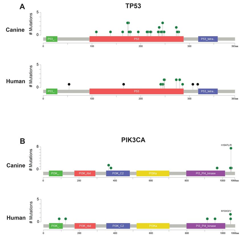
Author Manuscript Published OnlineFirst on September 30, 2019; DOI: 10.1158/1541-7786.MCR-19-0221
           Author manuscripts have been peer reviewed and accepted for publication but have not yet been edited.

Abstract
Angiosarcoma is a highly aggressive cancer of blood vessel-forming cells with few effective treatment options
and high patient mortality. It is both rare and heterogenous, making large, well powered genomic studies nearly
impossible. Dogs commonly suffer from a similar cancer, called hemangiosarcoma, with breeds like the golden
retriever carrying heritable genetic factors that put them at high risk. If the clinical similarity of canine
hemangiosarcoma and human angiosarcoma reflects shared genomic etiology, dogs could be a critically needed
model for advancing angiosarcoma research. We assessed the genomic landscape of canine hemangiosarcoma
via whole exome sequencing (47 golden retriever hemangiosarcomas) and RNA sequencing (74
hemangiosarcomas from multiple breeds). Somatic coding mutations occurred most frequently in the tumor
suppressor TP53 (59.6% of cases) as well as two genes in the PI3K pathway: the oncogene PIK3CA (29.8%)
and its regulatory subunit PIK3R1 (8.5%). The predominant mutational signature was the age-associated
deamination of cytosine to thymine. As reported in human angiosarcoma, CDKN2A/B was recurrently deleted
and VEGFA, KDR, and KIT recurrently gained. We compared the canine data to human data recently released
by The Angiosarcoma Project, and found many of the same genes and pathways significantly enriched for
somatic mutations, particularly in breast and visceral angiosarcomas. Canine hemangiosarcoma closely models
the genomic landscape of human angiosarcoma of the breast and viscera, and is a powerful tool for investigating
the pathogenesis of this devastating disease.

Implications
We characterize the genomic landscape of canine hemangiosarcoma and demonstrate its similarity to human
angiosarcoma.

                                                                                                                     2

         Downloaded from mcr.aacrjournals.org on January 6, 2021. © 2019 American Association for Cancer Research.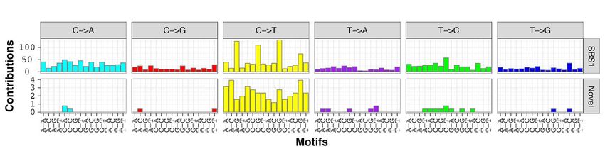
Author Manuscript Published OnlineFirst on September 30, 2019; DOI: 10.1158/1541-7786.MCR-19-0221
            Author manuscripts have been peer reviewed and accepted for publication but have not yet been edited.

Introduction
Angiosarcoma is an aggressive cancer of blood-vessel forming cells, associated with poor survival times (1–3).
There is an unmet need for new diagnostics and therapies for these patients. However, the rarity of this cancer
(approximately 0.01% of all cancers) (4,5) has limited large-scale genomic studies so far. Canine
hemangiosarcoma is a relevant clinical model for understanding the pathophysiology of human angiosarcoma.
The human and canine diseases share many clinical similarities, and hemangiosarcoma is common in dogs,
occurring in some breeds (notably the golden retriever) with a frequency up to 20% (6). This means that using
canine hemangiosarcoma as a model for human disease would yield sample cohorts of a magnitude inaccessible
using human data alone. However, for dogs to be an effective model of this disease in the era of precision
medicine, detailed genomic characterization of canine hemangiosarcoma must be undertaken, and the results
directly compared to existing and emerging genomic data from human angiosarcoma.

Angiosarcoma can form anywhere in the vasculature. In human patients, it most commonly occurs in the skin of
the head, neck, and scalp, the breast, the extremities, and less frequently in the liver, right auricle of the heart,
bone, and spleen (7). Prognosis is poor, with metastatic disease occurring in approximately 50% of cases (8),
and a median overall survival time of approximately 50 months for local disease and 10 months in metastatic
cases (9). Treatment involves surgical resection with wide margins, plus or minus radiation therapy, as well as
adjuvant chemotherapy in the metastatic disease setting (3). Many angiosarcomas are initially sensitive to
doxorubicin, paclitaxel, or targeted agents, but resistance to these therapies is virtually inevitable (3).

While most cases of angiosarcoma in humans occur without known cause, there are several known risk factors.
These tumors can arise secondary to radiation therapy for other cancers or chronic lymphedema (3). Other
known risk factors include UV irradiation, given the typical locations of cutaneous angiosarcomas on the head
and neck (10–12), as well as occupational exposure to vinyl chloride (13), arsenic exposure (14), and use of
anabolic steroids (15). Genetically, angiosarcoma is associated with familial syndromes including Li-Fraumeni
syndrome (TP53 mutations) (16) and Klippel-Trenaunay syndrome (PIK3CA mutations) (17). However, these
syndromes do not solely cause angiosarcoma (7), and angiosarcomas are not present in the majority of cases.

Canine hemangiosarcoma is the histopathological equivalent of human angiosarcoma (18), and follows a similar,
aggressive clinical course. In dogs, the most common tumor locations are the spleen, right auricle of the heart,
liver, and skin or subcutaneous tissue (19,20). The different anatomical distribution seen in the human and canine
diseases is likely due, at least in part, to the lack of secondary cases in dogs. Treatment protocols for dogs with
hemangiosarcoma similarly involve wide surgical resection, followed by adjuvant chemotherapy. Survival times
are short - a median of 4-6 months after surgical resection with adjuvant chemotherapy, with a 1 year survival
rate of approximately 10% (21–23). Biological risk factors have not yet been identified in dogs. Genetically, dog
breeds display differential predisposition to specific cancers, indicating that there are heritable risk factors that
have become common as a result of inbreeding based on selection or drift. In a previous genome-wide
association study, we identified several loci significantly associated with the risk of hemangiosarcoma in the
golden retriever (24).

Recent targeted next-generation sequencing of human angiosarcomas has begun to reveal the somatic
mutational spectrum of this disease. It has so far proven to be fairly heterogeneous - no pathognomonic
mutations or copy number aberrations occur in all cases, and tumors from different primary locations or with
different underlying etiologies have genomic differences. TP53 and genes in the MAPK pathway are frequently
mutated (25), and mutations in PLCG1 and PTPRB are common, particularly in secondary angiosarcomas
(25,26). Although the PI3K pathway is activated in some human angiosarcomas (27,28), PIK3CA mutations have
so far not been commonly reported in human angiosarcoma studies (26,29). Genes in the VEGF pathway are

                                                                                                                     3

         Downloaded from mcr.aacrjournals.org on January 6, 2021. © 2019 American Association for Cancer Research.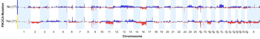
Author Manuscript Published OnlineFirst on September 30, 2019; DOI: 10.1158/1541-7786.MCR-19-0221
            Author manuscripts have been peer reviewed and accepted for publication but have not yet been edited.

frequently gained or amplified, including VEGFA, and KDR (25,28). The tumor suppressor CDKN2A is frequently
deleted, while the MYC oncogene is frequently amplified, most commonly in radiation-associated tumors (25,28).

In dogs, a recent whole-exome sequencing study of a small cohort of 20 hemangiosarcomas showed that the
top recurrently mutated genes were PIK3CA and TP53 (30). Earlier candidate gene studies of canine
hemangiosarcomas reported mutations in TP53 (31), PTEN (32), and PDGFRA and PDGFRB (33). An analysis
of somatic copy number aberrations in visceral hemangiosarcomas from five breeds found that VEGFA was
recurrently gained, while CDKN2A was frequently deleted (34). MYC copy number gain was infrequent, likely
reflective of the fact that secondary hemangiosarcomas are not seen in canine patients (34). While these earlier
studies provide clues as to the genetic features of canine hemangiosarcoma, there is very little of the genome-
wide data needed for comprehensive comparison to human angiosarcoma.

In order to assess the potential utility of hemangiosarcoma as a model for angiosarcoma at the molecular level,
we performed the largest exome sequencing study of hemangiosarcoma to date, and complemented our exome
data with oligonucleotide array comparative genomic hybridization (oaCGH) copy number data and RNA
sequencing (RNA-seq) data in partially overlapping cohorts of hemangiosarcoma cases. We then performed
comparative analyses of our results with those released by The Angiosarcoma Project direct-to-patient initiative.
In this way, we have created a detailed genomic profile of this cancer in the golden retriever breed, and begun
vetting this canine cancer as a comparative model for human angiosarcoma, and potentially other tumors.

Materials and Methods
Additional Materials and Methods can be found in the Supplementary Materials and Methods, and a workflow
in Figure S1.

Canine hemangiosarcoma sample collection for exome sequencing
Samples were obtained as part of necessary diagnostic procedures with owner consent. DNA from tumor tissue
and whole blood was collected from 47 golden retrievers with visceral hemangiosarcoma. Samples were
collected from dogs referred for treatment at the University of Minnesota (UMN) Veterinary Medical Center and
samples submitted to the Modiano laboratory for diagnostic assessment and/or use in research (n = 34), from
cases seen at the North Carolina State University (NCSU) Veterinary Hospital (n = 8), or from diagnostic samples
sent to Antech Diagnostics (n = 5). Cases where date of diagnosis was known (n = 36), were diagnosed between
2000 and 2014. Tumor samples were either frozen (n = 17), or formalin-fixed and paraffin-embedded (FFPE, n
= 30, Table S1). Procedures involving animal use were approved by the Institutional Animal Care and Use
Committees at the Broad Institute, UMN, or NCSU.

Sample preparation
DNA was extracted and sequencing libraries prepared using the Kapa Hyper Prep Kit (Table S2). For 66
samples, additional cycles of PCR were required to obtain sufficient DNA for exome capture, while libraries from
28 samples were re-constructed from source DNA using the standard protocol (See SI).

Exome capture
The Roche Nimblegen SeqCap-EZ capture canine exome (120705_CF3_Uppsala_Broad_EZ_HX1) was used
for hybrid exome capture, following the manufacturer’s protocol.

Sequencing and read alignments
The barcoded exome-captured libraries were multiplexed in pools of 8, and sequenced on the Illumina HiSeq
2500 to a target depth of 60x in the tumor and 30x in the normal, reaching a mean depth of 78x in the tumor and
63x in the normal. Reads were aligned to the CanFam3.1 reference genome using BWA. PCR duplicate reads

                                                                                                                     4

         Downloaded from mcr.aacrjournals.org on January 6, 2021. © 2019 American Association for Cancer Research.
Author Manuscript Published OnlineFirst on September 30, 2019; DOI: 10.1158/1541-7786.MCR-19-0221
Author manuscripts have been peer reviewed and accepted for publication but have not yet been edited.
were flagged using the Picard tool MarkDuplicates. Following the Genome Analysis Toolkit (GATK) Best
Practices, we then performed Base Quality Score Recalibration (BQSR) using a set of approximately 19 million
known canine germline variant locations.
Somatic variant calling
Somatic mutations were called using MuTect2, using the default settings with the addition of the --
dontUseSoftClippedBases option, which was added to avoid calling a large number of artifactual indels in our
FFPE-preserved samples (See SI). In order to further refine our set of somatic variant calls, and to avoid artifacts,
we also called variants using the GATK4 version of Mutect2, and kept only the consensus of calls which passed
in both the GATK3 and GATK4 versions. Both sets of calls were filtered using a “panel of normals” created using
all 47 normal (germline) samples, as well as germline samples from previous studies of canine lymphoma,
osteosarcoma, and melanoma. Calls were further filtered to exclude oxidation artifacts, and were removed if they
overlapped locations with known germline variants. Variants were further filtered if the position had low coverage
(defined as a read depth less than 20 in the normal, less than 40 in the tumor, or less than 4 alternate allele
reads in the tumor), or had excessive read depth (greater or equal to mean read depth + 5 x standard deviation),
to filter out potential alignment errors.
Variant annotation
Variants were annotated using the variant effect prediction program SnpEff v4.2. Where multiple effects were
predicted for a single variant, the most damaging predicted effect was selected. Coding variants were analyzed
using the smg function in Genome MuSiC 0.4 to determine which genes were significantly mutated above the
background rate. The merge-concurrent-muts option was applied to count multiple mutations in the same gene
within a sample as a single mutation, and a false discovery rate (FDR) threshold of 0.1 using the convolution
test (CT) was applied.
Canine mutational signature discovery
Mutational signatures, considering point mutations and their genomic context, were extracted using a Bayesian
non-negative matrix factorization (NMF) algorithm. The discovered signatures were compared to known
signatures in COSMIC (35), as well as those reported in the literature for human angiosarcoma (36). The overall
landscape of mutations was plotted for different groupings of samples using the SomaticSignatures package.
RNA-sequencing of canine angiosarcomas
Seventy-four snap-frozen samples were obtained from seventy-three dogs with hemangiosarcoma (41 golden
retrievers and 32 dogs from 13 other breeds or from mixed breeding)(Kim, et al., manuscript in preparation).
RNA-seq data from 51 of the hemangiosarcoma tissues had been published previously (37,38). RNA-seq
libraries were generated as described previously (37).
Somatic copy number detection in canine angiosarcomas
Analysis of somatic copy number aberrations (SCNAs) in our canine whole exome sequencing data was limited
by the inclusion of both frozen and formalin-fixed (FFPE) samples. The FFPE samples appeared to have an
increased number of amplifications and deletions when compared with the frozen samples, possibly as a result
of DNA fragmentation during the fixation process (39). While we were able to partially account for this using GC
correction (40), we were not able to completely remove the bias between the two groups (Figure S6). Hence,
we instead used oaCGH data to interrogate copy number changes in a cohort of 69 golden retrievers with
hemangiosarcoma tumors, of which 28 were also included in the exome sequencing cohort. SCNAs were called
from oligonucleotide array comparative genomic hybridization (oaCGH), as described previously (34), using a
~180,000-feature microarray (Agilent Technologies) with approximately 26 kb resolution throughout the dog
genome.
5
Downloaded from mcr.aacrjournals.org on January 6, 2021. © 2019 American Association for Cancer Research.Author Manuscript Published OnlineFirst on September 30, 2019; DOI: 10.1158/1541-7786.MCR-19-0221
Author manuscripts have been peer reviewed and accepted for publication but have not yet been edited.
Accessing human data from The Angiosarcoma Project for comparative analysis
We compared the somatic mutations and SCNAs present in our cohort of canine angiosarcomas to the results
reported by The Angiosarcoma Project, a direct-to-patient sequencing project run by the Count Me In initiative
(JoinCountMeIn.org). Somatic mutations and SCNAs derived from whole exome sequencing data from 48
samples from 36 patients were downloaded from CBioPortal, and somatic mutations classified as nonsense,
missense, or splice site were kept for analysis. For our analysis, we selected only the annotated primary tumor
site from each patient, including 30 tumors - 17 breast, five visceral (one each of right atrium, spleen, lung,
abdomen, and bladder wall), and eight head, face, neck, and scalp (HFNS) tumors.
Pathway analysis in canine and human data
Pathway analysis was performed on the canine and human mutational data using the DAVID Functional
Annotation Tool. For the overall enrichment analyses, all genes with coding nonsynonymous somatic mutations
were used. Functional annotation charts were created for KEGG pathways using the default options, using the
Benjamini-Hochberg method to control the false discovery rate. Analysis was performed independently for the
canine data and the human data, mapping genes from both species to Homo sapiens reference to avoid
confounding due to differences in gene annotation between the species. Canine Ensembl gene IDs were mapped
to their human 1:1 orthologs using Ensembl’s BioMart (41).
Results
Simple somatic mutations
TP53 and PIK3CA are commonly mutated in canine hemangiosarcomas
The seven significantly mutated genes (SMGs) in the canine hemangiosarcomas contained well-known cancer
genes, including TP53, as well as two genes in the PI3K pathway (Table 1, Figure 1). Tumor suppressor TP53
was most frequently mutated (28/47 cases, 59.5%), with all 28 cases carrying at least one mutation affecting the
DNA binding domain (Figure 2). Oncogene PIK3CA (14/47, 29.8%) and its regulatory subunit PIK3R1 (4/47,
8.5%) were both mutated. Ten of the fourteen cases with PIK3CA mutations had a mutation at amino acid
position 1047, a hotspot frequently mutated in many types of human cancers (42) (Figure 2). The remaining four
SMGs were ORC1 (4/47, 8.5%), RASA1 (4/47, 8.5%), ARPC1A (3/47, 6.4%), and ENSCAFG00000017407
(2/47, 4.3%), a “one-to-many” ortholog of human ATP5PD with 27 paralogs in the canine genome. Overall, 33
genes that were mutated at least once in the canine dataset are annotated as likely causal in the COSMIC
Cancer Gene Census (43), including TP53, PIK3CA, and PIK3R1 (Table S3).
Variant allele fraction and effect predictions
For further insight into whether the four SMGs not annotated in COSMIC are likely to be driver mutations, we
investigated the variant allele fraction (VAF) of each and whether missense mutations were predicted to be
deleterious or tolerated using the SIFT (44) score via Ensembl’s Variant Effect Predictor (45). The median VAF
for the four RASA1 mutations was 0.12 (range 0.08 - 0.23). Three mutations were nonsense mutations, with the
fourth a missense mutation predicted to to be deleterious (SIFT score 0), supporting a causal role for these
mutations. Similarly, the median VAF of the three ARPC1A mutations was 0.16 (range 0.12 - 0.17), with one
nonsense mutation, and two missense mutations at the same position, predicted to be deleterious (SIFT score
0). The median VAF for the four ORC1 mutations (all at the same position) was lower, at 0.08, and the change
was predicted to be tolerated (SIFT score 0.06). The two mutations in ENSCAFG00000017407 also had a low
median VAF (0.05), and were predicted to be tolerated (SIFT score 1).
Comparison with top mutated genes in human angiosarcoma
The most commonly mutated genes in the human data were TP53 (8/30 tumors, 29%), followed by KDR, LRP2,
RYR2, and ABCA13 with mutations in 7/30 tumors each, and PIK3CA, FLG, ASXL3, MYH14, and UNC13C,
6
Downloaded from mcr.aacrjournals.org on January 6, 2021. © 2019 American Association for Cancer Research.Author Manuscript Published OnlineFirst on September 30, 2019; DOI: 10.1158/1541-7786.MCR-19-0221
Author manuscripts have been peer reviewed and accepted for publication but have not yet been edited.
mutated in 6/30 tumors each. However, the distribution of these mutations varied by tumor location. In the human
data, breast tumors were the only location to carry PIK3CA mutations (n = 6 patients), and were the most
common location for KDR mutations. TP53 mutations occurred in all locations, but were more common in HFNS
and visceral tumors. KDR was much less frequently mutated in the canine cohort, with only one case having a
nonsynonymous mutation, predicted to be tolerated. Of the canine SMGs, two human tumors also had RASA1
mutations; one was a nonsense mutation and the other was predicted to be deleterious.
Differential patterns of mutations found in some tumor locations
The pattern of mutations in the PI3K gene family varied by tumor location in the canine data, as well. In the
canine cases, a slightly higher proportion of heart tumors (11/16, 68.8%) compared to splenic tumors (9/22,
40.9%) had PI3K alterations, although this difference was not significant (pchi-sq=0.09). Liver tumors had slightly
fewer overall alterations in the PI3K gene family (3/8, 37.5%), and were less likely to have PIK3CA mutations
(1/8, 12.5%), although this was not statistically significant.
Comparison of mutational burden by tumor location in canine and human data
We found fewer nonsynonymous mutations in the canine tumors than reported in the human tumors overall,
while the number of nonsynonymous mutations per sample varied significantly by tumor location in human cases.
In the canine cohort, there were a median of 22 nonsynonymous coding mutations per sample (range 1-149,
Table S1), which was closest to the human breast (median = 32, n = 17), and visceral (median = 40, n = 5)
tumors. In the dogs, there was a small difference in the total number of nonsynonymous mutations between
heart (median = 31, n = 15, outlier with 149 mutations removed) and splenic (n = 22, median = 17) tumors
(pTukeyHSD = 0.04). In the human data, there was a significant difference in the median number of nonsynonymous
mutations by tumor location, with head, face, neck, and scalp (HFNS, n = 8) tumors having a higher mutational
burden (median = 582) than breast or visceral tumors (pANOVA = 3.7 x 10⁻⁵)(46).
Comparative pathway analysis highlights similarity between canine and human tumors
We compared the gene functional annotations enriched in hemangiosarcoma to those enriched in breast and
visceral angiosarcoma and those enriched in HFNS angiosarcoma (Table 2). KEGG pathways enriched in
canine hemangiosarcoma overlapped with KEGG pathways enriched in both subtypes of angiosarcoma.
Pathways enriched in hemangiosarcoma included a larger fraction of those enriched in visceral and breast
angiosarcoma (71%, vs. 23% of pathways enriched in HFNS angiosarcoma), however, the total number of
pathways shared was greater between hemangiosarcoma and HFNS angiosarcoma (eight pathways). Pathways
shared between hemangiosarcoma and visceral and breast angiosarcoma included Rap1 signaling pathway
(pcanine = 6.5 x 10⁻³, pvisceral/breast = 7.9 x 10⁻³) and Central carbon metabolism in cancer (pcanine = 8.8 x 10⁻³,
pvisceral/breast = 2.7 x 10⁻²). Pathways shared between visceral hemangiosarcoma, visceral and breast
angiosarcoma, and HFNS angiosarcoma included Focal adhesion (pcanine = 3.7 x 10⁻³, pvisceral/breast = 2.3 x 10⁻²,
pHFNS = 7.9 x 10⁻⁴), Long-term depression (pcanine = 4.0 x 10⁻², pvisceral/breast = 6.5 x 10⁻³, pHFNS = 7.4 x 10⁻³), and
Calcium signaling pathway (pcanine = 3.3 x 10⁻², pvisceral/breast = 4.7 x 10⁻³, pHFNS = 1.7 x 10⁻²).
In addition, functional annotation enrichment analysis of the genes mutated in hemangiosarcoma, visceral and
breast angiosarcoma, and HFNS angiosarcoma found that they were enriched in many of the same protein
domains. Shared domains include Fibronectin Type III (pcanine = 7.1 x 10⁻⁹, pvisceral/breast = 5.2 x 10⁻⁵, pHFNS = 5.1 x
10-14), Epidermal growth factor-like domain (pcanine = 1.3 x 10⁻³, pvisceral/breast = 4.5 x 10⁻⁵, pHFNS = 6.1 x 10⁻¹⁶), and
Tyrosine protein kinase active site (pcanine = 6.0 x 10⁻⁴, pvisceral/breast = 3.4 x 10⁻³, pHFNS = 1.1 x 10⁻⁴). Both visceral
and breast and HFNS angiosarcomas were enriched in cadherins (pvisceral/breast = 5.0 x 10⁻⁴, pHFNS = 2.5 x 10⁻¹⁶),
HFNS highly so, while hemangiosarcoma was not (pcanine = 0.15). In addition, HFNS angiosarcoma was enriched
in ABC-transporters (p = 2.3 x 10⁻⁷), while visceral and breast angiosarcoma and canine hemangiosarcoma were
not (Table S7).
7
Downloaded from mcr.aacrjournals.org on January 6, 2021. © 2019 American Association for Cancer Research.Author Manuscript Published OnlineFirst on September 30, 2019; DOI: 10.1158/1541-7786.MCR-19-0221
Author manuscripts have been peer reviewed and accepted for publication but have not yet been edited.
Pathways and gene families previously reported in the angiosarcoma literature
Several pathways and gene families have been previously reported to be affected in the human angiosarcoma
literature. Mutations in both PLCG1 and PTPRB have been reported to be recurrent (26), particularly in
secondary angiosarcoma. In the human data, 12 patients had mutations in eight phospholipase C genes,
including PLCG1 (n = 5, Table 3). Eight canine cases (17%) had mutations in PLC genes, including one in
PLCG1. Nineteen protein tyrosine phosphatases were mutated in twelve samples in human, including two
patients with PTPRB mutations. While none of the canine tumors had somatic mutations in PTPRB, eight cases
(17%) harbored a mutation in one of nine other protein tyrosine phosphatase genes (Table 3).
The MAPK pathway has been reported to be frequently affected by mutations in angiosarcoma (25), and we
found the visceral and breast angiosarcomas to be enriched in mutations in MAPK pathway genes (p = 0.03,
Table 2). We noted mutations in 26 genes in the MAPK pathway in the canine cohort, including the SMGs TP53
and RASA1. In the human data, 65 genes in the MAPK pathway were mutated. Thirteen of these genes were
mutated in both the canine and human data (Table 3). Although we found mutations in genes involved in histone
methyltransferase/demethylase activity in both species, consistent with earlier human studies (47), we saw no
significant enrichment on a pathway level in either the canine or human cohorts (Table 3). Tyrosine protein
kinases were enriched in both hemangiosarcoma and angiosarcoma. In the human data, 52 genes in the GO
Protein Tyrosine Kinase Activity pathway were mutated, while 22 genes in this pathway were mutated in the
canine data, with 10 genes mutated in both human and canine samples (Table 3). Low-density lipoprotein
receptors were enriched for mutations in the canine data (p = 0.03), and LRP2 was one of the most frequently
mutated genes in the human dataset (mutated in 7/30 tumors). While mutations in the LDL receptor-related
protein family have not been widely reported in human angiosarcoma, they have recently been shown to play a
role in a variety of cancers (48). We found that four LDL receptor-related protein genes were mutated in both
hemangiosarcoma and angiosarcoma, with an additional two mutated in angiosarcoma only (Table 3).
Mutational signature of aging is present across all canine tumors
Analysis of mutational signatures, which capture the mutational landscape of tumors and are shaped by both
genetic factors and environmental exposure, revealed similarities between canine hemangiosarcoma and human
angiosarcoma, as reported by Thibodeau, et al. (36). We analyzed the signatures of trinucleotide mutational
frequencies present in golden retriever hemangiosarcomas, and found a strong signature of mutations arising
through spontaneous deamination of methylated cytosines in CpG islands, corresponding to COSMIC signature
SBS1 and generally associated with aging (35) (Figure 3, Figures S3-S5). In addition, we see a faint signature
not described in COSMIC (Figure 3). The overall mutational landscape was consistent across all canine
hemangiosarcoma tumors, regardless of tumor location or tissue preservation method (Figures S2 - S4).
Canine RNA-seq analysis validates exome mutations and extends findings to additional breeds
We validated our exome sequencing somatic mutation calls using RNA-seq data from a partially overlapping set
of dogs (n = 74 tumor samples from 73 dogs from 14 breeds, as well as mixed-breed dogs, Table S4). Thirteen
golden retrievers from the exome cohort with mutations in SMGs had the same tumor location included in the
RNA-seq data, with a total of 25 SMG mutations among them. We excluded 2 frameshift deletion variants from
the validation, as they showed evidence of deletion in the pileup data, but also unexpected alternate alleles. Of
the remaining 23 variants, we validated 15 (65%) mutations. Of the eight sites which were not validated, one
was a nonsense mutation, which may have been subject to nonsense-mediated decay, five had a variant allele
fraction (VAF) less than 0.1, and three variants came from samples with tumor purity estimate (≤ 35%) on
histological examination, suggesting that tumor heterogeneity and stromal contamination may be a contributing
factor to variants not being replicated (Table S5). In the RNA-seq data, four tumor samples from three dogs with
8
Downloaded from mcr.aacrjournals.org on January 6, 2021. © 2019 American Association for Cancer Research.Author Manuscript Published OnlineFirst on September 30, 2019; DOI: 10.1158/1541-7786.MCR-19-0221
Author manuscripts have been peer reviewed and accepted for publication but have not yet been edited.
variants in the SMGs were from a different tumor site than was included in the exome discovery cohort. In this
subset, we were able to detect the mutation discovered in the same individual, but a different tumor location in
4/8 (50%) cases. Overall, the group of variants which we were unable to validate came from tumor samples with
a significantly lower estimated tumor purity on histology (pt-test = 0.04), but this difference was not significant
based on tumor purity estimates from the program ESTIMATE (pt-test = 0.4, Table S5).
The RNA-seq cohort also confirmed that the TP53 and PI3K mutations were not breed-specific, but were present
across different breeds. Excluding the 19 golden retrievers who were also included in the exome cohort, we
looked to see whether somatic mutations corresponding to those observed in the exome cohort could be found
in other cases, which included golden retrievers (n = 22), Portuguese water dogs (PWD, n = 6), German
Shepherd Dogs (GSD, n = 6), mixes (n = 6), boxers (n = 2), Labrador retrievers (Labs, n = 2), Keeshonds (n =
2), and other breeds (n = 8, one each of American Staffordshire Terrier, Bernese Mountain Dog, Bichon Frise,
Briard, Bullmastiff, Gordon setter, Parson’s Russell Terrier, and Saluki). We found mutations at the same sites
as discovered in the exome cohort in TP53 (n = 14, 26%, 5 breeds), PIK3CA (n = 11, 20%, 6 breeds), and
PIK3R1 (n = 4, 7%, 4 breeds, Table S6).
Somatic copy number aberrations
SCNAs in canine hemangiosarcoma recurrently affect known cancer genes
We surveyed SCNAs in genes known to be involved in hemangiosarcoma and angiosarcoma. The genes most
recurrently affected by DNA copy number aberrations in the oaCGH data were VEGFA, with copy number gain
in 19% of cases, KDR, gained in 22%, KIT, gained in 17%, and the tumor suppressor CDKN2A/B, deleted in
22% (Table S8). The MYC oncogene had copy number gain in 9% of cases. Copy number aberrations in the
top significantly mutated genes were relatively rare (Figure 1A, Table S8).
Copy number gains in KDR and losses in AXIN1 are common in both dogs and humans
Comparison of SCNAs in the human data and canine oaCGH data revealed recurrent copy number aberrations
in known cancer genes KDR and AXIN1 in both species. Copy number gains in KDR occurred in approximately
27% of human samples, and 22% of canine samples. AXIN1 was lost in 20% of human and 22% of canine
samples. In addition, the genes PTK6, ARFRP1, and RTEL1 showed copy number loss in 17% of human and
canine samples, however, these genes are close together, and also showed copy number gains in a number of
canine samples (Table S9).
SCNA profiles differ among cases with and without PIK3CA mutations
We examined the SCNA profiles of hemangiosarcoma cases with and without TP53 and PIK3CA mutations in
the 28 cases with both exome sequencing and oaCGH data. There were no significant differences in the relative
frequency of any given CNA between the CNA profiles of cases with and without TP53 mutations. However,
significant differences were detected between cases with PIK3CA mutations and those without (Figure 4), using
a two-tailed Fisher’s Exact test and a minimum differential threshold of 25% between the two groups. A region
on chromosome 11 at approximately 22.6 Mb, near the UBE2B (ubiquitin-conjugating enzyme E2B) gene, was
deleted in 4/11 cases with PIK3CA mutations, and 0/17 without (p < 0.016). Similarly, the CDKN2B gene, located
distally on chromosome 11 at 41.2Mb, was deleted in 4/11 cases with PIK3CA mutations, and 0/17 cases without
(p < 0.016). A region on chromosome 24 at 21.2 Mb was gained in 7/17 cases without PIK3CA mutation and
0/11 with PIK3CA mutation (p < 0.023). This region overlaps the anti-apoptotic BCL2L1 gene. Broad copy
number gains along the length of chromosome 31 were more frequent in cases without PIK3CA mutations
compared to those with PIK3CA mutations.
Discussion
9
Downloaded from mcr.aacrjournals.org on January 6, 2021. © 2019 American Association for Cancer Research.Author Manuscript Published OnlineFirst on September 30, 2019; DOI: 10.1158/1541-7786.MCR-19-0221
Author manuscripts have been peer reviewed and accepted for publication but have not yet been edited.
Detailed molecular profiling of canine hemangiosarcoma has revealed both similarities and differences in the
genetic landscape between dogs and humans. In particular, visceral canine hemangiosarcoma showed strong
similarities to human angiosarcoma of the viscera and breast. Our findings have important implications for
comparative oncology, as the study of canine hemangiosarcoma has the potential to improve our understanding
of the pathophysiology of both canine hemangiosarcoma and human angiosarcoma, and to improve treatment
and outcomes in both species.
Tumor suppressor TP53 was the top significantly mutated gene in the canine data. The majority of mutations
occurred in the DNA binding domain, likely causing loss of function (Figure 2). TP53 was also the only
significantly mutated gene in the human Angiosarcoma Project data, and it has been frequently reported as
mutated in targeted sequencing studies of angiosarcoma (25).
We found that the PI3K pathway was commonly mutated in canine hemangiosarcoma. A total of 23 canine
tumors (48.9%) had at least one somatic mutation affecting this gene family (Figure 1A). Tumors with a mutation
in the PI3K family tended to have only one mutation in this family. The PI3K pathway is one of the most commonly
altered pathways in cancer, playing an important role in signal transduction leading to cell proliferation, survival,
differentiation, and regulation of metabolism and immunity (49,50). PIK3CA is an oncogene (51) that has been
shown to be mutated in human glioblastoma, breast, gastric, colorectal, lung, and endometrial cancers (52). Ten
of the 14 PIK3CA mutations in our canine cohort occurred at amino acid position 1047, a mutational hotspot in
many human cancers (42). Mutations within this domain have been shown to increase catalytic activity (53).
Of the less frequently mutated SMGs, PIK3R1 is annotated as a likely driver mutation by COSMIC. In addition,
variant allele fraction and SIFT scores support a potential role for RASA1 and ARPC1A as driver mutations.
RASA1 is a negative regulator of the RAS and MAPK pathways, and plays an important role in vascular formation
(54,55). Germline RASA1 mutations can cause capillary malformation - arteriovenous malformation syndrome
(56). Somatic mutations in this gene have been found in a subset of human basal cell carcinomas, and
expression has been correlated with survival in invasive ductal breast carcinomas and hepatocellular carcinomas
(57,58). ARPC1A plays an important role in regulating the actin cytoskeleton, which functions in the migration
and invasiveness of pancreatic carcinoma cells (59). There is less support for a causal role for ORC1 and
ENSCAFG00000017407, however, it is important to note that the SIFT score does not annotate activating
mutations - for example, the hotspot mutations in PIK3CA (known to be drivers in many human cancers) are also
predicted to be tolerated.
There were also potentially important differences in somatic mutations between the two species. In the human
data, mutations in TP53 and PIK3CA tended to be mutually exclusive, while we did not see this pattern in the
canine tumors. In addition, PIK3CA mutations were exclusively found in breast tumors in the small human data
set, while we found them to be common in cardiac and splenic tumors in dogs. Within the canine visceral tumors,
we found fewer mutations in PIK3CA in liver tumors. These differences in distribution of PIK3CA mutations by
tumor location may be due to genetic heterogeneity of the cancer, with tumors in different locations activating
the PI3K pathway at different points, or relying on alterations in different pathways to affect an essential protein
downstream. It is also possible that this difference is due to the small number of human visceral angiosarcomas
currently sequenced, and the small number of liver tumors in the canine cohort.
Another potentially important difference between the two species is that, while copy number gains in KDR are
common in both species, somatic mutations in this gene were seen in over 20% of human tumors, but in only
one canine tumor. As the KDR receptor is upstream of the PI3K pathway, it is possible that mutations in either
may lead to a similar phenotype. We also saw more copy number gains of VEGFA in our canine cohort (19%
vs. 0 in the human data), and as VEGFA is upstream of KDR, this copy number gain may serve a similar role to
10
Downloaded from mcr.aacrjournals.org on January 6, 2021. © 2019 American Association for Cancer Research.Author Manuscript Published OnlineFirst on September 30, 2019; DOI: 10.1158/1541-7786.MCR-19-0221
Author manuscripts have been peer reviewed and accepted for publication but have not yet been edited.
KDR mutations in the canine tumors. In addition, whole genome sequencing will be necessary to determine
whether there are common regulatory mutations affecting this gene in canine hemangiosarcoma.
In the human data, mutation rates were significantly different between different tumor locations, with head and
neck angiosarcomas having a much higher mutational burden, as reported by Painter, et al. (46). It is possible
that the higher mutational burden in these tumors is due to UV exposure. It would be interesting to compare
these findings to canine cutaneous and subcutaneous hemangiosarcomas, which were not included in the
current study. In the canine visceral hemangiosarcoma, a slight difference was found between mutational burden
in heart and splenic tumors. It is possible that the golden retrievers have a lower overall somatic mutation burden
than humans, as they likely have a higher germline risk burden, given the high incidence of hemangiosarcoma
in the breed. Further studies in dogs will be necessary to determine whether the lower mutation rate is correlated
with breed risk, or if in this case it is an artifact of how mutations were called.
Tumors in both dogs and humans were enriched for mutations in protein tyrosine kinases, which are important
regulators of cellular growth and division signals and are commonly mutated in cancers. There were also
recurrent mutations in the protein tyrosine phosphatase gene family in both species. This may suggest an
alternate mechanism of tyrosine kinase overactivation, as protein tyrosine phosphatases deactivate tyrosine
kinase signaling by dephosphorylating proteins in opposition to kinase phosphorylation (60). In addition,
Phospholipase C proteins play a crucial role in cellular signalling pathways by hydrolyzing phosphatidylinositol
4,5-bisphosphate (PIP2) into the second messengers DAG and IP3, passing on signals from receptor tyrosine
kinases (61). The canine hemangiosarcomas were enriched for phospholipase C genes, including PLCG1, and
PLCG1 mutations were recurrent in the human tumors.
The shared enriched pathways between canine and human angiosarcomas provide insight into disease
pathogenesis for both species. Tyrosine kinase inhibitors have been effective against angiosarcoma in the clinic,
but tumor heterogeneity and the development of resistance have limited their long-term utility. They have also
shown promise against hemangiosarcoma in vitro (62), but so far have been less promising in the veterinary
clinic (63). Future investigation of the interaction between the many affected pathways will help to determine the
potential for combination therapy targeting multiple of these pathways or a common downstream effector to
combat resistance.
A recent study in 13 radiation-induced and 3 spontaneous breast angiosarcomas detected the irradiation
signature, as well as the aging signature, and a unique C>T signature (36). The median age of patients in this
human cohort was 74.5 years (36). Canine hemangiosarcomas looked very similar, in that we primarily saw the
aging signature and low levels of a novel C>T signature. This novel signature bore some resemblance to the
signature reported in the human angiosarcomas, including higher levels of C>T mutations at C nucleotides
flanked by A-A or A-T, however, there were also differences, such as a high number of mutations flanked by T-
G in the dogs, and a low number of these in the human cohort. A larger study will be needed to decipher whether
this is a novel angiosarcoma-related signature or whether it represents noise. The lack of the irradiation signature
was anticipated as our canine data did not include any tumors secondary to radiation therapy.
We examined copy number changes in canine hemangiosarcoma in genes previously reported to be affected in
hemangiosarcoma and angiosarcoma. Most common were copy number gain of VEGFA and KDR, and loss of
CDKN2A/B. The MYC oncogene, which has been reported amplified in human angiosarcoma and canine
hemangiosarcoma, was only rarely gained in our dataset and showed no evidence of high-level amplification.
This makes sense, given that it is more common in radiation-induced tumors, which were not present in the
canine cohort.
11
Downloaded from mcr.aacrjournals.org on January 6, 2021. © 2019 American Association for Cancer Research.Author Manuscript Published OnlineFirst on September 30, 2019; DOI: 10.1158/1541-7786.MCR-19-0221
Author manuscripts have been peer reviewed and accepted for publication but have not yet been edited.
Our data suggest that visceral canine hemangiosarcoma could be developed as a model for primary human
angiosarcoma. Detailed molecular characterization of canine hemangiosarcoma revealed many similarities, but
also some important differences, between canine hemangiosarcoma and human angiosarcoma. Future work
should include analysis of larger sample sizes, including recalling all currently available human angiosarcoma
data using the same methods, in order to decipher potential molecular subtypes and to facilitate a more complete
comparison between tumors in different locations. An integrated understanding of the interaction between
mutations in the many enriched signaling pathways may be useful for determining treatment strategy, for
example, the feasibility of combinations of targeted inhibitors or the prevention of convergent resistance. Our
data suggest that clinical trials evaluating therapeutic approaches in dogs with this disease might also inform
human medicine.
Acknowledgments
The authors would like to thank all of the dogs and owners who participated in our research, as well as the
veterinarians who collected samples. The results included here include the use of data from The Angiosarcoma
Project (https://ascproject.org/), a project of Count Me In (https://joincountmein.org/), downloaded March 2019.
We would also like to thank Dr. Scott Moroff, Vice-President and Chief Scientific Officer of Antech Diagnostics,
for contributing tumor and paired blood samples to our research. This work was funded by American Kennel
Club (AKC) Canine Health Foundation (CHF) grants #422 (JFM), 1131 (JFM), and 1889-G (JFM, MB, KLT, EKK),
NIH grants R03CA191713 (JFM), P30CA077598 (NIH Comprehensive Cancer Center Support Grant to the
Masonic Cancer Center, University of Minnesota), R37CA218570 (KM, EKK), and R24OD018250 (KM, EKK),
National Canine Cancer Foundation (NCCF) grants DM06CO-003 (JFM) and JHK15MN-004 (JHK), and Morris
Animal Foundation grant D10CA-501 (JFM, MB, KLT). KLT is the recipient of a Distinguished Professor award
from the Swedish Research Council. IE is supported by a postdoctoral fellowship from the Swedish Medical
Research Council, SSMF. JFM is supported by the Alvin and June Perlman Chair in Animal Oncology at the
University of Minnesota. MB is supported in part by the Oscar J. Fletcher Distinguished Professorship in
Comparative Oncology Genetics at NC State University. ALS is supported by NCI R50 CA211249.
Data availability
The whole-exome sequencing data, as well as the RNA-seq data that has not been previously published, has
been submitted to the NCBI Sequence Read Archive (SRA). WES data: BioProject PRJNA552034, BioSamples
SAMN12173468 - SAMN12173561. RNA-seq data: BioProject PRJNA562916, BioSamples SAMN12659339 -
SAMN12659361.
12
Downloaded from mcr.aacrjournals.org on January 6, 2021. © 2019 American Association for Cancer Research.Author Manuscript Published OnlineFirst on September 30, 2019; DOI: 10.1158/1541-7786.MCR-19-0221
Author manuscripts have been peer reviewed and accepted for publication but have not yet been edited.
References
1. Penel N, Marréaud S, Robin Y-M, Hohenberger P. Angiosarcoma: state of the art and perspectives. Crit
Rev Oncol Hematol. 2011;80:257–63.
2. Abraham JA, Hornicek FJ, Kaufman AM, Harmon DC, Springfield DS, Raskin KA, et al. Treatment and
outcome of 82 patients with angiosarcoma. Ann Surg Oncol. 2007;14:1953–67.
3. Florou V, Wilky BA. Current and Future Directions for Angiosarcoma Therapy. Curr Treat Options Oncol.
2018;19:14.
4. Antonescu C. Malignant vascular tumors—an update. Mod Pathol. United States & Canadian Academy of
Pathology; 2014;27:S30.
5. Siegel R, Naishadham D, Jemal A. Cancer statistics, 2013. CA Cancer J Clin. 2013;63:11–30.
6. Glickman L, Glickman N, Thorpe - Retriever Club of America National … R, 1999. The Golden Retriever
Club of America National Health Survey 1998-1999. grca.org [Internet]. 1999; Available from:
https://www.grca.org/wp-content/uploads/2015/08/healthsurvey1-1.pdf
7. Young RJ, Brown NJ, Reed MW, Hughes D, Woll PJ. Angiosarcoma. Lancet Oncol. 2010;11:983–91.
8. Mark RJ, Poen JC, Tran LM, Fu YS, Juillard GF. Angiosarcoma: a report of 67 patients and a review of the
literature. Cancer: Interdisciplinary International Journal of the American Cancer Society. Wiley Online
Library; 1996;77:2400–6.
9. Lahat G, Dhuka AR, Hallevi H, Xiao L, Zou C, Smith KD, et al. Angiosarcoma: clinical and molecular
insights. Ann Surg. 2010;251:1098–106.
10. Stenbäck F. Cellular injury and cell proliferation in skin carcinogenesis by UV light. Oncology. 1975;31:61–
75.
11. Cioffi A, Reichert S, Antonescu CR, Maki RG. Angiosarcomas and other sarcomas of endothelial origin.
Hematol Oncol Clin North Am. 2013;27:975–88.
12. Maddox JC, Evans HL. Angiosarcoma of skin and soft tissue: a study of forty-four cases. Cancer.
1981;48:1907–21.
13. Makk L, Creech JL, Whelan JG Jr, Johnson MN. Liver damage and angiosarcoma in vinyl chloride
workers. A systematic detection program. JAMA. 1974;230:64–8.
14. Centeno JA, Mullick FG, Martinez L, Page NP, Gibb H, Longfellow D, et al. Pathology related to chronic
arsenic exposure. Environ Health Perspect. 2002;110 Suppl 5:883–6.
15. Falk H, Thomas LB, Popper H, Ishak KG. Hepatic angiosarcoma associated with androgenic-anabolic
steroids. Lancet. 1979;2:1120–3.
16. Calvete O, Martinez P, Garcia-Pavia P, Benitez-Buelga C, Paumard-Hernández B, Fernandez V, et al. A
mutation in the POT1 gene is responsible for cardiac angiosarcoma in TP53-negative Li-Fraumeni-like
families. Nat Commun. 2015;6:8383.
17. Ploegmakers MJM, Pruszczynski M, De Rooy J, Kusters B, Veth RPH. Angiosarcoma with malignant
peripheral nerve sheath tumour developing in a patient with Klippel--Trénaunay--Weber syndrome.
Sarcoma. Hindawi Publishing Corporation; 2005;9:137–40.
18. Kim J-H, Graef AJ, Dickerson EB, Modiano JF. Pathobiology of Hemangiosarcoma in Dogs: Research
Advances and Future Perspectives. Vet Sci China. 2015;2:388–405.
13
Downloaded from mcr.aacrjournals.org on January 6, 2021. © 2019 American Association for Cancer Research.Author Manuscript Published OnlineFirst on September 30, 2019; DOI: 10.1158/1541-7786.MCR-19-0221
Author manuscripts have been peer reviewed and accepted for publication but have not yet been edited.
19. Oksanen A. Haemangiosarcoma in dogs. J Comp Pathol. 1978;88:585–95.
20. Brown NO, Patnaik AK, MacEwen EG. Canine hemangiosarcoma: retrospective analysis of 104 cases. J
Am Vet Med Assoc. 1985;186:56–8.
21. Withrow SJ, Page RL. Withrow and MacEwen’s Small Animal Clinical Oncology. Elsevier Health Sciences;
2013.
22. Wendelburg KM, Price LL, Burgess KE, Lyons JA, Lew FH, Berg J. Survival time of dogs with splenic
hemangiosarcoma treated by splenectomy with or without adjuvant chemotherapy: 208 cases (2001-
2012). J Am Vet Med Assoc. 2015;247:393–403.
23. Sorenmo KU, Jeglum KA, Helfand SC. Chemotherapy of Canine Hemangiosarcoma With Doxorubicin and
Cyclophosphamide. J Vet Intern Med. 1993;7:370–6.
24. Tonomura N, Elvers I, Thomas R, Megquier K, Turner-Maier J, Howald C, et al. Genome-wide association
study identifies shared risk loci common to two malignancies in golden retrievers. PLoS Genet.
2015;11:e1004922.
25. Murali R, Chandramohan R, Möller I, Scholz SL, Berger M, Huberman K, et al. Targeted massively parallel
sequencing of angiosarcomas reveals frequent activation of the mitogen activated protein kinase pathway.
Oncotarget. 2015;6:36041–52.
26. Behjati S, Tarpey PS, Sheldon H, Martincorena I, Van Loo P, Gundem G, et al. Recurrent PTPRB and
PLCG1 mutations in angiosarcoma. Nat Genet. 2014;46:376–9.
27. Italiano A, Chen C-L, Thomas R, Breen M, Bonnet F, Sevenet N, et al. Alterations of the p53 and
PIK3CA/AKT/mTOR pathways in angiosarcomas: a pattern distinct from other sarcomas with complex
genomics. Cancer. 2012;118:5878–87.
28. Wagner MJ, Ravi V, Menter DG, Sood AK. Endothelial cell malignancies: new insights from the laboratory
and clinic. npj Precision Oncology. Nature Publishing Group; 2017;1:11.
29. Je EM, An CH, Yoo NJ, Lee SH. Mutational analysis of PIK3CA, JAK2, BRAF, FOXL2, IDH1, AKT1 and
EZH2 oncogenes in sarcomas. APMIS. 2012;120:635–9.
30. Wang G, Wu M, Maloneyhuss MA, Wojcik J, Durham AC, Mason NJ, et al. Actionable mutations in canine
hemangiosarcoma. PLoS One. 2017;12:e0188667.
31. Mayr B, Zwetkoff S, Schaffner G, Reifinger M. Tumour suppressor gene p53 mutation in a case of
haemangiosarcoma of a dog. Acta Vet Hung. 2002;50:157–60.
32. Dickerson EB, Thomas R, Fosmire SP, Lamerato-Kozicki AR, Bianco SR, Wojcieszyn JW, et al. Mutations
of phosphatase and tensin homolog deleted from chromosome 10 in canine hemangiosarcoma. Vet
Pathol. 2005;42:618–32.
33. Abou Asa S, Mori T, Maruo K, Khater A, El-Sawak A, Abd el-Aziz E, et al. Analysis of genomic mutation
and immunohistochemistry of platelet-derived growth factor receptors in canine vascular tumours. Vet
Comp Oncol. 2015;13:237–45.
34. Thomas R, Borst L, Rotroff D, Motsinger-Reif A, Lindblad-Toh K, Modiano JF, et al. Genomic profiling
reveals extensive heterogeneity in somatic DNA copy number aberrations of canine hemangiosarcoma.
Chromosome Res. 2014;22:305–19.
35. Forbes SA, Beare D, Gunasekaran P, Leung K, Bindal N, Boutselakis H, et al. COSMIC: exploring the
world’s knowledge of somatic mutations in human cancer. Nucleic Acids Res. 2015;43:D805–11.
14
Downloaded from mcr.aacrjournals.org on January 6, 2021. © 2019 American Association for Cancer Research.Author Manuscript Published OnlineFirst on September 30, 2019; DOI: 10.1158/1541-7786.MCR-19-0221
Author manuscripts have been peer reviewed and accepted for publication but have not yet been edited.
36. Thibodeau BJ, Lavergne V, Dekhne N, Benitez P, Amin M, Ahmed S, et al. Mutational landscape of
radiation-associated angiosarcoma of the breast. Oncotarget. 2018;9:10042–53.
37. Gorden BH, Kim J-H, Sarver AL, Frantz AM, Breen M, Lindblad-Toh K, et al. Identification of three
molecular and functional subtypes in canine hemangiosarcoma through gene expression profiling and
progenitor cell characterization. Am J Pathol. 2014;184:985–95.
38. Borgatti A, Koopmeiners JS, Sarver AL, Winter AL, Stuebner K, Todhunter D, et al. Safe and Effective
Sarcoma Therapy through Bispecific Targeting of EGFR and uPAR. Mol Cancer Ther. 2017;16:956–65.
39. Do H, Dobrovic A. Sequence artifacts in DNA from formalin-fixed tissues: causes and strategies for
minimization. Clin Chem. 2015;61:64–71.
40. Benjamini Y, Speed TP. Summarizing and correcting the GC content bias in high-throughput sequencing.
Nucleic Acids Res. 2012;40:e72.
41. Kinsella RJ, Kähäri A, Haider S, Zamora J, Proctor G, Spudich G, et al. Ensembl BioMarts: a hub for data
retrieval across taxonomic space. Database . 2011;2011:bar030.
42. Lawrence MS, Stojanov P, Mermel CH, Robinson JT, Garraway LA, Golub TR, et al. Discovery and
saturation analysis of cancer genes across 21 tumour types. Nature. 2014;505:495–501.
43. Sondka Z, Bamford S, Cole CG, Ward SA, Dunham I, Forbes SA. The COSMIC Cancer Gene Census:
describing genetic dysfunction across all human cancers. Nat Rev Cancer. 2018;18:696–705.
44. Ng PC, Henikoff S. SIFT: Predicting amino acid changes that affect protein function. Nucleic Acids Res.
2003;31:3812–4.
45. McLaren W, Gil L, Hunt SE, Riat HS, Ritchie GRS, Thormann A, et al. The Ensembl Variant Effect
Predictor. Genome Biol. 2016;17:122.
46. Painter C, Jain E, Dunphy M, Anastasio E, McGillicuddy M, Stoddard R, et al. Abstract B085: High
mutation burden and response to immune checkpoint inhibitors in angiosarcomas of the scalp and face.
Proceedings of the Fourth CRI-CIMT-EATI-AACR International Cancer Immunotherapy Conference:
Translating Science into Survival. American Association for Cancer Research; 2019. page B085–B085.
47. Kunze K, Spieker T, Gamerdinger U, Nau K, Berger J, Dreyer T, et al. A recurrent activating PLCG1
mutation in cardiac angiosarcomas increases apoptosis resistance and invasiveness of endothelial cells.
Cancer Res. 2014;74:6173–83.
48. Gonias SL, Karimi-Mostowfi N, Murray SS, Mantuano E, Gilder AS. Expression of LDL receptor-related
proteins (LRPs) in common solid malignancies correlates with patient survival. PLoS One.
2017;12:e0186649.
49. Fruman DA, Chiu H, Hopkins BD, Bagrodia S, Cantley LC, Abraham RT. The PI3K Pathway in Human
Disease. Cell. 2017;170:605–35.
50. Cully M, You H, Levine AJ, Mak TW. Beyond PTEN mutations: the PI3K pathway as an integrator of
multiple inputs during tumorigenesis. Nat Rev Cancer. 2006;6:184–92.
51. Chang HW, Aoki M, Fruman D, Auger KR, Bellacosa A, Tsichlis PN, et al. Transformation of Chicken Cells
by the Gene Encoding the Catalytic Subunit of PI 3-Kinase. Science. American Association for the
Advancement of Science; 1997;276:1848–50.
52. Samuels Y, Wang Z, Bardelli A, Silliman N, Ptak J, Szabo S, et al. High frequency of mutations of the
PIK3CA gene in human cancers. Science. 2004;304:554.
15
Downloaded from mcr.aacrjournals.org on January 6, 2021. © 2019 American Association for Cancer Research.You can also read