Potent ex vivo armed T cells using recombinant bispecific antibodies for adoptive immunotherapy with reduced cytokine release
←
→
Page content transcription
If your browser does not render page correctly, please read the page content below
Open access Original research
J Immunother Cancer: first published as 10.1136/jitc-2020-002222 on 13 May 2021. Downloaded from http://jitc.bmj.com/ on September 24, 2021 by guest. Protected by copyright.
Potent ex vivo armed T cells using
recombinant bispecific antibodies for
adoptive immunotherapy with reduced
cytokine release
Jeong A Park ,1 Brian H Santich,1 Hong Xu,1 Lawrence G Lum,2
Nai-Kong V Cheung 1
To cite: Park JA, Santich BH, ABSTRACT has been limited to hematological malignan-
Xu H, et al. Potent ex vivo Background T cell-based immunotherapies using cies and a few solid cancers with high tumor
armed T cells using recombinant chimeric antigen receptors (CAR) or bispecific antibodies
bispecific antibodies for
mutational burden. The immune suppressive
(BsAb) have produced impressive responses in tumor microenvironment (TME), cytokine
adoptive immunotherapy with
hematological malignancies. However, major hurdles release syndrome (CRS), life- threatening
reduced cytokine release.
Journal for ImmunoTherapy remained, including cytokine release syndrome,
neurotoxicity, and permanent on-target off-
of Cancer 2021;9:e002222. neurotoxicity, on-target off-tumor effects, reliance on
autologous T cells, and failure in most solid tumors. BsAb tumor side effects are persisting immunolog-
doi:10.1136/jitc-2020-002222
armed T cells offer a safe alternative. ical hurdles.5–8
►► Additional material is Methods We generated ex vivo armed T cells (EATs) Beyond CAR T cells, BsAb armed T cells
published online only. To view, using IgG-[L]-scFv-platformed BsAb, where the anti-CD3 have been investigated clinically.9–11 Using
please visit the journal online (huOKT3) scFv was attached to the light chain of a tumor- chemically conjugated anti-GD2 × anti-CD3
(http://dx.doi.org/10.1136/jitc- binding IgG. BsAb density on EAT, in vitro cytotoxicity, (hu3F8 × mouse OKT3; NCT02173093),
2020-002222). cytokine release, in vivo trafficking into tumors, and their anti-HER2 × anti-CD3 (trastuzumab × mouse
antitumor activities were evaluated in multiple cancer cell OKT3; NCT00027807), and anti- EGFR
Accepted 17 March 2021 lines and patient-derived xenograft mouse models. The
× anti-CD3 (cetuximab × mouse OKT3;
efficacy of EATs after cryopreservation was studied, and
gamma delta (γδ) T cells were investigated as unrelated
NCT04137536), BsAb armed T cells have
alternative effector T cells. been administered safely with minimal
Results The antitumor potency of BsAb armed T cells neurotoxicities or cytokine storm. They were
was substantially improved using the IgG-[L]-scFv BsAb safe at cell doses as high as 4×10¹⁰/dose in
platform. When compared with separate BsAb and T cell more than 170 patients in breast, prostate,
injection, EATs released less TNF-α, and infiltrated tumors and pancreatic cancers, as well as in hemato-
faster, while achieving robust antitumor responses. The in logical malignancies.9 11–13 However, robust
vivo potency of EAT therapy depended on BsAb dose for responses remained elusive due to insuffi-
arming, EAT cell number per injection, total number of EAT
cient T cell infiltration and persistence, and/
doses, and treatment schedule intensity. The antitumor
or impaired T cell function in the TME.
efficacy of EATs was preserved following cryopreservation,
and EATs using γδ T cells were safe and as effective as αβ We previously demonstrated that the
T cell-EATs. interdomain distance and cis- configuration
Conclusions EATs exerted potent antitumor activities of BsAb are critical for in vivo antitumor
against a broad spectrum of human cancer targets response.14 We take advantage of our struc-
with remarkable safety. The antitumor potency of tural design strategy for BsAb armed T cell.
© Author(s) (or their EATs depended on BsAb dose, cell number and total
employer(s)) 2021. Re-use We generate ex vivo IgG-[L]-scFv-platformed
permitted under CC BY-NC. No
dose, and schedule. EATs were equally effective after BsAb armed T cells (EATs) and test their
commercial re-use. See rights cryopreservation, and the feasibility of third-party γδ-EATs efficacy. First, we show that altering BsAb
and permissions. Published by offered an alternative for autologous T cell sources.
structural format significantly affects BsAb
BMJ.
1
armed T cell infiltration into tumors and
Pediatrics, Memorial Sloan
Kettering Cancer Center, New INTRODUCTION in vivo antitumor response, as expected by
York, New York, USA T cell-based immunotherapy using chimeric the relationship between tumor infiltrating
2
Medicine, University of Virginia, antigen receptor (CAR), immune checkpoint lymphocyte (TIL) density and clinical
Charlottesville, Virginia, USA inhibitors, or T cell-engaging bispecific anti- outcome.15 16 Second, using EATs, we inves-
Correspondence to body (T-BsAb) has shown major responses in tigate the optimal surface density of BsAb,
Dr Nai-Kong V Cheung; human cancers, including metastatic stage or cytokine release, and in vivo T cell trafficking
cheungn@mskcc.org relapsed disease.1–4 However, clinical success into tumors and persistence. Third, we study
Park JA, et al. J Immunother Cancer 2021;9:e002222. doi:10.1136/jitc-2020-002222 1
Open access
J Immunother Cancer: first published as 10.1136/jitc-2020-002222 on 13 May 2021. Downloaded from http://jitc.bmj.com/ on September 24, 2021 by guest. Protected by copyright.
the factors determining the antitumor efficacy of EAT System (Promega, Cat# DC8942) and periodically tested
therapy. Fourth, we test the stability and efficacy of EATs for mycoplasma infection using a commercial kit (Lonza,
after cryopreservation. Finally, we evaluate gamma delta Cat# LT07-318). The luciferase- labeled melanoma cell
T cells (γδ T cells) for EAT therapy as an off-the-shelf line M14Luc, osteosarcoma cell line 143BLuc, and neuro-
unrelated T cells that should avoid or minimize graft- blastoma cell line IMR32Luc were generated by retroviral
versus-host disease. infection with an SFG-GF Luc vector.
GD2-BsAb or HER2-BsAb was mainly used for arming
T cells. Hu3F8-BsAb specific for GD2 was built on the
METHODS IgG-[L]-scFv format, in which the anti- CD3 huOKT3
T cell expansion ex vivo single-chain variable fragment (ScFv) was linked to the
Serially expanded T cells from a single donor were used carboxyl end of the anti-GD2 hu3F8 IgG1 light chain,
for each individual experiment. Peripheral blood mono- where the N297A mutation was introduced to remove
nuclear cells (PBMCs) were separated from buffy coats glycosylation and the K322A to remove complement acti-
(New York Blood Center) by Ficoll. Naïve T cells were vation—a combination to reduce spontaneous cytokine
purified using Pan T Cell Isolation Kit (Miltenyi Biotec, release.18 HER2-BsAb built on the IgG-[L]-scFv format
Cat#130096535) and expanded by CD3/CD28 Dynabeads carried a heavy chain variable (VH) identical to that of
(Gibco, Cat#11 132D) for 7–14 days in the presence of trastuzumab IgG1, again with both N297A and K322A
30 IU/mL of interleukin-2 (IL-2). Expanded T cells were mutations to silence fragment crystallisable (Fc) func-
analyzed for their proportion of CD3(+), CD4(+), and tions.19 Hu3F8 × mOKT3 and Herceptin × mOKT3 chem-
CD8(+) T cells, and the fraction of CD4 or CD8 T cells ical conjugates were made as previously described by Sen
was allowed between 40% and 60% to maintain consis- et al.10 20 The other BsAbs were synthesized as previously
tency. Unless stated otherwise, these activated T cells were described (US Patent #62/896415).18 21–23 Anti-CD33/
used for all T cell experiments. anti-CD3 BsAb or anti-GPA33/anti-CD3 BsAb was used as
a control BsAb.22 Biochemistry data of these BsAbs are
Autologous T cell activation summarized in online supplemental table 1.
Naïve T cells were separated from cryopreserved periph-
eral blood stem cell collections with institutional review Antibody-dependent T cell-mediated cytotoxicity
board (IRB) approval. These cells were purified using T cell-mediated cytotoxicity was performed using 51Cr
Dynabeads untouched human T cell kit (Invitrogen, release,18 and EC50 was calculated using SigmaPlot soft-
Cat# 11 344D) and expanded with CD3/CD28 Dynabeads ware. Target cells were labeled with sodium 51Cr chro-
(Gibco, Cat#11 132D) and 30 IU/mL of IL-2 for 10–14 mate (51CrNa2CrO4; Amersham, Arlington Heights,
days. Illinois) at 100 µCi/106 cells at 37°C for 1 hour. After
washing twice, these radiolabeled target cells were plated
Gamma delta T cell activation in 96-well plates. BsAb armed T cells were added to target
γδ T cells were expanded in one of two ways: (1) fresh cells at decreasing effector to target cell (E:T) ratios, at
PBMCs separated from buffy coats were cultured with twofold dilutions from 50:1. After incubation at 37°C
2 µM of zoledronic acid (Sigma, #Cat 165800066) and for 4 hours, the released 51Cr was measured by a gamma
800 IU/mL of IL-2 for 12–14 days according to protocols; counter (Packed Instrument, Downers Grove, Illinois).
and (2) fresh PBMCs were cultured with 2 µM of zole- The percentage of specific lysis was calculated using the
dronic acid and 30 ng/mL of IL15Rα-IL15 for 12–14 days. formula where cpm represented counts per minute of
51
Cultured PBMCs were tested for their antigen expres- Cr released.
sion using antibodies against human CD3 (BioLegend, 100%×(experimental cpm−background cpm)
(total cpm−background cpm)
Cat# 300308, RRID:AB_314044), CD4 (BioLegend, Cat#
357410, RRID:AB_2565662), CD8 (BioLegend, Cat# Total release of 51Cr was assessed by lysis with 10%
300912, RRID:AB_314116), γδ T cell receptor (TCR) sodium dodecyl sulfate (SDS; Sigma, St Louis, Missouri,
(BioLegend, Cat# 331207, RRID:AB_1575111), and αβ Cat# 71736) and background release was measured in the
TCR (BioLegend, Cat# 306723, RRID:AB_2563001). The absence of effector cells and antibodies.
IL15Rα-IL15 complex was prepared as described.17
Cytokine release assays
Tumor cell lines Human T helper 1 (Th1) cell- released cytokines
Representative neuroblastoma cell line IMR-32 including IL-2, IL-6, IL-10, interferon (IFN)-γ and tumor
(ATCC- CCL-127), osteosarcoma cell line 143B necrosis factor (TNF)-α were measured using LEGEND-
(ATCC- CRL-8303) and U-2 OS (ATCC- HTB-96), plex Human Th1 Panel (BioLegend, Cat# 741035) in
primitive neuroectodermal tumor cell line TC-71 vitro and in vivo and analyzed by flow cytometry.
(ATCC-CRL-1598), prostate cancer cell line LNCaP-AR
(ATCC-CRL-1740), and melanoma cell line M14 (UCLA- T cell arming
SO-M14) were used. All cancer cells were authenticated Activated T cells were armed with each BsAb for
by short tandem repeats profiling using PowerPlex V.2.1 20 min at room temperature and washed twice with
2 Park JA, et al. J Immunother Cancer 2021;9:e002222. doi:10.1136/jitc-2020-002222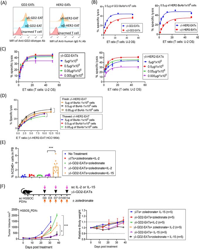
Open access
J Immunother Cancer: first published as 10.1136/jitc-2020-002222 on 13 May 2021. Downloaded from http://jitc.bmj.com/ on September 24, 2021 by guest. Protected by copyright.
phosphate-buffered saline (PBS). T cell surface BsAb Bioluminescence imaging
density was measured by mean fluorescence intensity Luc(+) T cell engraftment and trafficking were quan-
(MFI) using anti-idiotype antibody or anti-human IgG Fc tified after intravenous injection of 3 mg D- luciferin
antibody (BioLegend, Cat# 409303, RRID:AB_10900424). (Gold Biotechnology, Cat# LUCK-100) on different
MFIs were referenced to antibody binding capacity using days following T cell injection. Bioluminescence images
Quantum Simply Cellular microspheres (Bio-Rad, Cat# were acquired using IVIS Spectrum CT In Vivo Imaging
FCSC815A, RRID:AB_10061915). System (Caliper Life Sciences) and overlaid onto visible
light images, to allow Living Image V.2.60 (Xenogen)
Freeze-thaw of EATs to quantify bioluminescence in the tumor regions of
After arming, EATs were centrifuged at 500 g for 5 min at interest (ROI). The total flux (photon/s) in the ROI was
4°C, and the cell pellet was suspended in T cell freezing monitored.
medium (90% of fetal bovine serum (FBS) and 10%
Flow cytometry of blood, spleen and tumor
dimethyl sulfoxide (DMSO)) to achieve a cell concen-
Peripheral blood and tumors were collected and analyzed by
tration of 5×10⁷ cells/1 mL, chilled to 4°C and aliquoted
flow cytometry. Tumor tissue was dissociated into single cell
into 2 mL cryovials. Vials were immediately transferred to
suspensions as previously described.26 Antibodies against
freeze at −80°C for 24 hours before transferring to liquid
human CD3 (BioLegend, Cat# 300308, RRID:AB_314044),
nitrogen. After storage cryovials were thawed in a 37°C
CD4 (BioLegend, Cat# 357410, RRID:AB_2565662), CD8
water bath with gentle swirling for 1 min. The thawed cells
(BioLegend, Cat# 300912, RRID:AB_314116), and CD45
were transferred to F10 media and centrifuged at 500 g
(BioLegend, Cat# 304012, RRID:AB_314400) were used to
for 5 min. They were analyzed for viability, phenotype,
quantify T cell engraftment and subpopulations. Fluores-
antibody binding, and cytotoxicity.
cence of stained cells was acquired using either a BD FACS-
Calibur or a BD LSRFortessa (BD Biosciences, Heidelberg,
T cell transduction with tdTomato and click beetle red
Germany) and analyzed using FlowJo V.10.6.0 software
luciferase
(FlowJo, Ashland, Oregon).
T cells isolated from PBMCs were stimulated with CD3/
CD28 Dynabeads (Gibco, Cat#11 132D) for 24 hours. T Immunohistochemical staining
cells were transduced with retroviral constructs containing Fresh tumors were embedded in Tissue-Tek OCT (Miles
tdTomato and click beetle red luciferase in RetroNectin Laboratories, Elkhart, Indiana) and snap- frozen in
(Takara Bio, #Cat T100A/B)-coated six-well plates in the liquid nitrogen for storage at −80°C. The tumor sections
presence of IL-2 (100 IU/mL) and protamine sulfate were stained with mouse IgG3 mAb 3F8, as previously
(4 µg/mL). Transduced T cells were cultured for 8 days described.27 Paraffin- embedded tumor sections were
before use in animal experiments. tested for T cell infiltration using immunohistochemical
(IHC) staining of human CD3, CD4, and CD8 T cells
In vivo antitumor effects performed by the Molecular Cytology Core Facility of
Tumor cells suspended in Matrigel (Corning, Tewks- MSKCC using Discovery XT processor (Ventana Medical
bury, Massachusetts) were implanted in the flank of male Systems), as previously described.26 Histological images
BALB-Rag2−/−IL-2R-γc-KO (BRG) mice aged 6–10 weeks were acquired with NIS-Elements V.4.0 imaging software
old (Taconic Biosciences).24 The following tumor cell and Nikon ECLIPSE Ni-U microscope.
lines were used: 1×10⁶ of 143BLuc, 5×10⁶ of IMR32Luc,
5×10⁶ of M14Luc, 5×10⁶ of HCC1954, 5×10⁶ of LnCaP-AR, Statistics
and 5×10⁶ TC-71. Three osteosarcoma, two neuroblas- Statistical analysis of tumor growth or cytokine release was
toma, one Ewing sarcoma, and one breast cancer patient- conducted using area under the curve (AUC). Two-tailed
derived tumor xenografts (PDXs) established from Student’s t-test was used to determine statistical difference
fresh surgical specimens with Memorial Sloan Kettering between two sets of data, while one-way analysis of variance
Cancer Center (MSKCC) IRB approval were used for in with Tukey’s post-hoc test was used to determine statistical
vivo experiments (online supplemental table 2). Treat- differences among three or more sets of data. All statistical
ment was initiated after tumors were established, with analyses were performed using GraphPad Prism V.8.0 for
an average tumor volume of 100 mm3 when measured Windows (GraphPad Software, La Jolla, California; www.
using TM900 scanner (Piera, Brussels, Belgium). Before graphpad.com). P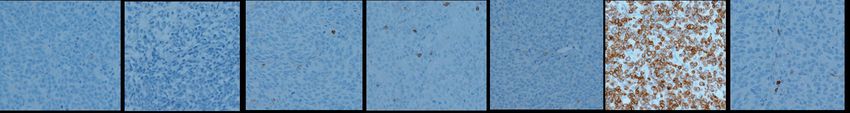
Open access
J Immunother Cancer: first published as 10.1136/jitc-2020-002222 on 13 May 2021. Downloaded from http://jitc.bmj.com/ on September 24, 2021 by guest. Protected by copyright.
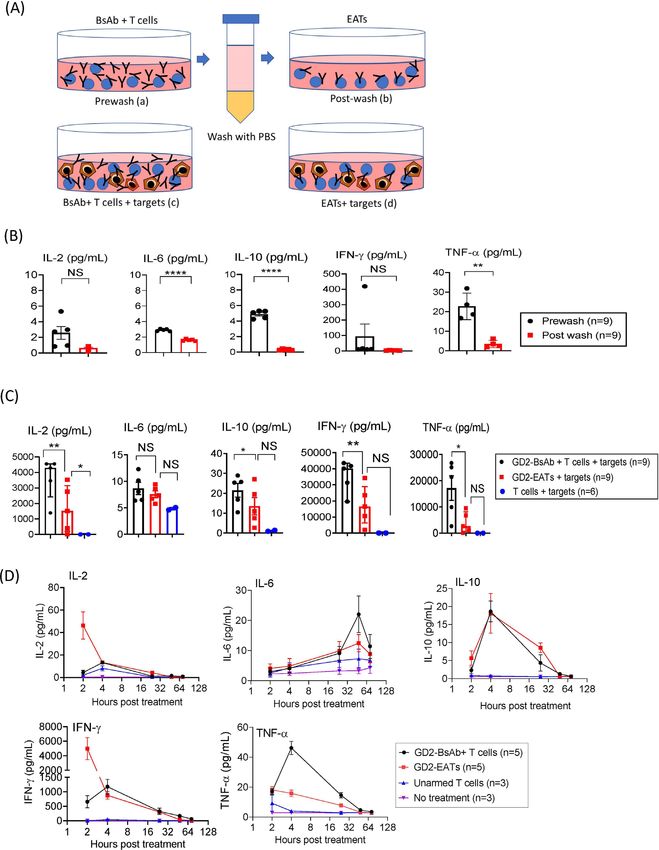
(A)
Monomeric BiTE Dimeric BiTE BiTE-Fc IgG Heterodimer IgG-[H]-scFv IgG-[L]-scFv IgG Conjugate
(B) (C)
GD2-BiTE monomer 1g= 20pmol Control BsAb
10000 BsAb on T cell 60 U-2 OS
GD2-BiTE dimer 1g=10pmol GD2-BiTE monomer
% specific lysis
GD2-BiTE dimer
GD2-BiTE-Fc 1g=7.7pmol
40 GD2-BiTE-Fc
GD2-IgG heterodimer 1g= 6.7pmol GD2-IgG heterodimer
1000
GD2-IgG-[H]-scFv 1g=5pmol 20 GD2-IgG-[H]-scFv
MFI
GD2-IgG-[L]-scFv 1g=5pmol GD2-IgG-[L]-scFv
0 GD2-IgG conjugate
100 GD2-IgG conjugate 1g=3.3pmol
-7 -6 -5 -4 -3 -2 -1 0 1 2
-4 -3 -2 -1 0 1 2 3 Log BsAb [Conc], pmol/1x106 cells
Log BsAb [Conc],pmol/1x106 cells
(D)
2000 2500 NBL50a PDX
OS1b_PDXs No treatment (n=5) No treatment (n=5)
Tumor Volume mm3
Tumor Volume mm3
2000 Unarmed T cells (n=5)
1500 Control-BsAb (n=5)
GD2-BiTE monomer (n=5) 1500 Control BsAb (n=5)
1000 1000 GD2-IgG heterodimer (n=5)
GD2-IgG heterodimer (n=5) ***
** GD2-IgG conjugate(n=5)
500 GD2-IgG conjugate (n=5) 500
GD2-IgG-[L]-scFv (n=5)
GD2-IgG-[L]-scFv (n=5) 0
0
0 20 40 60
0 7 14 21 28 35 Days post treatment
Days post treatment
(E)
GD2-BiTE monomer GD2-BiTE dimer GD2-BiTE-Fc GD2-IgG heterodimer GD2-IgG-[H]-scFv GD2-IgG-[L]-scFv GD2-IgG conjugate
(F) (G)
2000 80 U-2 OS
BsAb on T cell
% specific lysis
1500 60
40
MFI
1000 Control BsAb 1g = 5pmol
HER2-IgG-[L]-scFv 1g = 5pmol
500 HER2-IgG-[L]-scFv 1ug = 5pmol 20
HER2-IgG conjugate 1g = 3.3pmol
HER2-IgG conjugate 1ug = 3.3pmol 0
0
-3 -2 -1 0 1 2 -8 -6 -4 -2 0 2
Log BsAb [Conc],pmol/1x106 cells
Log BsAb [Conc], pmol/1x106 cells
(H)
2000 100
Tumor Volume mm3
OS1b_PDX
No treatment (n=5)
Percent survival
1500 No treatment (n=5)
Control BsAb (n=5) Control BsAb (n=5)
1000 ** 50 ****
HER2-IgG conjugate (n=5) HER2-IgG conjugate (n=5)
500 HER2-IgG-[L]-scFv (n=5)
HER2-IgG-[L]-scFv (n=5)
0 0
0 20 40 60 0 50 100 150 200
Days post treatment Days post treatment
Figure 1 Bispecific antibody platform has profound effects on the antitumor activity of bispecific antibody armed T cell
immunotherapy. (A) BsAb structural platform.10 14 62 (B) BsAb armed T cells were stained with anti-idiotype antibody, and
surface BsAb densities were measured as MFI by flow cytometry. (C) Antibody-dependent T cell-mediated cytotoxicity assay
of GD2-BsAb armed T cells at increasing BsAb arming doses. The effector to target cell ratio (E:T ratio) was 10:1. (D) 2×10⁷
of T cells were armed with 2 µg of each BsAb and administered intravenously two doses per week for 2–3 weeks in GD2(+)
osteosarcoma PDX and GD2(+) neuroblastoma PDX mouse models. In vivo antitumor responses were compared among groups.
(E) Immunohistochemical staining of CD3(+) tumor infiltrating lymphocytes in neuroblastoma PDX tumors treated with a variety
of GD2-BsAb armed T cells (on day 10 after the beginning of treatment). (F) Surface BsAb density (MFI) of HER2-BsAb armed
T cells. (G) HER2-BsAb armed T cell induced cytotoxicity against osteosarcoma cell line U-2 OS at an E:T ratio of 10:1. (H)
HER2-BsAb armed T cells (2 µg of HER2-BsAb/2×107 of T cells) were administered intravenously twice per week for 3 weeks in
HER2(+) osteosarcoma PDX-bearing mice. In vivo antitumor effect was compared. **P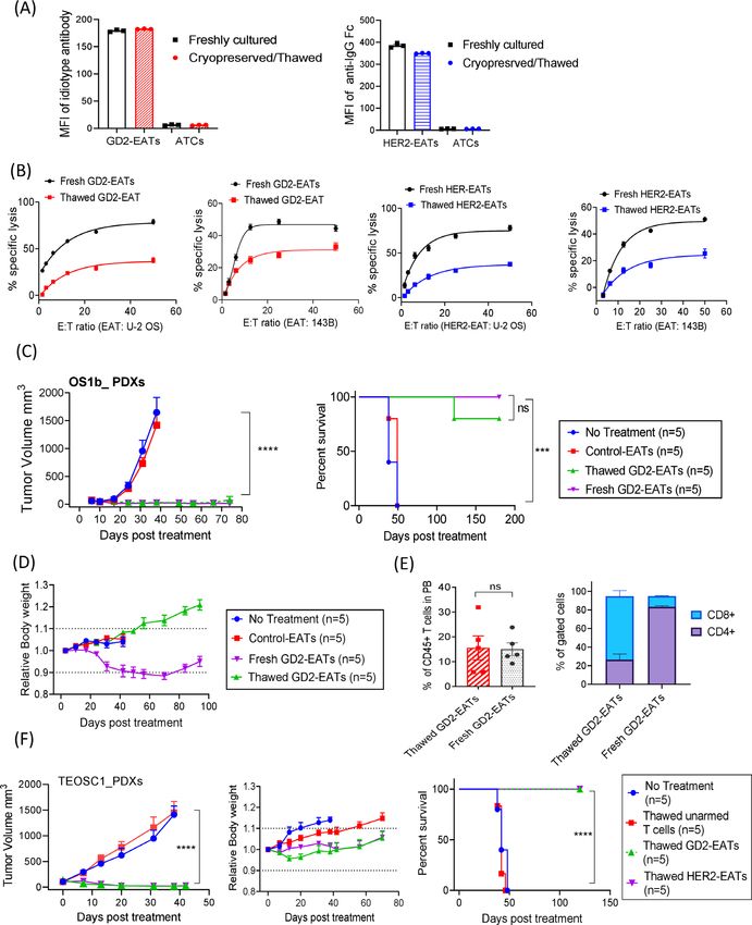
Open access
J Immunother Cancer: first published as 10.1136/jitc-2020-002222 on 13 May 2021. Downloaded from http://jitc.bmj.com/ on September 24, 2021 by guest. Protected by copyright.
properties of GD2- BsAbs are summarized in online low, IFN-γ and TNF-α did increase with the increment of
supplemental table 3. Surface BsAb densities (measured GD2-BsAb (online supplemental figure 2A), which were
by MFIs) on T cells were analyzed in figure 1B. While removed after the washing steps (online supplemental
chemical conjugate (IgG conjugate) armed T cells had figure 2B). Next, after co- culture with target cells, T
lower MFIs, other GD2- BsAb armed T cells showed cell cytokine levels were measured again and compared
comparable MFIs. In vitro cytotoxicity assay showed that among groups. The cytokines surged after exposure to
IgG-[L]-scFv or monomeric bi- specific T- cell engagers target cells. Both GD2-EATs and T cells coincubated with
(BiTE) armed T cells had the strongest in vitro potency, GD2-BsAb (GD2-BsAb plus T cells) released more cyto-
while dimeric BiTE or IgG conjugate armed T cells had kines in proportion to BsAb dose (online supplemental
comparable cytotoxicity (figure 1C). To compare in vivo figure 2C,D). However, GD2-EATs produced significantly
antitumor potency, a fixed dose of T cells armed with less amount of IL-2 (p=0.0071), IL-10 (p=0.0203), IFN-γ
different GD2-BsAb formats (2 µg of each BsAb/2×107 T (p=0.0043), and TNF-α (p=0.0162) compared with GD2-
cells) was intravenously administered into PDX-bearing BsAb plus T cells. Finally, we analyzed in vivo cytokine
mice (figure 1D and online supplemental figure 1). All levels at different time points after GD2-EAT or GD2-
EATs were well tolerated irrespective of BsAb formats, but BsAb plus T cell injection (figure 3D and online supple-
only IgG-[L]-scFv GD2-BsAb armed T cells (GD2-EATs) mental figure 3). GD2-EATs showed an early IL-2 and
exerted significant and durable antitumor responses IFN-γ release, while GD2-BsAb plus T cells showed a surge
prolonging survival. The in vivo efficacy of GD2- BsAb of TNF-α at 4 hours and IL-6 at 48 hours. The AUC for
armed T cells strongly correlated with the density of TNF-α was significantly higher in GD2-BsAb plus T cells
tumor infiltrating CD3(+) T cells (TILs) by IHC staining compared with GD2-EATs (p=0.0033).
of PDXs (figure 1E); GD2-EATs (T cells armed with GD2-
IgG-[L]-scFv) showed substantially more abundant TILs EATs facilitate T cell trafficking into tumors
compared with those armed with other BsAb formats. T cell trafficking and persistence in tumors play an essen-
Alongside GD2-BsAb armed T cells, we also compared tial role in tumor immunity. To quantitate how efficiently
the efficacy between HER2-EATs (armed with HER2-IgG- EATs traffic into tumors and persist in vivo, we gener-
[L]-scFv) and HER2-IgG conjugate (Herceptin × OKT3) ated luciferase transduced T cells and armed with GD2-
armed T cells proven safe in clinical trials.9 28 HER2-EATs BsAb. Luc(+) GD2-EATs or Luc(+) unarmed T cells were
showed higher BsAb densities (MFIs) (figure 1F) and intravenously administered into neuroblastoma PDX-
more potent tumor cell killing than HER2-IgG conjugate bearing mice. Without GD2-BsAb arming, Luc(+) T cells
armed T cells (figure 1G). HER2-EATs were much more did not localize to tumors and dissipated. In contrast,
effective in vivo for tumor response (p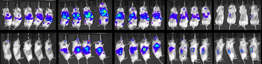
Open access
J Immunother Cancer: first published as 10.1136/jitc-2020-002222 on 13 May 2021. Downloaded from http://jitc.bmj.com/ on September 24, 2021 by guest. Protected by copyright.
Figure 2 Ex vivo arming of T cells with IgG-[L]-scFv-platformed BsAb. (A) Antibody-dependent T cell-mediated cytotoxicity
assay of GD2-EATs and HER2-EATs at increasing effector to target ratios (E:T ratios) and at increasing BsAb doses. (B) Surface
BsAb density on EAT was analyzed using anti-human IgG Fc-specific antibody and anti-mouse quantum beads. Geometric
MFIs of GD2-EATs and HER2-EATs were measured with increasing arming dose of either GD2-BsAb or HER2-BsAb, and BsAb
density (MFI) on EAT was referenced to antibody binding capacity (ABC). (C) Comparison of in vitro cytotoxicity between EATs
and soluble BsAb with identical E:T ratios, for both anti-GD2 and anti-HER2 systems. BsAb, bispecific antibodies; EATs, ex vivo
armed T cells; GD2, disialoganglioside; HER2, human epidermal growth factor receptor 2; MFI, mean fluorescence intensities.
assessment in humanized mouse models. Since autolo- (online supplemental figure 5). HER2-EATs were tested
gous T cell-PDX pairs were in short supply, we proceeded against osteosarcoma PDXs (TEOS1C), breast cancer
with the rest of EAT studies using random donor T cells. PDXs (M37), and osteosarcoma cell line (143BLuc)
xenografts (online supplemental figure 6). Beyond
EATs exerted significant antitumor effects in a variety of tumor
GD2 and HER2, EATs targeting other cancer antigens
targets
including STEAP1 (six transmembrane epithelial antigen
In vivo antitumor effects of EATs were tested in a panel of
xenograft mouse models. Four to six doses of EATs (10 µg prostate-1) on Ewing sarcoma cell line (TC71), PSMA
of each BsAb/2×107 cells) were administered intrave- (prostate membrane antigen) on prostate cancer cell
nously with a frequency of two doses per week. GD2-EATs line (LNCaP-AR), and EGFR (epidermal growth factor
were tested against neuroblastoma PDXs (Piro20Lung), receptor) on osteosarcoma cell line (143B) were tested
neuroblastoma cell line (IMR32Luc) xenografts, and against each target cell line xenograft (online supple-
melanoma cell line (M14Luc) xenografts, respectively mental figure 7); in each instance, EATs exerted significant
6 Park JA, et al. J Immunother Cancer 2021;9:e002222. doi:10.1136/jitc-2020-002222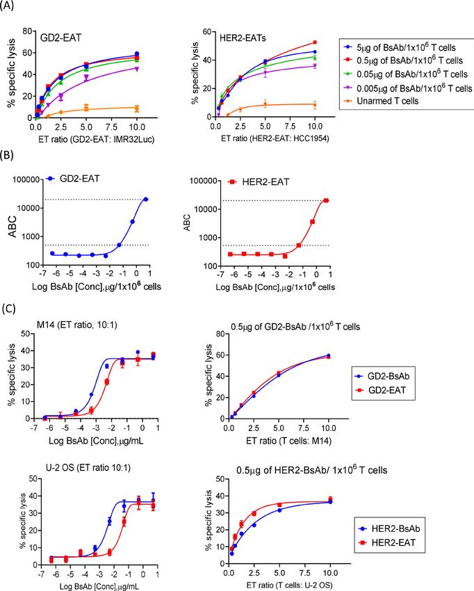
Open access
J Immunother Cancer: first published as 10.1136/jitc-2020-002222 on 13 May 2021. Downloaded from http://jitc.bmj.com/ on September 24, 2021 by guest. Protected by copyright.
Figure 3 Ex vivo arming of T cells could significantly reduce cytokine release. (A) Th1 cell cytokines (IL-2, IL-6, IL-10, IFN-γ,
and TNF-α) released by T cells were measured in each step of arming: (a) in the supernatants after 20 min of incubation with
GD2-BsAb (prewash), (b) after the second washing step (postwash), and (c and d) after incubation with target cells (GD2(+) M14
melanoma cell line) at 37℃ for 4 hours. (B) Cytokine release was compared between prewash supernatants (a) and postwash
supernatants (b). (C) Cytokine release was compared between GD2-BsAb plus T cells (c), GD2-EATs (d) and unarmed T cells
after exposure to target antigen. (D) After intravenous injection of GD2-EATs (armed with 10 µg of GD2-BsAb/2×107 cells) or
GD2-BsAb (10 µg) plus unarmed T cells (2×10⁷ cells) into osteosarcoma PDX-bearing mice, serum Th1 cytokine levels were
measured at different time points. *POpen access
J Immunother Cancer: first published as 10.1136/jitc-2020-002222 on 13 May 2021. Downloaded from http://jitc.bmj.com/ on September 24, 2021 by guest. Protected by copyright.
(A)
Supine
(lung)
Lateral
(tumor)
Day 1 Day 2 Day 3 Day 4 Day 7
(B)
Supine
(lung)
Lateral
(tumor)
Day 1 Day 2 Day 3 Day 4 Day 7
(C) (D)
Bioluminescence in tumors Tumor response by treatment
10 9 3000
Total flux (photon/sec)
Tumor Volume mm3
10 8 2000
GD2-EATs (n=5) GD2-EATs (n=5)
1000
10 7 GD2-BsAb + unarmed GD2-BsAb + T cells (n=5)
T cells (n=5) 200
150 Unarmed T cells (n=5)
10 6 Unarmed T cells (n=5) 100
50
10 5 0
0 5 10 15 20 0 20 40
Days post treatment Days post treatment
(E) (F)
Bioluminescence in tumors Tumor response by treatment
10 9 3000
Tumor Volume mm3
10 8 2000
Photon (p/s)
GD2-EATs (n=5) GD2-EATs (n=5)
10 7 1000 HER2-EATs (n=5)
HER2-EATs (n=5) 800
10 6 Unarmed T cells (n=4) Unarmed T cells (n=4)
400 No treatment (n=2)
10 5 No treatment (n=4)
10 4 0
0 10 20 30 0 10 20 30 40 50
Days post treatment
Days post treatment
(G)
Day1 WARNING: Saturated Luminescent Image Day3 Image
Day 5 Day 7 Day 14 Day 21 Day 28
Min = -7038.7 WARNING: Saturated Luminescent Image WARNING: Saturated Luminescent Image
Max = 8.4566e+06
6 p/sec/cm^2/sr 6
10
9
10
7 10
8
10
7
7 8 8
7 10 3
6 6
6
7 2
5 6 7 4
5
6 2 4
4 5
4 6
3 4 2 10
3 2
3
7 6 6
2 2 10 10 5
9 8
2 4 5
8
7
6 10
3 8
6 6 4
5 10 6
10
9 10
6 5 2
8 7
6 4
2 4
7 7
6 5 6 5
5 10
5 4 5 3
10 7
4 3 4
8
6
6 10 2
3 2 5
3 4
2 4
7 10
4
3
2
2 6 8
2
5 2 5 6
Color Bar 10 10
6
Color Bar
Min = 20000 Color Bar 5
Min = 20000 4
10 Color Bar
Max = 1e+06 Min = 1e+05 Min = 1e+06 Max = 1e+07 10
4 Color Bar
Max = 1e+07 Color Bar Min = 5000
Min = 1e+05 Max = 3e+07 3
Max = 1e+07 Max = 1e+06
Color Bar
bkg sub bkg sub Min = 7000 2
flat-fielded bkg sub flat-fielded Max = 2e+06
bkg sub bkg sub
GD2-EATs
cosmic flat-fielded flat-fielded cosmic
Click # JP20201202143800 User: cosmic bkg sub cosmic
Click # JP20201215123834 User: JP
flat-fielded
Wed, Dec 02, 2020 14:38:09 Group: EXP 445 Click # JP20201204155353 User: JP flat-fielded Tue, Dec 15, 2020 12:38:43 Group: E445 cosmic
Clickcosmic
# JP20201206162517 User: JP 6
Em filter=Open
Bin:M (8), FOV23.2, f1, 2m
Experiment: TEOSC 1
Comment1:
WARNING: Saturated G3 Image
Luminescent
Fri, Dec 04, 2020 15:54:01
Em filter=Open
Group: EXP 450
Experiment: nbl50a Click # JP20201206162517 User: JP
Sunday, Dec 06, 2020 16:25:26 Group: E445 Em filter=Open
Bin:M (8), FOV23.2, f1, 2m
Experiment: TEOSC1
Comment1: G3 day14
Click # JP20201221105451
bkg sub User: JP 10
Em filter=Open Experiment: TEOSC1
Camera: IS1308N6213, Andor, iKon Comment2: 2 MIN+TUMOR Bin:M (8), FOV23.2, f1, 1m Comment1: G3 Sunday, Dec 06, 2020 16:25:26 Group: E445 Bin:M (8), FOV23.2, f1, 1m Comment1: G3 Camera: IS1308N6213, Andor, iKon Comment2: tumor 2 min Mon, Dec 21, 2020 10:55:00
flat-fielded Group: E445
Camera: IS1308N6213, Andor, iKon Comment2:
WARNING: Saturated chest
Luminescent 1 min
Image Em filter=Open Experiment: TEOSC1 Em filter=Open cosmic Experiment: TEOSC1 7
Camera: IS1308N6213, Andor, iKon Comment2: TUMOR 1 MIN
6 Bin:M (8), FOV23.2, f1, 1m WARNING: Saturated Luminescent
Comment1: Image
G3 Click # JP20201218123947 User: JP Bin:M (8), FOV23.2, f1, 2m Comment1: G3 DAY20 6
10 6
9
8
Camera: IS1308N6213, Andor, iKon Comment2: TUMOR 1 MIN Fri, Dec7
18, 2020 12:39:55 Group: E450 Camera: IS1308N6213, Andor, iKon Comment2: tumor 2 min 10 5
7
10
7 10
Em 8filter=Open Experiment: TEOSC1
7 10
6 7 Bin:M (8), FOV23.2, f1, 2m Comment1: G3 DAY 17 6 4
7
10 7 6 5
5
6 6 Camera: IS1308N6213, Andor, iKon Comment2: tumor 2 min 4
4 5
7
6 5
4 3
2 3
4 5 4
3
4 2
3 3 2
3
2
2
2 6 5
2 10 9
10 5
8
2
8 10
6
7
5 6 6
10 10 10 4 6 6
5
9
8
7 7
10
6
7
5 5
4
10
6 6 2 4
Color Bar
6 7 3
5 6 5
5 3
Min = 1e+05
4 5 4
5 2
4
4 10
3 3 8
3 3 6
2 Max = 1e+07
2 2 4
4 10
2 2
4 6
Color Bar 5 5 2 10
Min = 20000 10 10 9
5 8 Color Bar
Max = 1e+06 Color Bar 10 Color Bar Color Bar 7 Min = 6000
Min = 1e+05 Min = 1e+05 Min = 20000 Max = 1e+06
Color Bar 6
Max = 1e+07 Min = 1e+05 Max = 1e+07 Max = 1e+07
5 bkg sub
Max = 1e+07
Color Bar
Min = 5000
flat-fielded
bkg sub
flat-fielded Max = 2e+05 cosmic
bkg sub
cosmic bkg sub bkg sub bkg sub
flat-fielded
flat-fielded bkg sub flat-fielded flat-fielded Click # JP20201209145027 User: JP
HER2-EATs
Click # JP20201202144538 User: cosmic
cosmic flat-fielded cosmic cosmic
Wed, Dec 02, 2020 14:45:47
Em filter=Open
Group: EXP 445
Experiment: TEOSC 1 Click # JP20201204154809 User: JP cosmic
Click # JP20201209145027 User: JP Click # JP20201215124239 User: JP Wed, Dec 09, 2020 14:50:36 Click # JP20201221105931 Group: E445 User: JP
Mon, bkg
Dec sub
21, 2020 10:59:40 Group: E445
Bin:M (8), FOV23.2, f1, 2m Comment1: G4 Fri, Dec 04, 2020 15:48:18 Group: EXP 450 Click # JP20201206161758 User: JP Wed, Dec 09, 2020 14:50:36 Group: E445 Tue, Dec 15, 2020 12:42:47
Em filter=Open
Group: E445
Experiment: TEOSC1
Em filter=Open flat-fielded
Em filter=Open Experiment: TEOSC1
Experiment: TEOSC1
Camera: IS1308N6213, Andor, iKon Comment2: TUMOR 2 MIN Em filter=Open Experiment: nbl50a Sunday, Dec 06, 2020 16:18:07 Group: E445 Em filter=Open Experiment: TEOSC1 Bin:Mcosmic
Bin:M (8), FOV23.2, f1, 1m Comment1: G4 Em filter=Open Experiment: TEOSC1 Bin:M (8), FOV23.2, f1, 2m Comment1: G4 day8 Bin:M (8), FOV23.2, f1, 2m Comment1: G4 day14
Click # JP20201218124344
Bin:M (8), FOV23.2,
User: JP
f1, 2m (8), FOV23.2, f1, 2m Comment1: G4
Camera: IS1308N6213, Andor, iKon
day8G4 DAY20
Comment1:
Comment2: tumor 2 min
Camera: IS1308N6213, Andor, iKon Comment2: tumor 2 min
Camera: IS1308N6213, Andor, iKon Comment2: tumor 1 min Bin:M (8), FOV23.2, f1, 1m
Camera: IS1308N6213, Andor, iKon
Comment1: G4
Comment2: TUMOR 1 MIN
Camera: IS1308N6213, Andor, iKon Comment2: tumor 2 min
Fri, Dec 18, 2020 12:43:54 Camera: IS1308N6213,
Group: E450 Andor, iKon Comment2: tumor 2 min
Em filter=Open Experiment: TEOSC1
Bin:M (8), FOV23.2, f1, 2m Comment1: G4 DAY 17
Camera: IS1308N6213, Andor, iKon Comment2: tumor 2 min
Figure 4 EATs showed faster tumor homing kinetics than BsAb-directed unarmed T cells, rapidly bypassing lung
sequestration. Luciferase transduced T cells were expanded and armed with BsAb (Luc(+) GD2-EATs). Luc(+) GD2-EATs
(10 µg of GD2-BsAb/2×107 T cells) or Luc(+) unarmed T cells (2×10⁷ cells) with or without GD2-BsAb (10 µg) were administered
intravenously into GD2(+) neuroblastoma PDX mice when the average tumor volume reached 100 mm3. (A) Bioluminescence
images of GD2-EATs trafficking into tumors over days. (B) Bioluminescence images of GD2-BsAb directed unarmed T cell
trafficking into tumors over days. (C) Quantitation of T cell infiltration into tumors over time measured by bioluminescence
(n=5 mice/group) expressed as total flux or radiance (photons/s) per pixel integrated over the tumor contour (ROI). (D) Tumor
growth curves of individual mice treated with GD2-EATs, GD2-BsAb plus unarmed T cells, or unarmed T cells. To test the in
vivo persistence of target antigen-specific EATs, Luc(+) GD2-EATs (armed with 10 µg of GD2-BsAb/2×107 T cells) or Luc(+)
HER2-EATs (10 µg of HER2-BsAb/2×107 T cells) were intravenously administered into osteosarcoma TEOS1C PDX-bearing
mice. Additional two doses of non-luciferase transduced GD2-EATs or HER2-EATs were administered on day 7 and day 14. (E)
Quantitation of bioluminescence of Luc(+) EATs in tumors post-treatment. (F) Tumor growth curves of individual mice treated
with GD2-EATs, HER2-EATs, or unarmed T cells, or no treatment. (G) Bioluminescence images of Luc(+) GD2-EATs (upper) or
Luc(+) HER2-EATs (lower) in tumors over time. Bioluminescence of Luc(+) EATs was detected over 28 days postinjection. BsAb,
bispecific antibodies; EATs, ex vivo armed T cells; GD2, disialoganglioside; HER2, human epidermal growth factor receptor 2;
PDX, patient-derived tumor xenograft; ROI, region of interest.
8 Park JA, et al. J Immunother Cancer 2021;9:e002222. doi:10.1136/jitc-2020-002222Open access
J Immunother Cancer: first published as 10.1136/jitc-2020-002222 on 13 May 2021. Downloaded from http://jitc.bmj.com/ on September 24, 2021 by guest. Protected by copyright.
antitumor effects, without weight loss or adverse events GD2-EATs were fixed at 2 µg GD2-BsAb/2×107 cells. Dose-
during follow-up period (online supplemental figure 8). dense schedule contributed to improved tumor control
over standard or low-intensity treatment schedules. Arm
Factors determining in vivo efficacy of EATs 3 showed the most potent antitumor effect against rapidly
BsAb dose for arming growing neuroblastoma PDXs compared with arm 2 (stan-
In vitro tumor cell killing by EATs showed the best dard) or arm 1 (low-intensity schedules) (p=0.0276 and
efficacy at arming doses between 0.05 µg and 5 µg of p=0.0001, respectively), which translated into survival
BsAb/1×106 T cells, corresponding to BsAb surface densi- benefit (both p1 µg of BsAb/2×107 T cells (0.05 µg/1×106 survival
cells)) for consistent treatment outcome (figure 5A). To test the effect of repeat EAT dosing on long-term
When delivered by EATs, 1 µg of BsAb was substantially remission and whether supplemental BsAb could elimi-
inferior to 10 µg or 100 µg of BsAb. In contrast to separate nate the necessity of repeat EAT, we tested three different
BsAb injection, where high-dose BsAb (100 µg of HER2- treatment schedules on osteosarcoma PDXs (figure 5D
BsAb) showed inferior antitumor effect compared with and online supplemental figure 11A): arm 1, two doses
low-dose BsAb (p=0.0056) (online supplemental figure of EATs followed by six doses of intravenous BsAb; arm 2,
9A), 100 µg of HER2-BsAb was equally effective as 10 µg four doses of EATs followed by four doses of BsAbs; and
of HER2-BsAb when delivered by EATs (p=0.2448). arm 3, eight doses of EATs. In contrast to the rapid tumor
To investigate the BsAb dose and T cell response growth with no treatment or eight doses of unarmed T
relationship, CD3(+) T cells were incubated with or cells, two doses of GD2-EATs or HER2-EATs successfully
without target cells at 37°C for 4 hours in the presence ablated tumors. However, additional doses of EATs were
of increasing BsAb dose (5×10⁻⁵ µg to 50 µg of HER2- beneficial in maintaining long-term remission. Contrary
BsAb/1×106 T cells). T cells were analyzed for activation to the mice treated with two doses of GD2-EATs showing
(CD69), exhaustion (TIM-3), activation induced T cell short-term response, those treated with eight doses of
apoptosis and death (annexin V and 7-aminoactinomycin GD2-EATs, two of five mice showed sustained remission
D, 7-AAD) and compared with HER2-EATs under identical past 6 months, demonstrating statistically significant
conditions (online supplemental figure 9B). While CD69 survival benefit over two doses or four doses of GD2-
and TIM-3 expression rose when HER2-BsAb was above EATs supplemented with identical doses of GD2- BsAb
0.05 µg/1×106 T cells, 7- AAD or annexin V expression (p=0.0018 and p=0.0089, respectively). Eight doses of
started to increase above 0.5 µg/1×106. All four markers HER2-EATs also showed a benefit in sustaining long-term
were consistently and significantly lower for HER2-EATs remission over two doses or four doses of HER2-EATs
when compared with HER2-BsAb plus T cells, in agree- supplemented with HER2-BsAb (p=0.0018 and p=0.0494,
ment with the in vivo antitumor effect of high-dose BsAb respectively) (online supplemental figure 11B). When we
when delivered as soluble BsAb versus EATs. analyzed the relapsed tumors, those treated with GD2-
EATs lost their GD2 expression (online supplemental
Infused EAT cell number figure 12), suggesting that tumor antigen downregulation
Next, we evaluated the effect of infused EAT cell number or loss following treatment led to immune evasion.
on antitumor response in vivo (figure 5B). At a fixed
arming dose of 0.5 µg of BsAb/1×106 cells, different cell Expanding the clinical utility of EAT therapy
numbers of GD2-EAT or HER2-EAT (5×10⁶ cells, 1×10⁷ EATs retain antitumor properties after cryopreservation
cells, and 2×10⁷ cells) were administered twice weekly for We tested EATs for their viability, BsAb surface density,
2 weeks. The antitumor effect consistently increased with and tumoricidal activities after a freeze-thaw process. After
the cell number of EATs infused. The in vivo antitumor thawing at 37°C, EATs remained over 85% viable, irre-
effect of 2×10⁷ of GD2-EATs was significantly superior spective of whether they were frozen for 2 hours at −80°C
to 5×10⁶ or 1×10⁷ of GD2-EATs (p=0.0018 and p=0.0366, or up to 12 months in liquid nitrogen. When the thawed
respectively). The effect of 2×10⁷ of HER2-EATs was also EATs were stained with anti- idiotype antibody or anti-
superior to 5×10⁶ or 1×10⁷ of HER2-EATs (p=0.0077 and human IgG Fc antibody, BsAb surface density remained
p=0.0340, respectively). This antitumor response was comparable with freshly armed EATs (fresh EATs) by
correlated with the percentage of human CD45(+) TILs, MFIs (figure 6A). The CD4:CD8 ratio was preserved after
which was obvious with 2×10⁷ of GD2-EATs, but negligible freeze-thaw (online supplemental figure 13A). However,
with 5×10⁶ of GD2-EATs (online supplemental figure 10). the thawed CD4(+) GD2-EATs expressed more apoptosis
marker than CD8(+) GD2-EATs compared with freshly
Treatment schedule armed GD2-EATs. Although the maximal killing of target
To evaluate the impact of treatment interval on the in vivo cells did diminish after freeze- thaw (50% of maximal
efficacy of EATs, we tested three different schedules: arm killing efficacy of fresh EATs) due to not enough recovery
1, low intensity (1 dose/week); arm 2, standard (2 doses/ time after thawing, antigen specificity and the potency of
week); or arm 3, dose-dense (3 doses/week) (figure 5C). EAT were maintained (figure 6B).
Park JA, et al. J Immunother Cancer 2021;9:e002222. doi:10.1136/jitc-2020-002222 9Open access
J Immunother Cancer: first published as 10.1136/jitc-2020-002222 on 13 May 2021. Downloaded from http://jitc.bmj.com/ on September 24, 2021 by guest. Protected by copyright.
(A)
PiroLung20 PDX 143B xenografts
2500
2000 BsAb dose for EAT
Tumor Volume mm3
Tumor Volume mm3 BsAb dose for EAT
* (per 2x10⁷ T cells) 2000 (per 2x10⁷ T cells)
1500
Unarmed T cells (n=5) 1500 **
Unarmed T cells (n=5)
1000 **** 10g of GD2-BsAb (n=5) ****
1000 1g of HER2-BsAb (n=5)
1g of GD2-BsAb (n=5) *
500 * 500 10g of HER2-BsAb(n=5)
No treatment (n=5)
100g of HER2-BsAb (n=5)
0 0
0 20 40 60 0 10 20 30 40
Days post treatment Days post treatment
(B)
2500 OS1b_PDX 2500 OS1b PDX
Tumor Volume mm3
Tumor Volume mm3
2000 2000
ns 20x106 unarmed T cells (n=5)
1500 2x10⁷ unarmed T cells (n=5) 1500
** 5x10⁶ HER2-EATs (n=5)
1000 ** 5x10⁶ GD2-EATs (n=5) 1000
* 10x106 HER2-EATs (n=5)
1x10⁷ GD2-EATs (n=5) 500
500 20x106 HER2-EATs (n=5)
2x10⁷ GD2-EATs (n=5)
0 0
0 10 20 30 40 -10 0 10 20 30 40 50
Days post treatment
Days post treatment
(C) Unarmed T cells
Arm 1
GD2-EATs
Arm 2
Arm 3
sc NBL50a D0 D2 D4 D7 D14
PDX
2500
Tumor Volume mm3
100
2000
Percent survival
No Treatment (n=5) No Treatment (n=5)
1500 Unarmed T cells (n=5)
Unarmed T cells (n=5)
1000 50 **** arm 1 (n=5)
*** arm 1 (n=5)
* arm 2 (n=5)
500 arm 2 (n=5) arm 3 (n=5)
0 arm 3 (n=5) 0
0 7 14 21 0 10 20 30
Days post treatment Days post treatment
(D)
Unarmed T cells
Arm 1 GD2-BsAb
Arm 2
Arm 3 GD2-EATs
sc OS1b PDX D0 D3 D7 D10 D14 D17 D21 D24
2000
Tumor Volume mm3
100
No Treatment (n=5)
Percent survival
1500 No Treatment (n=5)
Unarmed T cells (n=5) **** Unarmed T cells (n=5)
1000 **** arm 1 (n=5)
* arm 1 (n=5) 50
500 arm 2 (n=5) arm 2 (n=5)
arm 3 (n=5) ** arm 3 (n=5)
0 0
0 20 40 60 80 0 50 100 150 200
Days post treatment Days post treatment
Figure 5 Factors determining the in vivo efficacy of EATs. (A) The effect of BsAb dose on in vivo efficacy of EATs. 2×10⁷ of T
cells were armed with increasing dose of GD2-BsAb or HER2-BsAb and their antitumor effects were compared. (B) The effect
of EAT cell number on the in vivo efficacy of EATs. At a fixed arming dose of 0.5 µg of BsAb/1×106 cells, increasing numbers of
GD2-EAT or HER2-EAT (5×10⁶ cells, 1×10⁷ cells, and 2×10⁷ cells) were administered. (C) The effect of EAT treatment schedule
on the in vivo efficacy of EATs. GD2-EATs (2 µg of GD2-BsAb/2×107 cells) were administered as low intensity (1 dose/week
in arm 1), standard (2 doses/week in arm 2), or dose-dense (3 doses/week in arm 3) and their in vivo antitumor response
and survival were compared. (D) The effect of total number of doses of EATs on long-term tumor control and survival. Arm
1, GD2-EATs (10 µg of GD2-BsAb/2×107 cells) were administered twice weekly for 1 week, and GD2-BsAb (10 µg/dose) were
followed twice weekly for 3 weeks; arm 2, four doses of GD2-EATs followed by four doses of GD2-BsAb; arm 3, eight doses
of GD2-EATs. *POpen access
J Immunother Cancer: first published as 10.1136/jitc-2020-002222 on 13 May 2021. Downloaded from http://jitc.bmj.com/ on September 24, 2021 by guest. Protected by copyright.
Figure 6 Cryopreserved EATs retained target antigen-specific cytotoxicity and exerted a comparable antitumor activity. (A)
Geometric mean fluorescence intensities of BsAb on GD2-EATs (0.5 µg of GD2-BsAb/106 cells) and HER2-EATs (0.5 µg of HER2-
BsAb/106 cells) before and after cryopreservation. (B) Antibody-dependent T cell-mediated cytotoxicity assay of GD2-EATs and
HER2-EATs against GD2(+) and HER2(+) osteosarcoma cell lines before and after cryopreservation (n=6). (C) In vivo antitumor
response of thawed GD2-EATs. T cells were first armed with GD2-BsAb (10 µg of BsAb/2×107 cells) before cryopreservation.
These cells were then thawed and administered into mice bearing GD2(+) osteosarcoma PDX to compare their in vivo antitumor
potency with freshly armed GD2-EATs derived from the same donor. Four to six doses of cryopreserved (thawed) GD2-EATs
or freshly armed (fresh) GD2-EATs were administered intravenously twice per week. (D) Relative body weights over time after
treatment with fresh or thawed GD2-EATs. (E) Flow cytometry analyses of peripheral blood (PB) T cells in the mice treated
with fresh or thawed GD2-EATs on day 35 after treatment. (F) In vivo antitumor response of thawed GD2-EATs or HER2-EATs
against telangiectatic osteosarcoma PDXs. T cells were first armed with GD2-BsAb or HER2-BsAb (10 µg of BsAb/2×107 cells)
before cryopreservation. Six doses of GD2-EATs or HER2-EATs were given twice a week for 3 weeks with SC interleukin-2.
Both thawed EATs significantly suppressed tumor growth without weight loss, improving survival (pOpen access
J Immunother Cancer: first published as 10.1136/jitc-2020-002222 on 13 May 2021. Downloaded from http://jitc.bmj.com/ on September 24, 2021 by guest. Protected by copyright.
In vivo antitumor activities of thawed EATs were evalu- γδ-GD2-EATs and γδ-HER2-EATs did not produce signif-
ated in two different osteosarcoma PDX models. Thawed icant antitumor response irrespective of additional zole-
GD2- EATs exerted potent and comparable antitumor dronate. Flow cytometry analyses of T cells in the blood
effect to fresh GD2- EATs (p=0.1633) and significantly and tumors after γδ-EAT treatment presented substan-
prolonged survival compared with controls (p=0.0002) tially fewer human CD45(+) T cells in the circulation
(figure 6C). Four of five mice treated with thawed GD2- and in the tumors compared with αβ-EATs (p=0.0006 and
EATs showed long-term remission past 6 months post- p=0.0007, respectively), suggesting poor in vivo survival of
treatment. IHC staining of the tumors treated with thawed γδ-EATs (online supplemental figure 15B).
GD2-EATs showed diffuse infiltration of CD4(+) T cells as However, when exogenous IL-15 (as IL15Rα-IL15
well as CD8(+) T cells (online supplemental figure 13B), complex) instead of IL-2 was used as T cell survival cyto-
supporting their intact homing properties. Interestingly, kine, the in vivo frequencies of γδ-EATs significantly
while the mice treated with fresh GD2-EATs developed increased (figure 7E). γδ T cells expanded from fresh
mild to moderate GVHD 1–2 months post- treatment, PBMCs using 2 µM of zoledronate plus 30 ng/mL of IL-15
the mice treated with thawed GD2-EATs did not present for 12–14 days were armed with GD2-BsAb or HER2-BsAb
clinical signs of GVHD throughout the entire follow-up and administered intravenously into osteosarcoma PDX-
period, maintaining body weight, good coat condition bearing mice, with 5 µg of subcutaneous IL-15 or 1000 IU
and general activity (figure 6D). When blood samples of IL-2 (figure 7F and online supplemental figure 16A).
were analyzed on day 45 post- treatment (figure 6E), While γδ-GD2-EATs and γδ-HER2- EATs sustained with
the fresh GD2-EATs-treated mice showed CD4(+) T cell IL-2 failed to suppress tumor, same γδ-EATs sustained with
predominance with signs of xenogeneic GVHD, but the IL-15 exerted significant antitumor effects without toxic-
thawed GD2-EATs-treated mice displayed a predominance ities or weight loss during follow-up period. The PDXs
of CD8(+) T cells without signs of GVHD, correlating treated with γδ-GD2-EATs plus IL-15 showed a diffuse T
with their clinical manifestations. The comparable in vivo cell infiltration into tumors, in contrast to tumors treated
potency of thawed EATs was confirmed in the second by γδ-GD2- EATs plus IL-2 or unarmed γδ-EATs plus
tumor model (figure 6F); both thawed GD2-EATs and IL-15 (online supplemental figure 16B). Furthermore,
thawed HER2-EATs exerted significant antitumor effect cryopreserved γδ-EATs showed strong in vivo antitumor
against osteosarcoma PDXs (pOpen access
J Immunother Cancer: first published as 10.1136/jitc-2020-002222 on 13 May 2021. Downloaded from http://jitc.bmj.com/ on September 24, 2021 by guest. Protected by copyright.
Figure 7 EAT as a versatile platform to harness γδ T cells. (A) Flow cytometry of GD2-BsAb or HER2-BsAb on γδ T cells (γδTs)
or unselected polyclonal T cells (mostly αβ T cells (αβTs)) after arming with GD2-BsAb or HER2-BsAb. (B) ADTC assays of γδ-
GD2-EATs and γδ-HER2-EATs compared with αβ-GD2-EATs and αβ-HER2-EATs. Non-specific tumor cell killing by unarmed γδ
T cells and unarmed αβ T cells (background) were subtracted. (C) ADTC assay of γδ-GD2-EATs and γδ-HER2-EATs at increasing
effector to target ratios (E:T ratios) and at increasing BsAb arming doses. (D) In vitro cytotoxicity against breast cancer cell line
HCC1954 was compared between fresh γδ-HER2-EATs and thawed γδ-HER2-EATs at increasing E:T ratios and at increasing
BsAb arming doses. (E) Flow cytometry analyses of peripheral blood T cells after second dose of γδ-GD2-EATs plus zoledronate
and supplementary IL-2 or IL-15. (F) Three doses of γδ-GD2-EATs (10 µg of GD2-BsAb/2×107 cells) were administered with
supplementary IL-2 or IL-15 to treat osteosarcoma PDXs and the antitumor response was compared among groups. *POpen access
J Immunother Cancer: first published as 10.1136/jitc-2020-002222 on 13 May 2021. Downloaded from http://jitc.bmj.com/ on September 24, 2021 by guest. Protected by copyright.
infiltrate ‘cold’ tumors to exert their antitumor effect. CD4(+) GD2-EATs did have higher levels of apoptosis
Despite similar antitumor properties among BsAb armed marker than CD8(+) GD2- EATs, which was consistent
T cells in vitro, only ‘EATs’, T cells armed with the IgG-[L]- with the in vivo dominance of CD8(+) T cell population
scFv-platformed BsAb, showed strong infiltration into following treatment. CD4(+) T cell dysfunction including
tumors to produce durable antitumor responses which reduced IFN-γ production has been reported in the cryo-
translated into prolonged survival. Although genetically preserved product of T cells, PBMCs or cord blood.48–50
engineered T cells expressing CD19-BiTE or CAR T cells To improve antitumor activity and to reduce treatment-
secreting EGFR-BiTE had shown success in preclinical related toxicity, optimizing cryopreservation protocols
models,34 35 T cells ex vivo armed with GD2-BiTE have for EATs would be the next front.
never been as successful. This may attribute to the short Another limitation of CAR T cell therapy is the need
half-life of BiTE or the nature of the tumor target, partly for autologous T cells. Alternative effector T cells with low
explaining why there have been no successful reports of alloreactivity including human leukocyte antigen (HLA)-
armed T cells using BiTE despite the clinical success of
identical donor’s activated T cells,51 52 clonally committed
Blincyto.36 Besides, protein size, distance of the epitope
Epstein-Barr virus (EBV)-specific cytotoxic T cells,53 and
to the membrane, purity, and binding affinities to tumor
innate-like lymphocytes such as γδ T cells or natural killer
and to CD3 T cells strongly affect the biodistribution and
T (NKT) cells could be potential ‘off- shelf’ cyto-
the-
in vivo antitumor activities of T-BsAb.37–41 Since all GD2-
therapy products. When obtained from healthy donors,
BsAb platforms were built using the same anti-GD2 and
anti-CD3 VH and variable light chain (VL) sequences, the probability of successful large-scale ex vivo expansion
monovalent binding to CD3 or to target in monomeric of effector cells with consistent product quality should be
BiTE and IgG heterodimer (online supplemental table high. γδ T cells are known to have innate antitumor prop-
3) might affect the stability of BsAb armed T cells in vivo. erties and significantly reduced alloreactivity or ‘on-target
However, despite the multivalency in dimeric BiTE, BiTE- off-tumor’ toxicities.32 In our studies, γδ-EATs could
Fc, IgG-[H]-scFv, and IgG conjugate, none of these plat- suppress osteosarcoma xenografts in the presence of the
forms was able to achieve the potency of IgG-[L]-scFv, IL15Rα-IL15 cytokine complex without toxicities. IL-15
implicating additional factors besides binding avidity. enhanced proliferation of T cells in vivo and improved
We previously described the critical importance of inter- potency of T cell-based immunotherapy.54 55 For γδ T cell
domain distance and cis- configuration of BsAb,14 now treatment, poor in vivo survival has been a major chal-
confirmed with these extensive series of EAT-based exper- lenge. Supplementary zoledronate and IL-15/IL- 15Ra
iments. The mechanism behind the in vivo superiority of enabled γδ-EATs to survive and to proliferate in vivo to
the IgG-[L]-scFv format is likely to be more complex. exert significant antitumor effects. By employing optimal
One of the major toxicities of T cell-based immuno- cytokines to prolong in vivo survival,56 γδ-EATs deserve to
therapy is CRS,42 typically associated with surge of IFN-γ, be explored in more depth. EATs using γδ T cells should
IL-6, and TNF-α, although elevations of IL-2, granulocyte- provide a powerful platform to harness third- party or
macrophage colony-stimulating factor (GM-CSF), IL-10, haploidentical donor’s effector T cells with minimal
IL-8, and IL-5 have also been reported.43 44 IL-6 and graft-versus-host side effects, with the additional benefit
TNF-α have been implicated as the central mediators of cytotoxic mechanisms through TNF-related apoptosis-
of CRS.42 When the anti-CD28 antibody TGN1412 was inducing ligand (TRAIL), NKG2D receptor, and FcγRIII
administered to healthy subjects, CRS developed in an (CD16).57 58
hour, coinciding with peak cytokine levels: TNF-α, as an Neurotoxicity is the other major hurdle that has
initial trigger, was followed by INF-γ and IL-10, IL-8, IL-6, plagued CAR T cell and BsAb.4 Since GD2 is expressed
IL-4, IL-2, and IL-1β.45 When BsAb engages polyclonal
in mouse brain, several studies have reported neurotox-
T cells to undergo synchronous activation, TNF-α acts
icity in mice when CAR T cells were used.59 Although we
as the initial signal for monocyte activation, resulting
did not directly compare EATs and CAR T cells in this
in release of IL-6 and IL-1, preventable by anti-TNF-α
manuscript, we previously compared the efficacy of GD2-
antibodies without compromising antitumor activity.46
CAR T cells and GD2 IgG-[L]-scFv BsAb built with iden-
However, the absence of cytokine storm in clinical studies
of BsAb armed T cell is notable.12 13 47 T cells exposed to tical anti-GD2 IgG sequence.60 GD2-BsAb-driven T cells
BsAb produced TNF-α during the initial 20 min of BsAb were more efficient than GD2-CAR T cells in vivo. More
arming; after washing, EATs released significantly less importantly, while GD2-CAR T cells caused neurological
cytokines, particularly TNF-α, in vitro and in vivo without damage, neither GD2- BsAb nor GD2- EATs caused any
61
affecting their trafficking ability or tumoricidal activity. neurotoxicity in mouse models.
We also showed that thawed EATs maintained the In conclusion, ex vivo arming of T cells with IgG-[L]-
viability and their affinity to target antigens and to CD3(+) scFv BsAb is a potential strategy beyond CAR T cell tech-
T cells. The cryopreservation did not affect the frequency nology to use proteins instead of genes to modify T cells.
of CD4 or CD8 T cells in EATs. The thawed GD2-EATs While avoiding CRS and neurotoxicity, the potential to
preserved their target antigen specificity and were effec- use cryopreserved alternative effector T cells other than
tive in vitro and in vivo. When thawed, the cryopreserved αβ T cells should expand the clinical utility of EATs.
14 Park JA, et al. J Immunother Cancer 2021;9:e002222. doi:10.1136/jitc-2020-002222You can also read