In vitro Differentiation of Human iPSC-derived Cardiovascular Progenitor Cells (iPSC-CVPCs)
←
→
Page content transcription
If your browser does not render page correctly, please read the page content below
Bio-protocol 10(18): e3755.
                                           www.bio-protocol.org/e3755           DOI:10.21769/BioProtoc.3755

    In vitro Differentiation of Human iPSC-derived Cardiovascular Progenitor Cells (iPSC-CVPCs)
              Agnieszka D’Antonio-Chronowska1, *, Matteo D’Antonio1 and Kelly A. Frazer1, 2

Department of Pediatrics, University of California, San Diego, La Jolla, USA; 2Institue for Genomic
1

Medicine, University of California, San Diego, La Jolla, USA
*For correspondence: adantoniochronowska@health.ucsd.edu

[Abstract] Induced pluripotent stem cell derived cardiovascular progenitor cells (iPSC-CVPCs) provide
an unprecedented platform for examining the molecular underpinnings of cardiac development and
disease etiology, but also have great potential to play pivotal roles in the future of regenerative medicine
and pharmacogenomic studies. Biobanks like iPSCORE (Stacey et al., 2013; Panopoulos et al., 2017),
which contain iPSCs generated from hundreds of genetically and ethnically diverse individuals, are an
invaluable resource for conducting these studies. Here, we present an optimized, cost-effective and
highly standardized protocol for large-scale derivation of human iPSC-CVPCs using small molecules
and purification using metabolic selection. We have successfully applied this protocol to derive iPSC-
CVPCs from 154 different iPSCORE iPSC lines obtaining large quantities of highly pure cardiac cells.
An important component of our protocol is Cell confluency estimates (ccEstimate), an automated
methodology for estimating the time when an iPSC monolayer will reach 80% confluency, which is
optimal for initiating iPSC-CVPC derivation, and enables the protocol to be readily used across iPSC
lines with different growth rates. Moreover, we showed that cellular heterogeneity across iPSC-CVPCs
is due to varying proportions of two distinct cardiac cell types: cardiomyocytes (CMs) and epicardium-
derived cells (EPDCs), both of which have been shown to have a critical function in heart regeneration.
This protocol eliminates the need of iPSC line-to-line optimization and can be easily adapted and scaled
to high-throughput studies or to generate large quantities of cells suitable for regenerative medicine
applications.
Keywords: Human induced pluripotent stem (iPSC), Cardiovascular progenitor cells (CVPCs),
cardiomyocytes (CMs), Epicardium cells (EPDCs), Human induced pluripotent stem cell-derived
cardiovascular progenitor cells (iPSC-CVPCs), Human induced pluripotent stem cell-derived
cardiomyocytes cells (iPSC-CMs), Human induced pluripotent stem cell-derived epicardium derived
cells (iPSC-EPDCs), Cardiovascular disease, Heart, Differentiation, Genetic studies, Small molecules,
Pharmacogenomics, Regenerative medicine

[Background] Cardiovascular diseases (CVDs) remain the leading cause of death worldwide and
account for about 30% of all mortality causes globally. Coronary artery disease (CAD) and myocardial
infarction (MI) are among the most common CVDs, and in the USA alone, every 40 s someone suffers
a heart attack (Association, 2016; Heron, 2017; WHO, 2018; Benjamin et al., 2019; D'Antonio-
Chronowska et al., 2019a). Heart failure results in the death of cardiac muscle cells and is the
consequence of morphological and functional changes (cardiac remodeling: necrosis, scar formation,

Copyright © 2020 The Authors; exclusive licensee Bio-protocol LLC.                                       1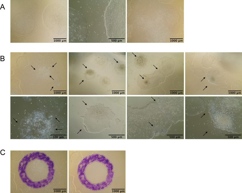
Bio-protocol 10(18): e3755.
                                           www.bio-protocol.org/e3755          DOI:10.21769/BioProtoc.3755

inflammation, fibrosis, dilation and reshaping) that occur in response to pre-existing cardiac conditions,
including CAD, MI, hypertension, cardiomyopathy, myocarditis and abnormal cardiac valve function
(Cohn et al., 2000; Reed et al., 2017). Heart failure is commonly treated with beta blockers, ACE
inhibitors and aldosterone antagonists which partially reverse cardiac remodeling and thereby improve
prognosis (Reis Filho et al., 2015), but does not result in the regeneration of cardiac tissue. There are
currently several ongoing clinical trials, including ESCORT and DREAM-HF (Menasche et al., 2015 and
2018; Borow et al., 2019), which are aimed at evaluating the effectiveness of transplanting iPSC-derived
Cardiovascular Progenitor Cells (iPSC-CVPCs) or embryonic stem cell derived CVPCs (ESC-CVPCs)
as a therapeutic treatment for heart failure. The ability to generate iPSC-CVPCs in large quantities, as
is required for regenerative medicine, using biological material obtained directly from the patient would
enable autologous transplantations and thereby eliminate the need of immunosuppression. Thus, the
development of a robust and cost-effective protocol for generating large amounts of high-quality iPSC-
CVPCs without requiring individualized optimization for each iPSC line is imperative for the
advancement of future therapeutic treatments of heart failure.
  Large collections of iPSC-CVPCs (D'Antonio-Chronowska et al., 2019b) generated from genetically
and ethnically diverse individuals could also be used for cost-effective large-scale testing of drugs for
cardiotoxicity or proarrhythmic effects. Previous studies (Burridge et al., 2016) and initiatives like CiPA
Project (Blinova et al., 2017) have shown the utility of iPSC-CVPCs for testing drugs for cardiotoxicity,
which, if scaled to examine large collections of iPSC-CVPCs derived from both healthy or disease
bearing individuals, could greatly improve the efficiency of testing new drugs for safety, and in turn
decrease the cost of drug development.
  We have previously demonstrated the feasibility of using a highly standardized protocol for
successfully deriving high quality iPSC-CVPCs from hundreds of iPSC lines reprogrammed from
ethnically diverse individuals (D'Antonio-Chronowska et al., 2019b). In this study, we performed 193
differentiations to derive iPSC-CVPCs from 154 iPSCORE iPSC lines (Panopoulos et al., 2017) from
144 individuals. We obtained large numbers of high quality cells, specifically, on average we derived 1.5
x 108 (and up to 6 x 10⁸) cells from a 450 cm2 culture with median cardiac troponin T (cTnT; TNNT2)
positive cells of 89.2%. Importantly, while previous differentiation studies acknowledged cellular
heterogeneity and the presence of beating cardiomyocytes and non-contractile cell types, the origin and
cellular identity of the non-contractile cells had not been addressed. We characterized the 154 iPSC-
CVPCs lines using single cell RNA-seq and bulk RNAs-seq and determined that across all the iPSC-
CVPC samples there were two distinct fetal-like cardiac cell types: cardiomyocytes (CMs) and
epicardium-derived cells (EPDCs), which were present in varying proportions. Of note, both CMs and
EPDCs have been show to contribute to the post-infarction heart regeneration (Bargehr et al., 2019).
Moreover, our previous studies have shown how molecular characterization of iPSC-CVPCs can result
in the identification of genetic variants that contribute to heart development and cardiac pathologies
(Benaglio et al., 2019).
  Protocols to derive cardiac cells from ESCs or iPSCs have been developed to mimic the processes
naturally occurring during cardiogenesis. Initially, cardiac cells were derived as embryoid bodies cultures,

Copyright © 2020 The Authors; exclusive licensee Bio-protocol LLC.                                      2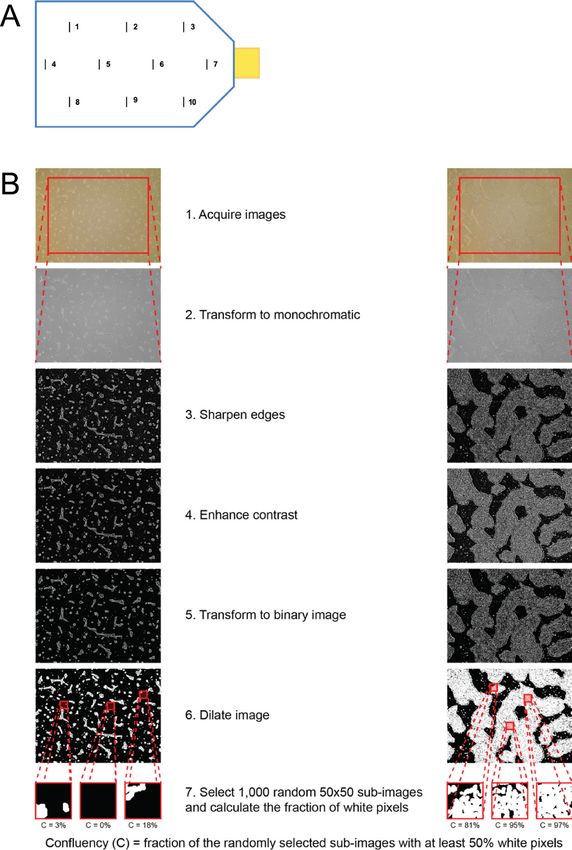
Bio-protocol 10(18): e3755.
                                           www.bio-protocol.org/e3755           DOI:10.21769/BioProtoc.3755

first as spontaneous differentiations by culturing ESCs in medium containing 20% fetal calf serum or by
stimulation with several reagents known to enhance cardiac differentiation like dimethyl sulfoxide,
retinoic acid, or 5-aza-2’-deoxycytidine, after which beating cardiac cells were manually or mechanically
purified (Maltsev et al., 1993; Burridge et al., 2014). Differentiation efficiency was greatly improved by
the development of directed differentiation protocols that incorporated recombinant proteins including
fibroblast growth factor 2, transforming growth factor β, superfamily growth factors activin A and BMP4,
vascular endothelial growth factor and the WNT inhibitor DKK-1 proteins (Schneider and Mercola, 2001;
Marvin et al., 2001; Beqqali et al., 2006; Laflamme et al., 2007), and by modification of the format of cell
differentiation from embryoid bodies to monolayer culture (Paige et al., 2010; Lian et al., 2012). Further
advancements were made by the introduction of small molecule protocols (Lian et al., 2012 and 2013)
and chemically defined differentiation media (Burridge et al., 2014). Finally, by taking advantage of the
adaptation of the developing heart to metabolize lactate, we and others were able to eliminate all non-
cardiac cells (Burridge et al., 2014; D'Antonio-Chronowska et al., 2019b; Tohyama et al., 2013).
Importantly, previous studies have optimized differentiation protocols to derive cardiac cells from a
limited number of iPSC or ESC lines, and in most cases utilized small format culture vessels. Here, we
present an optimized, cost-effective and highly standardized protocol which we applied to derive iPSC-
CVPCs from 154 genetically and ethnically diverse human iPSC lines in large-sized culture flasks. We
optimized the concentration of IWP-2 used to drive the cardiac cell differentiation, which resulted in
improved formation of a thick cardiac syncytium and strong wave-like beating (Video 1) (D'Antonio-
Chronowska et al., 2019b). We also demonstrated that simple mechanical disruption of the cardiac
syncytium prior to metabolic purification of iPSC-CVPCs using lactate results in improved selection and
virtually pure cardiac cells (CMs and EPDCs). Additionally, we developed Cell confluency estimates
(ccEstimate), an automated method for estimation of cell confluency during the monolayer stage.
ccEstimate estimates the point in time for each iPSC line when the monolayer will reach 80% of
confluency, which is optimal condition at which to initiate iPSC-CVPC differentiation. Thereby ccEstimate
overcomes some of the technical issues in standardizing a differentiation protocol across iPSC lines
which have widely varying growth rates. The derived iPSC-CVPCs beat synchronously, are positive for
multiple cardiac markers and can be used directly for molecular or electrophysiological assays like
multielectrode array (MEA), or they can be cryopreserved for future analysis. Our optimized protocol
allowed us to derive high quality iPSC-CVPC samples from 154 iPSC lines generated from ethnically
diverse individuals under identical culturing conditions without the requirement of any individualized
optimization steps.

Materials and Reagents

A. iPSC Cell Culture as described also in detail in D’Antonio-Chronowska et al. (2019)
    1. 6-well plates (Corning, catalog number: 3506)
    2. Syringe filter 0.2 μm (VWR, catalog number: 28145-501)
    3. Soft-Ject® 3-Part Disposable Syringe, Air-Tite-3 ml (VWR, catalog number: 89215-234)

Copyright © 2020 The Authors; exclusive licensee Bio-protocol LLC.                                       3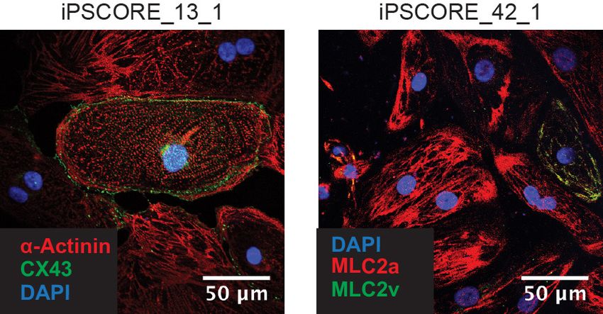
Bio-protocol 10(18): e3755.
                                           www.bio-protocol.org/e3755         DOI:10.21769/BioProtoc.3755

    4. 5 ml Borosilicate serological pipettes (Fisher Scientific, catalog number: 1367827E)
    5. 5 ml Serological pipettes (Bio Pioneer, catalog number: GEX0050-S01)
    6. 10 ml Serological pipettes (Bio Pioneer, catalog number: GEX0100-S01)
    7. P20 pipette tips sterile with filter (Fisher Scientific, catalog number: 21-403-00) or other
         equivalent sterile tips with filter compatible with pipettes
    8. P1000 pipette tips sterile with filter (Fisher Scientific, catalog number: 21-403-02) or other
         equivalent P1000 sterile tips with filter compatible with pipettes
    9. 15 ml conical tubes (Bio Pioneer, catalog number: CNT-15R)
    10. iPSC cells
    11. UltraPureTM DNase/RNase-Free Distilled Water (Thermo Fisher Scientific, catalog number:
         10977023)
    12. Corning® Matrigel® Growth Factor Reduced (GFR) Basement Membrane Matrix (Corning,
         catalog number: 354230)
    13. mTeSRTM 1 (Stem Cell Technologies, catalog number: 85850)
    14. DMEM/F-12 medium (Thermo Fisher Scientific, catalog number: 11330057)
    15. Dispase II, powder (Thermo Fisher Scientific, catalog number: 17105041)
    16. 200 proof Ethanol denatured (i.e., VWR, catalog number: 71002-398)–to prepare 70% ethanol
    17. 70% ethanol (see Recipes: Table 20)
    18. Matrigel solution (Matrigel) (see Recipes: Table 1)
    19. 10 mM ROCK inhibitor, Y-27632 dihydrochloride solution (ROCK Inhibitor) (see Recipes: Table
         3)
    20. 10x Dispase (see Recipes: Table 4)
    21. mTeSRTM 1 complete medium (mTeSR) (see Recipes: Table 5)

B. PSC passaging using Versene–Versene I passage
    1. 6-well plates (Corning, catalog number: 3506)
    2. Automated cell counter slides (Bio-Rad Laboratories, catalog number: 1450019) or a
         hemocytometer (Hausser Scientific, catalog number: 1483) or equivalent
    3. 5 ml Serological pipettes (Bio Pioneer, catalog number: GEX0050-S01)
    4. P20 pipette tips sterile with filter (Fisher Scientific, catalog number: 21-403-00) or other
         equivalent P200 sterile tips with filter compatible with pipettes
    5. P1000 pipette tips sterile with filter (Fisher Scientific, catalog number: 21-403-02) or other
         equivalent P1000 sterile tips with filter compatible with pipettes
    6. 15 ml conical tubes (Bio Pioneer, catalog number: CNT-15R)
    7. iPSC cells
    8. UltraPureTM DNase/RNase-Free Distilled Water (Thermo Fisher Scientific, catalog number:
         10977023)
    9. Corning® Matrigel® Growth Factor Reduced (GFR) Basement Membrane Matrix (Corning,
         catalog number: 354230)

Copyright © 2020 The Authors; exclusive licensee Bio-protocol LLC.                                     4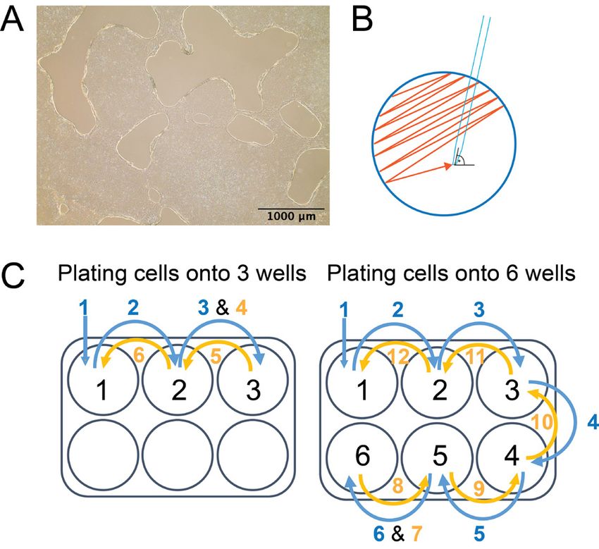
Bio-protocol 10(18): e3755.
                                           www.bio-protocol.org/e3755          DOI:10.21769/BioProtoc.3755

    10. mTeSRTM 1 (Stem Cell Technologies, catalog number: 85850)
    11. DMEM/F-12 medium (Thermo Fisher Scientific, catalog number: 11330057)
    12. Versene® (EDTA) 0.02% (Lonza, catalog number: 17-711E)
    13. 200 proof Ethanol denatured (i.e., VWR, catalog number: 71002-398)–to prepare 70% ethanol
    14. 70% ethanol (see Recipes: Table 20)
    15. Matrigel solution (Matrigel) (see Recipes: Table 1)
    16. 10 mM ROCK inhibitor, Y-27632 dihydrochloride solution (ROCK Inhibitor) (see Recipes: Table
         3)
    17. mTeSRTM 1 complete medium (mTeSR) (see Recipes: Table 5)

C. iPSC passaging using Versene - Versene II passage
    1. 100 mm tissue culture dishes (Corning, catalog number: 430167)
    2. Automated cell counter slides (Bio-Rad Laboratories, catalog number: 1450019) or a
         hemocytometer (Hausser Scientific, catalog number: 1483) or equivalent
    3. 5 ml Serological pipettes (Bio Pioneer, catalog number: GEX0050-S01)
    4. P20 pipette tips sterile with filter (Fisher Scientific, catalog number: 21-403-00) or other
         equivalent sterile tips with filter compatible with pipettes
    5. P1000 pipette tips sterile with filter (Fisher Scientific, catalog number: 21-403-02) or other
         equivalent P1000 sterile tips with filter compatible with pipettes
    6. 15 ml conical tubes (Bio Pioneer, catalog number: CNT-15R)
    7. iPSC cells
    8. UltraPureTM DNase/RNase-Free Distilled Water (Thermo Fisher Scientific, catalog number:
         10977023)
    9. Corning® Matrigel® Growth Factor Reduced (GFR) Basement Membrane Matrix (Corning,
         catalog number: 354230)
    10. mTeSRTM 1 (Stem Cell Technologies, catalog number: 85850)
    11. DMEM/F-12 medium (Thermo Fisher Scientific, catalog number: 11330057)
    12. Versene® (EDTA) 0.02% (Lonza, catalog number: 17-711E)
    13. 200 proof Ethanol denatured (i.e., VWR, catalog number: 71002-398)–to prepare 70% ethanol
    14. 70% ethanol (see Recipes: Table 20)
    15. Matrigel solution (Matrigel) (see Recipes: Table 1)
    16. 10 mM ROCK inhibitor, Y-27632 dihydrochloride solution (ROCK Inhibitor) (see Recipes: Table
         3)
    17. mTeSRTM 1 complete medium (mTeSR) (see Recipes: Table 5)

D. Monolayer plating is also described in detail in D’Antonio-Chronowska et al. (2019a)
    1. 100 mm tissue culture dishes (Corning, catalog number: 430167)
    2. Automated cell counter slides (Bio-Rad Laboratories, catalog number: 1450019) or a
         hemocytometer (Hausser Scientific, catalog number: 1483) or equivalent

Copyright © 2020 The Authors; exclusive licensee Bio-protocol LLC.                                      5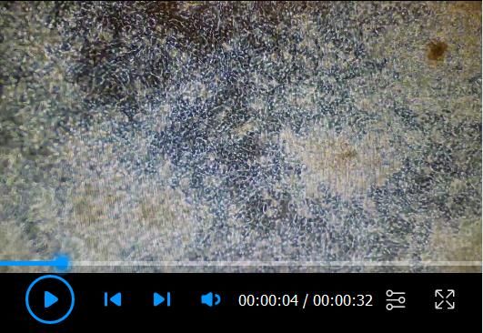
Bio-protocol 10(18): e3755.
                                           www.bio-protocol.org/e3755         DOI:10.21769/BioProtoc.3755

     3. 5 ml Serological pipettes (Bio Pioneer, catalog number: GEX0050-S01)
     4. 10 ml Serological pipettes (Bio Pioneer, catalog number: GEX0100-S01)
     5. P20 pipette tips sterile with filter (Fisher Scientific, catalog number: 21-403-00) or other
         equivalent P20 sterile tips with filter compatible with pipettes
     6. P200 pipette tips sterile with filter (Fisher Scientific, catalog number: 21-403-01) or other
         equivalent P200 sterile tips with filter compatible with pipettes
     7. P1000 pipette tips sterile with filter (Fisher Scientific, catalog number: 21-403-02) or other
         equivalent P1000 sterile tips with filter compatible with pipettes
     8. 15 ml conical tubes (Bio Pioneer, catalog number: CNT-15R)
     9. 50 ml conical tubes (Bio Pioneer catalog number: CNT-50R)
     10. Corning® Matrigel® Growth Factor Reduced (GFR) Basement Membrane Matrix (Matrigel)
         (Corning, catalog number: 354230)
     11. mTeSRTM 1 (Stem Cell Technologies, catalog number: 85850)
     12. DMEM/F-12 medium (Thermo Fisher Scientific, catalog number: 11330-057)
     13. Accutase (Innovative Cell Technologies, Inc., catalog number: AT 104)
     14. Trypan Blue Solution, 0.4% (Thermo Fisher Scientific, catalog number: 15250061)
     15. ROCK inhibitor, Y-27632 dihydrochloride (Selleck hem, catalog number: S1049)
     16. iPSC cell culture
     17. 200 proof Ethanol denatured (i.e., VWR, catalog number: 71002-398)–to prepare 70% ethanol
     18. 70% ethanol (see Recipes: Table 20)
     19. Matrigel solution (see Recipes: Table 1)
     20. 10 mM ROCK inhibitor, Y-27632 dihydrochloride solution (see Recipes: Table 3)
     21. mTeSRTM 1 complete medium (see Recipes: Table 5)

E. Estimation of optimal time for initiation of iPSC-CVPCs differentiation using ccEstimate
     1. iPSCs monolayer
     2. Marker pen resistant to 70% ethanol

F.   iPSC-CVPCs differentiation
     1. T150 tissue culture flasks, vented (Sigma, catalog number: Z707929)
         Note: At the time of preparation of this manuscript Z707929 was no longer available. The same
         flasks are available under the catalog number Z707511-36EA (Sigma, catalog number:
         Z707511-36EA).
     2. Automated cell counter slides (Bio-Rad Laboratories, catalog number: 1450019) or a
         hemocytometer (Hausser Scientific, catalog number: 1483) or equivalent
     3. 10 ml Serological pipettes (Bio Pioneer, catalog number: GEX0100-S01)
     4. 25 ml Serological pipettes (Bio Pioneer catalog number: GEX250-S01)
     5. 50 ml Serological pipettes (Bio Pioneer, catalog number: GEX500-S01)
     6. P20 pipette tips sterile with filter (Fisher Scientific, catalog number: 21-403-00) or other

Copyright © 2020 The Authors; exclusive licensee Bio-protocol LLC.                                     6
Bio-protocol 10(18): e3755.
                                           www.bio-protocol.org/e3755         DOI:10.21769/BioProtoc.3755

         equivalent P20 sterile tips with filter compatible with pipettes
    7. P200 pipette tips sterile with filter (Fisher Scientific, catalog number: 21-403-01) or other
         equivalent P200 sterile tips with filter compatible with pipettes
    8. P1000 pipette tips sterile with filter (Fisher Scientific, catalog number: 21-403-02) or other
         equivalent P1000 sterile tips with filter compatible with pipettes
    9. Cell scraper (VWR International, catalog number: 179707)
    10. 15 ml conical tubes (Bio Pioneer, catalog number: CNT-15R)
    11. 50 ml conical tubes (Bio Pioneer, catalog number: CNT-50R)
    12. 125 ml Nalgene® PET sterile bottle (Fisher Scientific, catalog number: 342040-0125)
    13. Nalgene® Cryogenic vials (Thermo Fisher Scientific, catalog number: 5000-1020)
    14. iPSCs monolayer
    15. Corning® Matrigel® Growth Factor Reduced (GFR) Basement Membrane Matrix (Matrigel)
         (Corning®, catalog number: 354230)
    16. RPMI 1640 medium (Thermo Fisher Scientific, catalog number: 11875119)
    17. RPMI 1640 medium, no glucose (Thermo Fisher Scientific, catalog number: 11879020)
    18. 1x Dulbecco’s phosphate buffered saline (DPBS; PBS) without calcium and magnesium
         (Thermo Fisher Scientific, catalog number: 14190250)
    19. B-27TM Supplement, minus insulin (Thermo Fisher Scientific, catalog number: A1895601)
    20. B-27TM Supplement (50x), serum free (Thermo Fisher Scientific, catalog number: 17504044)
    21. FBS (Omega Scientific, catalog number: FB-02) or equivalent, or KnockOutTM Serum
         Replacement (KOSR) (Thermo Fisher Scientific, catalog number: 10828028)
    22. MEM Non-Essential Amino Acids Solution 100x (Thermo Fisher Scientific, catalog number:
         11140050)
    23. Penicillin-Streptomycin (10,000 U/ml) (Thermo Fisher Scientific, catalog number: 15140122)
    24. CHIR-99021 (CT99021) (CHIR99021) HCl (Selleckchem, catalog number: S2924)
    25. IWP-2 (Tocris, catalog number: 3533)
    26. Sodium L-lactate (Sigma, catalog number: 71718-10G)
    27. HEPES sodium salt solution 1M, BioReagent, suitable for cell culture (Sigma, catalog number:
         H3662-100ML)
    28. Accutase (Innovative Cell Technologies, Inc., catalog number: AT 104)
    29. Trypan Blue Solution, 0.4% (Thermo Fisher Scientific, catalog number: 15250061)
    30. Dimethyl Sulfoxide (DMSO) (Sigma, catalog number: D2650-100ML)
    31. Liquid nitrogen
    32. 200 proof Ethanol denatured (i.e., VWR, catalog number: 71002-398) – to prepare 70% ethanol
    33. 70% ethanol (see Recipes: Table 20)
    34. RPMI Minus (-) medium (see Recipes: Table 6)
    35. RPMI Plus (+) medium (see Recipes: Table 7)
    36. RPMI Lactate medium (see Recipes: Table 8)
    37. iPSC-CVPCs Harvest Medium (see Recipes: Table 9)

Copyright © 2020 The Authors; exclusive licensee Bio-protocol LLC.                                     7
Bio-protocol 10(18): e3755.
                                           www.bio-protocol.org/e3755          DOI:10.21769/BioProtoc.3755

    38. iPSC-CVPCs 2x freezing medium (see Recipes: Table 10)
    39. 10 mM CHIR-992021 solution (see Recipes: Table 11)
    40. 5 mM IWP-2 solution (see Recipes: Table 12)
    41. 1M Sodium L-lactate solution (see Recipes: Table 13)

G. Flow cytometry
    1. 96-well round bottom assay plates (Genesee Scientific, catalog number: 25-224)
    2. CorningTM FalconTM Test Tube with Cell Strainer Snap Cap (Fisher Scientific, catalog number:
         352235)
    3. CorningTM CostarTM Sterile Disposable Reagent Reservoirs (Fisher Scientific, catalog number:
         4870)
    4. 5 ml Serological pipettes (Bio Pioneer, catalog number: GEX0050-S01)
    5. 10 ml Serological pipettes (Bio Pioneer, catalog number: GEX0100-S01)
    6. P20 pipette tips sterile with filter (Fisher Scientific, catalog number: 21-403-00) or other
         equivalent P20 sterile tips with filter compatible with pipettes
    7. P200 pipette tips sterile with filter (Fisher Scientific, catalog number: 21-403-01) or other
         equivalent P200 sterile tips with filter compatible with pipettes
    8. P1000 pipette tips sterile with filter (Fisher Scientific, catalog number: 21-403-02) or other
         equivalent P1000 sterile tips with filter compatible with pipettes
    9. P20 sterile pipette tips without filter (VWR, catalog number: 83009-694) or other equivalent P20
         sterile tips without filter compatible with pipettes
    10. (Optional) P200 sterile pipette tips without filter (VWR, catalog number: 89495-378) or other
         equivalent P20 sterile tips without filter compatible with pipettes
    11. 1x Dulbecco’s phosphate buffered saline (PBS) without calcium and magnesium (Thermo Fisher
         Scientific, catalog number: 14190250)
    12. Methanol, ACS reagent, ≥ 99.8% (Sigma, catalog number: 179337-4L-PB)
    13. Bovine Serum Albumin (BSA) (Sigma, catalog number: A2153-100G)
    14. (Optional) NaN3 (Sigma, catalog number: S2002-5G)
    15. 37% Formaldehyde (Sigma, catalog number: F-1635-500ML)
    16. TritonTM X-100 (Sigma, catalog number: X-100-500ML)
    17. Goat serum, New Zealand origin (Thermo Fisher Scientific, catalog number: 16210064)
    18. Mouse monoclonal anti-Troponin T, Cardiac Isoform (cTNT; TNNT2) Ab-1antibody, clone 13-11
         (Thermo Fisher Scientific, catalog number: MS-295-P0)
    19. Mouse IgG1 antibody (Thermo Fisher Scientific, catalog number: MG100)
    20. Goat-anti-Mouse Alexa FluorTM 488 conjugated antibody (Thermo Scientific, catalog number: A-
         11001)
    21. FACS Buffer (see Recipes: Table 14)
    22. FACS-FIX Buffer (see Recipes: Table 16)
    Note: For antibody working concentration, see Recipes: Table 21.

Copyright © 2020 The Authors; exclusive licensee Bio-protocol LLC.                                      8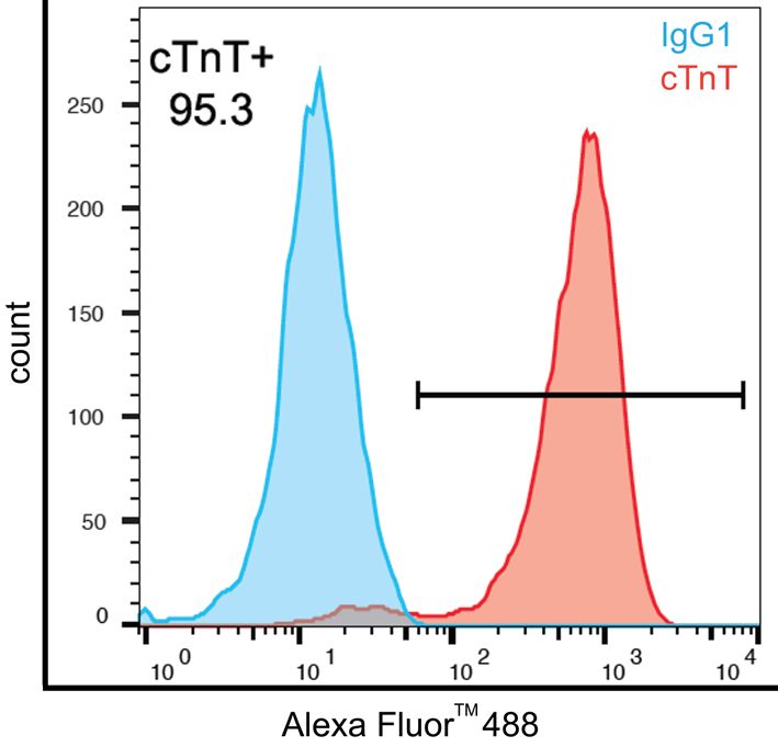
Bio-protocol 10(18): e3755.
                                           www.bio-protocol.org/e3755        DOI:10.21769/BioProtoc.3755

H. Immunofluorescence
    1. Millicell EZ SLIDE 8-well glass slides (Millipore, catalog number: PEZGS0816)
    2. Coverslip glass slides (Fisherbrand, catalog number: 12-545-F [coverslip thickness #1]).
         Different coverslip thickness may be used if required
    3. 5 ml Serological pipettes (Bio Pioneer, catalog number: GEX0050-S01)
    4. 10 ml Serological pipettes (Bio Pioneer, catalog number: GEX0100-S01)
    5. P20 pipette tips sterile with filter (Fisher Scientific, catalog number: 21-403-00) or other
         equivalent P20 sterile tips with filter compatible with pipettes
    6. P200 pipette tips sterile with filter (Fisher Scientific, catalog number: 21-403-01) or other
         equivalent P200 sterile tips with filter compatible with pipettes
    7. 1x Dulbecco’s phosphate buffered saline (PBS) without calcium and magnesium (Thermo Fisher
         Scientific, catalog number: 14190250)
    8. Bovine Serum Albumin (BSA) (Sigma, catalog number: A2153-100G)
    9. Paraformaldehyde (PFA) (Sigma, catalog number: 158127-100G)
    10. Tween® 20 (Sigma, catalog number: P9416-100ML)
    11. TritonTM X-100 (Sigma, catalog number: X100-500ML)
    12. Gelatin from porcine skin (Sigma, catalog number: G1890-100G)
    13. Mouse monoclonal anti-α-Actinin (Sarcomeric) antibody, clone EA-53 (Sigma, catalog number:
         A7811)
    14. Rabbit polyclonal anti-Connexin 43 (CX43/GJA1) antibody (Invitrogen, catalog number: 71-
         0700)
    15. Rabbit polyclonal anti-Myosin Light Chain 2 (MYL2; MLC2v) antibody (Proteintech, catalog
         number: 10906-1-AP)
    16. Mouse monoclonal anti-Myosin Atrial Light Chain 2 (MYL7; MLC2a) antibody clone 56F5
         (Synaptic Systems, catalog number: 311 011)
    17. Donkey anti-Rabbit Alexa FluorTM 488 conjugated antibody (Invitrogen, catalog number: A21206)
    18. Goat anti-Mouse Alexa FluorTM 568 conjugated antibody (Invitrogen, catalog number: A-11004)
    19. ProLong® Gold Antifade Reagent with DAPI (Cell Signaling Technologies, catalog number: 8961)
    20. 0.1% Gelatin solution (see Recipes: Table 2)
    21. 4% PFA solution in PBS
    22. IF Perm Buffer II (see Recipes: Table 17)
    23. IF Blocking Buffer II (see Recipes: Table 18)
    24. IF Staining Buffer (see Recipes: Table 19)
    Note: For antibody working concentration, see Recipes: Table 21.

Equipment

A. iPSC Cell Culture
    1. Biosafety cabinet (Labconco, model: Logic+)

Copyright © 2020 The Authors; exclusive licensee Bio-protocol LLC.                                    9
Bio-protocol 10(18): e3755.
www.bio-protocol.org/e3755 DOI:10.21769/BioProtoc.3755
2. Incubator with humidity and gas control set to maintain 37 °C and 95% humidity in an atmosphere
of 5% CO2 in air (Panasonic, model: MCO-170AICUVH-PA)
3. Water bath (Thermo Scientific, model: Precision)
4. Tissue culture centrifuge with rotors for 15 ml conical tubes and 50 ml conical tubes (Thermo
Scientific, model: Legend RT+)
5. Phase contrast inverted microscope (objectives: x4, x10, x20) (Olympus, model: CKX41SF)
6. (Optional) Phase contrast inverted microscope with camera (objectives: x4, x10, x20) (Thermo
Scientific, model: EVOS XL Core)
7. Microscope Object marker (Nikon, model: MBW10020)
8. Pipette aid (Labnet, catalog number: FastPetteTM V2 P2000) or other equivalent available
pipette aid
9. P20 Micropipette (Rainin, catalog number: 17014392) or other available P20 pipette
10. Non-frost-free freezer -20 °C
11. Refrigerator 2-8 °C
B. iPSC passaging using Versene (Versene I and Versene II passage)
1. Biosafety cabinet (Labconco, model: Logic+)
2. Incubator with humidity and gas control set to maintain 37 °C and 95% humidity in an atmosphere
of 5% CO2 in air (Panasonic, model: MCO-170AICUVH-PA)
3. Tissue culture centrifuge with rotors for 15 ml conical tubes and 50 ml conical tubes (Thermo
Scientific, model: Legend RT+)
4. Phase contrast inverted microscope (objectives: x4, x10, x20) (Olympus, model: CKX41SF)
5. (Optional) Phase contrast inverted microscope with camera (objectives: x4, x10, x20) (Thermo
Scientific, model: EVOS XL Core)
6. Pipette aid (Labnet, catalog number: FastPetteTM V2 P2000) or other equivalent available
pipette aid
7. P20 Micropipette (Rainin, catalog number: 17014392) or other available P20 pipette
8. P200 Micropipette (Rainin, catalog number: 17014391) or other available P200 pipette
9. P1000 Micropipette (Rainin, catalog number: 17014382) or other available P1000 pipette
10. Automated cell counter (Bio-Rad, model: TC20) or a hemocytometer (Hausser Scientific, catalog
number: 1483) or equivalent
11. Non-frost-free freezer -20 °C
12. Refrigerator 2-8 °C
C. Monolayer plating
1. Biosafety cabinet (Labconco, model: Logic+)
2. Incubator with humidity and gas control set to maintain 37 °C and 95% humidity in an atmosphere
of 5% CO2 in air (Panasonic, model: MCO-170AICUVH-PA)
3. Tissue culture centrifuge with rotors for 15 ml conical tubes and 50 ml conical tubes (Thermo
Copyright © 2020 The Authors; exclusive licensee Bio-protocol LLC. 10Bio-protocol 10(18): e3755.
www.bio-protocol.org/e3755 DOI:10.21769/BioProtoc.3755
Scientific, model: Legend RT+)
4. Phase contrast inverted microscope (objectives: x4, x10, x20) (Olympus, model: CKX41SF)
5. (Optional) Phase contrast inverted microscope with camera (objectives: x4, x10, x20) (Thermo
Scientific, model: EVOS XL Core)
6. Pipette aid (Labnet, catalog number: FastPetteTM V2 P2000 or other equivalent available pipette
aid
7. P20 Micropipette (Rainin, catalog number: 17014392) or other available P20 pipette
8. P200 Micropipette (Rainin, catalog number: 17014391) or other available P200 pipette
9. P1000 Micropipette (Rainin, catalog number: 17014382) or other available P1000 pipette
10. Automated cell counter (Bio-Rad, model: TC20) or a hemocytometer (Hausser Scientific, catalog
number: 1483) or equivalent
11. Non-frost-free freezer -20 °C
12. Refrigerator 2-8 °C
D. iPSC-CVPCs differentiation and cryopreservation
1. Biosafety cabinet (Labconco, model: Logic+)
2. Incubator with humidity and gas control set to maintain 37 °C and 95% humidity in an atmosphere
of 5% CO2 in air (Panasonic, model: MCO-170AICUVH-PA)
3. Tissue culture centrifuge with rotors for 15 ml conical tubes and 50 ml conical tubes (Thermo
Scientific, model: Legend RT+)
4. Phase contrast inverted microscope (objectives: x4, x10, x20) (Olympus, model: CKX41SF)
5. (Optional) Phase contrast inverted microscope with camera (objectives: x4, x10, x20) (Thermo
Scientific, model: EVOS XL Core)
6. Pipette aid (Labnet, catalog number: FastPetteTM V2 P2000) or other equivalent available
pipette aid
7. P20 Micropipette (Rainin, catalog number: 17014392) or other available P20 pipette
8. P200 Micropipette (Rainin, catalog number: 17014391) or other available P200 pipette
9. P1000 Micropipette (Rainin, catalog number: 17014382) or other available P1000 pipette
10. Automated cell counter (Bio-Rad, model: TC20) or a hemocytometer (Hausser Scientific, catalog
number: 1483) or equivalent.
11. Mr. Frosty freezing container (Corning, model: CoolCell® FTS30)
12. Refrigerator 2-8 °C
13. Non-frost-free freezer -20 °C
14. Freezer -80 °C
15. Liquid nitrogen vapor tank
E. Estimation of optimal time for initiation of iPSC-CVPCs differentiation using ccEstimate
1. Phase contrast inverted microscope with camera (objective: x4) (Thermo Scientific, model:
EVOS XL Core) or equivalent or automatic imaging system
Copyright © 2020 The Authors; exclusive licensee Bio-protocol LLC. 11Bio-protocol 10(18): e3755.
www.bio-protocol.org/e3755 DOI:10.21769/BioProtoc.3755
2. Any computer with R 3.5.1 and the R package EBImage (Pau et al., 2010) installed and 4GB
RAM
F. Flow cytometry
1. Pipette aid (Labnet, catalog number: FastPetteTM V2 P2000) or other equivalent available
pipette aid
2. P20 Micropipette (Rainin, catalog number: 17014392) or other available P20 pipette
3. P200 Micropipette (Rainin, catalog number: 17014391) or other available P200 pipette
4. P1000 Micropipette (Rainin, catalog number: 17014382) or other available P1000 pipette
5. P200 Multichannel micropipette (Rainin, catalog number: 17013805) or other available
multichannel P200 pipette
6. Tissue culture centrifuge with rotors suitable to centrifuge 96-well plates (Thermo Scientific,
model: Legend RT+)
7. Refrigerator 2-8 °C
8. Non-frost-free freezer -20 °C
9. Flow cytometer (BD Biosciences, model: FACSCanto II) or equivalent.
G. Immunofluorescence
1. Pipette aid (Labnet, catalog number: FastPetteTM V2 P2000) or other equivalent available
pipette aid
2. P20 Micropipette (Rainin, catalog number: 17014392) or other available P20 pipette
3. P200 Micropipette (Rainin, catalog number: 17014391) or other available P200 pipette
4. P1000 Micropipette (Rainin, catalog number: 17014382) or other available P1000 pipette
5. Refrigerator 2-8 °C
6. Non-frost-free freezer -20 °C
7. Confocal laser scanning fluorescence microscope (Olympus, FluoView1000)
Software
1. FlowJo (Version 10) (FlowJo, LLC, https://www.flowjo.com/)
2. FlowView ASW V03.01.03.03 or V4.2a (Olympus Life Science, https://www.olympus-
lifescience.com/en/support/downloads/)
3. ccEstimate available on request
4. R 3.5.1
5. R package EBImage (Pau et al., 2010)
Copyright © 2020 The Authors; exclusive licensee Bio-protocol LLC. 12Bio-protocol 10(18): e3755.
www.bio-protocol.org/e3755 DOI:10.21769/BioProtoc.3755
Procedure
A. iPSC cell culture is also described in detail in D’Antonio-Chronowska et al. (2019a)
1. Thaw iPSC cells
a. Prepare 12 ml of mTeSR containing 10 μM ROCK Inhibitor.
b. Transfer 9 ml of mTeSR containing 10 μM ROCK Inhibitor into a sterile conical tube labeled
with the name of the line.
c. Remove vial of cryopreserved cells from liquid nitrogen tank. Keep vial on dry ice.
d. Place and shake gently in a 37 °C water bath until a pea-sized ice crystal remains (around
2 min).
e. Wipe off excess water from the vial, spray with 70% ethanol before placing in the hood.
f. Remove thawed cells from the vial and add gently into 9 ml mTeSR containing 10 μM ROCK
Inhibitor in a conical tube. Wash the vial with 1-2 ml of mTeSR containing 10 μM ROCK
Inhibitor. Collect all cells in the same conical tube.
g. Centrifuge cells for 5 min at 53 x g (500 RPM in a Sorvall 75006445 rotor with 75006441 K
buckets) at room temperature.
h. Aspirate supernatant, and gently resuspend cell pellet in 2 ml of mTeSR containing 10 μM
ROCK Inhibitor (1 cryovial is thawed into 1 well of 6-well plate).
Note: The iPSC cells are cryopreserved in clumps and cannot be counted. Therefore, we
recommend to cryopreserve cells from 1 well of a 6-well plate into 1 cryovial, and then to
thaw cells from one cryovial onto 1 well of a 6-well plate.
i. Label a Matrigel plate with name of line, clone and passage number. Aspirate DMEM/F-12
from Matrigel-coated plate. Add +1 to the passage number after thawing.
Note: Do not add +1 to the passage number if the passage number was increased during
cryopreservation of iPSCs.
j. Plate cells resuspended in 2 ml into one well of a Matrigel-coated 6-well plate (final volume
2 ml/well).
k. 24 h after plating, observe cells. Wash cells gently with DMEM/F-12 (2 ml/well) to remove
cell debris and feed using fresh mTeSR medium without ROCK Inhibitor (2 ml/well).
l. Daily, observe the iPSCs, remove the differentiated cells, and change the medium with fresh
mTeSR (2 ml/well).
Note: It is critical to maintain iPSC culture differentiation free. For details on how to mark
and remove differentiated iPSC cells, please refer below to procedure: 2. iPSC passaging
using Dispase (Steps A2c-A2d) and Figure 1.
m. Cells should reach 80-90% of confluency and be ready for passage in about 5 days.
iPSCs can be passaged using multiple methods, depending on the needs. For routine iPSC
culture and expansion, cells should be passaged using Dispase, which cleaves the proteins of
extracellular matrix used for iPSC culture (Matrigel). Dispase will dissociate iPSCs into clumps
Copyright © 2020 The Authors; exclusive licensee Bio-protocol LLC. 13Bio-protocol 10(18): e3755.
www.bio-protocol.org/e3755 DOI:10.21769/BioProtoc.3755
of cells, which is a gentle method of iPSC expansion and maintenance. We recommend
passaging iPSC with Dispase at the ratio 1:2 (one well split onto 2 wells) or 1:3 (one well split
onto 3 wells), which allows to easily schedule iPSC cell maintenance. Cells passaged with
Dispase should reach about 80% confluency in about 4-5 days. Other ratios also can be used
(i.e., 1:1 or 1:4), however we recommend testing it for individual lines.
When iPSC cells have to be expanded at much higher ratios or using a defined cell number,
iPSC should be dissociated as single cells using Versene or Accutase. Please note that single
cell passage is very stressful for iPSCs and leads to cell apoptosis. To prevent dissociation-
induced apoptosis of iPSC, reagents like ROCK inhibitor, Y-27632 dihydrochloride (or others)
have to be used. We do not recommend dissociating iPSCs into single cells for routine
expansion or prior to cryopreservation.
Versene is a non-enzymatic, gentle method of single cell dissociation which acts by chelating
metal ions (mainly magnesium and calcium) required by integrins to maintain cell-cell and cell-
extracellular matrix contacts. We have routinely expanded over 150 different iPSC lines with
Versene prior to plating large-scale monolayers, however we do not recommend dissociating
cells using Versene in more than two consecutive passages. Accutase is an enzymatic reagent
that allows for very efficient single cell dissociation. Although Accutase is a gentle reagent,
commonly used for iPSC, we recommend using Accutase only for monolayer plating.
Notes:
a. It is critical for a successful iPSC-CVPCs differentiation to use heathy and pluripotent iPSC
cells and use iPSC which were culture was maintained free of differentiation.
b. Optimal cell number will vary depending on the scale of differentiation. Differentiation at the
scale described in this protocol requires that cells are expanded gradually, by performing
two Versene passages (Passages: Versene I and Versene II).
2. iPSC passaging using Dispase
a. Prepare 1x (2 mg/ml) Dispase solution by adding 9 ml DMEM/F-12 to 1 ml of 10x Dispase
(20 mg/ml).
b. Allow 1x Dispase solution to come to room temperature.
Note: 1x Dispase solution can be stored at 4 °C for maximum 2 weeks.
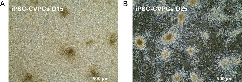
Mark any areas of differentiation in the well(s) to be split, using the Microscope Object
marker refer to Figure 1 for representative image of healthy pluripotent colonies (A) and
differentiated colonies which need to be removed (B).
c. Aspirate spent media. Aspirate marked areas of differentiation, if any, by gently tapping a
Pasteur pipette within the marked circle (Figure 1C). Wash with 2 ml of DMEM/F-12 per well.
Copyright © 2020 The Authors; exclusive licensee Bio-protocol LLC. 14Bio-protocol 10(18): e3755.
www.bio-protocol.org/e3755 DOI:10.21769/BioProtoc.3755
Figure 1. Pluripotent and differentiated iPSC colonies. A. Healthy and pluripotent iPSC
colonies fuse into larger colonies: before fusion (left and middle) and after fusion (right).
Healthy and pluripotent iPSC colonies have a smooth, flat, colony surface, are very compact
with tightly growing cells that have almost invisible cell membranes (borders between cells),
and uniform, well defined, colony edges. Cells have high nucleus:cytoplasm ratios with clearly
visible nucleoli. Almost any deviation from this morphology may suggest loss of pluripotency
and may indicate unwanted differentiation. B. Examples of differentiated iPSC cell which need
to be removed from the culture. Black arrows indicate differentiated cells. Cells of various iPSC
lines may have diverse morphology upon differentiation and these are only a few
representative examples. C. Differentiated colony has been marked in purple dye using
Microscope Object marker (left) and aspirated with a Pasteur pipette (right).
d. Add 1 ml of 1x Dispase in each well to be split. Incubate at 37 °C for 5 min.
e. Check morphology of colonies after 5 min.
When edges of the colonies are slightly curled up, cells are ready to be passaged. If edges
of colonies are not curled up, incubate cells at 37 °C for another 1-2 min. Do not incubate
with Dispase for longer than 8 min. Refer to Figure 2A for representative image of colonies
with edges curled up.
f. Aspirate Dispase from all wells.
Note: iPSC colonies passaged with Dispase will remain fully attached to the well. Only the
edges of the colonies will be slightly curled up indicating that cells are ready for the next
steps.
g. Rinse the wells gently 3 times with DMEM/F12 (2 ml/well).
Copyright © 2020 The Authors; exclusive licensee Bio-protocol LLC. 15Bio-protocol 10(18): e3755.
www.bio-protocol.org/e3755 DOI:10.21769/BioProtoc.3755
h. Add 1 ml of mTeSR media to each well to be passaged.
i. Use a glass serological pipette to detach colonies. Hold the pipette at a 90° angle to the
surface of the plate. Scrape across the surface of the 6-well plate in the motion outlined in
Figure 2B (start from top left side of the well and zig-zag tightly down to bottom-right side,
then turn plate clockwise or counterclockwise and scrape again). Scrape until at least 90%
of the colonies are detached from the well.
j. Wash plate with the volume of mTeSR required to bring cells up to the final volume needed
to seed a new Matrigel-coated vessel. Calculate the final volume considering 2 ml per each
well to be seeded with passaged cells. For example, if cells are to be passaged 1 to 3 the
final volume will be 6 ml, therefore the volume of mTeSR used to wash the plate is 5 ml.
k. Seed cells on a new Matrigel plate plating 1 ml of cell suspension per well and then add 1
more ml of cell suspension to each well (Figure 2C). Plate cells dropwise across the entire
surface of the well to ensure uniform plating.
l. Observe seeded cells under microscope to ensure even plating.
m. Place in a 37 °C incubator. Shake the plate in T-shape to homogenously distribute the
colonies pieces in the well.
n. Twenty-four hours after plating gently, wash cells with DMEM/F-12 before adding fresh
mTeSR medium.
Note: For a healthy and efficient iPSC culture, it is critical to plate cells uniformly. Plate cells
uniformly across the entire surface of the well and, when plating multiple wells plate cells
uniformly across all wells.
Figure 2. Passaging iPSC with Dispase. A. Edges of the iPSCORE_2_1 iPSC colonies
are curled up after 5 min incubation with Dispase. B. Pattern of movement of a glass
serological pipette during the iPSCs passaging with Dispase. After scraping the well in one
direction, turn the plate clockwise or counterclockwise by 90° and scrape remaining iPSC
colonies again. About 90% cells should be detached from the well. C. Example of plating
Copyright © 2020 The Authors; exclusive licensee Bio-protocol LLC. 16Bio-protocol 10(18): e3755.
www.bio-protocol.org/e3755 DOI:10.21769/BioProtoc.3755
cells when passaging iPSC cells from 2 wells onto 6 wells. Add 1 ml of cell suspension into
each well following a pattern: when plating cells onto three wells: 1-2-3-3-2-1 or when plating
cells onto six wells: 1-2-3-4-5-6-6-5-4-3-2-1. Please refer to the section on iPSC passaging
using Dispase for details. (Described also in detail in D’Antonio-Chronowska et al., 2019a)
B. Passaging iPSC using Versene (Versene I)
Notes:
a. After thawing an iPSC line, passage cells with Dispase at least once before passaging cells with
Versene.
b. Optimal cell number will vary depending on the scale of iPSC-CVPC differentiation.
Differentiation at the scale described in this protocol requires cells from 1-2 wells for Versene I
passage.
1. Remove 6-well plates from the incubator. When iPSC cells are at around 80% confluency (cells
are ready for a passage), iPSC cells are ready for Versene I passage. Mark all differentiated
cells, which need to be removed (Figure 1).
2. Aspirate the spent medium. Remove all marked differentiated cells and wash cells with
DMEM/F-12 (2 ml/well).
3. Aspirate DMEM/F-12 and add 1 ml of room temperature Versene® (EDTA) 0.02% to well of a 6-
well plate. Incubate cell for 5 min at 37 °C.
4. After 5 min of incubation check cells under microscope to ensure the cells are ready–individual
cell borders should be visible. (If the cells are not ready, return cells to incubator and allow cells
to act for another 30 s-1 min. Do not allow Versene incubation exceed total of 8 min.
5. Gently aspirate the Versene from each well. DO NOT wash cells with DMEM/F12.
Note: Individual cells incubated with Versene will be clearly visible. Cells will remain attached to
the surface of the well however the plate should be handled gently.
6. Add 1 ml per well of mTeSR containing 5 μM ROCK inhibitor and re-suspend cells as single
cells without scraping plate surface, by pipetting up and down using a P1000 pipette. Pipette
cells 10-12 times, turn the dish by 180° (upside down) and pipette 5 more times. Collect cells
from all wells in a 15 ml conical tube. You should not see any cell clumps.
7. Wash well (wells) twice with 3 ml of mTeSR containing 5 μM ROCK Inhibitor. Collect all cells in
the same conical tube.
8. Centrifuge the cells at 53 x g (500 RPM in a Sorvall 75006445 rotor with 75006441 K buckets)
for 5 min at room temperature. Aspirate the supernatant and resuspend cells in 3-5 ml of mTeSR
containing ROCK Inhibitor.
9. Mix the pooled cell suspension by inverting 20 times or more if necessary. Perform the live cell
count using 0.4% Trypan Blue Solution.
Note: iPSC cell viability should not be lower than 80%.
Copyright © 2020 The Authors; exclusive licensee Bio-protocol LLC. 17Bio-protocol 10(18): e3755.
www.bio-protocol.org/e3755 DOI:10.21769/BioProtoc.3755
10. Prepare required number of cells. For a Versene I passage on three wells of a 6-well plate,
prepare 7 ml of cell suspension containing 1.05 x 10⁶ cells in a 15 ml conical tube. Mix cell
suspension very well by inverting the tube 20 times.
Note: Optimal cell numbers will vary depending on the scale of differentiation. iPSC-CVPCs
differentiated at the scale described in this protocol require plating 9.0 x 10⁵ live cells onto three
wells of a 6-well plate during the Versene I passage (3.0 x 10⁵ live cells per each well of a 6-
well plate).
11. Add 2 ml of cell suspension per each of the three well of a 6-well plate. Add cells dropwise using
a 5 or 10 ml pipette.
Note: It is critical to plate cells uniformly on the entire surface of the plate. To help distribute the
cells uniformly plate one dish at the time and shake the newly plated dish in a cross shape (T-
shape).
12. Place plates in the incubator without stacking the plates. Incubate the cells until next morning,
at 37 °C, 5% CO2.
13. Next day change medium for fresh mTeSR without ROCK inhibitor (2 ml/well). iPSC after
Versene I passaged requires culturing cells for about 3-4 days until the culture reaches 80%
confluency. Change medium daily with fresh mTeSR. Maintain differentiation-free iPSC culture.
C. Passaging iPSC using Versene (Versene II)
Note: Optimal cell number will vary depending on the scale of differentiation. iPSC-CVPCs
differentiation at the scale described in this protocol requires cells from 3 wells for Versene II
passage.
1. Remove 6-well plates from the incubator. When iPSC cells are at around 80% confluency (cells
are ready for a passage), iPSC cells are ready for Versene II passage. Mark all differentiated
cells, which need to be removed (Figure 1).
2. Aspirate the spent medium. Remove all marked differentiated cells and wash cells with
DMEM/F-12 (2 ml/well).
3. Aspirate DMEM/F-12 and add 1 ml of room temperature Versene® (EDTA) 0.02% to well of a 6-
well plate. Incubate cell for 5 min at 37 °C.
4. After 5 min of incubation check cells under microscope to ensure the cells are ready–individual
cell borders should be visible. (If the cells are not ready, return cells to incubator and allow cells
to act for another 30 s-1 min. Do not allow Versene incubation exceed total of 8 min.
5. Gently aspirate the Versene from each well. DO NOT wash cells with DMEM/F12
6. Add 1 ml per well of mTeSR containing 5 μM ROCK inhibitor and re-suspend cells as single
cells without scraping plate surface, by pipetting up and down using a P1000 pipette. Pipette
cells 10-12 times, turn the dish by 180° (upside down) and pipette 5 more times. Collect cells
from all wells in a 15 ml conical tube. You should not see any cell clumps.
7. Wash all wells twice with 5 ml of mTeSR containing 5 μM ROCK Inhibitor. Collect all cells in the
same conical tube.
Copyright © 2020 The Authors; exclusive licensee Bio-protocol LLC. 18Bio-protocol 10(18): e3755.
www.bio-protocol.org/e3755 DOI:10.21769/BioProtoc.3755
8. Centrifuge the cells at 53 x g (500 RPM in a Sorvall 75006445 rotor with 75006441 K buckets)
for 5 min at room temperature. Aspirate the supernatant and resuspend cells in 5-7 ml of mTeSR
containing ROCK Inhibitor.
9. Mix the pooled cell suspension by inverting 20 times or more if necessary. Perform the live cell
count using 0.4% Trypan Blue Solution.
Note: iPSC cell viability should not be lower than 80%.
10. Prepare required number of cells. For a Versene II passage on three 100 mm dishes, prepare
33 ml of cell suspension containing 6.6 x 10⁶ cells in a 50 ml conical tube. Mix cell suspension
very well by gently inverting the tube 20 times.
Note: Optimal cell number will vary depending on the scale of differentiation. iPSC-CVPCs
differentiated at the scale described in this protocol require plating 6.0 x 10⁶ live cells onto three
100 mm dishes during the Versene II passage (6.0 x 10⁶ live cells per each 100 mm dish).
11. Add 10 ml of cell suspension per each of the three 100 mm dishes. Add cells dropwise using a
10 ml pipette. Before plating each of the 100 mm dishes, mix cells by inverting tubes 5-10 times.
Note: It is critical to plate cells uniformly on the entire surface of the plate. To help distribute the
cells uniformly plate one dish at the time and shake the newly plated dish in a cross shape (T-
shape).
12. Place dishes in the incubator without stacking the plates. Incubate the cells until next morning,
at 37 °C, 5% CO2.
13. Next day change medium for fresh mTeSR without ROCK inhibitor (10 ml/100 mm dish). iPSC
after Versene II passaged requires culturing cells for about 3-4 days until the culture reaches
80% confluency. Change medium daily with fresh mTeSR. Maintain differentiation-free iPSC
culture.
D. Monolayer plating
1. The day before plating monolayer mark 10 spots (views) at the bottom of each of the three T150
flasks using a marker resistant to 70% ethanol (Figure 3A). Coat three T150 flasks with Matrigel
(20ml per flask). Place flasks in the incubator.
2. Prepare 140-150 ml of mTeSR medium containing 5 μM ROCK inhibitor.
3. Add 25 ml mTeSR medium containing 5 μM ROCK inhibitor to each of three T150 flasks coated
overnight with matrigel. Place flasks in the incubator.
4. Remove 6-well plates from the incubator. When iPSC cells are at around 80% confluency (cells
are ready for a passage), iPSC cells are ready for Monolayer. Mark all differentiated cells, which
need to be removed (Figure 1).
5. Aspirate the spent medium. Remove all marked differentiated cells and wash cells with
DMEM/F-12 (2 ml/well).
6. Aspirate DMEM/F-12 and add 5 ml of room temperature Accutase to each 100 mm dish.
Incubate cell for 8 min at 37 °C.
Copyright © 2020 The Authors; exclusive licensee Bio-protocol LLC. 19Bio-protocol 10(18): e3755.
www.bio-protocol.org/e3755 DOI:10.21769/BioProtoc.3755
7. After 8 min of incubation, add 5 ml per well of mTeSR containing 5 μM ROCK inhibitor and re-
suspend cells as single cells without scraping plate surface, by pipetting up and down using a
P1000 pipette. Pipette cells 10-12 times, turn the dish by 180° (upside down) and pipette 5 more
times. Collect cells from all wells in a 50 ml conical tube. You should not see any cell clumps.
Note: DO NOT aspirate the iPSC cells after incubation with Accutase. iPSC cells will be
detached from the surface of the well.
8. Wash all wells twice with 5 ml of mTeSR containing 5 μM ROCK Inhibitor. Collect all cells in the
same conical tube.
9. Centrifuge the cells at 53 x g (500 RPM in a Sorvall 75006445 rotor with 75006441 K buckets)
for 7 min at room temperature. Aspirate the supernatant and resuspend cells in 10 ml of mTeSR
containing ROCK Inhibitor.
10. Mix the pooled cell suspension by inverting 20 times or more if necessary. Perform the live cell
count using 0.4% Trypan Blue Solution.
Note: iPSC cell viability should be not lower than 80%.
11. Prepare required number of cells. Optimal cell number will vary depending on the scale of
differentiation. iPSC-CVPCs differentiation protocol requires 3.66 x 10⁴ live cells per cm2 (5.5 x
106 per one 150 cm2 flask). For three T150 flasks, prepare in a 50 ml conical tube 33 ml of cell
suspension containing 1.815 x 10⁷ cells. Mix cell suspension very well by inverting the tube 20
times.
12. Add 10 ml of cell suspension per T150 flask dropwise using a 10 ml pipette to each of the three
flasks containing 25 ml of mTeSR with ROCK Inhibitor.
Note: It is critical to plate cells uniformly on the entire surface of the flask. To help distribute the
cells uniformly plate one flask at the time, adding cells dropwise to the entire surface of the flask.
This step may require practice.
13. Once 10ml of cells was added shake the newly plated flask in a cross shape (T-shape). Place
the flask at a clean, leveled surface (table) and shake the flask again in a cross shape (T-shape).
Note: Start with stronger movement and continually decrease the shaking, finishing with a very
gentle movement. This step may require practice.
14. Place flasks in the incubator without stacking them. Incubate the cells until next morning, at
37 °C, 5% CO2.
15. Next day change medium for fresh mTeSR without ROCK inhibitor (35 ml/T150 flask).
Monolayer for iPSC-CVPCs cell differentiation requires culturing cells for about 4-5 days until
the monolayer reaches 80% confluency. Change medium with fresh mTeSR daily.
E. Estimation of monolayer confluency and optimal time for initiation of iPSC-CVPCs differentiation
using ccEstimate
Variable growth rates across different iPSC lines results in them reaching optimal confluency at the
monolayer stage at different time points (i.e., faster growing lines will obtain the optimal confluency
earlier) and hence impact differentiation outcome. To enable the differentiation of large number of
Copyright © 2020 The Authors; exclusive licensee Bio-protocol LLC. 20Bio-protocol 10(18): e3755.
www.bio-protocol.org/e3755 DOI:10.21769/BioProtoc.3755
different iPSC lines we developed cell confluency Estimates (ccEstimate), an automatic pipeline,
that analyzes images of monolayer-grown cells, determines their confluency at various timepoints
and predicts when the cells will reach 80% confluency. We also used ccEstimate after the initiation
of differentiation, i.e., after addition of CHIR99021, to measure the actual cell confluency in an
unbiased, i.e. operator independent, way.
ccEstimates are performed by first dividing each T150 flask into 10 sections (Figure 3) and
acquiring images for each section every 24 h after cells are plated as a monolayer. The final images
are acquired immediately after treatment with CHIR99021, which occurs when confluency is at 80%
(Day 0). The time required for cells to reach 80% confluency is estimated on the basis of the
confluency curve derived for each section in each flask. To digitally measure iPSC confluency,
ccEstimate performs image analysis using the EBImage package in R (Pau et al., 2010). Images
are read using the readImage function. As lighting may be different between the center and the
border of an image, only the central part of the image is retained. To separate cells from the
background and calculate confluency (i.e., the fraction of the surface of the flask that is covered by
cells) the following operations are performed. Confluency measurement data is collected for at least
the first three days after plating as monolayer to train a generalized linear model (GLM) using the
function glm in R to estimate when cells must be treated with CHIR99021. Estimation is performed
separately for each flask section and CHIR is added to all three flasks associated to a given line
when at least 75% of sections have 80% confluency (Figure 3).
Using ccEstimate, allows one to start differentiation at the same confluency level for each iPSC
line, thereby reducing or neutralizing the effects of different growth rates. Based on our data, on
average, each line required 4.23 ± 1.12 days after plating a monolayer to reach 80% confluency.
The correlation between the number of days required to reach 80% confluency and the %CM
population was -0.05, suggesting that iPSC growth rate does not affect differentiation outcome
(D'Antonio-Chronowska et al., 2019b).
1. Mark the spots (views) at the bottom of the vessel. In case of using T150 flask mark 10 spots
as indicated in Figure 3A.
2. Take images of the cells starting from 24 h after plating monolayer. Provide the following
nomenclature to the file:
UDID_NNN_SUBJECT_CLONE_PASSAGE_MONO_DAY_FLASK_VIEW_DATE_
Example: UDID_001_iPSCORE-2-3_C5_P22_MONO_D1_FL1_VIEW1_20150723_
Where:
UDID – Unique Differentiation Identifier
NNN – UDID number
SUBJECT – Subject ID from whom iPSC was derived (iPSC line name)
CLONE – iPSC clone number
PASSAGE – iPSC passage number
MONO – indicates Monolayer stage
Copyright © 2020 The Authors; exclusive licensee Bio-protocol LLC. 21You can also read