Very small embryonic-like stem cells are the elusive mouse endometrial stem cells- a pilot study
←
→
Page content transcription
If your browser does not render page correctly, please read the page content below
Gunjal et al. Journal of Ovarian Research (2015) 8:9
DOI 10.1186/s13048-015-0138-2

 RESEARCH                                                                                                                                         Open Access

Very small embryonic-like stem cells are the elusive
mouse endometrial stem cells- a pilot study
Pranesh Gunjal1, Deepa Bhartiya1*, Siddhanath Metkari2, Dhananjay Manjramkar2 and Hiren Patel1

  Abstract
  Background: Endometrium undergoes dramatic growth, breakdown and regeneration throughout reproductive
  period in mammals. Stem cells have been implicated in the process however their origin, nature, anatomical
  localization and characterization still remain obscure. Classical concept of presence of stem cells in the basal layer
  of endometrium was recently challenged when side population and label retaining cells were found to be distributed
  throughout endometrium. We have earlier reported very small embryonic-like stem cells (VSELs) in adult mammalian
  ovary and testis as a small population of cells with nuclear OCT-4 along with progenitors (spermatogonial stem cells
  and ovarian germ stem cells) with cytoplasmic OCT-4. Present study was undertaken to gauge presence of VSELs in
  bilaterally ovariectomized mouse uterus and their modulation by hormones.
  Methods: Bilaterally ovariectomized mice were subjected to sequential estradiol and progesterone treatment in order
  to induce proliferation, differentiation and remodeling (regeneration). Stem cells were studied in tissue smears after H &
  E staining and after sorting using SCA-1 by immuno-localization and qRT-PCR studies (Oct-4A, Nanog and Sca-1). Flow
  cytometry studies were also undertaken to confirm the presence of VSELs in mouse uterus.
  Results: Two distinct populations of stem cells with dark stained nucleus and high nucleo-cytoplasmic ratio were
  detected in ovariectomized mouse uterus. These cells were sorted using SCA-1 and comprised smaller VSELs with
  nuclear expression of OCT-4 and slightly bigger, more abundant progenitors termed as endometrial stem cells
  (EnSCs) with cytoplasmic OCT-4. RT-PCR studies showed presence of pluripotent transcripts (Oct-4, Sca-1) and flow
  cytometry confirmed the presence of 0.069% of LIN-/CD45-/SCA-1+ VSELs. These stem cells were distinctly regulated
  during endometrial growth, differentiation and regeneration as evidenced by qRT-PCR results.
  Conclusions: VSELs are present in normal uterus and also under conditions of atrophy induced by bilateral
  ovariectomy. Marked increase in EnSCs is associated with endometrial growth and regeneration. Further studies
  are warranted to define the niche for these stem cells and whether EnSCs arising from the pluripotent VSELs are
  common progenitors for epithelial and stromal cells or not remains to be addressed. Results of the present study
  will help in better understanding of endometrial pathologies and their management in the future.
  Keywords: Uterus, Endometrium, Stem cells, Regeneration, VSELs, OCT-4

Introduction                                                                            to various developing body organs and persist through-
A novel population of pluripotent stem cells termed very                                out life as a backup population of pluripotent stem cells
small embryonic-like stem cells exists in various adult                                 to maintain tissue homeostasis [3-5]. VSELs are very
body organs as shown by seminal contributions made by                                   small in size (smaller than red blood cells), have a char-
Ratajczak’s group [1,2]. These cells are believed to be the                             acteristic spherical shape, high nucleo-cytoplasmic ratio,
primordial germ cells or their precursors which while                                   exhibit intensely Hematoxylin stained large nucleus sur-
migrating to the gonadal ridges via the dorsal mesentery                                rounded by a thin rim of cytoplasm, are positive for
during early embryonic development, possibly migrate                                    alkaline phosphatase, can be isolated in mice as LIN-/
                                                                                        CD45-/SCA-1+ cells by flow cytometry, express pluripo-
* Correspondence: deepa.bhartiya@yahoo.in                                               tent transcripts including nuclear Oct-4, Nanog, Sox-2
1
 Stem Cell Biology Department, National Institute for Research in
Reproductive Health, Mumbai 400 012, India                                              and cell surface SSEA-1 and have long telomeres. VSELs
Full list of author information is available at the end of the article

                                           © 2015 Gunjal et al.; licensee BioMed Central. This is an Open Access article distributed under the terms of the Creative
                                           Commons Attribution License (http://creativecommons.org/licenses/by/4.0), which permits unrestricted use, distribution, and
                                           reproduction in any medium, provided the original work is properly credited. The Creative Commons Public Domain
                                           Dedication waiver (http://creativecommons.org/publicdomain/zero/1.0/) applies to the data made available in this article,
                                           unless otherwise stated.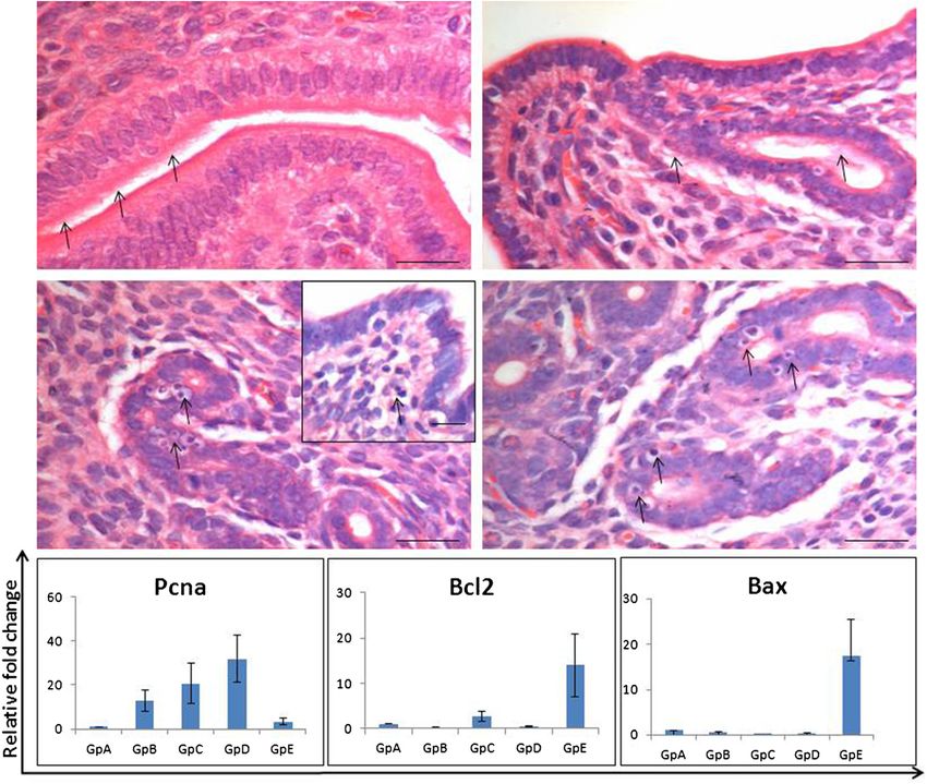
Gunjal et al. Journal of Ovarian Research (2015) 8:9                                                               Page 2 of 15

express pluripotent markers, self-renew and have the                Present study was undertaken to investigate whether
potential to differentiate into all three germ layers in          similar populations of VSELs and endometrium specific
mice [6] and humans [7], however; unlike pluripotent              progenitors exist in the mouse uterus and if they do,
embryonic stem cells, they do not form teratoma nor               whether they are modulated by sex hormones. Uterine
compliment developing embryos. This is due to the                 endometrium is a dynamic tissue in the body which under-
novel epigenetic mechanism of imprint erasure on pater-           goes regular proliferation, differentiation, growth, break-
nally imprinted DMRs (H19-Igf2, RasGRF1) exhibited by             down and shedding and again regenerates more than 400
VSELs [3,8] as they appear slightly later during embry-           times during the reproductive life in humans [20]. After the
onic development (in epiblast-stage embryo) compared              endometrium is shed as part of the physiologic, normal
to ES cells derived in vitro from the inner cell mass. ES         28 days menstrual cycle, it regenerates to a thickness of
cells undergo symmetric cell divisions in vitro, are im-          4-7 mm within 4-10 days [21]. Besides it also undergoes
mortal in nature, form teratoma and compliment devel-             extensive growth during pregnancy to accommodate the
oping embryo in contrast to VSELs which exhibit                   growing fetus and following hormone replacement therapy
extreme quiescence in vitro and possibly undergo asym-            in menopausal women. Stem cells have been implicated in
metric cell divisions in vivo to self-renew and give rise         the process of endometrium remodeling, regeneration and
to progenitors which expand in large numbers and fur-             also during various disease conditions like endometriosis
ther differentiate into specific cell types depending on          and endometrial hyperplasia, carcinoma, leiomyomas and
their location.                                                   adenomyosis [22]. Data is now emerging using various
  We have reported relatively quiescent, pluripotent              approaches on endometrial stem cells and their possible
VSELs with nuclear OCT-4 in adult mammalian testis                location however, a clear consensus on their origin, nature,
and ovary [9,10]. Besides VSELs, there exists another             anatomical location and character is still lacking [23].
population of tissue specific progenitors derived from              Bilaterally ovariectomized mice model has been used
the VSELs which are slightly bigger in size, have cyto-           in the present study as it allows manipulation of endo-
plasmic OCT-4 and are more active including spermato-             metrial cells in response to the steroids administered
gonial stem cells (SSCs) in testis and ovarian germ stem          and has been used by various investigators in the past
cells (OGSCs) in ovary. This existence of two stem cells          [24-26]. This model was recently used as a functional
populations in gonads is in agreement with similar con-           model of endometrial breakdown and repair [27] that
cept of quiescent and active stem cell populations pro-           mimics events of menstruation in women [24,28,29]. It
posed in bone marrow, skin and gut [11,12]. Stem cells            is intriguing to point out here that pluripotent tran-
are lodged in the ovary surface epithelium and in the             scripts have been reported in both mice [30] and human
testicular seminiferous epithelium. VSELs have remained           [31-35] endometrium. However, most of the groups have
elusive so far because of their very small size and are           reported cytoplasmic OCT-4 which is not crucial for
not easily visualized in paraffin sections; rather we first de-   stemness and has resulted in lot of confusion in the
tected them in smears prepared after enzymatic digestion          available literature [36,37]. But since cells with cytoplas-
of the gonadal tissue. VSELs located in the ovary surface         mic OCT-4 have been reported in the uterus, we specu-
epithelium express gonadotropin (follicle-stimulating hor-        lated that there may also exist a small population of
mone, FSH) receptors and undergo self-renewal and germ            pluripotent stem cells with nuclear OCT-4A which gives
cell nest formation after FSH treatment [13-16]. Similarly,       rise to the cells with cytoplasmic OCT-4. Our study is a
Kucia et al. [3] reported that bone marrow VSELs express          preliminary report and further studies to functionally
mRNA for several pituitary and gonadal hormone recep-             characterize the stem cells and their ability to differenti-
tors and administration of sex hormones directly stimu-           ate into glands and stromal cells are ongoing.
lates expansion (∼2–3x) of VSELs and HSCs in bone
marrow associated with increased BrdU incorporation.              Materials and methods
Because of their quiescent nature, VSELs survive total            The study was approved by Institute Animal Ethics
body radiation in mouse bone marrow (HSCs are                     Committee and was carried out using in-house bred
destroyed) [17] and also chemotherapy in mice testes              eight-weeks old Swiss mice housed in the Experimental
(SSCs, spermatocytes and haploid sperm get destroyed)             Animal Facility. They were kept in a temperature and
[18] and ovaries (OGSCs, follicles get destroyed) [19].           humidity controlled room on a 12 hr light/12 hr dark-
On providing a healthy microenvironment (by way                   ness cycle with free access to food and water.
of inter-tubular transplantation of healthy Sertoli or
mesenchymal cells) resulted in restoration of spermato-           Study design
genesis in chemoablated testis [18]. Similarly the VSELs          Presence of VSELs in mouse uterus
in chemoablated ovaries retain potential to initiate neo-         Despite the presence of VSELs in the ovarian [38] and
oogenesis and germ cells cluster formation [19].                  testicular [39] smears, they cannot be easily observed in
Gunjal et al. Journal of Ovarian Research (2015) 8:9                                                                                           Page 3 of 15

histological sections due to their very small size. Indeed                      factor). Oct-4 (representing total Oct-4 inclusive of all
these stem cells have been described as the ‘lost pearls’                       transcripts of Oct-4) transcript which is suggestive of
that are smaller than red blood cells in size and easily get                    the presence of VSELs and their immediate descendant
missed as debris [40]. Thus similar to our earlier efforts of                   ‘progenitors’ was also studied.
detecting VSELs in smears and later in sections, whole
uterine tissue was used to prepare smears to search for the                     Modulation of VSELs by different hormonal conditions
VSELs since we were sure neither of their presence nor                          To study whether VSELs are modulated by different
their exact location/niche in uterus. A normal mouse                            hormonal conditions during endometrial atrophy, pro-
uterus was used to prepare cell suspension by enzymatic                         liferation, differentiation and remodeling, the mice
digestion. Once the VSELs were detected in the smears                           were subjected to bilateral ovariectomy under aseptic
and also by flow cytometry as SCA-1 positive cells in the                       conditions. Fourteen days after bilateral ovariectomy,
size range of 3-5 μm, the whole study was planned and                           mice were divided into 5 groups (Figure 1) including (i)
various experiments were undertaken. Detailed enumer-                           control OVX group which received only sesame oil
ation of VSELs in mouse uterus as LIN-/CD45-/SCA-1+                             injections (ii) estradiol (E) (iii) progesterone (P) (iv) E + P
cells was done at the Stem Cell Institute at James Graham                       (v) 48 hrs after withdrawal of E + P. These different groups
Brown Cancer Center, University of Louisville by PG (refer                      represented uterine atrophy (Gp A); proliferation (ex-
to Additional file 1 for details).                                              pected after E and not after P treatment, Gp B & C);
                                                                                secretory phase (E + P group, Gp D) and remodeling
Characterization of uterine VSELs                                               (48 hrs of withdrawal of E + P, Gp E). Various events like
The uterine smears were used to study expression of octa-                       uterine proliferation and apoptosis was confirmed by
mer binding transcription factor (OCT-4) specific for                           qRT-PCR using specific markers. Proliferating cell nuclear
pluripotent state [41] in VSELs. The cell suspension after                      antigen (Pcna) was used as a marker for proliferation. It is
enzymatic digestion was subjected to immuno-magnetic                            a key cell-cycle regulator and has an important role to play
enrichment of VSELs using SCA-1. Both the positive and                          during DNA repair and replication. Ratio of Bcl-2/Bax
negative SCA-1 sorted fractions were subjected to RT-                           was used to confirm apoptosis. Bcl-2 is a proto-oncogene
PCR to detect presence of pluripotent transcripts includ-                       and Bax is a pro-apoptotic protein that induces apoptosis
ing Oct-4A (specific for nuclear OCT-4A isoform), Sca-1                         by homodimerization and heterodimerization with Bcl-2.
(stem cell antigen-1) and Nanog (homeobox transcription                         VSELs specific transcripts (Oct4, Oct4A, Nanog, Sca-1)

 Figure 1 Treatment schedule and gross appearance of uteri at the time of sacrifice. Various treatment groups (Gp) were used to study
 how mouse uterine stem cells are modulated by hormones under varying conditions of endometrium including atrophy, growth and remodeling/
 regeneration. Mice were bilaterally ovariectomized (on Day 0) and 14 days later were subjected to various hormonal treatments as shown. Gp A
 received no hormonal treatment, were sacrificed on Day 25 and the uterus appeared atrophied evident by its very thin appearance compared
 to other groups. Gp B, D & E received estradiol (E, 100 ng/day) priming for six days (D14-19). Gp B was treated with 50 ng/ml estradiol on Day 25 and
 sacrificed 24 hrs later to study the proliferative effect of estradiol on endometrial cells. Gp C was not primed with estradiol, only received progesterone
 (P, 1 mg/day) on Days 21-24 and sacrificed 24 hrs later to study the effect of progesterone alone on the endometrium. Gp D received estradiol priming,
 progesterone on Days 21-24 and also estradiol (50 ng) on Day 24, and was sacrificed 24 hrs after the treatment on Day 25 was the E + P treated
 receptive phase endometrium with maximal growth. Gp E reflected condition of remodeling/regeneration resulting by withdrawal of hormones. These
 mice were treated as Gp D and were sacrificed 48 hrs after Gp D i.e. on Day 27.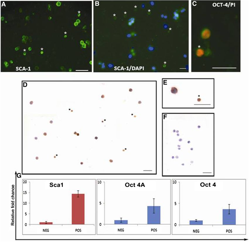
Gunjal et al. Journal of Ovarian Research (2015) 8:9                                                           Page 4 of 15

were also studied by qRT-PCR in various treatment                on gonadal stem cells that VSELs (with abundant eu-
groups to evaluate whether they are differentially modu-         chromatin) do not stain easily with DAPI compared to
lated under different conditions.                                the progenitors [38] because of the presence of abun-
                                                                 dant open euchromatin and DAPI selectively stains
Methods                                                          specifically to A-T base pairs, producing a much stron-
Bilateral ovariectomy and hormone treatment                      ger signal on the heterochromatin than on the eu-
Bilateral ovariectomy was performed by expert veterinar-         chromatin [43,44].
ians (SM and DM) using standard protocols. Beta-estradiol
(E, Sigma-Aldrich, Germany) stock for injections was             Enrichment of SCA-1 positive cells
prepared by dissolving 1 mg estradiol in 100 μl of ethanol       Single cell suspension was prepared from uterine tissue
and making up to 1 ml with sesame oil. 10 mg progester-          as described above. SCA-1 positive cells were immuno-
one (P, Sigma) was dissolved in 100 μl of chloroform and         magnetically enriched using FITC tagged SCA-1 anti-
final volume made to 1 ml by adding 900 μl of sesame oil.        body and Easysep kit (Stem Cell Technology, Canada)
Hormones were injected subcutaneously in a volume of             according to manufacturer’s instructions. Briefly 108
100 μl. Two weeks after bilateral ovariectomy, the mice          cells were immuno-stained with SCA-1 antibody (BD
were subjected to various treatment schedules as shown in        Life Sciences, USA) for 15 minutes at room temperature
Figure 1. Mice in Gp A received only sesame oil injections       (RT), followed by incubation with FITC selection cock-
and sacrificed on day 25 whereas Gp B, D & E were                tail for 15 minutes. The cell suspension was then incu-
initially primed by daily estradiol (100 ng/day) injec-          bated with magnetic nanoparticles for 10 minutes at RT
tions for 6 days (Day 14-19 after ovariectomy). Mice in          and placed on a magnetic particle concentrator. After
Gp B were injected estradiol (50 ng) on day 24 and               isolation, enriched SCA-1+ cells fraction as well as the
sacrificed 24 hrs later on day 25. Similarly mice in GpC         negative fraction were used for making smears and for
were injected progesterone (1 mg/Kg) on days 21-24               RNA extraction.
and sacrificed 24 hrs later on day 25. Gp D was initially
primed with estradiol and then injected progesterone             Immuno-staining of OCT-4 on smears
on days 21-24 along with estradiol (50 ng/day) on day            The cells smears were washed with PBS and then incu-
24 and then sacrificed on day 25. Mice in group E                bated for 30 min in 3% hydrogen peroxide followed by
received similar treatment as Gp D and were sacrificed           antigen retrieval in boiling sodium citrate saline buffer
on day 27, 48 hrs after Gp D. Estradiol priming for six          of pH 6 for 5 min. After cooling, the smears were
days was essential as it is well known that progesterone         washed with PBS for 5 min and then blocked with 10%
alone is incapable of expressing its own receptors [42].         normal goat serum in PBS. After removing excess block-
The tissue collected after various treatments was proc-          ing reagent, the slides were incubated with primary anti-
essed appropriately to make uterine tissue cell smears,          body (polyclonal OCT4 from Millipore) for two hours at
for histology flow cytometry and RNA extraction. For-            room temperature. The detection was done using anti-
malin fixed uterine tissue was processed and embedded            rabbit Vecta ABC kit (Vector Laboratories, USA) accord-
in paraffin using standard protocols. 5 μm thick sec-            ing to manufacturer’s instructions. Color development was
tions were prepared and stained with Hematoxylin and             done using 3,3' diaminobenzidine (DAB) (Biogenex, USA).
Eosin for studying the histo- architecture of uterus             After counterstaining with Hematoxylin, the slides were
after different hormonal administration. The represen-           observed under Nikon 90i microscope. Representative
tative areas were photographed and data recorded.                areas were photographed. Total protein isolated from
                                                                 atrophied uterus was subjected to Western blotting for
Smears preparation from uterine tissue                           OCT-4 (Additional file 1: Figure S2).
Briefly the uterine tissue was minced into small pieces
and then incubated with collagenase (1 mg/ml) & Trypsin          Immuno-fluorescence and confocal microscopy
(1 mg/ml) and DNAse (0.5 mg/ml) in DMEM culture                  SCA-1 sorted cells were used for studying expression of
media at 37°C for 30 minutes. After filtering through            OCT-4 by immuno-fluorescence. For this, following
40 μm filter, the filtrate was used to make smears. After        washes with 0.5% BSA solution, the cells were perme-
air drying, the smears were fixed in 4% PFA, washed 2-3          abilized using 0.3% Triton-X 100 for 5 minutes. Blocking
times with phosphate buffer saline (PBS), air dried and          was done with 3% BSA solution for 1 hour. Smears were
stored at 4°C till further use. These smears were stained with   incubated with polyclonal OCT-4 antibody which stains
Hematoxylin and Eosin using standard method. The smears          both the isoforms of OCT-4 (Abcam) at 4°C overnight.
were also stained with 4',6-diamidino-2-phenylindole,            Next day the smears were washed with wash buffer and
dihydrochloride (DAPI) for few seconds to demonstrate            incubated with fluorescent tagged secondary alexafluor-
the presence of VSELs, as we know from our experience            488 antibody for 2 hours at RT in dark. The slides were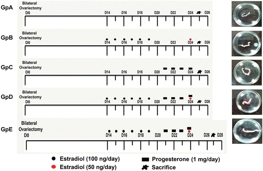
Gunjal et al. Journal of Ovarian Research (2015) 8:9                                                      Page 5 of 15

then washed thrice with PBS and counter-stained with       1x Dream Taq buffer (Fermentas) and 0.2 mM dNTPs in
DAPI for 15 minutes. Images were captured by laser         a G-STORM thermocycler. Amplification was carried
scanning confocal microscope (Carl Zeiss, Germany).        out for 35 cycles, with each cycle consisting of denatur-
                                                           ation at 94°C for 30 sec, annealing at the specified
RT-PCR and qRT-PCR studies                                 temperature for each set of primers (Table 1) for 20 sec,
Details of primers used in the present study are shown     and extension at 72°C for 30 sec. The products were
in Table 1.                                                analyzed on 2% agarose gel stained with 0.5 μg/ml
                                                           ethidium bromide (Bangalore Genei, India). The product
RNA isolation and cDNA synthesis                           size was approximated using a 100-bp DNA ladder
The uterine tissue was collected in Trizol (Invitrogen,    (Bangalore Genei). The negative control did not include
Carlsbad, CA, USA) for RNA extraction by standard          cDNA in the reaction mixture and cDNA from mouse
protocol and then treated with DNase I (Amersham Bio-      ovary tissue was used as positive control.
sciences, Piscataway, NJ) at 37°C for 30 min to remove       Quantitative RT- PCR (qRT-PCR): Later, using the
any genomic DNA contamination. First-strand cDNA           standardized annealing temperature, qRT-PCR was car-
was synthesized using the cDNA synthesis Kit (Bio-Rad,     ried out to study how these transcripts are regulated by
USA) according to the manufacturer’s instructions.         steroid treatment. The expression levels of these gene
Briefly, 1-2 μg of total RNA was incubated with 5X reac-   transcripts in relation to housekeeping gene transcript
tion mix and reverse transcriptase mix. The reaction was   18S were estimated by CFX96 real-time PCR system
carried out in G-STORM thermocycler (Gene Technolo-        (Bio-Rad Laboratories, USA) using SYBR Green chemis-
gies, Braintree, UK). The reaction mix was first incu-     try (Bio-Rad). The amplification conditions included
bated at 25°C for 5 min, then at 42°C for 30 min and       initial denaturation at 94°C for 3 min followed by 40 -
finally at 85°C for 5 min. for cDNA synthesis.             cycles comprising of denaturation at 94°C for 30 seconds,
  RT-PCR for pluripotent stem cells specific markers: In   primer annealing for 20 sec and extension at 72°C for
order to demonstrate the presence of VSELs by RT-PCR,      30 sec. The final step included incubation at 94°C for
various transcripts (Oct4, Oct4A, Nanog, Sca1) were        20 s to remove any secondary structures. The fluores-
amplified using a temperature gradient to establish the    cence emitted at each cycle was collected during the
optimal annealing temperature which varies from tissue     extension step of each cycle. The homogeneity of the
to tissue. Briefly, the cDNA mix (2 μl) was amplified      PCR amplicons was verified by running the products
using 0.2 mM of each primer as described (Table 1),        on 2% agarose gels and also by studying the melt curve.
1.25 unit of DreamTaq DNA polymerase (Fermentas) in        All PCR amplifications were carried out in duplicate.
Table 1 List of primer sequence used for RT-PCR and qRT-PCR
Gene name                   Primer sequence                    Annealing temperature (°C)           Amplicon size (bp)
Pcna                        F:GATGCCGTCGGGTGAATTTG             55                                   182
                            R:TCTCTATGGTTACCGCCTCCT
Bax                         F:GTTTCATCCAGGATCGAGCAG            55                                   488
                            R:CATCTTCTTCCAGATGGTGA
Bcl-2                       F:CCTGTGGATGACTGAGTACC             55                                   128
                            R:GAGACAGCCAGGAGAAATCA
Oct-4                       F: CCTGGGCGTTCTCTTTGGAAAGGTG       66                                   198
                            R: GCCTGCACCAGGGTCTCCGA
Oct-4A                      F: CCATGTCCGCCCGCATACGA            61                                   235
                            R: GGGCTTTCATGTCCTGGGACTCCT
Nanog                       F: CAGGAGTTTGAGGGTAGCTC            61                                   223
                            R: CGGTTCATCATGGTACAGTC
Sca-1                       F: AGAGGAAGTTTTATCTGTGCAGCCC       66                                   276
                            R:TCCACAATAACTGCTGCCTCCTGA
18S                         F:GGAGAGGGAGCCTGAGAAAC             61                                   198
                            R: CCTCCAATGGATCCTCGTTA
GAPDH                       F: GTCCCGTAGACAAAATGGTGA           58                                   435
                            R: TGCATTGCTGACAATCTTGAG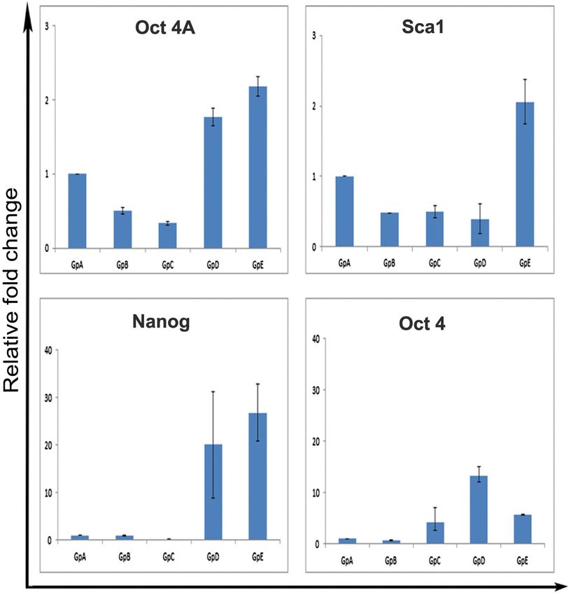
Gunjal et al. Journal of Ovarian Research (2015) 8:9 Page 6 of 15
Mean Ct values generated in each experiment using the rim of cytoplasm (Figure 2A). These cells were distinct
CFX Manager software (Bio-Rad) were used to calculate from the somatic cells with pale stained nucleus sur-
the mRNA expression levels. Since ΔCt is inversely pro- rounded by abundant cytoplasm. The stem cells appear
portional to relative mRNA expression levels, the levels similar to the VSELs reported by our group in testis
were calculated manually by the ΔCt method. and ovary (Bhartiya et al., [9]). Flow cytometry studies
confirmed the presence of 0.0685% of LIN-/CD45-/
Results SCA-1+ VSELs in mouse uterus (Additional file 1:
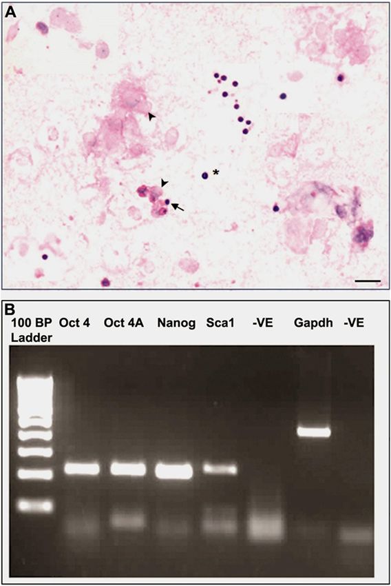
Presence of VSELs in mouse uterus Figure S1). RT-PCR analysis (Figure 2B) confirmed the
H & E stained smears of uterine cell suspension clearly presence of pluripotent transcripts of expected size for
showed the presence of very small sized cells with dark Oct4, Oct4A, Sca1 and Nanog in the ovariectomized
Haematoxylin stained nucleus surrounded by a thin uterine tissue (Gp A).
Figure 2 Detection of VSELs in mouse uterus. Panel A Hematoxylin and Eosin stained uterine smears prepared after enzymatic digestion of
the tissue. Note the presence of distinct spherical stem cells with dark stained nucleus with high nucleo-cytoplasmic ratio. In contrast, the somatic
uterine cells have pale stained nucleus surrounded by abundant cytoplasm. Please note that 5-6 different fields have been combined to make this
figure. Scale bar represent 20 μm. Lower Panel B shows RT-PCR results for pluripotent markers Oct 4A, Nanog, Sca-1, Oct 4 and Gapdh in bilaterally
ovariectomized uterus (Gp A). Results show that pluripotent stem cells exist in atrophied endometrium.Gunjal et al. Journal of Ovarian Research (2015) 8:9 Page 7 of 15 Characterization of uterine VSELs were stained with DAPI within 2 secs. These results are Uterine cell suspension obtained by enzymatic digestion similar to our earlier results where ovarian VSELs also was used to enrich SCA-1 positive VSELs. SCA-1 sorted poorly stain with DAPI [38]. Immuno-localization for cells under fluorescent microscope showed very distinct OCT-4 on sorted stem cells showed the presence of two surface staining for SCA-1 (Figure 3A) and did not stain distinct cell types expressing OCT-4 including few smaller with DAPI (Figure 3B) compared to somatic cells which cells with nuclear OCT-4 and large number of cells with Figure 3 Characterization of uterine stem cells. A. SCA-1 sorted cells under microscope appear green and are of two distinct sizes including small sized cells which possibly are the very small embryonic-like stem cells VSELs (asterix) and slightly bigger endometrial stem cells EnSCs. B. DAPI stained smear shows that the somatic cells negative for SCA-1 stain nicely with DAPI whereas the stem cells do not stain with DAPI. Similar data was obtained for ovarian stem cells33; these cells do not stain with DAPI within 2-3 secs possibly because being pluripotent they have open chromatin. C. Higher magnification of OCT-4 stained cells with propidium iodide used as a counterstain shows cells have distinct nuclear staining of OCT-4. D. OCT-4 immuno-localization on the smears. Note the presence of two distinct sized cells which stain positive for OCT-4 including the VSELs (asterix) and slightly bigger EnSCs. E. Nuclear versus cytoplasmic OCT-4 staining can be clearly observed at higher magnification. F. Negative control. Scale bars represent 20 μm. Lower panel G shows the qRT-PCR results of negative (NEG) and positive (POS) fractions after immuno-magnetic sorting of SCA-1 positive cells. As evident the stem cell markers Sca1, Oct 4A and Oct 4 are highly enriched in the positive fraction.
Gunjal et al. Journal of Ovarian Research (2015) 8:9 Page 8 of 15
cytoplasmic OCT-4 (Figure 3C-E). qRT-PCR analysis Lower panel) confirmed the histological findings. Pcna
showed that the pluripotent transcripts (Sca-1, Oct-4A (marker suggestive of DNA synthesis and proliferation)
and Oct-4) were enriched in the immuno-magnetically showed minimal expression in Gps A and E (after ovari-
positively sorted (POS) compared to the negative (NEG) ectomy and during hormone withdrawal). Whereas it
fraction suggesting that pluripotent VSELs do exist in was variably expressed in groups B-D. Bcl-2 was also
the uterus and can be sorted using SCA-1 (Figure 3, variably expressed in all groups whereas Bax transcripts
bottom panel). were observed only in Gp E. The ratio of Bax and Bcl-2
Thus above results showed the presence of LIN-/ (suggestive of apoptosis) were increased in Gp E only
CD45-/SCA + VSELs ranging in size from 3-5 μm with confirming that withdrawal of E + P for 48 hrs resulted
nuclear OCT-4 and expressing Oct-4, Nanog and Sca-1 in remodeling of the endometrium and involved apop-
mRNA transcripts in mouse uterus. Two distinct sizes of tosis. Thus we successfully obtained a mouse model to
stem cells with nuclear OCT-4 in smaller VSELs and study how VSELs are modulated in an atrophied endo-
slightly bigger cells with cytoplasmic OCT-4 were visual- metrium (Gp A), proliferative endometrium (Gp B com-
ized. The results are similar to the VSELs reported in pared to Gp C), secretory endometrium (Gp D) and
testis and ovary by our group. Interestingly the bigger endometrium which is undergoing remodeling and
‘progenitors’ (immediate descendants of VSELs) with cyto- regeneration (Gp E).
plasmic OCT-4 are the spermatogonial stem cells (SSCs) Quantitative RT-PCR studies for pluripotent markers
in testis, ovarian stem cells (OSCs/OGSCs) in ovary and Oct-4A, Nanog and Sca-1 revealed an interesting pattern.
we term them as endometrial stem cells (EnSCs) in the These transcripts were detected in all the groups however;
endometrium. Since we have observed EnSCs in smears, their expression was maximum in Gp A and E. Also Oct-4
their true shape and localization remains to be studied in (suggestive of the presence of ‘progenitors’ EnSCs) was
the endometrium. They could possibly be the cytoplasmic maximally expressed in the Gp D. It was interesting to
OCT-4 positive mesenchymal cells described by several note that the ratio of Oct-4 to Oct-4A was equal in the
groups in the endometrium [45,46]. ovariectomized samples suggesting that stem cells in
ovariectomized sample mainly comprised of VSELs. In
Modulation of VSELs by hormones contrast Oct-4 was more than ten-fold increase in Gp D
Effect of steroid treatment on mouse uterus: As evident, compared to a two-fold increase in Oct-4A, suggesting a
the uterus responded well to the treatment, being thin higher proportion of progenitors with cytoplasmic OCT-4
and atrophied after bilateral ovariectomy and showed when the endometrium undergoes maximum growth/dif-
variable morphology after various treatments (Figure 1). ferentiation and gets ready for implantation to occur.
Histology studies (Figures 4 & 5) showed expected
changes in the uterine histology with maximum atrophy Discussion
(evident by the size in cross-section) observed post Different strategies have been employed by various investi-
ovariectomy (Gp A). The uterus showed growth and ex- gators to detect endometrial stem cells as shown in Table 2
tensive proliferation after estradiol treatment (Gp B) com- but a clear picture of the true identity of endometrial
pared to progesterone alone (Gp C). Combined estradiol stem cells is yet to emerge. Present study for the first
and progesterone (E + P) resulted in differentiation and time shows the presence of LIN-CD45-SCA+ very small
a receptive endometrium (Gp D) with characteristic embryonic-like stem cells (VSELs) in the mouse uterus.
narrowing of lumen and secretory activity in the lumen. VSELs (comprising 0.07% of the total cell population as
Withdrawal of E + P for 48 hrs (after mice in Gp D were judged by flow cytometry) were observed in uterine cell
sacrificed) resulted in extensive remodeling (Gp E) asso- smears and could be enriched by immuno-magnetic
ciated with reduction in epithelial cell height (Figure 4). sorting using a cell surface antigen SCA-1. Uterine
At higher magnification (Figure 5) luminal epithelium VSELs are small in size (3-5 μm), spherical in shape,
showed characteristic features of a receptive stage in Gp have high nucleo-cytoplasmic ratio, stain intensely with
D comprising of tall epithelial cells with oval shaped nuclei Hematoxylin but poorly with DAPI and express nuclear
in the middle, sub-nuclear vacuolation and secretory activ- OCT-4, cell surface SCA-1 and pluripotent transcripts
ity at the luminal surface. Whereas in Gp E the luminal including Oct4A, Sca1 and Nanog. Similar VSELs exist
epithelial cells reduced in size due to withdrawal of hor- in various adult mouse organs [1], human testes [39],
mones resulting in increased nucleo-cytoplasmic ratio. ovaries [38], bone marrow [2,47] and cord blood [48,49].
Increased vacuolation and apoptotic bodies (in luminal We succeeded to detect VSELs in uterine smears but
and glandular epithelium as well as in the stromal com- the exact location/niche of these cells in the uterus still
partment) could be observed at places, suggestive of remains elusive because of their very small size and for
remodeling, repair and regeneration (Figure 5B-E). The the same reason they have evaded the reproductive biol-
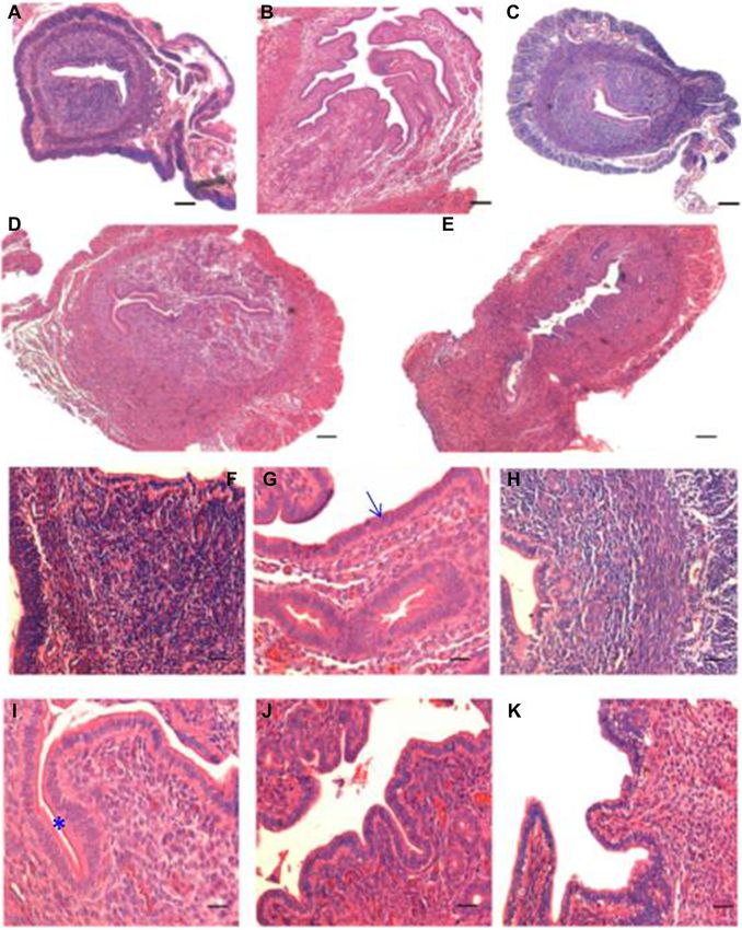
effect of steroid treatment at transcript level (Figure 5 ogists till date.Gunjal et al. Journal of Ovarian Research (2015) 8:9 Page 9 of 15 Figure 4 (See legend on next page.)
Gunjal et al. Journal of Ovarian Research (2015) 8:9                                                                                     Page 10 of 15

 (See figure on previous page.)
 Figure 4 Histology of mouse uterus of different treatment groups. A and F Ovariectomized GpA. B and G Estradiol alone GpB. C and H
 Progesterone alone GpC D and I E + P GpD E, J and K E + P withdrawal GpE. As evident the ovariectomized uterus was atrophied (A) and the
 cells were tightly packed (F) whereas estradiol treatment resulted in increased size of the uterus (B) with increased proliferation in both the
 glands and stromal cells (G). Epithelial cells were tall and columnar and frequent mitotic figures were observed. Luminal epithelium appeared
 highly folded, glands were also bigger and lot of edema in the stromal compartment. Progesterone treatment exerted minimal effect and the
 uterus remained small in size (C) with densely packed cells (H). E + P treatment resulted in a receptive endometrium with characteristic narrowing of
 uterine lumen (D), tall epithelial cells lined the lumen and the glands and lot of edema and increased vascularity was evident in the stromal
 compartment. In contrast to the apically placed nuclei after estradiol treatment (G), in E + P treated section the nuclei are more centrally placed
 (I), suggestive of a receptive condition when nutrients required for successful implantation shift to the apical end of the epithelial cells. Also
 secretory activity was evident on the apical surface of luminal epithelial cells. Withdrawal of E + P resulted in repair and remodeling suggested
 by the presence of a compacted uterus (E), small epithelial cells with increased nucleo-cytoplasmic ratio and dense compact stromal cells
 (J & K). Scale bars represent 20 μm.

 Figure 5 Higher magnification of uterine sections of Gp D and E. Luminal epithelium in Gp D (A) comprised tall, columnar cells with
 centrally placed nuclei. Note the presence of secretory activity in the lumen. Withdrawal of E + P (C-D) resulted in reduction in epithelial cell
 height with increased nucleo-cytoplasmic ratio and the nuclei were apically placed. The nuclei appeared more apical and signs for remodeling
 and apoptotic bodies were evident in luminal and glandular epithelium and also in the stromal compartment. Scale bar represent 20 μm. Bottom
 panel shows the qRT-PCR results of specific transcripts reflecting proliferation (Pcna) and apoptosis (Bax and Bcl2). As evident Pcna was minimally
 expressed in Gp A and E whereas Bax and Bcl2 were unregulated in Gp E suggestive of increased apoptosis, in agreement with the histology data.
Gunjal et al. Journal of Ovarian Research (2015) 8:9                                                                          Page 11 of 15

Table 2 Various approaches utilized to detect and study                     give rise to both the beta and acinar cells [58]. Ability of
endometrial stem cells                                                      VSELs to give rise to SSCs, OSCs, HSCs, MSCs, EnSCs
Various approaches       Salient observations and relevant references       and pancreatic progenitors reflects their pluripotent state.
Classic concept          Stem cells may reside in the basal region of       Ability of VSELs to give rise to three germ layers has
                         endometrium which continuously repairs the         already been reported in mice [6] and humans [7]. Further
                         functional layer [27,50]
                                                                            detailed studies are required to understand stem cells-
Side population          Side population cells are distributed throughout   niche interaction as recently suggested [59,60].
(SP) analysis            endometrium [51-53]
                                                                               Two stem cell populations comprising of a quiescent and
Label retaining          Mouse LRCs decline with age, exist in lower        actively dividing stem cells have been reported in adult
cells (LRCs)             region of stroma and exhibit undifferentiated
                         markers including OCT-4 [25]                       somatic tissues including gut epithelium, hair follicle, bone
                         LRCs detected in luminal epithelium and
                                                                            marrow etc. [11,12]. Similarly we have found that VSELs are
                         stromal cells adjacent to the luminal              the quiescent stem cells in the testis and ovaries whereas
                         epithelium [54] in mouse endometrium               the SSCs and OGSCs divide rapidly and undergo clonal
                         LRCs detected only in the glandular epithelium     expansion prior to initiating differentiation [9,10]. That
                         [22] in humans                                     VSELs comprise the quiescent population of stem cells gets
Clonogenic assays        Human endometrial stromal progenitor cells         further credence from the reports showing that they survive
                         can be cultured for more than 15 passages and      chemotherapy in mice testis [18], ovary [19] and total body
                         show high clonogenic efficiency (15%) [55]
                                                                            irradiation in bone marrow [17] whereas the actively divid-
                         Single endometrial stromal cell can form
                         cellular colonies that can be further serially
                                                                            ing progenitors are destroyed. These findings are intriguing
                         cloned and differentiated into various             and suggest that VSELs could be the quiescent stem cell
                         mesodermal lineages [56]                           population in various adult organs. VSELs have also been
Other relevant results   Pluripotent markers have also been reported        implicated in cancers [61] and are possibly the elusive
                         in human endometrial and endometriotic             cancer-initiating cells responsible for recurrence (as they are
                         samples [27-30]
                                                                            likely to be spared of oncotherapy because of their quiescent
                         Bone marrow cells may be implicated in             nature). Further studies are required to investigate this.
                         endometrial repair [47,57]
                                                                               Cells with cytoplasmic OCT-4 and other pluripotent
                                                                            transcripts have been reported by various investigators
   Besides the VSELs with nuclear OCT-4, we also detected                   in mouse as well as human endometrial and endometriotic
slightly bigger cells with cytoplasmic OCT-4 which we                       tissue [32-35]. There exists lot of confusion because these
term as the endometrial stem cells (EnSCs). These are                       studies have failed to distinguish and appreciate the
most likely the immediate multipotent descendants or                        relevance of cytoplasmic versus nuclear OCT-4 staining
‘progenitors’ which arise by differentiation of pluripotent                 [36,37]. The small sub-population of VSELs with nuclear
VSELs and are expected to be multipotent and give rise to                   OCT-4 remained undetected in the mouse uterus till now
various endometrial cell types. Differential OCT-4 expres-                  possibly because they exist in very few numbers, are of very
sion pattern (obtained using a polyclonal antibody which                    small size and also because VSELs tend to get lost during
cross-reacts with both OCT-4A and OCT-4B isoforms)                          processing as suggested earlier while studying cord blood
has been very useful to differentiate between pluripotent                   and bone marrow stem cells [48]. Mesenchymal stem cells
VSELs, the immediate descendants with cytoplasmic                           have been reported in the endometrium [45,46]. It has been
OCT-4 and the differentiated progeny which show loss of                     suggested that the MSCs (negative for CD45, CD34 and
OCT-4. It is intriguing that similar VSELs exist in ovary,                  positive for CD146, PDGF-receptor-Beta, CD29, CD44,
testis, bone marrow and uterus but the immediate descen-                    CDE73, CD90 and CD105) get further differentiated into
dants are distinct and give rise to different cell types based              stromal and glandular cells. Evidence to support that VSELs
on where the VSELs are located; spermatogonial stem cells                   may give rise to the MSCs has been elegantly demonstrated
(SSCs) in testis differentiate to form the haploid sperm,                   by Taichman et al. [62] wherein they reported that Sca+Lin-
ovarian stem cells (OSCs) differentiate into haploid oo-                    CD45- cells could differentiate into multiple mesenchymal
cytes, hematopoietic (HSCs) and mesenchymal (MSCs)                          lineages in their study model. Similarly in umbilical cord
stem cells give rise to various kind of blood cells and simi-               tissue we have earlier reported that VSELs (with nuclear
larly the uterine stem cells (EnSCs) are expected to differ-                OCT-4) and MSCs (with cytoplasmic OCT-4) co-exist in
entiate into various endometrial cell types. Results suggest                the Wharton’s jelly [48]. Existence of two stem cell popula-
that the microenvironment/niche where the VSELs are                         tions has also been postulated in the endometrium [54] and
located is crucial and dictates the fate of stem cells. We                  the endometrial stromal cells are considered lineage cells of
recently reported that in mouse pancreas after partial                      endometrial mesenchymal stem cells.
pancreatectomy – VSELs give rise to OCT-4 & PDX-1                              Results of the present study further show that uterine
co-expressing progenitors which then differentiate and                      VSELs and the progenitors EnSCs are implicated during
Gunjal et al. Journal of Ovarian Research (2015) 8:9 Page 12 of 15 endometrial atrophy, growth and remodeling/regeneration. remains preserved even in atrophic endometrium [63]. Presence of VSELs in ovariectomized atrophied uterus is in Oct-4A and Oct-4 transcripts were equally expressed in agreement with earlier report that a pool of stem cells Gp A (Figure 6) suggesting that stem cell population Figure 6 qRT-PCR results for pluripotent transcripts showing variable effects of hormone treatment. Oct-4A, Nanog, Sca-1 (pluripotent markers for VSELs) and Oct-4 (all Oct-4 transcripts including Oct-4B indicating presence of EnSCs) in various treatment groups. The pluripotent transcripts are observed in all the treated groups suggesting presence of stem cells in all the conditions, but are modulated by steroid treatment. They are up-regulated in GpA, compared to Gp B and C and are maximally expressed in Gp D and E. Oct-4 is more than 10 fold increased in GpD suggesting presence of EnSCs. Note the scale of Y-axis to appreciate differences between groups. Results suggest that stem cells are present in atrophic endometrium and stem cells activity is greatly increased in E + P group when maximum growth is observed and remain elevated in GpE associated with remodeling and repair. Ratio of Oct-4 to Oct-4A is almost 1 in GpA suggesting most of stem cells are the VSELs whereas in GpD the ratio is greatly increased suggesting presence of large number of progenitors. Results are compilation of more than three biological samples and error bars represent standard error.
Gunjal et al. Journal of Ovarian Research (2015) 8:9                                                                       Page 13 of 15

comprise mostly of VSELs in the atrophied uterus, how-        of autologus bone marrow derived mesenchymal stem
ever they are unable to differentiate possibly because of     cells in women with Asherman’s syndrome resulted in
a compromised microenvironment/niche, a situation             conception. Similarly, Alawadhi et al. [73] reported that
similar to presence of VSELs in chemoablated testis and       transplantation of bone marrow derived stem cells can
ovary [18,19]. VSELs and EnSCs are present in the             regenerate mouse model of Asherman’s syndrome and
uterus in the E and P treated groups but their numbers        improve fertility rates. Available literature suggests that
are greatly increased in E + P and the withdrawal group.      VSELs are mobilized whenever any organ function is
Total Oct-4 transcript is 5-15 fold increase compared to      compromised [74-78] and thus endogenous VSELs are ex-
Oct-4A suggesting rapid expansion of progenitors (EnSCs)      pected to be present in the uterus affected by Asherman’s
under these conditions. VSELs give rise to EnSCs which        syndrome (similar to their presence in atrophied endomet-
multiply in large numbers and further differentiate into      rium in the present study, Gp A). Transplanted bone
various cell types to bring about growth (Gp D) and           marrow stem cells act as a source of growth factors/cyto-
remodeling/regeneration (Gp E) of endometrium. In the         kines thus facilitating endogenous VSELs to regenerate
past, stem cells have been implicated in the process of       the damaged endometrium. In addition, few transplanted
endometrium remodeling, regeneration and also during          stem cells may also get involved in regeneration and thus
various disease conditions like endometriosis and endo-       Y chromosome positive cells in both stromal and epithelial
metrial hyperplasia, carcinoma, leiomyomas and adeno-         compartment have been reported by Alawadhi et al. [73].
myosis [22]. Thus it becomes pertinent to use a more          This highlights the fact that a similar stem cell population
quantitative approach like flow cytometry and other           exists in the endometrium and bone marrow stem cells.
cellular and molecular techniques to evaluate VSELs           Similar to improvement of the thin endometrium by
and EnSCs in the endometrial tissue from normal and           transplanting bone marrow stem cells, surviving endogen-
various pathological conditions. Similar to modulation        ous VSELs in chemoablated testis underwent differen-
of endometrial VSELs by steroid hormones, VSELs in            tiation (spermatogenesis) when a healthy niche was
adult mammalian ovaries are modulated by follicle             provided by way of transplanting healthy Sertoli or mesen-
stimulating hormone [13-16].                                  chymal cells [18]. We speculate that if the uterine function
  Several studies [64,65] suggest that stem cells (isolated   of the mice (with Y chromosome positive cells engrafted
as side population SP cells which efflux Hoechst 33342        after transplantation) is compromised by subjecting the
and also express CD133) from normal endometrium may           mice to bilateral ovariectomy or by any other means
be involved in the development of endometrial cancer.         (thereby creating regenerative pressure), one may observe
Kato et al. [66] provided first direct evidence to support    clonal expansion, differentiation and increased numbers of
that SP cells contribute to endometrial carcinogenesis and    Y chromosome bearing cells. Further studies need to be
are associated with elevated OCT-4 expression. Endomet-       undertaken to delineate endogenous stem cells biology
rial hyperplasia (considered as a precursor to endometrial    and also Y chromosome bearing cells.
cancer) was observed in mice after neonatal exposure to          To conclude, we have shown for the first time the pres-
estradiol and VSELs altered biology was implicated [67].      ence of two stem cell populations in mouse uterus includ-
Thus it is likely that the VSELs which exist in normal        ing pluripotent VSELs and their immediate descendants
endometrium are possibly the embryonic remnants re-           EnSCs. Data has also been generated to show that these
sponsible for initiation of endometrial cancer as has been    stem cells are modulated by hormones and thus play an im-
proposed earlier [61].                                        portant role during endometrial growth and regeneration.
  Engraftment of donor cells in the endometrium (in both      VSELs could also be the cancer initiating cells in the endo-
glands and stroma) has been reported in women undergo-        metrium. Further studies are urgently required to study
ing single-antigen, HLA-mismatched, bone marrow trans-        VSELs and EnSCs during endometrial normal biology, can-
plantation and also in mice [57,68,69]. Nagori et al. [57]    cer and other pathologies.
reported beneficial effect of autologus bone marrow cell
transplantation in case of Asherman syndrome. Gargett         Additional file
and Healy [70] suggested that main mechanism resulting
in beneficial effect of bone marrow cells demonstrated by      Additional file 1: Isolation and flow cytometric analysis of uterine
Nagori’s group, could possibly be to improve the niche         VSELs.
rather than true regeneration. Resident stem cells may
play a major role in regeneration rather than the bone        Competing interests
marrow cells because the engraftment of XY donor de-          The authors declare that they have no competing interests.
rived cells of bone marrow origin has been found to be
                                                              Authors’ contributions
very poor (
Gunjal et al. Journal of Ovarian Research (2015) 8:9 Page 14 of 15
preparation; SM and DM performed the surgeries; HP helped with few 16. Bhartiya D, Sriraman K, Gunjal P, Modak H. Gonadotropin treatment augments
experiments. All read and approved the final manuscript draft. postnatal oogenesis and primordial follicle assembly in adult mouse ovaries?
J Ovarian Res. 2012;5(1):32–45.
17. Ratajczak J, Wysoczynski M, Zuba-Surma E, Wan W, Kucia M, Yoder MC, et al.
Acknowledgements Adult murine bone marrow-derived very small embryonic-like stem cells
The study was financially supported by Indian Council of Medical Research, differentiate into the hematopoietic lineage after coculture over OP9 stromal
Government of India, New Delhi, India. Help of Ms Harshada is acknowledged cells. ExpHematol. 2011;39(2):225–37.
for doing the histology procedures. Help of Prof Ratajczak from Stem Cell 18. Anand S, Bhartiya D, Sriraman K, Patel H, Manjramkar DD. Very small
Institute at James Graham Brown Cancer Center, University of Louisville, embryonic-like stem cells survive and restore spermatogenesis after
Louisville, KY 40202, USA is acknowledged for flow cytometry results. busulphan treatment in mouse testis. J Stem Cell Res Ther. 2014;4:216.
Study was supported by Institute Core Support provided by ICMR. doi:10.4172/2157-7633.1000216.
19. Sriraman K, Bhartiya D, Anand S and Bhutda S. Mouse ovarian very small
Author details embryonic-like stem cells resist chemotherapy and retain ability to initiate
1
Stem Cell Biology Department, National Institute for Research in oocyte-specific differentiation. Reproductive Sciences (In press).
Reproductive Health, Mumbai 400 012, India. 2Experimental Animal Facility, 20. Gargett CE, Chan RW, Schwab KE. Hormone and growth factor signaling in
National Institute for Research in Reproductive Health, JM Street, Parel, endometrial renewal: role of stem/progenitor cells. Mol Cell Endocrinol.
Mumbai 400 012, India. 2008;288(1–2):22–9.
21. McLennan CE, Rydell AH. Extent of endometrial shedding during normal
Received: 12 November 2014 Accepted: 23 February 2015 menstruation. Obstet Gynecol. 1965;26(5):605–21.
22. Maruyama T, Masuda H, Ono M, Kajitani T, Yoshimura Y. Human uterine
stem/progenitor cells: their possible role in uterine physiology and pathology.
Reproduction. 2010;140(1):11–22.
References
23. Maruyama T. Endometrial stem/progenitor cells. J ObstetGynaecol Res.
1. Zuba-Surma EK, Kucia M, Wu W, Klich I, Lillard Jr JW, Ratajczak J, et al. Very
2014;40(9):2015–22.
small embryonic-like stem cells are present in adult murine organs: image
24. Brasted M, White CA, Kennedy TG, Salamonsen LA. Mimicking the events of
stream-based morphological analysis and distribution studies. Cytometry A.
menstruation in the murine uterus. Biol Reprod. 2003;69(4):1273–80.
2008;73A(12):1116–27.
2. Ratajczak MZ, Zuba-Surma E, Wojakowski W, Suszynska M, Mierzejewska K, 25. Heryanto B, Rogers PA. Regulation of endometrial endothelial cell
Liu R, et al. Very small embryonic-like stem cells (VSELs) represent a real proliferation by oestrogen and progesterone in the ovariectomized mouse.
challenge in stem cell biology: Recent pros and cons in the midst of a lively Reproduction. 2002;123(1):107–13.
debate. Leukemia. 2014;28(3):473–84. 26. Finn CA, Pope M. Vascular and cellular changes in the decidualized
3. Mierzejewska K, Borkowska S, Suszynska E, Suszynska M, Poniewierska-Baran A, endometrium of the ovariectomized mouse following cessation of
Maj M, et. al. Hematopoietic stem/progenitor cells express several functional hormone treatment: a possible model for menstruation. J Endocrinol.
sex hormone receptors-novel evidence for a potential developmental link 1984;100(3):295–300.
between hematopoiesis and primordial germ cells. Stem Cells Dev. 2015 27. Kaitu'u-Lino TJ, Ye L, Gargett CE. Reepithelialization of the uterine surface
Jan 21. [Epub ahead of print] PMID: 25607657 arises from endometrial glands: evidence from a functional mouse model of
4. Shin DM, Liu R, Klich I, Wu W, Ratajczak J, Kucia M, et al. Molecular signature breakdown and repair. Endocrinology. 2010;151(7):3386–95.
of adult bone marrow-purified very small embryonic-like stem cells supports 28. Kaitu'u-Lino TJ, Morison NB, Salamonsen LA. Neutrophil depletion retards
their developmental epiblast/germ line origin. Leukemia. 2010;24(8):1450–61. endometrial repair in a mouse model. Cell Tissue Res. 2007;328(1):197–206.
5. Kucia M, Wu W, Ratajczak MZ. Bone marrow-derived very small embryonic-like 29. Kaitu'u-Lino TJ, Morison NB, Salamonsen LA. Estrogen is not essential for full
stem cells: their developmental origin and biological significance. Dev Dyn. endometrial restoration after breakdown: lessons from a mouse model.
2007;236(12):3309–20. Endocrinology. 2007;148(10):5105–11.
6. Kucia M, Reca R, Campbell FR, Zuba-Surma E, Majka M, Ratajczak J, et al. A 30. Cervelló I, Martínez-Conejero JA, Horcajadas JA, Pellicer A, Simón C.
population of very small embryonic-like (VSEL) CXCR4(+)SSEA-1(+)Oct-4+ stem Identification, characterization and co-localization of label retaining cell
cells identified in adult bone marrow. Leukemia. 2006;20(5):857–69. population in mouse endometrium with typical undifferentiated markers.
7. Havens AM, Sun H, Shiozawa Y, Jung Y, Wang J, Mishra A, et al. Human and Hum Reprod. 2007;22(1):45–51.
murine very small embryonic-like cells represent multipotent tissue progenitors, 31. Chang JH, Au HK, Lee WC, Chi CC, Ling TY, Wang LM, et al. Expression of
in vitro and in vivo. Stem Cells Dev. 2014;23(7):689–701. the pluripotent transcription factor OCT4 promotes cell migration in
8. Shin DM, Zuba-Surma EK, Wu W, Ratajczak J, Wysoczynski M, Ratajczak MZ, endometriosis. Fertil Steril. 2013;99(5):1332–1339.e5.
et al. Novel epigenetic mechanisms that control pluripotency and quiescence 32. Pacchiarotti A, Caserta D, Sbracia M, Moscarini M. Expression of oct-4 and
of adult bone marrow-derived Oct4(+) very small embryonic-like stem cells. c-kit antigens in endometriosis. Fertil Steril. 2011;95(3):1171–3.
Leukemia. 2009;23(11):2042–51. 33. Bentz EK, Kenning M, Schneeberger C, Kolbus A, Huber JC, Hefler LA, et al.
9. Bhartiya D, Unni S, Parte S, Anand S. Very small embryonic-like stem cells OCT-4 expression in follicular and luteal phase endometrium: a pilot study.
(VSELs): Implications in reproductive biology. Biomed Res Int. Reprod Biol Endocrinol. 2010;8:38–44.
2013;2013:682326. PMID: 23509758. 34. Forte A, Schettino MT, Finicelli M, Cipollaro M, Colacurci N, Cobellis L, et al.
10. Bhartiya D, Parte S, Patel H, Anand S, Sriraman K, Gunjal P. Pluripotent very Expression pattern of stemness-related genes in human endometrial and
small embryonic-like stem cells in adult mammalian gonads. In: Mariusz R, endometriotic tissues. Mol Med. 2009;15(11–12):392–401.
Kursad T, editors. “Adult stem cell therapies: alternatives to plasticity”, stem 35. Matthai C, Horvat R, Noe M, Nagele F, Radjabi A, van Trotsenburg M, et al.
cell biology and regenerative medicine, pp 191-209. New York: Springer; 2014. Oct-4 expression in human endometrium. Mol Hum Reprod.
ISSN: 2196-8985ISBN: 978-1-4939-1000-7, doi:10.1007/978-1-4939-1001-4_11. 2006;12(1):7–10.
11. Li L, Clevers H. Coexistence of quiescent and active adult stem cells in 36. Wang X, Dai J. Concise review: isoforms of OCT4 contribute to the
mammals. Science. 2010;327:542–5. confusing diversity in stem cell biology. Stem Cells. 2010;28(5):885–93.
12. De Rosa L, De Luca M. Cell biology: dormant and restless skin stem cells. 37. Liedtke S, Enczmann J, Waclawczyk S, Wernet P, Kögler G. Oct4 and its
Nature. 2012;489:215–7. pseudogenes confuse stem cell research. Cell Stem Cell. 2007;1(4):364–6.
13. Patel H, Bhartiya D, Parte S, Gunjal P, Yedurkar S, Bhatt M. Follicle stimulating 38. Parte S, Bhartiya D, Telang J, Daithankar V, Salvi V, Zaveri K, et al. Detection,
hormone modulates ovarian stem cells through alternatively spliced receptor characterization, and spontaneous differentiation in vitro of very small
variant FSH-R3. J Ovarian Res. 2013;6:52–66. embryonic-like putative stem cells in adult mammalian ovary. Stem Cells
14. Parte S, Bhartiya D, Manjramkar DD, Chauhan A, Joshi A. Stimulation of Dev. 2011;20(8):1451–64.
ovarian stem cells by follicle stimulating hormone and basic fibroblast 39. Bhartiya D, Kasiviswanathan S, Unni SK, Pethe P, Dhabalia JV, Patwardhan S,
growth factor during cortical tissue culture. J Ovarian Res. 2013;6(1):20–9. et al. Newer insights into premeiotic development of germ cells in adult
15. Bhartiya D, Sriraman K, Parte S, Patel H. Ovarian stem cells: absence of human testis using Oct-4 as a stem cell marker. J Histochem Cytochem.
evidence is not evidence of absence. J Ovarian Res. 2013;6(1):65–70. 2010;58(12):1093–106.You can also read