The structural basis for monoclonal antibody 5D2 binding to the tryptophan-rich loop of lipoprotein lipase
←
→
Page content transcription
If your browser does not render page correctly, please read the page content below
The structural basis for monoclonal antibody 5D2 binding to the
tryptophan-rich loop of lipoprotein lipase

 John G. Luza, Anne P. Beigneuxb, DeeAnn K. Asamotoc, Cuiwen Heb, Wenxin Songb,
 Christopher M. Allanb, Jazmin Moralesb, Yiping Tub, Adam Kwokb, Thomas Cottlea,
 Muthuraman Meiyappand, Loren G. Fongb, Judy E. Kimc, Michael Plouge,f, Stephen
 G. Youngb,g,†, and Gabriel Birranea,†

 a
 Division of Experimental Medicine, Beth Israel Deaconess Medical Center, Boston, MA
 02215;
 b
 Department of Medicine, David Geffen School of Medicine, University of California, Los
 Angeles, CA 90095;
 c

 Downloaded from www.jlr.org by guest, on November 8, 2020
 Department of Chemistry and Biochemistry, University of California, San Diego, CA
 92093;
 d
 Analytical Development, Pharmaceutical Sciences, Takeda Pharmaceutical Company,
 Lexington, MA 02421;
 e
 Finsen Laboratory, Rigshospitalet, DK–2200 Copenhagen N, Denmark;
 f
 Biotech Research and Innovation Centre (BRIC), University of Copenhagen, DK-2200
 Copenhagen N, Denmark;
 g
 Department of Human Genetics, David Geffen School of Medicine, University of
 California, Los Angeles, CA 90095

Running title: Binding of antibody 5D2 to LPL
†
 Correspondence: Stephen G. Young (e-mail: sgyoung@mednet.ucla.edu; phone: 310-825-4934)
and Gabriel Birrane (e-mail: gbirrane@bidmc.harvard.edu; phone: 617-667-0025).

Abbreviations: LPL, lipoprotein lipase; GPIHBP1, glycosylphosphatidylinositol-anchored high-
density lipoprotein binding protein 1; ELISA, enzyme-linked immunosorbent assay; CHO,
Chinese hamster ovary; SUV, small unilamellar vesicle; DMPC, 1,2-dimyristoyl-sn-glycero-3-
phosphocholine; NATA, N-acetyl tryptophanamide; UVRR, ultraviolet resonance Raman; SPR,
surface plasmon resonance; CDR, complement-determining region

Luz et al.
 Page 2 of 41

Abstract For three decades, the lipoprotein lipase (LPL)–specific monoclonal antibody
5D2 has been used to investigate LPL structure/function and intravascular lipolysis. 5D2 has
been used to measure LPL levels, block the triglyceride hydrolase activity of LPL, and
prevent the propensity of concentrated LPL preparations to form homodimers. Two early
studies on the location of the 5D2 epitope reached conflicting conclusions, but the more
convincing report suggested that 5D2 binds to a tryptophan (Trp)-rich loop in the carboxyl
terminus of LPL. The same loop had been implicated in lipoprotein binding. Using surface
plasmon resonance, we showed that 5D2 binds with high affinity to a synthetic LPL peptide
containing the Trp-rich loop of human (but not mouse) LPL. We also showed, by both
fluorescence and ultraviolet resonance Raman spectroscopy, that the Trp-rich loop binds

 Downloaded from www.jlr.org by guest, on November 8, 2020
lipids. Finally, we used X-ray crystallography to solve the structure of the Trp-rich peptide
bound to a 5D2 Fab fragment. The Trp-rich peptide contains a short a-helix, with two
tryptophans projecting into the antigen recognition site. A proline substitution in the a-helix,
found in mouse LPL, is expected to interfere with several hydrogen bonds, explaining why
5D2 cannot bind to mouse LPL.

Supplementary key words: Antibodies l lipoprotein lipase l lipid metabolism l protein structure
l triglycerides l X-ray crystallography;
Luz et al.
 Page 3 of 41

 The lipoprotein lipase (LPL)–specific mouse monoclonal antibody 5D2, created by the
laboratory of John Brunzell (1-3), has been a key reagent for investigating lipoprotein lipase (LPL)
for more than 30 years. 5D2 was generated by immunizing mice with bovine LPL but binds LPL
from multiple vertebrate species (including rat LPL but not mouse LPL) (2, 3). 5D2 was initially
used to develop immunoassays for LPL in human plasma (1). Subsequently, the epitope for 5D2
was localized to the carboxyl-terminal domain of LPL (downstream from the larger amino-
terminal domain containing LPL’s catalytic triad) (3). Nevertheless, 5D2 was shown to block 95%
of LPL’s catalytic activity against a triolein substrate (4). That observation, together with the
observation that 5D2 does not block LPL’s activity against a soluble substrate, suggested that 5D2
could bind to lipid-binding sequences in LPL and thereby interfere with lipid delivery to LPL’s

 Downloaded from www.jlr.org by guest, on November 8, 2020
catalytic domain (4).

 Defining the location of 5D2’s epitope has had a topsy-turvy history. Initially, the laboratory
of John Brunzell proposed, based on experiments with an LPL synthetic peptide, that 5D2 binds
to LPL residues 423–432 (5), but subsequent studies cast doubt on that finding. For example,
Lookene et al. (6) found that mutating two tryptophans (W420, W421) in a carboxyl-terminal Trp-
rich motif markedly reduced 5D2 binding as well as LPL’s ability to hydrolyze the triglycerides
in triglyceride emulsion particles. Studies by Williams et al. (7) revealed that mutating W420 and
W421 abolished the ability of the carboxyl terminus of LPL to bind lipoproteins. The importance
of the tryptophans for LPL–lipoprotein interactions was confirmed by Goulbourne et al. (8). These
studies implied that the Trp-rich loop is relevant to 5D2 binding as well as lipoprotein binding—
either directly or indirectly by disrupting the overall conformation of the enzyme.

 Soon after Lookene et al. (6) showed that LPL residues W420 and W421 were important for
5D2 binding, Chang et al. (2) found, using competitive immunoassays, that 5D2 bound to a
synthetic peptide corresponding to LPL’s Trp-rich motif (residues 411–423) but not to the
sequences proposed initially by Brunzell’s group (residues 423–432). 5D2 did not bind to a
synthetic peptide corresponding to the Trp-rich motif of mouse LPL, which contained a Ser-to-Pro
substitution at residue 418. Recently, Kristensen et al. (9) demonstrated, using synthetic peptides
Luz et al.
 Page 4 of 41

and surface plasmon resonance studies, that 5D2 binds to LPL’s Trp-rich motif, and they went on
to quantify the impact of each amino acid residue in the Trp-rich loop for 5D2 binding affinity.

 Studies using 5D2 to probe LPL structure have also had a topsy-turvy history. Brunzell’s
laboratory reported that LPL could be detected with a “single antibody” sandwich ELISA in which
5D2 was used both to capture LPL and to detect the bound LPL (3). That observation led them to
infer that LPL must be a homodimer. Additional studies with antibody 5D2 led them to infer that
LPL’s catalytic activity actually depends on the assembly of LPL into homodimers (3). The
concept that LPL was a homodimer had already been proposed (10-12), but the immunochemical
studies by Brunzell and coworkers (3) were crucial for dogmatizing this concept. The notion that
LPL is active only as a homodimer was universally accepted (4, 13-15), and newer discoveries in

 Downloaded from www.jlr.org by guest, on November 8, 2020
the field were invariably interpreted within the framework of LPL being a homodimer (16, 17). In
recent years, however, both of the early inferences from 5D2-based immunoassays—that LPL is a
homodimer and that homodimer formation is essential for catalytic activity—have proven to be
incorrect. Using 5D2-based ELISAs and density gradient ultracentrifugation studies, Beigneux et
al. (18) showed that freshly secreted, catalytically active LPL is monomeric. Detection of fresh
LPL in a “single-antibody” 5D2 sandwich ELISA was negligible, consistent with LPL being a
monomer (18). The fact that freshly secreted LPL, as well as low concentrations of purified LPL,
are monomeric was confirmed by highly standardized density gradient ultracentrifugation studies
(18).

 Subsequent small angle X-ray scattering studies and X-ray crystallography studies (19)
revealed that purified LPL, when present at high protein concentrations, assumes a head-to-tail
homodimer conformation (19). Two partner LPL monomers interacted reciprocally at a single site;
the Trp-rich loop in the carboxyl terminus of one LPL monomer was buried in the catalytic pocket
in the amino terminus of the partner LPL monomer (19). The same head-to-tail LPL dimer
interactions were observed by cryo-EM in large oligomeric helical LPL fibrils (20). This head-to-
tail homodimer conformation is incompatible with LPL activity, as it would not allow interactions
of lipoproteins with the Trp-rich motif, nor would it allow triglyceride hydrolysis by the catalytic
domain (18, 19).

Luz et al.
 Page 5 of 41

 The fact that 5D2 binds to LPL’s carboxyl-terminal Trp-rich loop led Kristensen et al. (9) to
predict that the binding of 5D2 to LPL would abolish the LPL–LPL interactions observed in the
crystal structure and thereby trap LPL in a monomeric conformation. Indeed, purified preparations
of LPL, even at high protein concentrations, were monomeric in the presence of 5D2 Fab
fragments, and the monomeric LPL remained catalytically active against a soluble substrate (9).
Thus, the field’s understanding of LPL structure had come full-circle. Older studies with 5D2 were
interpreted as showing that LPL is a homodimer and that homodimers are required for catalytic
activity (3). The more recent studies with 5D2 revealed that catalytically active LPL is actually
monomeric (18) and that 5D2 abolishes LPL’s propensity for homodimer formation (9).

 Now, three decades after antibody 5D2 was created, two issues require clarification. First, the

 Downloaded from www.jlr.org by guest, on November 8, 2020
structural basis for 5D2 binding to the Trp-rich loop in LPL has never been defined, nor have there
been insights into why 5D2 fails to bind to mouse LPL. A second issue requiring attention is the
role of LPL’s Trp-rich loop in binding lipids. The Trp-rich loop is clearly important for lipoprotein
binding and triglyceride hydrolysis in the context of full-length LPL (6), but evidence that the
tryptophans are directly involved in lipid binding has been lacking. In the current study, we
investigated both issues.

Luz et al.
 Page 6 of 41

 MATERIALS AND METHODS

Production of monoclonal antibody 5D2

 The hybridoma for 5D2 was a gift from Dr. John Brunzell (University of Washington). The
cells were adapted and cultured in a 50:50 mixture of PFHM II (Gibco) and DMEM (Gibco) media
containing 10% FBS (GE Healthcare), 5% L-glutamine, 5% penicillin/streptomycin, and 5%
sodium pyruvate. Medium from four T175 flasks (Corning) was harvested every 3–4 days,
centrifuged to remove detached cells, and filtered through a 0.22-µm Stericup filter
(MilliporeSigma). Antibodies in the medium were concentrated by precipitation with 50%
ammonium sulfate and centrifugation (4000 ´ g for 30 min); the pellet was resuspended and

 Downloaded from www.jlr.org by guest, on November 8, 2020
dialyzed against 20 mM sodium phosphate buffer (pH 7.0). After centrifugation (4000 ´ g for 10
min) to remove insoluble material, the supernatant was loaded onto a Protein G–agarose (Sigma
Aldrich) column. The column was washed with a minimum of 10 column volumes of 20 mM
sodium phosphate buffer (pH 7.0), and antibodies eluted with 100 mM glycine buffer (pH 2.8).
The eluate (containing 5D2) was immediately neutralized with 1 M Tris (pH 9.0). The purity of
the antibody was established by SDS-PAGE and Coomassie Blue staining; the binding of the
antibody to human LPL was established by western blotting. Protein concentration of the 5D2
antibody was determined by the Bradford protein assay.

Mammalian expression vectors

 The complete coding sequence (including the native signal peptide) of human (h), mouse (m),
chicken (ck), zebrafish (z), and frog (x) LPL were cloned in frame with a carboxyl-terminus V5
tag into the pcDNA6/V5-His vector (ThermoFisher Scientific) for expression in mammalian cells
under a CMV promoter (21). All plasmids were sequenced verified after cloning, and the LPL
amino acid sequences started and ended as follows:
h: NH2–MESKALLVLT..//..HDKSLNKKSG–COOH
m: NH2–MESKALLLVV..//..CHDKSLKKSG–COOH
ck: NH2–MERGRGMGKT..//..SKENSAHESA–COOH
z: NH2–MMFNKGRVSS..//..HGSSFKQNNE–COOH
x: NH2–MSSGEFLTLF..//..NEHAKKKQEW–COOH
Luz et al.
 Page 7 of 41

Western blot studies of 5D2 binding to LPL

 CHO cells (5 ´ 106) were electroporated with an expression vector for V5-tagged human,
mouse, chicken, zebrafish, and frog (Xenopus tropicalis) LPL and then seeded in a 6-well plate.
On the next day, total cell lysates were prepared in a buffer (150 mM NaCl, 1.0% IPEGAL CA-
630, 0.5% sodium deoxycholate, 0.1% SDS, 50 mM Tris pH 8.0). Proteins were separated by SDS-
PAGE and then transferred to a sheet of nitrocellulose membrane. LPL proteins were detected with
an IRDye800-conjugated monoclonal antibody against the V5 tag (Thermo Fisher Scientific, 2
µg/ml) and IRDye680-conjugated 5D2 (4 µg/ml). The signal for each antibody was quantified with
an Odyssey infrared scanner (LI-COR).

 Downloaded from www.jlr.org by guest, on November 8, 2020
Generating 5D2 Fab fragments

 5D2 Fab fragments were prepared using the mouse IgG1 Fab and F(ab¢)2 fragmentation kit
(Thermo Fisher Scientific). Briefly, 5D2 (8 mg/ml) was passed through a Zeba Spin desalting
column that was pre-equilibrated with digestion buffer and incubated in the presence of 25 mM
cysteine with Ficin-agarose at 37°C for 5 h to create Fab fragments. The Ficin-agarose was
removed by centrifugation and non-digested IgG1 and Fc fragments were removed with a Protein
A-agarose spin column. Fab fragments were concentrated with a 3K centrifugal filter (Amicon)
and further purified by size-exclusion chromatography with a Superdex 200 10/300 column (GE
Healthcare) equilibrated with PBS on an ÄKTA Pure HPLC (GE Healthcare) at a flow rate of 0.4
ml/min. The size-exclusion fractions corresponding to the Fab fragments were collected, pooled,
and purity verified by SDS-PAGE. The protein concentration of the purified Fab fragments was
determined with the BCA Protein Assay (Thermo Fisher Scientific).

 The ability of purified 5D2 Fab fragments to bind to GPIHBP1-bound LPL was verified with
an LPL–GPIHBP1 “co-plating assay” (21). RJ-1 cells (CHO pgsA-745 cells in which the hamster
gene for LPL had been knocked out) (5 ´ 105 cells) were electroporated with either 0.5 µg of a
plasmid for S-protein–tagged versions of wild-type human GPIHBP1 or GPIHBP1-W109S. These
cells were then mixed with RJ-1 cells (5 ´ 105) that had been electroporated with 0.5 µg of a
plasmid for V5-tagged human LPL and plated on coverslips in a 24-well plate. After 24 h, the cells
were cooled on ice for 10 min and washed twice with PBS/Ca2+/Mg2+. Cells were then incubated
Luz et al.
 Page 8 of 41

with 20 µg/ml 5D2 Fab fragments in PBS/Ca2+/Mg2+ for 1 h at 4°C. A control set of cells was
incubated with PBS/Ca2+/Mg2+. The cells were then washed twice with PBS/Ca2+/Mg2+ and
processed for immunocytochemistry. Cells were fixed with 3% paraformaldehyde (PFA) for 15
min, permeabilized with 0.2% Triton X-100 for 5 min, and blocked with 10% donkey serum in
PBS/Ca2+/Mg2+ for 1 h. Cells were then incubated overnight at 4°C with a goat polyclonal antibody
against the S-protein tag (Abcam, 1:800). The control set of cells that were not incubated with 5D2
Fab fragments were incubated overnight at 4°C with a mouse monoclonal antibody against the V5
tag (Thermo Fisher Scientific, 1:50). Cells were then incubated for 30 min with an Alexa488-
conjugated donkey anti–mouse IgG (LI-COR Biosciences, 1:800) and an Alexa568-conjugated
donkey anti–goat IgG (LI-COR Biosciences, 1:800). After washing, the cells were fixed with 3%

 Downloaded from www.jlr.org by guest, on November 8, 2020
PFA for 15 min and stained with DAPI to visualize DNA. Images were recorded with an LSM 700
confocal microscope on an Axiovert 200M stand and processed with Zen 2010 software (all from
Zeiss). The exposure conditions for each construct were identical. These studies (Fig. S1) showed
binding of the 5D2 Fab fragments to GPIHBP1-bound LPL on the surface of cells.

Assessing the impact of a serine–proline interchange at LPL amino acid 418 on 5D2 binding

 A point mutation (p.S418P) was introduced into a V5-tagged human LPL expression vector
with the QuikChange Site-Directed Mutagenesis Kit (Agilent Technologies). The primer
sequences were: 5¢–TTCATACTTCAGTTGGCCTGACTGGTGGAGCAGT–3¢ and
5¢–ACTGCTCCACCAGTCAGGCCAACTGAAGTATGAA–3¢ [underlined nucleotides indicate
the location of the missense mutation; bolded nucleotides show sites where several changes were
introduced (without changing the amino acid sequence) to optimize oligonucleotide melting
temperature]. Using the same methods, a p.P418S point mutation was introduced into an
expression vector for V5-tagged mouse LPL. The primer sequences were: 5¢–
ACTCCTACTTCAGCTGGTCAGACTGGTGGAGCAG–3¢ and 5¢–CTGCTCCACCAGT
CTGACCAGCTGAAGTAGGAGT–3¢ (underlined nucleotides indicate the location of the
missense mutation). Transfected cells were plated onto glass cover slips and were grown in Ham’s
F-12 medium (Thermo Fisher Scientific) containing 2 mM L-glutamine and 10% (vol/vol) fetal
bovine serum (HyClone). After 24 h, cells were washed three times (10-min each) with
Luz et al.
 Page 9 of 41

PBS/Ca2+/Mg2+ and fixed for 15 min in PBS/Ca2+/Mg2+ containing 3% (wt/vol) PFA. Cells were
permeabilized with 0.2% Triton X-100 in PBS/Ca2+/Mg2+ for 5 min. After blocking with 10%
donkey serum in PBS/Ca2+/Mg2+ for 1 h, cells were incubated for 1 h at room temperature with an
Alexa 647-conjugated antibody against the V5 tag (11.6 µg/ml) and Alexa 488–5D2 (20 µg/ml) in
PBS/Ca2+/Mg2+ containing 3% donkey serum and 0.2% Triton X-100. After washing three times
in PBS/Ca2+/Mg2+ (10-min each), cells were fixed with 3% (wt/vol) PFA in PBS/Ca2+/Mg2+ for 15
min and then stained with 10 µg/ml DAPI in PBS/Ca2+/Mg2+ for 5 min. Cells were then rinsed
with PBS/Ca2+/Mg2+ three times and mounted with ProLong gold antifade mounting media
(Thermo Fisher Scientific). Images were recorded with an LSM700 confocal fluorescence
microscope (Zeiss) and processed with Zen 2010 software (Zeiss).

 Downloaded from www.jlr.org by guest, on November 8, 2020
Analysis of 5D2–LPL binding by surface plasmon resonance

 To define the species selectivity of 5D2, we used a Biacore T200 (GE Healthcare) to measure
the real-time binding kinetics of synthetic 14-mer LPL peptides spanning the tryptophan-rich lipid-
binding sequences from six vertebrate species. We immobilized 5D2 (5 µg/ml in 10 mM NaOAc,
pH 5.0) on a CM5 sensor chip (GE Healthcare) with N-hydroxysuccinimide and N-ethyl-N-(3-
(diethylamino)propyl)-carbodiimide, which yielded a surface density of 1548 resonance units
(RU), corresponding to 10.3 fmol of 5D2/mm2. Kinetic rate constants for the LPL synthetic
peptides were determined with single-cycle protocols (22); five twofold dilutions of the peptide
were injected for 200 s without intervening regeneration and followed by a dissociation phase of
2,500 s. Interactions were measured at 40 µl/min in 10 mM HEPES, 150 mM NaCl, 3 mM EDTA,
and 0.05% (v/v) surfactant P-20 at pH 7.4 at 20°C. Two consecutive injections with 10 µl of 20
mM H3PO4 at the end of each single cycle were used to regenerate the chip. Double blank-
referenced data were fit by nonlinear regression to a simple bimolecular interaction model with
BiacoreT200 Evaluation 3.0 software (22). Association (kon) and dissociation (koff) rate constants,
the KD (koff/kon), as well as the binding capacity (Rmax) were generated assuming pseudo first-order
reaction conditions.

Crystallization of 5D2 Fab with and without an LPL peptide
Luz et al.
 Page 10 of 41

 The apo 5D2 Fab (13 mg/ml) was crystallized in the space group C2 at 18°C by vapor diffusion
by mixing 0.1 µl of protein with 0.1 µl of reservoir solution containing 100 mM 2-(N-morpholino)
ethanesulfonic acid pH 6.5, 150 mM ammonium sulfate, and 20–24% PEG 4000. The LPL14
peptide (human LPL residues 410–423; KSDSYFSWSDWWSS) was synthesized and purified
(Tufts University Core Facility). A complex of LPL14 and 5D2 Fab was formed by mixing the
peptide (10 mM in DMSO) and 5D2 Fab at a 2:1 molar ratio. Crystals in the space group P21 were
grown by vapor diffusion at 18°C using a well solution containing 16–22% PEG 3350 and 250
mM sodium thiocyanate. Apo and Fab–LPL14 crystals were cryocooled by immersion in liquid
nitrogen after a brief soak in the crystallization solutions supplemented with 25% ethylene glycol.

Data collection, structure determination, and refinement

 Downloaded from www.jlr.org by guest, on November 8, 2020
 X-ray diffraction data were collected on the Apo 5D2 Fab crystal to a maximum resolution of
2.25 Å at synchrotron beamline I24 at Diamond Light Source (Dicot, UK). Strong anisotropy and
elongated diffraction spots limited the effective resolution to 2.85 Å. The dataset was processed
with XDS (23) and Staraniso (24) in the AutoPROC suite (25) in space group C2. The structure
was solved by molecular replacement using Phaser (26) with PDB ID 5D8J (27) as the search
model. The asymmetric unit contained one copy of 5D2 Fab, and the structure was refined using
Phenix (28) to an Rfactor and Rfree of 25.3% and 30.8%, respectively.

 Data for the 5D2 Fab–LPL14 complex was collected at the 17-ID-1 beamline at NSLS II
(Brookhaven National Laboratory, NY). After analyzing a single crystal by rastering, X-ray
diffraction data to 2.74 Å were collected along a screw axis and processed in space group P21 with
HKL2000 (29). Using Phaser (26), the first of two Fab molecules in the asymmetric unit was found
by molecular replacement using the structure of the apo 5D2 Fab as the model. Two of the IgG
domains of the second copy were placed in a subsequent round of molecular replacement with the
first solution fixed. However, due to the difference in elbow angles, the remaining two Ig domains
did not fit the electron density map. Attempts to identify a molecular replacement solution for the
remaining two Ig domains (constant) using Phaser and MolRep (30) failed, possibly due to weaker
electron density for these domains. As density for several b-strands was apparent in the electron
density maps, Buccaneer (31) was used to autobuild several b-strands fragments into the electron
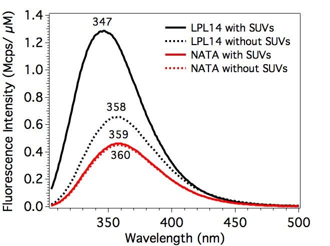
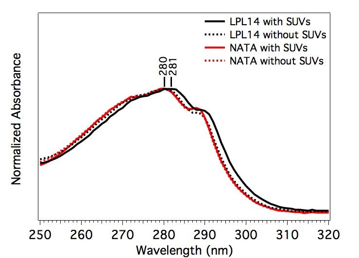
Luz et al.
 Page 11 of 41

density map. The complete structural model was built using COOT (32) and refined with
REFMAC5 (33) at 2.71 Å resolution to an Rfactor of 20.8% and an Rfree of 24.3% (Table 2).
Validation of the models was carried out using MolProbity (34).

Preparation of small unilamellar vesicles (SUVs)

 To generate SUVs, a 25 mg aliquot of 1,2-dimyristoyl-sn-glycero-3-phosphocholine (DMPC
from Avanti Polar Lipids) dissolved in chloroform was dried under nitrogen gas for at least 4 h.
The dried lipid was re-suspended in 5 ml of 20 mM potassium phosphate (KPi) buffer (pH 8.0),
yielding a lipid concentration of 5 mg/ml. The lipid solution was sonicated with an ultrasonic
microtip probe at 50% duty cycle with 30% maximum amplitude for 30 min. The SUVs (~50 nm

 Downloaded from www.jlr.org by guest, on November 8, 2020
diameter) were passed through a 0.22-µm filter and equilibrated overnight at 40°C before use in
experiments. A 5 mg/ml solution served as the stock SUV solution and was further diluted for
experiments.

Samples for optical spectroscopy

 LPL14 peptide was dissolved in 20 mM KPi buffer at pH 8.0 to create solutions of 10 µM
peptide in the absence and presence of 1 mg/ml SUVs; the total volume of each sample was 3.6
ml. Sample solutions of the model compound N-acetyl tryptophanamide (NATA) were also
prepared. These NATA solutions contained 30 µM NATA in KPi buffer (pH 8.0) in the presence
and absence of 1 mg/ml SUVs. A blank solution of 1 mg/ml SUVs in KPi buffer was also
generated. This SUV-only solution was used for background subtraction of the absorbance,
fluorescence, and UVRR spectra. All samples containing SUVs (peptide + SUVs, NATA + SUVs,
and SUVs-only) were allowed to equilibrate at 37°C for at least 1 h prior to collection of
measurements. Peptide and NATA samples that did not contain SUVs were incubated at room
temperature for at least 30 min prior to measurements. All reagents (NATA and buffer salts) were
purchased from Sigma-Aldrich and Thermo Fisher Scientific.

UV-Vis absorption spectroscopy

 The absorption spectra of LPL14 and NATA samples were acquired using an Agilent 8453
UV-Visible absorption spectrometer. The absorption features from LPL14 and NATA were
Luz et al.
 Page 12 of 41

isolated by subtraction of an SUV-only spectrum from the raw spectra of peptide and NATA. The
concentrations were determined using the 280-nm molar absorption coefficient, ε280, for
tryptophan (5,500 M–1cm–1) and tyrosine (1,490 M–1cm–1) (35) The calculated value of ε280 for
LPL14 was 17,990 M–1cm–1 based on the presence of one tyrosine and three tryptophan residues.
All absorption spectra were acquired using a 1-cm path length.

Steady-state fluorescence spectroscopy

 The fluorescence spectra were acquired using a Jobin Yvon-SPEX Fluorolog FL3-11
spectrofluorometer (Horiba). All samples were held in a quartz cuvette sealed with a Teflon cap
and maintained at 37°C during collection of the spectra. The samples were excited with 290 nm

 Downloaded from www.jlr.org by guest, on November 8, 2020
light along the 2 mm path, and emission was collected along the 10 mm path from 305 to 550 nm.
The excitation and emission bandpass were set to 2 nm. Spectra were collected at 1-nm increments
with 0.8-sec integration time. Fluorescence spectra of SUV-only and buffer-only samples were
also collected, and these background spectra were subtracted from the raw fluorescence spectra of
peptide and NATA to eliminate signal from buffer and SUV scattering.

UV resonance Raman spectroscopy (UVRR)

 The UVRR Ti:Sapphire laser apparatus has been described previously (36). UVRR spectra of
LPL14 and NATA were acquired with a 228 nm UV excitation beam. The concentrations of LPL14
were 11 µM and 9 µM in the presence and absence of SUVs, respectively. NATA concentrations
were 33 µM and 32 µM in the presence and absence of SUVs, respectively. To isolate signal from
LPL14 and NATA, UVRR spectra of SUVs-only and buffer-only were also acquired and
subtracted to remove contributions from vesicle and buffer scattering. All UVRR spectra were
collected for 20 min. The UV power at the sample was 1.2 mW and the flow rate was set to 0.16
ml/min to ensure fresh sample with each laser pulse and avoid sample degradation.

Peptide folding

 The LPL14 peptide sequence was submitted to the PEP-FOLD3 server
(http://bioserv.rpbs.univ-paris-diderot.fr/services/PEP-FOLD3/) using default parameters
Luz et al.
 Page 13 of 41

(number of simulations=100; sort by sOPEP). The top five models were superimposed by
secondary structure matching (37) and analyzed in COOT (32).

 Downloaded from www.jlr.org by guest, on November 8, 2020
Luz et al.
 Page 14 of 41

 RESULTS

Binding of 5D2 to LPL

 We analyzed, by surface plasmon resonance (SPR), the binding affinity of immobilized 5D2
to a 14-mer Trp-rich human LPL synthetic peptide (LPL14; KSDSYFSWSDWWSS) and 14-mer
peptides corresponding to the same Trp-rich loop in mouse, bovine, rat, chicken, and megabat
(“flying fox,” Pteropus vampyrus) LPL. The binding affinity of 5D2 to the human and bovine LPL
peptides was very high (KD of 0.19 and 0.78 nM, respectively) (Table 1, Fig. 1). The binding
affinity of 5D2 for the chicken LPL peptide was also very high, despite the fact that 5 of 14 residues
differed from the human sequence (Table 1). The binding affinity for the rat LPL peptide was

 Downloaded from www.jlr.org by guest, on November 8, 2020
modestly reduced, likely due to a Ser-to-Arg substitution in the Trp-rich loop (Table 1). In an
earlier study (9), changing that serine in human LPL (Ser-416) to Ala reduced the binding affinity
for 5D2. By SPR, 5D2 did not bind to the Trp-rich peptide from mouse LPL, where Ser-418 is
replaced with a Pro (Table 1, Fig. 1). 5D2 also did not bind to the Trp-rich LPL peptide from
megabat (Table 1, Fig. 1), which has the same Ser-to-Pro substitution. In earlier SPR studies (9),
replacing Ser-418 in human LPL with Ala reduced the affinity of 5D2 binding to a synthetic LPL
peptide.

 We used western blotting to assess 5D2 binding to V5-tagged full-length human, mouse,
chicken, zebrafish, and frog LPL (Fig. 2). We observed avid 5D2 binding (relative to the binding
of a V5 antibody) to human LPL, but there was no binding of 5D2 to mouse, zebrafish, or frog
LPL (Fig. 2). All three of those species contain substitutions in the WSDWW motif (417–421)
that is important for 5D2 binding (2, 9). We did detect 5D2 binding to full-length chicken LPL
(Fig. 2), but the binding was lower than we would have expected from the SPR studies (Table 1,
Fig. 1). This discrepancy suggests that the strength of 5D2 binding to full-length LPL differs from
the 14-mer LPL peptide (LPL14). Such a conclusion is consistent with findings from Kristensen
et al. (9), who observed that 5D2 binds to the properly folded carboxyl-terminal domain of human
LPL (residues 340–475) with greater affinity than to a 14-mer human LPL synthetic peptide.

 Given that affinity of 5D2 binding to synthetic peptides and properly folded LPL can differ,
we examined 5D2 binding to full-length mouse and human LPL. We transfected CHO cells with
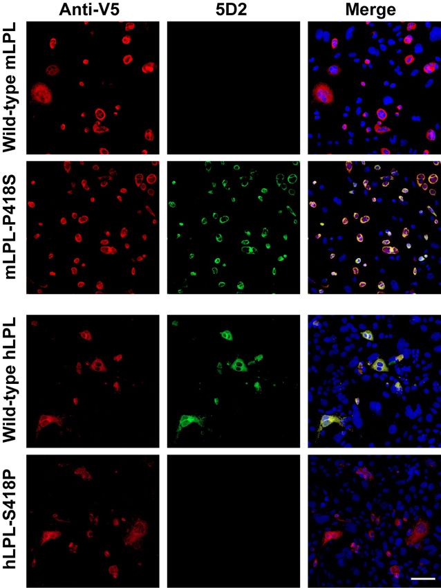
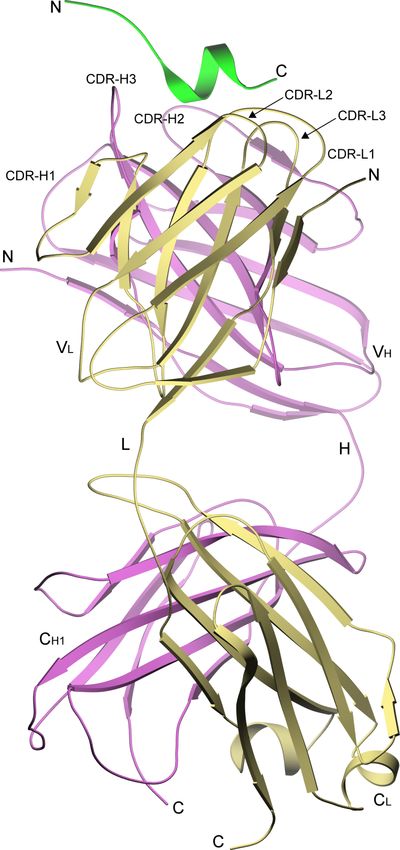
Luz et al.
 Page 15 of 41

a V5-tagged full-length mouse LPL vector or with a mutant vector in which Pro-418 had been
changed to Ser (the residue found in human LPL). By immunocytochemistry, 5D2 failed to bind
to full-length wild-type mouse LPL but bound avidly to the mutant mouse LPL with the
p.Pro418Ser substitution (mLPL-P418S) (Fig. 3). In parallel, we tested 5D2 binding to full-length
human LPL and a mutant human LPL containing a p.Ser418Pro substitution. 5D2 bound avidly to
the wild-type human LPL, but there was no binding to the mutant human LPL with the p.Ser418Pro
substitution (hLPL-S418P) (Fig. 3).

Structure of the 5D2 Fab fragment bound to the human LPL14 peptide

 We purified Fab fragments of 5D2, verified that they bound GPIHBP1-bound LPL (Fig. S1),

 Downloaded from www.jlr.org by guest, on November 8, 2020
and then used X-ray crystallography to solve the Fab structure, both in the presence and absence
of the 14-mer LPL peptide (LPL14). The structure revealed a typical Fab tertiary fold comprised
of four Ig domains, two from the light chain and two from the heavy chain and contained one Fab
molecule per crystallographic asymmetric unit. In the Apo 5D2 structure, indexing and scaling of
diffraction images for the apo 5D2 structure was complicated by anisotropic diffraction and
elongated spots leading to elevated Rwork and Rfree factors (Table 2); however, the electron density
maps were easily interpretable and allowed for an unambiguous assignment of main-chain and
side-chain atoms. Interestingly, the electron density maps for the Apo 5D2 structure showed
evidence for two conformations in the side chain of W69 of the heavy chain (part of the antigen
binding site) (Fig. S2). Apart from differences in the IgG hinge angle, the Apo and LPL14-bound
5D2 structures were very similar; the variable domains superimpose with a root-mean-square
(RMS) deviation of 0.61 Å in main-chain atom positions (Fig. S3). The 5D2–LPL14 complex
contained two molecules per crystallographic asymmetric unit which are almost identical to each
other, with an RMS deviation of 0.31 Å in main-chain atom positions within the variable domains.
LPL14 was located in the canonical antigen-binding site at the interface between the heavy and
light chains and shaped at its periphery by residues from the complement-determining region
(CDR) loops (Fig. 4). The LPL14 peptide was located parallel to the axis of the antigen recognition
site, with a hydrophobic face comprised of side chains from F415, W417, W420, and W421
oriented towards the light chain and a polar face (comprised of side chains from S416, S418, D419,
Luz et al.
 Page 16 of 41

and S422) oriented towards the heavy chain (Fig. 5). The N-terminus of LPL14 arches over the
top of the heavy chain CDR-H3 loop, with the side chain of D412 in LPL14 folding back to form
a hydrogen bond with the guanidinium group of the R121 side chain in the 5D2 heavy chain. A C-
terminal a-helical turn in LPL14 (residues 416–421) was nestled into the base of the antigen
binding site (Fig. 5).

 The interaction of LPL14 with 5D2 is biased towards heavy chain contacts. A total of 193
intermolecular interactions between LPL14 and the heavy chain were identified within a 4.5 Å
limit by CONTACT from the CCP4 suite (38), whereas only 37 interactions were identified
between LPL14 and the light chain. In addition, 454 Å2 of the peptide surface area was buried by
the heavy chain, whereas only 131 Å2 was buried by the light chain, as determined by the PISA

 Downloaded from www.jlr.org by guest, on November 8, 2020
server (39). Electron densities for LPL14 residues K410, S411, and S423 were absent and were
not included in the model.

 W417 and W421 of LPL14 (within the a-helical turn) extend into a cleft in the antigen-
recognition site, with the LPL14 side chains forming edge-to-face p-stacking interactions with
light chain W112 (Fig. 6) and additional hydrophobic interactions with the side chains of H54,
W69, and H118 of the heavy chain (Fig. 6) and L117 of the light chain. The indole nitrogen of
LPL W417 forms a hydrogen bond with the main-chain carbonyl of heavy chain N122, and the
indole nitrogen of LPL W421 forms a hydrogen bond with the backbone carbonyl of W112 of the
light chain (Fig. 5). S418 in LPL14 is located within the a-helical turn of the peptide (Fig. 6), and
both the main-chain nitrogen and side-chain hydroxyl are within hydrogen bonding distance of the
side-chain carboxylate of heavy chain D120 (Fig. 5).

 Finding a short a-helix in LPL14 was in line with predictions regarding the structure of the
peptide. We submitted the sequence of the LPL14 peptide to the PEP-FOLD3 server (40), and five
models were returned. All five had an amino-terminal region of undefined secondary structure and
a carboxyl-terminal a-helix similar to that observed in the LPL14–5D2 crystal structure (Fig. S4).
The top model was very similar to the crystal structure, with an RMS deviation for backbone atoms
of residues in the a-helical region (residues 416–421) of 0.13 Å (Fig. S4). While the side chains
Luz et al.
 Page 17 of 41

of F415, W417, and W421 were observed as different conformers in the crystal structure and the
PEP-FOLD3 models, the-a carbons were nearly identical.

 There are eight atom pairs in LPL14 and 5D2 common to both molecules in the asymmetric
unit that are within hydrogen bonding distance of each other (Table S1). Of the polar atoms within
hydrogen bonding distance, only one pair is found between the LPL peptide and the light chain
(LPL W421 indole nitrogen with light chain W112 carbonyl). One pair includes LPL14 main chain
atoms and 5D2 side chain atoms, and three pairs include LPL14 side chain atoms and 5D2 main
chain atoms. There is one hydrogen bond between backbone atoms of both chains—heavy chain
D120 carbonyl oxygen to LPL W417 amide nitrogen. The remaining three potential hydrogen
bonds are found between pairs of side chain atoms. The network of hydrogen bonds extends across

 Downloaded from www.jlr.org by guest, on November 8, 2020
the span of the peptide that was visualized, with the exception of LPL14 residues Y414, D419, and
W420 (where the side chains point away from the binding site) and F415 (which cannot form a
side-chain hydrogen bond). Of note, framework residue I52 of the heavy chain, which is present
in only 0.74% of Kabat database IgG sequences (www.bioinf.org.uk/abs/seqtest.html), forms
extensive van der Waals contacts with several LPL14 residues (W417, S418, W421) (Fig. 6).
Electron densities for LPL residues K410, S411, and S423 were absent, suggesting that they are
not particularly important for 5D2 binding.

 Earlier SPR studies found that converting F415, W417, or W421 to an Ala reduced 5D2
binding affinity by more than three orders of magnitude (9). All three aromatic side chains
contribute significantly to both the intermolecular hydrophobic packing interactions in the antigen
recognition site (Fig. 5, Fig. 6) as well as to intramolecular hydrophobic packing that shapes the
aromatic face of the peptide. In the same SPR study (9), changing LPL S418 or W420 to Ala
decreased binding by 50-fold. The p.Ser418Ala substitution would eliminate a hydrogen bond
between the side-chain hydroxyl of Ser-418 and the side-chain carboxylate of heavy chain D120
(Fig. 5). The W420A substitution would eliminate hydrophobic packing with the phenyl group of
F415.

 The conformation of the peptide itself appears to be stabilized by a network of hydrogen bonds
which are conserved between the two copies in the asymmetric unit, except for that formed with
Luz et al.
 Page 18 of 41

the main-chain carbonyl atom of Ser-418 (Table S2). The carboxyl-terminal a-helical turn is
stabilized by main-chain hydrogen bonds formed between residues S416 and W420 and between
W417 and W421. Our experiments demonstrated that 5D2 does not bind to mouse LPL (which
contains a Pro at residue 418 rather than a Ser), either in the context of the 14-mer synthetic LPL
peptide or full-length LPL (Fig. 1, Fig. 3). Also, when Ser-418 in human LPL is replaced with a
Pro, the binding of 5D2 is abolished (Fig. 3). These observations are explained by our crystal
structure. Both the backbone nitrogen and side-chain hydroxyl of Ser-418 in human LPL14 are
within hydrogen bonding distance of the side-chain carboxylate of heavy chain D120 (Fig. 5).
Substitution of Ser-418 with a Pro would eliminate crucial hydrogen bonds and introduce a
hydrophobic side chain which would clash with the side chain of D120. Also, the side-chain

 Downloaded from www.jlr.org by guest, on November 8, 2020
hydroxyl of Ser-418 is within hydrogen bonding distance to the side-chain hydroxyl of Ser-416,
thereby contributing to the internal order of the bound LPL14 structure (Fig. 5). While the Pro at
residue 418 interferes with 5D2 binding, there is no reason to suspect that it would interfere with
the role of the Trp-rich loop in enzyme function. Interestingly, the PEP-FOLD3 server (40)
predicted a short a-helix in the mouse 14-mer LPL peptide, with one side enriched in hydrophobic
amino acids (similar to the crystal structure and the PEP-FOLD3 prediction for human LPL14)
(Fig. S5).

The Trp-rich 5D2 peptide (LPL14) interacts directly with lipids

 Spectroscopy revealed tryptophan–lipid interactions between LPL14 and SUV bilayers. The
absorption spectrum of LPL14 at 250–320 nm primarily reflects LPL14’s three tryptophan
residues. In the presence of SUVs, the absorption spectrum exhibited a 1-nm red shift from 280 to
281 nm, relative to LPL14 in buffer (Fig. 7). This shift was only observed in LPL14, and not the
model tryptophan compound NATA. This 1-nm shift for LPL14 indicates that the tryptophan
residue(s) are in a more hydrophobic environment in the presence of SUVs (41). This change in
environment from aqueous to lipid bilayer is corroborated by fluorescence spectra. The tryptophan
residues in LPL14 exhibited a fluorescence maximum at 358 nm in buffer and shifted to 347 nm
in the presence of SUVs, and there was a concomitant increase in the fluorescence intensity (i.e.,
quantum yield) (Fig. 8). This 11-nm blue shift and increase in the quantum yield upon incubation
Luz et al.
 Page 19 of 41

with SUVs indicates that the tryptophan residues are in a more hydrophobic lipid environment
(42).

 Vibrational spectroscopy in the form of UVRR supported the absorption and fluorescence
findings. UVRR is an ideal tool because excitation at 228 nm enhances the signal from tryptophan
without obfuscation from other residues or buffer. The UVRR bands reflect different properties of
the indole side chain. The tryptophan W7 Fermi doublet band at 1300 to 1400 cm–1 is particularly
informative because it is a sensitive marker for local hydrophobicity, and the ratio of the peak
intensities of 1366 to 1345 cm–1 (RFD) increases when tryptophan is in a hydrophobic environment
(36). The RFD value for LPL14 in the presence of SUVs is 1.6, which is significantly larger than
the values in buffer and for NATA (Fig. 9). This finding supports the notion that the tryptophan

 Downloaded from www.jlr.org by guest, on November 8, 2020
residue(s) in LPL14 directly interact with the more hydrophobic environment of the lipid bilayer.
Luz et al.
 Page 20 of 41

 DISCUSSION

 We defined the structure of 5D2 bound to a 14-mer peptide (LPL14) corresponding to the Trp-
rich loop within the carboxyl terminus of human LPL. Eleven of the 14 LPL residues were
identified in the electron density map. The carboxyl-terminal portion of the peptide formed a short
amphipathic a-helix that was positioned in the cleft of the antigen recognition site, with the
hydrophobic face (three tryptophans and a phenylalanine) oriented towards the light chain and a
polar face (three serines and an aspartic acid) pointing towards the heavy chain. In our crystal
structure, a short a-helix was observed in the LPL14 peptide bound to 5D2. However, at this point,
it is unclear whether this a-helix is an invariant feature in native LPL or whether it simply
represents one of several transient conformations that is detected and then trapped by 5D2 binding.

 Downloaded from www.jlr.org by guest, on November 8, 2020
Favoring the latter possibility is the observation that this segment in native LPL (specifically, the
peptide FSWSNW) is not protected from deuterium uptake in hydrogen–deuterium exchange
studies unless the LPL is bound to Fab 5D2 (9). If the a-helical conformation were an invariant
feature of native LPL, we would have expected to find that the backbone LPL amide hydrogens
were protected from deuterium uptake (even in the absence of bound 5D2). The Trp-rich loop in
full-length LPL was visualized in a recent cryo-EM LPL structure (20) and appeared to show an
a-helical conformation. However, that conformation was presumably a consequence of the
repetitive head-to-tail homodimer LPL configuration in the cryo-EM structure, where the Trp-rich
loop is buried in the catalytic pocket of a partner LPL molecule. In our X-ray crystal structure of
the LPL–GPIHBP1 complex (19), LPL’s Trp-rich loop was also buried in the catalytic pocket of
a partner LPL molecule and not clearly visualized. In considering the latter structure, there is little
reason to believe that GPIHBP1 binding would have changed conformation of the LPL’s Trp-rich
loop, simply because the Trp-rich loop and the binding site for GPIHBP1 are located on opposite
sides of the LPL molecule (19). 5D2 binds avidly to GPIHBP1-bound LPL, as shown in earlier
studies (9, 43) and in Fig. S1 of the current study.

 Our studies demonstrated that LPL’s Trp-rich loop is a crucial part of the 5D2 epitope, but it
is possible that other LPL sequences influence, either directly or indirectly, the strength of 5D2
binding. First, by SPR, 5D2 bound with higher affinity to the carboxyl-terminal region of human
Luz et al.
 Page 21 of 41

LPL (residues 340–475) than to the LPL14 synthetic peptide (9). Second, by SPR, the KD for 5D2
binding to the human and chicken 14-mer peptides were similar, but by western blotting 5D2 binds
more avidly to full-length human LPL than to full-length chicken LPL.

 Studies by Williams et al. (7) revealed that mutating W420 and W421 in the carboxyl terminus
of LPL markedly reduced LPL’s ability to bind lipoproteins. Subsequent studies by Lookene et al.
(6) revealed that mutating W420 and W421 markedly reduced triglyceride hydrolysis, suggesting
that the tryptophans were either directly involved in binding lipids or that the tryptophan mutations
had simply disrupted LPL conformation. Our current studies support the former possibility. When
we incubated LPL14 with small unilamellar vesicles (SUVs), the peak emission spectra by
fluorescence spectroscopy shifted from 358 to 347 nm, whereas no shift occurred when SUVs

 Downloaded from www.jlr.org by guest, on November 8, 2020
were added to the model compound N-acetyl-tryptophanamide (NATA). These findings implied
direct interactions of the hydrophobic Trp-rich loop with lipids, which is consistent with the fact
that tryptophans often participate in the binding of proteins to membrane bilayers (44). Moreover,
ultraviolet resonance Raman (UVRR) spectra indicated that the tryptophans in LPL14 interacted
directly with lipids. The hydrophobicity of LPL’s tryptophan-rich loop also underlies the
propensity of LPL, at high concentrations, to form head-to-tail homodimers (19). In those
homodimers, the interaction of the Trp-rich loop with the catalytic pocket shields the hydrophobic
tryptophans from the aqueous environment (19). The binding of 5D2 to LPL also shields the
tryptophan-rich loop and thereby prevents the formation of homodimers (9).

 5D2 was generated by immunizing laboratory mice (Mus musculus) with bovine LPL. For that
reason, it is not particularly surprising that 5D2 does not detect mouse LPL. Studies of 5D2 binding
to LPL peptides indicated that the substitution of a Pro for Ser in the Trp-rich loop of mouse LPL
played a role in the absence of 5D2 binding to mouse LPL (2), but it was not clear that this
substitution was solely responsible, particularly in the setting of full-length LPL. Our studies
settled that issue. When Pro-418 in full-length mouse LPL was changed to Ser, 5D2 bound to
mouse LPL avidly, and when Ser-418 in human LPL was replaced with Pro, 5D2 binding to human
LPL was abolished. The crystal structure allowed us to make sense of these observations.
Replacing Ser-418 with Pro would eliminate the hydrogen bonds and introduce a hydrophobic side
Luz et al.
 Page 22 of 41

chain which would clash with the side chain of D120 of the 5D2 heavy chain. Interestingly, the
Ser-to-Pro substitution in the Trp-rich loop of LPL is found in multiple mouse species (Mus pahari,
Mus caroli, Mus spicilegus, Mus spretus) and in the megabat (Pteropus vampyrus). By SPR, there
was no binding of 5D2 to a synthetic peptide corresponding to the Trp-rich loop from megabat
LPL. The appearance of a Pro in the Trp-rich loop in the megabat LPL likely appeared
independently during mammalian evolution, as phylogenetic analyses have revealed that bats are
more closely related to horses than to mice or humans (45). Interestingly, mice and megabats are
both voracious eaters, consuming a large percentage of their body weight each day in food, mainly
in the form of carbohydrates. The carbohydrates are likely transformed into triglycerides and used
for fuel, but plasma triglyceride levels are low in both mice and the megabat (46, 47). There is no

 Downloaded from www.jlr.org by guest, on November 8, 2020
reason to believe that the Pro in the Trp-rich loop interferes with triglyceride hydrolysis. Whether
the proline residue in mouse and megabat LPL might actually improve LPL function (e.g., by
optimizing LPL’s capacity to bind lipids and hydrolyze triglycerides) is unknown.
Luz et al.
 Page 23 of 41

Data Availability Statement

Data are contained in the manuscript. If questions, please contact the corresponding authors,
Stephen G. Young (e-mail: sgyoung@mednet.ucla.edu; phone: 310-825-4934) and Gabriel
Birrane (e-mail: gbirrane@bidmc.harvard.edu; phone: 617-667-0025). As noted in Figure 2, the
coordinates for APO 5D2 (PDB ID 6WT3) and 5D2-LPL11 (PDB ID 6WN4) have been deposited
in the PDB.

 Downloaded from www.jlr.org by guest, on November 8, 2020
Luz et al.
 Page 24 of 41

Acknowledgments

The authors thank John Brunzell for sharing the 5D2 hybridoma and Gry Ellis Rasmussen for
technical assistance. We would like to thank the staff at the AMX beamline 17-ID-1 at the NSLS
II, I24 beamline at the Diamond Light Source, and Helix Biostructures for assistance with X-ray
data collection. This work was supported by Grants HL090553, HL087228, and HL125335 from
the National Heart, Lung, and Blood Institute; Transatlantic Network Grant 12CVD04 from the
Fondation Leducq; and NOVO Nordisk Foundation Grant NNF17OC0026868.

 Downloaded from www.jlr.org by guest, on November 8, 2020
Luz et al.
 Page 25 of 41

 REFERENCES

1. Babirak, S. P., P. H. Iverius, W. Y. Fujimoto, and J. D. Brunzell. 1989. Detection and
 characterization of the heterozygote state for lipoprotein lipase deficiency. Arteriosclerosis
 9: 326–334.

2. Chang, S.-F., B. Reich, J. D. Brunzell, and H. Will. 1998. Detailed characterization of the
 binding site of the lipoprotein lipase-specific monoclonal antibody 5D2. J. Lipid Res. 39:
 2350–2359.

3. Peterson, J., W. Y. Fujimoto, and J. D. Brunzell. 1992. Human lipoprotein lipase:
 relationship of activity, heparin affinity, and conformation as studied with monoclonal

 Downloaded from www.jlr.org by guest, on November 8, 2020
 antibodies. J Lipid Res 33: 1165–1170.

4. Wong, H., R. C. Davis, T. Thuren, J. W. Goers, J. Nikazy, M. Waite, and M. C. Schotz. 1994.
 Lipoprotein lipase domain function. J. Biol. Chem. 269: 10319–10323.

5. Liu, M. S., Y. Ma, M. R. Hayden, and J. D. Brunzell. 1992. Mapping of the epitope on
 lipoprotein lipase recognized by a monoclonal antibody (5D2) which inhibits lipase activity.
 Biochim Biophys Acta 1128: 113–115.

6. Lookene, A., N. B. Groot, J. J. Kastelein, G. Olivecrona, and T. Bruin. 1997. Mutation of
 tryptophan residues in lipoprotein lipase. Effects on stability, immunoreactivity, and
 catalytic properties. J Biol Chem 272: 766–772.

7. Williams, S. E., I. Inoue, H. Tran, G. L. Fry, M. W. Pladet, P.-H. Iverius, J.-M. Lalouel, D.
 A. Chappell, and D. K. Strickland. 1994. The carboxyl-terminal domain of lipoprotein lipase
 binds to the low density lipoprotein receptor-related protein/a2-macroglobulin receptor
 (LRP) and mediates binding of normal very low density lipoproteins to LRP. J. Biol. Chem.
 269: 8653–8658.

8. Goulbourne, C. N., P. Gin, A. Tatar, C. Nobumori, A. Hoenger, H. Jiang, C. R. Grovenor,
 O. Adeyo, J. D. Esko, I. J. Goldberg, K. Reue, P. Tontonoz, A. Bensadoun, A. P. Beigneux,
Luz et al.
 Page 26 of 41

 S. G. Young, and L. G. Fong. 2014. The GPIHBP1-LPL complex is responsible for the
 margination of triglyceride-rich lipoproteins in capillaries. Cell Metab 19: 849–860.

9. Kristensen, K. K., K. Z. Leth-Espensen, H. D. T. Mertens, G. Birrane, M. Meiyappan, G.
 Olivecrona, T. J. D. Jorgensen, S. G. Young, and M. Ploug. 2020. Unfolding of monomeric
 lipoprotein lipase by ANGPTL4: Insight into the regulation of plasma triglyceride
 metabolism. Proc Natl Acad Sci U S A 117: 4337–4346.

10. Iverius, P. H., and A. M. Ostlund-Lindqvist. 1976. Lipoprotein lipase from bovine milk.
 Isolation procedure, chemical characterization, and molecular weight analysis. J Biol Chem
 251: 7791–7795.

 Downloaded from www.jlr.org by guest, on November 8, 2020
11. Garfinkel, A. S., E. S. Kempner, O. Ben-Zeev, J. Nikazy, S. J. James, and M. C. Schotz.
 1983. Lipoprotein lipase: size of the functional unit determined by radiation inactivation. J.
 Lipid Res. 24: 775–780.

12. Olivecrona, T., G. Bengtsson-Olivecrona, J. C. Osborne, Jr., and E. S. Kempner. 1985.
 Molecular size of bovine lipoprotein lipase as determined by radiation inactivation. J. Biol.
 Chem. 260: 6888–6891.

13. Lookene, A., L. Zhang, M. Hultin, and G. Olivecrona. 2004. Rapid subunit exchange in
 dimeric lipoprotein lipase and properties of the inactive monomer. J Biol Chem 279: 49964–
 49972.

14. Zhang, L., A. Lookene, G. Wu, and G. Olivecrona. 2005. Calcium triggers folding of
 lipoprotein lipase into active dimers. J. Biol. Chem. 280: 42580–42591.

15. Wang, H., and R. H. Eckel. 2009. Lipoprotein lipase: from gene to obesity. Am J Physiol
 Endocrinol Metab 297: E271–288.

16. Sukonina, V., A. Lookene, T. Olivecrona, and G. Olivecrona. 2006. Angiopoietin-like
 protein 4 converts lipoprotein lipase to inactive monomers and modulates lipase activity in
 adipose tissue. Proc. Natl. Acad. Sci. USA 103: 17450–17455.
Luz et al.
 Page 27 of 41

17. Doolittle, M. H., and M. Peterfy. 2010. Mechanisms of lipase maturation. Clin Lipidol 5: 71–
 85.

18. Beigneux, A. P., C. M. Allan, N. P. Sandoval, G. W. Cho, P. J. Heizer, R. S. Jung, K. L.
 Stanhope, P. J. Havel, G. Birrane, M. Meiyappan, J. E. t. Gill, M. Murakami, K. Miyashita,
 K. Nakajima, M. Ploug, L. G. Fong, and S. G. Young. 2019. Lipoprotein lipase is active as
 a monomer. Proc Natl Acad Sci U S A 116: 6319–6328.

19. Birrane, G., A. P. Beigneux, B. Dwyer, B. Strack-Logue, K. K. Kristensen, O. L. Francone,
 L. G. Fong, H. D. T. Mertens, C. Q. Pan, M. Ploug, S. G. Young, and M. Meiyappan. 2019.
 Structure of the lipoprotein lipase-GPIHBP1 complex that mediates plasma triglyceride

 Downloaded from www.jlr.org by guest, on November 8, 2020
 hydrolysis. Proc Natl Acad Sci U S A 116: 1723–1732.

20. Gunn, K. H., B. S. Roberts, F. Wang, J. D. Strauss, M. J. Borgnia, E. H. Egelman, and S. B.
 Neher. 2020. The structure of helical lipoprotein lipase reveals an unexpected twist in lipase
 storage. Proc Natl Acad Sci U S A 117: 10254–10264.

21. Beigneux, A. P., B. S. Davies, S. Tat, J. Chen, P. Gin, C. V. Voss, M. M. Weinstein, A.
 Bensadoun, C. R. Pullinger, L. G. Fong, and S. G. Young. 2011. Assessing the role of the
 glycosylphosphatidylinositol-anchored high density lipoprotein-binding protein 1
 (GPIHBP1) three-finger domain in binding lipoprotein lipase. J Biol Chem 286: 19735–
 19743.

22. Leth, J. M., and M. Ploug. 2020. Determination of binding kinetics of intrinsically disordered
 proteins by surface plasmon resonance. Methods in Molecular Biology. Chapter 31 in the
 volume, Intrinsically Disordered Proteins: Methods and Protocols, Kragelund, B.B., and
 Skriver, K. (Eds). Springer, ISBN 978-1-0716-0523-3.

23. Kabsch, W. 2010. Xds. Acta Crystallogr D Biol Crystallogr 66: 125–132.

24. Tickle, I. J., C. Flensburg, P. Keller, W. Paciorek, A. Sharff, C. Vonrhein, and G. Bricogne.
 2018. STARANISO. Global Phasing Ltd., Cambridge, United Kingdom.
 (http://staraniso.globalphasing.org/).
Luz et al.
 Page 28 of 41

25. Vonrhein, C., C. Flensburg, P. Keller, A. Sharff, O. Smart, W. Paciorek, T. Womack, and G.
 Bricogne. 2011. Data processing and analysis with the autoPROC toolbox. Acta Crystallogr
 D Biol Crystallogr 67: 293–302.

26. McCoy, A. J., R. W. Grosse-Kunstleve, P. D. Adams, M. D. Winn, L. C. Storoni, and R. J.
 Read. 2007. Phaser crystallographic software. J Appl Crystallogr 40: 658–674.

27. Burak, M. F., K. E. Inouye, A. White, A. Lee, G. Tuncman, E. S. Calay, M. Sekiya, A.
 Tirosh, K. Eguchi, G. Birrane, D. Lightwood, L. Howells, G. Odede, H. Hailu, S. West, R.
 Garlish, H. Neale, C. Doyle, A. Moore, and G. S. Hotamisligil. 2015. Development of a
 therapeutic monoclonal antibody that targets secreted fatty acid-binding protein aP2 to treat

 Downloaded from www.jlr.org by guest, on November 8, 2020
 type 2 diabetes. Sci Transl Med 7: 319ra205.

28. Liebschner, D., P. V. Afonine, M. L. Baker, G. Bunkoczi, V. B. Chen, T. I. Croll, B. Hintze,
 L. W. Hung, S. Jain, A. J. McCoy, N. W. Moriarty, R. D. Oeffner, B. K. Poon, M. G. Prisant,
 R. J. Read, J. S. Richardson, D. C. Richardson, M. D. Sammito, O. V. Sobolev, D. H.
 Stockwell, T. C. Terwilliger, A. G. Urzhumtsev, L. L. Videau, C. J. Williams, and P. D.
 Adams. 2019. Macromolecular structure determination using X-rays, neutrons and electrons:
 recent developments in Phenix. Acta Crystallogr D Struct Biol 75: 861–877.

29. Otwinowski, Z., and W. Minor. 1997. Processing of X-ray diffraction data collected in
 oscillation mode. Methods Enzymol. 276: 307–326.

30. Vagin, A., and A. Teplyakov. 2010. Molecular replacement with MOLREP. Acta Crystallogr
 D Biol Crystallogr 66: 22–25.

31. Cowtan, K. 2006. The Buccaneer software for automated model building. 1. Tracing protein
 chains. Acta Crystallogr D Biol Crystallogr 62: 1002–1011.

32. Emsley, P., B. Lohkamp, W. G. Scott, and K. Cowtan. 2010. Features and development of
 Coot. Acta Crystallogr D Biol Crystallogr 66: 486-501.

33. Murshudov, G. N., A. A. Vagin, and E. J. Dodson. 1997. Refinement of macromolecular
 structures by the maximum-likelihood method. Acta Crystallogr. D 53: 240–255.
Luz et al.
 Page 29 of 41

34. Williams, C. J., J. J. Headd, N. W. Moriarty, M. G. Prisant, L. L. Videau, L. N. Deis, V.
 Verma, D. A. Keedy, B. J. Hintze, V. B. Chen, S. Jain, S. M. Lewis, W. B. Arendall, 3rd, J.
 Snoeyink, P. D. Adams, S. C. Lovell, J. S. Richardson, and D. C. Richardson. 2018.
 MolProbity: More and better reference data for improved all-atom structure validation.
 Protein Sci 27: 293–315.

35. Pace, C. N., F. Vajdos, L. Fee, G. Grimsley, and T. Gray. 1995. How to measure and predict
 the molar absorption coefficient of a protein. Protein Sci 4: 2411–2423.

36. Asamoto, D. K., and J. E. Kim. 2019. UV resonance Raman spectroscopy as a tool to probe
 membrane protein structure and dynamics. Methods Mol Biol 2003: 327–349.

 Downloaded from www.jlr.org by guest, on November 8, 2020
37. Krissinel, E., and K. Henrick. 2004. Secondary-structure matching (SSM), a new tool for
 fast protein structure alignment in three dimensions. Acta Crystallogr D Biol Crystallogr 60:
 2256–2268.

38. Winn, M. D., C. C. Ballard, K. D. Cowtan, E. J. Dodson, P. Emsley, P. R. Evans, R. M.
 Keegan, E. B. Krissinel, A. G. Leslie, A. McCoy, S. J. McNicholas, G. N. Murshudov, N. S.
 Pannu, E. A. Potterton, H. R. Powell, R. J. Read, A. Vagin, and K. S. Wilson. 2011. Overview
 of the CCP4 suite and current developments. Acta Crystallogr D Biol Crystallogr 67: 235–
 242.

39. Krissinel, E., and K. Henrick. 2007. Inference of macromolecular assemblies from crystalline
 state. J Mol Biol 372: 774–797.

40. Maupetit, J., P. Derreumaux, and P. Tuffery. 2010. A fast method for large-scale de novo
 peptide and miniprotein structure prediction. J Comput Chem 31: 726–738.

41. Bailey, J. E., G. H. Beaven, D. A. Chignell, and W. B. Gratzer. 1968. An analysis of
 perturbations in the ultraviolet absorption spectra of proteins and model compounds. Eur J
 Biochem 7: 8–14.

42. Kang, G., I. López-Peña, S. Bhakta, and J. E. Kim. 2013. Probing membrane protein structure
 and dynamics by fluorescence spectroscopy. Encyclopedia of Analytical Chemistry:
You can also read