A rapidly deployable individualized system for augmenting ventilator capacity
←
→
Page content transcription
If your browser does not render page correctly, please read the page content below
REPORTS

                                                                                                               Cite as: S. Srinivasan et al., Sci. Transl. Med.
                                                                                                                      10.1126/scitranslmed.abb9401 (2020).

CORONAVIRUS

A rapidly deployable individualized system for augmenting
ventilator capacity
Shriya Srinivasan1,2,3, Khalil B Ramadi1,2,3, Francesco Vicario8, Declan Gwynne1,2,3, Alison Hayward2,3,4, David
Lagier7, Robert Langer1,3,5,6,·Joseph J. Frassica8,9, Rebecca M. Baron10, Giovanni Traverso1,2,3*
1Department of Mechanical Engineering, Massachusetts Institute of Technology, Cambridge, MA 02139, USA. 2Division of Gastroenterology, Hepatology and Endoscopy,

Brigham and Women’s Hospital, Harvard Medical School, Boston, MA 02115, USA. 3David H. Koch Institute for Integrative Cancer Research, Massachusetts Institute of
Technology, Cambridge, MA 02139, USA. 4Division of Comparative Medicine, Massachusetts Institute of Technology, Cambridge, MA 02139, USA. 5Department of Chemical
Engineering, Massachusetts Institute of Technology, Cambridge, MA 02139, USA. 6Institute for Medical Engineering and Science, Massachusetts Institute of Technology,

                                                                                                                                                                       Downloaded from http://stm.sciencemag.org/ by guest on December 27, 2020
Cambridge, MA 02139, USA. 7Department of Anesthesia, Critical Care and Pain Medicine, Massachusetts General Hospital and Harvard Medical School, Boston, MA, 02139.
8Philips Research North America, Cambridge, MA 02141, USA. 9Institute for Medical Engineering and Science, Massachusetts Institute of Technology, Cambridge, MA

02139, USA. 10Division of Pulmonary and Critical Care Medicine, Brigham and Women’s Hospital, Harvard Medical School, Boston, MA 02115, USA.
*Corresponding author. Email: cgt20@mit.edu, ctraverso@bwh.harvard.edu

Strategies to split ventilators to support multiple patients requiring ventilatory support have been
proposed and used in emergency cases in which shortages of ventilators cannot otherwise be remedied by
production or procurement strategies. However, the current approaches to ventilator sharing lack the
ability to individualize ventilation to each patient, measure pulmonary mechanics, and accommodate
rebalancing of the airflow when one patient improves or deteriorates, posing safety concerns to patients.
Potential cross-contamination, lack of alarms, insufficient monitoring, and inability to adapt to sudden
changes in patient status have prevented widespread acceptance of ventilator sharing. We have developed
an individualized system for augmenting ventilator efficacy (iSAVE) as a rapidly deployable platform that
uses a single ventilator to simultaneously and more safely support two subjects. The iSAVE enables
subject-specific volume and pressure control and the rebalancing of ventilation in response to improvement
or deterioration in an individual’s respiratory status. The iSAVE incorporates mechanisms to measure
pulmonary mechanics, mitigate cross-contamination and backflow, and accommodate sudden flow changes
due to subject interdependencies within the respiratory circuit. We demonstrate these capacities through
validation using closed- and open-circuit ventilators on linear test lungs. We show that the iSAVE can
temporarily ventilate two pigs on one ventilator as efficaciously as each pig on its own ventilator. By
leveraging off-the-shelf medical components, the iSAVE could rapidly expand the ventilation capacity of
healthcare facilities during emergency situations such as pandemics.

INTRODUCTION                                                                         chain, recent coronavirus disease 2019 (COVID-19) hospitali-
    Ventilators are vital equipment for critical-care condi-                         zation rates, and predictive models, estimates of the shortfall
tions, surgical interventions, and procedures requiring anes-                        of ventilators within the U.S. range between 45,000 - 160,000
thesia. Ventilators assist with respiration by moving air into                       (1–4). Globally, the World Health Organization (WHO) esti-
and out of the lungs and are extensively used in the treatment                       mates that fewer than 2,000 ventilators are available across
of patients with acute respiratory distress syndrome (ARDS)                          the 41 African countries (5) to support a population of 1.2 bil-
and respiratory failure. Pandemics of infections that cause se-                      lion people. Coupled with high population density and rela-
vere respiratory dysfunction can lead to a rapid increase in                         tive poverty, pandemics can make the procurement, building,
the volume of patients requiring ventilation, exceeding the                          or buying of new, emergency ventilators challenging for some
number of ventilators available in healthcare facilities and                         countries. Low-cost and emergency ventilators have been de-
necessitating difficult triage decisions (1, 2). The most recent                     veloped to address the cost barrier associated with the acqui-
publicly available data (2010) reports that about 62,000 full-                       sition of new ventilators at scale. However, their
featured mechanical ventilators are available within the                             manufacturing and deployment relies on supply, assembly,
United States, with an additional 12,700 stockpiled in the                           and distribution chains which can be disrupted by public
Centers for Disease Control and Prevention strategic national                        health crises involving lockdowns and import/export re-
stockpile (SNS) (1). Based on quantified disruptions in supply                       strictions. The use of new ventilator designs has also raised

First release: 18 May 2020                                             stm.sciencemag.org              (Page numbers not final at time of first release) 1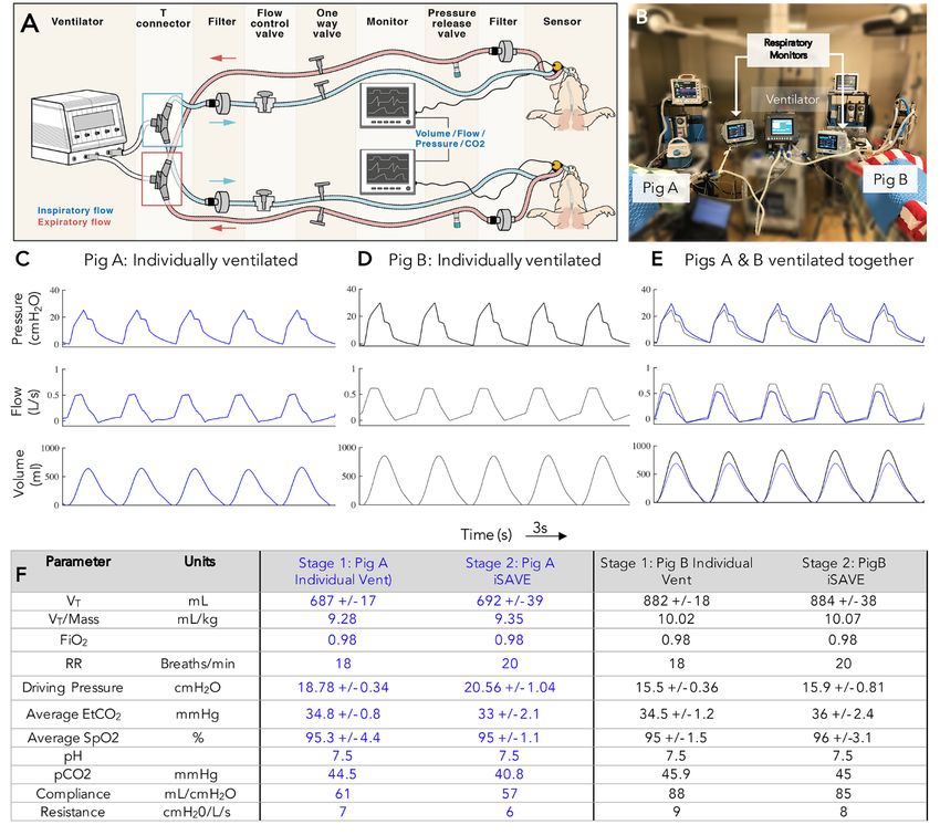
safety concerns, as clinical staff would need to operate unfa-     RESULTS
miliar technology. Given both supply and implementation            Design of the iSAVE
hurdles that may delay rapid deployment of low-cost ventila-           The iSAVE uses a series of valves and flow regulators in
tors, other strategies warrant consideration.                      parallel limbs to effectively maintain the desired tidal volume
    Ventilator sharing, or dividing the airflow from one ven-      (VT) and positive end-expiratory pressure (PEEP) for each pa-
tilator among multiple patients, has been previously per-          tient (Fig. 1, tables S1 and S2) under volume control mode. In
formed in a few emergency cases (6). By using readily              closed-circuit ventilators (Fig. 1), Y or T connectors are used
available tubing and ventilatory equipment, ventilator shar-       to multiplex individual inspiratory channels for each patient.
ing can be immediately implemented to expand the capacity          Each inspiratory channel consists of a filter, flow control
of existing ventilators with which clinicians are familiar. In     valve, a one-way flow valve, and standard sensors (pressure,
previously proposed configurations, multiplexing ventilation       flow, capnostat) in series. The expiratory limb consists of a
involved connecting multiple outflow tracts to the ventilator      filter, pressure release valve, and one-way valve prior to con-
to divide flow amongst patients (6–9) wherein the compli-          nection to Y or T connectors which are routed back to the

                                                                                                                                        Downloaded from http://stm.sciencemag.org/ by guest on December 27, 2020
ance (C) and resistance (R) of each patient’s pulmonary sys-       ventilator. The iSAVE can be configured to both open-circuit
tem became part of the same circuit and drove the balance of       ventilators (which consist of only an inspiratory limb with
airflow. This patient interdependence poses various safety         passive expiration) and closed-circuit ventilators (which pos-
concerns: (i) Independent control of volume and pressure to        sess inspiratory and expiratory limbs). Open-circuit ventila-
each patient is not possible, which is important for lung-pro-     tors can be used with the same circuit shown in Fig. 1, except
tective ventilation and the standard of care for ARDS; (ii)        that the expiratory limb would be connected to a Whisper
alarm monitoring becomes challenging due to the complex            Swivel (Philips Respironics) exhalation adaptor or equivalent
circuit configuration; (iii) sudden events such as pneumotho-      valve for the expiratory flow port. We used positive expiratory
rax, tube occlusion, or disconnection of an endotracheal tube,     pressure (PEP) threshold devices as one-way valves, set to
causes potentially harmful rebalancing of ventilation; and         their highest setting, because these are readily available in
(iv) changes in one patient’s condition (clinical improvement      hospitals. See tables S1 and S2 for a full list of supplies.
or deterioration) results in an automatic change to ventila-           The flow control valve is used to allocate the appropriate
tion of other patients. Other practical challenges include         VT to each patient. Filters on each limb mitigate cross-con-
monitoring, routine measurement of pulmonary mechanics,            tamination between individual patient circuits and filter ex-
overcoming ventilator self-tests/calibration, and the risk of      pired gas before release into the room through the pressure
exposure due to a break in the circuit that aerosolizes respir-    release valve, thus limiting pathogen exposure to healthcare
atory-borne infectious agents when adding and removing pa-         workers. The one-way valves prevent backflow and mitigate
tients. For these reasons, medical associations including the      over-distention in cases of rapid flow change. PEEP valves en-
American Association for Respiratory Care (AARC) issued a          able the individualized control of PEEP for each patient and
joint statement explicitly advising clinicians against the shar-   function as a pop-off valves to release excess pressure. Pres-
ing of mechanical ventilators with current approaches (10).        sure, flow, and CO2 sensors are positioned on the patient Y
    To expand ventilator capacity while incorporating the          piece and data, visualized on a separate patient monitor, is
constraints associated with supply chain/distribution limita-      used for setup, titration, and monitoring. During initial
tions, we engineered the individualized system for augment-        setup, the respiratory rate (RR), PEEP, FiO2 (fraction of in-
ing ventilator efficacy (iSAVE), which repurposes medical-         spired oxygen), inspiration:expiration (I:E) ratio, and the
grade valves, sensors, and filters to allow a single ventilator    sum of the VT for each patients is set on the ventilator under
to provide personalized volume and pressure support to at          volume control mode. Then, the flow control and PEEP valves
least two patients. The use of repurposed components can en-       can be titrated to individualize VT and PEEP. The exhaled vol-
hance ventilator capacity independent of supply chain limi-        ume and minute ventilation alarms on the ventilator are set
tations. Here, we describe the design and validation of the        according to the sum of the exhaled tidal volumes from both
iSAVE through benchtop and in vivo tests sharing a single          patients and enable the ventilator to alarm in response to
ventilator among two pigs. We hypothesized that our system         sudden change in either patient’s status (shunt, occlusion, or
could maintain specified ventilation parameters to each sub-       disconnection of the endotracheal tube). Through this design,
ject amidst static and dynamic changes in resistance and           the iSAVE overcomes many of the aforementioned challenges
compliance. We simulated clinical scenarios, focusing on           of splitting ventilation (Table 1).
those relevant to the management of ARDS, and validated the        Benchtop testing of the iSAVE using linear test lungs
safety mechanisms of the system. Leveraging off-the-shelf              The iSAVE was connected to three common models of in-
components, the iSAVE can rapidly expand existing ventila-         tensive care unit (ICU) ventilators (Hamilton, Puritan Ben-
tion capacity of healthcare facilities.                            nett, and Philips) (fig. S1). Testing confirmed the circuit was

First release: 18 May 2020                              stm.sciencemag.org      (Page numbers not final at time of first release)   2
generalizable and could be used to ventilate two linear test        along with an increase in the inspiratory time enabled the
lungs.                                                              desired flow to be achieved while maintaining lower pres-
Individualized management of ventilation and patient                sures (Fig. 2D). The aforementioned benchtop tests were also
interdependence                                                     performed with an open-circuit ventilator, yielding similar
    We tested the ability of the iSAVE to meet the diverse VT       results (figs. S2 to S4).
needs that may be presented by patients of varying sizes and        Managing abrupt changes in respiratory status and
respiratory mechanics. We first delivered VT of 800 mL to two       adjusting the number of subjects on the iSAVE
test lungs with healthy (C = 50 mL/cmH2O, R = 5 cmH2O/L/s)              Whereas subacute changes in resistance and compliance
and diseased (C = 20 mL/cmH2O, R = 5 cmH2O/L/s) pulmo-              can be accommodated, it is vital that the iSAVE enables
nary characteristics. By titrating the flow control valve, we       alarms in response to acute changes for patient safety. The
were able to achieve differential VT spanning ratios from           ventilator alarm was set to detect changes in the overall ex-
50:50 to 15:85 (Fig. 2A). However, at ratios more dispropor-        piratory volume. We mechanically occluded tubing of one
tionate than 20:80, pressure exceeded 40 cmH2O, which is            lung to simulate an instantaneous change in resistance,

                                                                                                                                         Downloaded from http://stm.sciencemag.org/ by guest on December 27, 2020
supratherapeutic.                                                   which created a reduction of flow in one channel and spike
Accommodating lung compliance and resistance                        in pressures/volumes of the other (fig. S5). This successfully
changes                                                             caused the ventilator alarm to activate instantaneously. We
    Respiratory mechanics for patients with ARDS can vary           also simulated the loss of the endotracheal tube to one lung,
considerably and evolve rapidly throughout the course of dis-       resulting in a leak in the system and yielding minimized flow
ease and recovery. Assuming that patients are initially             to the other lung. This activated the main ventilator leak
matched, clinical improvement or deterioration will yield           alarm. We immediately closed the valve to the disconnected
mismatches in airflow, requiring individualized management          affected lung’s circuit, effectively closing the leak and remov-
for optimized therapy. We tested the ability of the iSAVE to        ing the test lung from the circuit. We then titrated the other
compensate for static changes in compliance and resistance          lung’s valve to deliver the desired volume (Fig. 2E). This pro-
of one test lung while minimizing effects on the other lung.        cess of shutting airflow to one segment of the circuit could be
For all tests, we (i) measured the baseline ventilation values,     used in cases such as cardiac arrest or weaning from the ven-
(ii) performed an intervention to change compliance or re-          tilator to remove a patient from shared ventilation without
sistance, (iii) noted any safety features or alarms that were       leaking air and thereby mitigating potential aerosolization of
activated, and (iv) titrated valves to restore ventilation to       infectious agents.
baseline (within 5% error).                                             A practical challenge to be managed by this system is the
    We decreased the compliance of one of the test lungs            addition of patients without excessive disruption to other pa-
(from C = 50 mL/cmH2O to C = 15 mL/cmH2O), simulating               tients. We simulated the addition of a second patient to a
the parameters characteristic of ARDS (Fig. 2B). Flow was           shared ventilator while minimizing deleterious effects to the
quickly diverted, resulting in a disproportionate volume be-        original patient with a series of protocolized steps using arti-
ing delivered to the healthy lung (C = 50 mL/cmH2O). The            ficial lungs (fig. S6).
PEEP valve released excess volume during the period of titra-       Cross-contamination
tion, preventing overdistention of the healthy lung. The de-            It is critical that the iSAVE prevent cross-contamination
sired volume of 400 mL/lung was restored by titrating the           under potentially turbulent or unusual airflow patterns
flow control valve. We next began with two lungs simulating         caused by shared ventilation, particularly for use in patients
ARDS (C = 20 mL/cmH2O, R = 5 cmH2O/L/s) and increased               with highly airborne and infectious pathogens. Contamina-
the compliance of one lung (from C = 20 mL/cmH2O to C =             tion patterns were tested by nebulizing trypan blue into the
40 mL/cmH2O), which created a shift in the volumes deliv-           airflow tract of one artificial lung and testing for contami-
ered. Adjustment of the flow control valve restored the de-         nants in each segment of the circuit using a closed-circuit
sired flow to both lungs (Fig. 2C).                                 ventilator (fig. S7A). Even at unrealistic conditions yielding
    We then performed ventilation (300 mL/lung) under high          turbulent flows at high pressures [nebulization at 40 cmH2O,
resistances, simulating the physiology characteristic of the        10 min of continuous nebulization, 5 mL of nebulized parti-
comorbidities commonly associated with ARDS, including              cles, VT = 300 mL, RR = 30 breaths per minute (bpm)], no
bacterial pneumonia, asthma, chronic obstructive pulmonary          cross contamination of filters was visually observed (fig. S7B)
disease/emphysema, and presence of viscous airway secre-            or detected in wipe tests of each segment of the circuit (fig.
tions. With both lungs simulating ARDS (C = 20 mL/cmH2O,            S7C).
R = 5 cmH2O/L/s), the resistance of one lung was increased          Measuring pulmonary mechanics
ten-fold (R = 5 cmH2O/L/s to R = 50 cmH2O/L/s), causing a               With shared ventilation, pulmonary mechanics used to
drastic reduction in the flow to the lung. Titration of the valve   optimize ventilation become challenging to evaluate for each

First release: 18 May 2020                               stm.sciencemag.org      (Page numbers not final at time of first release)   3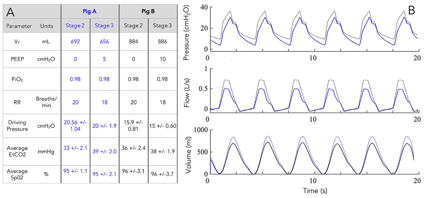
patient. The measurement of plateau pressure (Pplat)–the baseline and after titration.
pressure maintained during inspiration when flow is equal to After euthanasia, the animal’s lungs were filled with 750
zero and approximates the alveolar pressure – can be used to mL of saline, effectively decreasing their compliance and re-
calculate the lungs’ R and C. On closed-circuit ventilators sulting in a lower VT. With repeated breaths, flow was further
with iSAVE, the end-inspiratory hold feature can be used to diminished despite efforts by the automatic adjustments of
yield the Pplat. On open-circuit ventilators with iSAVE, an ad- the ventilator in increasing the pressure (fig. S8E). Titration
ditional flow valve can be added to the expiratory channel of the flow valve enabled restoration of the desired volume.
and briefly closed at the end of inspiration to yield Pplat. While We also simulated scenarios such as tube clogging and aspi-
connected to two artificial test lungs using a closed-circuit ration, in which the endotracheal tube of the artificial lung
ventilator, we performed an inspiratory hold and visualized was mechanically obstructed. This immediately increased
Pplat. Because of the circuits’ interdependence, Pplat was uni- flow to the animal by 30% (fig. S8F) and was quickly resolved
form between both lungs; However, PEEP and tidal volumes by closing the valve to the artificial lung circuit and adjusting
differed. Thus, accurate compliances can be computed. In flow to the animal.
Downloaded from http://stm.sciencemag.org/ by guest on December 27, 2020
three separate trials, we simulated this procedure with artifi- Validating iSAVE ventilation against standard ventila-
cial lungs of varying compliances. Computed compliances tion
were ≤ 11% error of the set compliances (table S3). We then investigated whether the iSAVE could ventilate
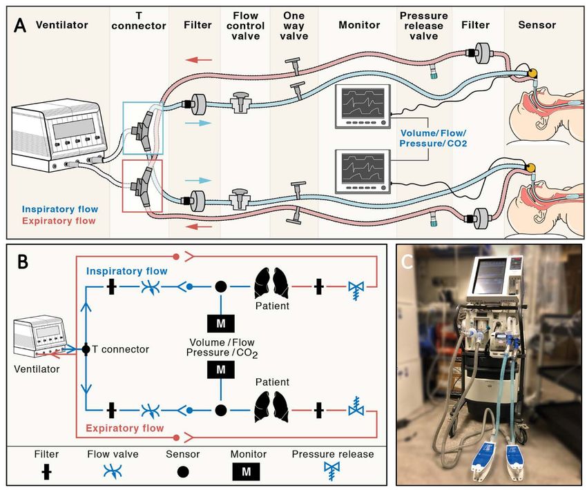
In vivo testing two large animals (Yorkshire swine, 74 kg and 88 kg) as ade-
Individualized management of ventilation and patient quately as ventilating each animal on its own closed-circuit
interdependence ventilator (Fig. 3, A and B) in a 3-stage experiment. In stage
To evaluate the practical management of rebalancing ven- 1, pigs were ventilated individually. Pig A required a VT = 690
tilation using the iSAVE when ventilating real lungs, which mL, whereas Pig B required a VT = 880 mL. We ventilated
exhibit variable respiratory mechanics, we performed closed- each animal for about 45 min under stable EtCO2 (33-38
circuit ventilation using a large animal (Yorkshire swine, 70 mmHg), SpO2 (93-99%), desired tidal volumes and arterial
kg) alongside an artificial lung (fig. S8A). The pig possesses a blood gasses (Fig. 3, C and D). In stage 2, we ventilated both
lung capacity of 5 to 6 L, similar to human lungs, and serves animals on the same closed-circuit ventilator using iSAVE.
as a translational preclinical model for this testing. We varied Representative individual pressure, flow, and volume traces
the lung mechanics of either the porcine or artificial lung (1 are provided in Fig. 3E and the means and standard devia-
L capacity) to test the system performance, delivering VT = tions (SD) of 300 breathing cycles are provided in Fig. 3F. No
500 mL from the ventilator. The iSAVE delivered differential significant differences were observed between stage 1 and
volumes to each channel. Ratios of 50:50, 40:60, and 30:70 stage 2 and all values were within the physiologically healthy
are presented in fig. S8B. range for swine. The difference in tidal volume between stage
Accommodation to static compliance changes and al- 1 and 2 was 5 mL for Pig A and 2 mL for Pig B. The difference
tered respiratory status in driving pressure between stages was 1.8 cmH2O for Pig A
To evaluate the system’s response to changes in lung com- and 0.4 cmH2O for Pig B. Blood electrolytes, anion gap, glu-
pliance, VT was initially distributed from the ventilator cose, blood urea nitrogen (BUN), hematocrit, and hemoglo-
equally to each channel (600 mL total, 300 mL per channel). bin remained stable throughout (table S4). We measured the
The compliance of the artificial lung was decreased (from C respiratory mechanics of both animals during both stages of
= 120 mL/cmH2O to C = 60 mL/cmH2O), resulting in a greater ventilation. During shared ventilation, Pplat was simultane-
allocation of volume (~100 mL) to the porcine lung (fig. S8C). ously determined using the end-inspiratory hold feature for
By titrating the flow valve, VT distribution was restored both animals. The computed compliances and resistances
within 3 breaths. (based on the Pplat from the main ventilator) matched those
During the course of recovery from ARDS, lung compli- displayed by the individual respiratory monitors.
ance increases–thus, we tested a range of such scenarios (C = In the third stage, we ventilated the two animals with dif-
20, 50, 60, and 120 mL/cmH2O). To model an extreme case, ferential PEEPs (Pig A: PEEP = 5 cmH2O, Pig B: PEEP = 10
we adjusted the artificial lung compliance from C =20 cmH2O, Fig. 4, A and B) using iSAVE on a closed-circuit ven-
mL/cmH2O to C = 120 mL/cmH2O: iSAVE immediately di- tilator. During ventilation, we simulated several practically
verted flow away from the higher resistance in the animal’s challenging scenarios including: (i) Adding an animal to the
lungs toward the less-resistant artificial lung (fig. S8D). Valve iSAVE circuit; (ii) quickly removing an animal from the cir-
adjustment restored the desired flow to the animal’s lungs cuit without leaking the airflow (containing isoflurane) into
and artificial lung. Throughout these tests, end tidal carbon the atmosphere; and (iii) adjusting ventilation parameters as
dioxide (EtCO2) and oxygen saturation (SpO2) were main- the animal’s respiratory mechanics changed. In all cases,
tained between 38-41 mmHg and 91-98%, respectively, at iSAVE enabled stable management of ventilation.
First release: 18 May 2020 stm.sciencemag.org (Page numbers not final at time of first release) 4DISCUSSION two individuals (1:2 ratio). Ventilator volume can be linearly
In this study, we demonstrate how the iSAVE enables in- divided to additional patients. Thus, the maximal capacity of
dividualized management of ventilation using valves, sen- this system is defined by:
sors, and alarms. Through benchtop and in vivo testing, we
Capacity = VT,Ventilator(VT,Patient 1+VT,Patient 2+VT,Patient 3+…) Eq. 1
demonstrate that we can not only individualize tidal volume
and PEEP, but also rebalance ventilation to accommodate Since most ICU ventilators provide up to 2500 mL, it is
changes in respiratory mechanics in one channel that could estimated that at least 6 individuals could be simultaneously
otherwise jeopardize flow to a second connected channel. The ventilated. However, physical and practical challenges will
data show that the system can support the flows and pres- limit implementation, particularly in the ICU setting. Dead
sures required to manage the ventilation of lungs with prop- space, the volume of air that doesn’t participate in gas ex-
erties of ARDS for two individuals over several hours. We change, accumulated in tubing cannot be greater than the
tested several clinical scenarios associated with multiplexing lowest individual tidal volume required. Further, with the ad-
ventilation to address alarm mechanisms, connection and dition of more than two patients to one ventilator circuit, de-
Downloaded from http://stm.sciencemag.org/ by guest on December 27, 2020
disconnection processes, mitigation of contamination risk, termining flow changes and titration will entail a more
and measurement of pulmonary mechanics. Moreover, the involved process, requiring monitors for each patient. In the
stability of tidal volume, PEEP, respiratory mechanics (re- case of n patients, data from n-1 circuits would be required to
sistance and compliance) and gas exchange (SpO2, EtCO2), as make adjustments. If the supply of respiratory monitors is
indicated by low standard deviation values during the venti- constrained, this limitation could potentially be overcome by
lation of two pigs, suggest that it may be possible to integrate sharing monitors among patients and using monitors to per-
iSAVE in a protective ventilation strategy. form frequent checks.
The iSAVE’s pressure release valve, flow control valve, Based on the iSAVE’s individualization capacity, matching
one-way valve, pressure sensor, and CO2 sensor are commer- criteria for patients may not need to be as stringent as other
cially available medical-grade parts commonly found in hos- protocols designed to multiplex ventilation without individu-
pitals, lowering the barrier to implementation. In the event alized control (10). Nevertheless, certain ventilatory parame-
of shortages, these parts could be procured from plumbing ters (RR, FiO2, I:E) will remain shared amongst patients.
and ventilation departments in hardware stores and auto- Additionally, patients need to remain sedated (and/or para-
claved for sterile usage. If standard adaptors do not interface lyzed) to prevent spontaneous breathing, which would lead
these parts, adaptors can be made from standard piping or to asynchrony in the system. Thus, patients should be
three-dimensional printing. As specified, the iSAVE permits matched as closely as possible in terms of degree of illness
individualized ventilation through alteration in tidal volume and ventilatory needs to optimize functioning. A list of rec-
for two channels. Future iterations could incorporate closed- ommendations for patient stratification is provided in table
loop control, directly modulating flow according to flow me- S5. These guidelines were derived from the highest bounds of
ter readings and valves to allow for independent control of variance tolerable by the iSAVE from our in vitro and preclin-
RR and FiO2 in each patient circuit. For ventilators requiring ical testing, with a safety factor of 2-3, in combination with
closed-loop flow control, standard flow sensors can be placed feedback from standardized treatment protocols (14). Other
in line and rerouted through stopcocks to enable normal considerations include hemodynamic stability, anticipated
functioning of the ventilator’s self-check/calibration mecha- invasive ventilation time, proning, co-infection, and the logis-
nisms (fig. S9). In the envisioned setup, individual patient tics of space allocation for patients. Ideally, patients would be
monitors (such as the Philips NM3) would be used to set the ventilated in negative pressure rooms, with their heads
initial conditions of the valves, perform periodic checks, and placed as close as possible to the ventilator. Recovering pa-
determine changes in the circuit. Standard humidification tients would need to be transitioned to an individual ventila-
devices for airstreams would be connected in series with the tor when spontaneous breathing becomes viable (11).
iSAVE on the inspiratory channel. In addition to the compo- This study only tested the iSAVE on test lungs and in a
nents described here, a whistle ring could be added to the small number of large animals without active disease pathol-
pressure release valve to provide an auditory alarm when ogy. Further, ventilation was performed for a short period of
high pressure develops in one segment (fig. S10). Independ- time in vivo to serve as a proof of concept. Our overall goal is
ent monitoring mechanisms should also be developed. to ensure that the iSAVE can be reliably implemented and
Although we tested scenarios involving only two individ- used across intensive care clinical settings to address ventila-
uals and splitting among two channels under preclinical con- tor shortages. To this end, further steps must be taken to ad-
ditions, in theory the iSAVE should be able to accommodate dress current limitations prior to clinical implementation.
more than two channels. Our testing thus far has provided a Due to differences in performance characteristics (mecha-
proof of concept for the iSAVE using a single ventilator with nism of pressure/flow monitoring) of ICU ventilators, this
First release: 18 May 2020 stm.sciencemag.org (Page numbers not final at time of first release) 5approach must be tested across a range of ventilators. The they related to specific ventilator models. We performed ven-
iSAVE must be evaluated in conditions reflecting the real-life tilation of one pig and one test lung, wherein we modulated
variability of intensive care practice. The procurement and the respiratory mechanics of each lung to test the manage-
sterilization processes for non-standard components must be ment of rebalancing differential ventilation to both lungs. We
addressed. Toward facilitating this evaluation by our team then performed the simultaneous ventilation of two pigs to
and others, we have assembled a list of components, instruc- evaluate the iSAVE’s performance in an in vivo setting, simu-
tions for assembly, links to three-dimensional printable adap- lating several clinical scenarios. Animal experiments were ap-
tors (barb, push-to-connect, or luer lock fittings), and clinical proved by the Committee on Animal Care at the
consideration guidelines (https://i-save.mit.edu/). Modules Massachusetts Institute of Technology (MIT). No blinding or
to train personnel in using this system should also be devel- randomization was performed as this was a proof-of-concept
oped. Although this system mitigates several challenges asso- study.
ciated with splitting ventilation, it has several drawbacks, Open-circuit ventilator assembly
unknown limitations, and does not fully address safety con- A Philips Trilogy portable ventilator was used as a repre-
Downloaded from http://stm.sciencemag.org/ by guest on December 27, 2020
cerns associated with this and other approaches for ventilator sentative open-circuit ventilator for testing. Standard flex
splitting. Ventilator sharing is strongly discouraged by criti- corrugated tubing (22 mm outer diameter) was used to con-
cal care practitioners due to safety concerns. In a setting of nect two respiratory circuits, each consisting of a (i) ball valve
severe shortage of ventilators, sharing a ventilator among two (EKWB G ¼’’ Nickel, Microcenter), (ii) bacterial/viral filter
patients could potentially mitigate the need to triage which (Main Flow Bacterial/Viral Filter, Teleflex Medical), (iii) pres-
patients receive ventilatory support and which do not. This sure sensor (TD160D, Biopac Systems), (iv) airflow sensor
may present societal value in comparison to potentially sav- (BSL Medium Airflow Transducer SS11LA and AFT 20, Biopac
ing or prioritizing one life. Further ethical and policy-based Systems), and (v) Whisper Swivel valve (passive exhalation
discussions are necessary prior to implementation. The au- port, Philips). This was connected to the expiratory side of a
thors caution that the iSAVE approach is not a standard of Y-piece to allow for the inclusion of a bacterial/viral filter be-
clinical care and that this is a preclinical study. Rigorous fur- fore venting exhaled gas to the ambient. See the circuit dia-
ther testing and validation of this approach is necessary be- gram provided in Fig. 1A. The airflow and pressure sensors
fore implementation could be considered under the were connected to DA100C differential amplifiers with 1000x
Emergency Use Authorization issued by the U.S. Food and gain and processed through the MP160WSW signal pro-
Drug Administration (13). The techniques and approaches cessing unit (Biopac Systems) sampling at 2 kHz. Volume
described in this preclinical study do not represent any rec- control mode was employed, and settings were adjusted to
ommendation or alteration in the recommended use of de- deliver the desired VT, RR, and PEEP. Artificial linear test
vices that were studied in this article. lungs (IngMar Medical) were used for simulations with the
Here we have demonstrated that the iSAVE provides a so- open-circuit ventilator.
lution for expansion of respiratory support using readily Closed-circuit ventilator assembly
available medical-grade materials and exiting ventilators. The iSAVE is designed to work with closed-circuit ventila-
The iSAVE can at least double existing ventilator capacity tors currently found in ICUs. To this end, the system was
while retaining personalized ventilation settings for two in- tested on a Philips VX850 ventilator, a Hamilton G5 ventila-
dividuals. Healthcare systems worldwide could potentially tor, and Puritan Bennett 840 ventilator (fig. S1). These venti-
benefit from this system as they strive to care for the increas- lators were chosen as they represent the most common
ing volume of patients with ARDS associated with COVID-19 brands used in the United States. Y-connectors were con-
infection. nected to the inspiratory and expiratory limbs of the ventila-
tor to enable individual channels for each lung. In the
MATERIALS AND METHODS inspiratory limbs, the following components were connected
Study design in series: (i) a bacterial/viral filter (Main Flow Bacterial/Viral
The goal of this study was to design and validate a system Filter, Teleflex Medical), (ii) ball valve (EKWB G ¼’’ Nickel,
using medical-grade filters, valves, and sensors to provide in- Microcenter), (iii) one-way valve (a Threshold PEP positive
dividualized ventilation using one ventilator shared among expiratory pressure device, Philips, was used as a surrogate
at least two subjects. We performed benchtop testing of the for one-way valves), (iv) pressure sensor, (v) flow sensor, and
iSAVE using linear test lungs to ensure its ability to provide (vi) capnostat adaptor. In the expiratory limb, the following
differential ventilation, accommodate for changes in respira- components were connected in series: (i) a bacterial/viral fil-
tory mechanics, and manage acute changes to patient status. ter, (ii) a pressure release valve (PEEP valve), and (iii) an op-
We also evaluated issues of cross contamination, adding and tional one-way valve prior to connection with the ventilator
removing subjects from the circuits, and circuit design as (Threshold PEP). Polytetrafluoroethylene tubing or rubber
First release: 18 May 2020 stm.sciencemag.org (Page numbers not final at time of first release) 6adapters were used to adapt the ball valves to the standard 2- parameters, including the maximal combination designed to
mm outer diameter corrugated flex tubing. A dual adult lung create the most turbulent flow conditions: valves: fully open
simulator (Model 5600i, Michigan Instruments) was used to to fully closed: airflow pressure used to nebulize: 1-2800
perform simulations with the closed-circuit ventilator. Pres- cmH2O, duration of nebulization: 1 – 10 min, VT setting: 100-
sure and flow were displayed and recorded by the Philips 900 mL per lung, PEEP: 2 – 10 cmH2O, and RR: 2 – 30 bpm.
NM3 monitors and associated sensors, one for each channel. After each test, filters were manually inspected for contami-
For ventilators requiring continuous flow measurements for nation and a wipe test was performed in each of the segments
calibration and closed-loop control, such as the Hamilton G5, of tubing. Each wipe was then incubated in diH2O for 5 min.
the standard flow sensors can be reconfigured using stop- 1 mL from each sample was used to perform UV-vis absorp-
cocks as elaborated in fig. S9. tion spectroscopy at a wavelength of 580 nm. Each sample
Setup and testing protocol was measured in triplicate.
The following main ventilator settings were selected: (i) In vivo testing
Volume control mode is selected with triggering turned off; The first experiment was performed as part of a terminal
Downloaded from http://stm.sciencemag.org/ by guest on December 27, 2020
(ii) RR is determined based on the minute ventilation needs procedure on a 70 kg female swine (n = 1). The iSAVE was
for both sets of lungs; (iii) FiO2 is set to the desired value to connected to a veterinary anesthesia ventilator (Model 200IE,
be shared for both patients; (iv) I:E ratio is set with consid- Hallowell EMC) delivering 2% isoflurane in oxygen. One in-
eration of the tau (τ = RC) parameter of each patient, which spiratory circuit was connected to the anesthesia machine de-
must initially be estimated for each patient. The longer τ will livering gas to the animal while the other inspiratory circuit
dictate the expiratory window and prevent autoPEEP; and (v) was connected to an artificial linear test lung (IngMar Medi-
the alarm for minute volume was set to the following lower cal). Pressure and flow measurements were recorded on the
and upper bounds [10% of RR(VT1 + VT2), 10% of RR(VT1 + inspiratory limb of the animal. A VetTrends Vital Signs Mon-
VT2)]. Next, we checked for leaks and alarms by first testing itor was utilized to measure SpO2, EtCO2, RR, and other phys-
the ability of the system to perform ventilation without leaks. iological parameters. 600 mL of VT were equally distributed
We then disconnected a lung to ensure the activation of between the animal and test lung. A respiratory rate between
standard alarms on the ventilator. To produce variable tidal 18-20/min was set on the ventilator. We carried out the same
volumes, starting with both flow valves fully open, we meas- tests outlined in the benchtop testing protocol, modulating
ured the flow and pressure delivered to each lung. Then, we the parameters of the test lung to validate the capabilities of
gradually closed one of the valves, measuring the distribution the iSAVE to restore the system to baseline. After the animal
of flow, to map the range of volume distribution capable of was euthanized, to acutely change the compliance of the pig’s
the system. To maintain VT after static changes to compliance lung we used an endoscope (Pentax) to deliver 750 mL of
and resistance: We modulated the compliance and/or re- phosphate buffered saline (PBS, Sigma Aldrich) into the left
sistance of one test lung; measured the effect of the interven- and right bronchi. Ventilation was performed and the valves
tion; and titrated the valve until the baseline parameters were titrated to achieve desired flow parameters.
were reached. Resistors (Rp5, Rp20, Rp50, and Rp500 Mich- In the second experiment, one 74 kg and one 88 kg female
igan Instruments) were used for all benchtop testing. We per- swine were used in a survival approach. These pigs were se-
formed these tests with baseline parameters set to those of (i) dated with 0.25 mg/kg (5 mg/mL) midazolam and 0.03 mg/kg
a healthy lung (C = 50 cmH2O, R = Rp5) and (ii) a lung with (0.5 mg/mL) dexmedetomidine. The experiment was divided
ARDS (C = 20 cmH2O, R = Rp5-50). The flow valve was the into three stages: Individual ventilation of each animal, PEEP
main variable we modulated but, in some cases, the ventila- = 0 (stage 1); iSAVE ventilation (differential VT, PEEP = 0)
tor’s tidal volume or inspiration:expiration ratio was also ad- (stage 2); and iSAVE ventilation (differential VT, differential
justed. These cases are specifically mentioned in the results PEEP) (stage 3). Ventilation was performed with the Philips
section. Last, to maintain desired ventilation after dynamic Respironics Esprit ventilator in series with the anesthesia
changes to compliance and resistance, we simulated acute machine. The target tidal volume for each animal was calcu-
changes by disconnecting or clamping tubes and monitoring lated at 10 mL/kg, although ventilation was optimized to
the ventilator’s response. The flow valves were also titrated maintain SpO2 > 94% and EtCO2 ~ 30-35 mmHg in all stages.
to return the circuit to its baseline. The PEEP setting on the main ventilator was set to 0 cmH2O.
Cross-contamination testing After 30 min of stable ventilation, arterial blood gases were
5 mL of Trypan Blue (0.4%, 15250061, Thermo Fisher) was measured using an i-STAT handheld blood analyzer (Abbott).
placed in a nebulizer attached to the patient inflow/outflow Respiratory flow, pressure and volume were continuously
segment of one of two circuits containing a test lung. Air from logged using the Philips NM3 monitors as well as Biopac sys-
a separate line was used to nebulize droplet particles into the tems described above. Reported values represent the mean
main tubing. We then performed tests ranging the following and standard deviations of 300 breathing cycles. In stage 3,
First release: 18 May 2020 stm.sciencemag.org (Page numbers not final at time of first release) 7the PEEP settings were changed by adjusting the PEEP valve impact of high-flow nasal cannula (HFNC) on coughing distance: Implications on
to enable the animals to be ventilated with differential PEEP its use during the novel coronavirus disease outbreak. Can. J. Anesth. Can. Anesth.
10.1007/s12630-020-01634-3 (2020). doi:10.1007/s12630-020-01634-3
values (Pig A: PEEP = 5 cmH2O, Pig B: PEEP = 10 cmH2O). Medline
Compliance and resistance were measured for each animal 5. R. D. Truog, C. Mitchell, G. Q. Daley, The toughest triage – Allocating ventilators in
by performing an end-inspiratory pause of 1 s to ensure that a pandemic. N. Engl. J. Med. NEJMp2005689 (2020).
the iSAVE enabled these measurements. The computed com- doi:10.1056/NEJMp2005689 Medline
6. M. L. Ranney, V. Griffeth, A. K. Jha, Critical supply shortages – The need for
pliance and resistance values matched those which were ventilators and personal protective equipment during the Covid-19 pandemic. N.
measured on the monitor. The duration of each stage lasted Engl. J. Med. 382, e41 (2020). doi:10.1056/NEJMp2006141 Medline
about 45 min. 7. S. Fink, Worst-Case Estimates for U.S. Coronavirus Deaths. N. Y. Times (2020),
Data and statistical analysis (available at https://www.nytimes.com/2020/03/13/us/coronavirus-deaths-
estimate.html).
Pressure and flow data from the Biopac system was ana- 8. R. Maclean, S. Marks, 10 African Countries Have No Ventilators. That’s Only Part of
lyzed using MATLAB (2018a). Flow data was integrated every the Problem. N. Y. Times (2020), (available at
respiratory cycle to derive the flow. In cases where pressure https://www.nytimes.com/2020/04/18/world/africa/africa-coronavirus-
ventilators.html).
Downloaded from http://stm.sciencemag.org/ by guest on December 27, 2020
and flow data were not simultaneously obtained from both
9. G. Neyman, C. B. Irvin, A single ventilator for multiple simulated patients to meet
limbs of the circuit, data were derived based on the settings disaster surge. Acad. Emerg. Med. Off. J. Soc. Acad. Emerg. Med. 13, 1246–1249
on the main ventilator and the measurements on one limb. (2006). doi:10.1197/j.aem.2006.05.009 Medline
Homoscedastic, two-tailed t tests were performed to assess 10. R. D. Branson, T. C. Blakeman, B. R. Robinson, J. A. Johannigman, Use of a single
significance on the data from the contamination testing with ventilator to support 4 patients: Laboratory evaluation of a limited concept.
Respir. Care 57, 399–403 (2012). doi:10.4187/respcare.01236 Medline
a threshold of P < 0.05. A homoscedastic, two-tailed, t test 11. Joint Statement on Multiple Patients Per Ventilator, (available at
was used to compare the values from 300 breathing cycles https://www.asahq.org/about-asa/newsroom/news-releases/2020/03/joint-
between the two conditions for the in vivo study with a statement-on-multiple-patients-per-ventilator).
threshold of P < 0.05. 12. Working Protocol for Supporting Two Patients with a Single Ventilator. GNYHA,
(available at https://www.gnyha.org/news/working-protocol-for-supporting-
two-patients-with-a-single-ventilator/).
SUPPLEMENTARY MATERIALS 13. C. for D. and R. Health, Emergency Use Authorizations. FDA (2020) (available at
stm.sciencemag.org/cgi/content/full/scitranslmed.abb9401/DC1 http://www.fda.gov/medical-devices/emergency-situations-medical-
devices/emergency-use-authorizations).
Fig. S1. Photographs of the iSAVE setup
14. NHLBI ARDS Network | Tools, (available at http://www.ardsnet.org/tools.shtml).
Fig. S2. Differential tidal volume and PEEP delivery on the iSAVE using an open-cir-
cuit ventilator and test lungs
Fig. S3. Accommodation to changes in compliance of one test lung using the iSAVE Acknowledgments: We thank K. Wasco, K. Graap, and the Biopac team for extreme
and an open-circuit ventilator responsiveness for provision of sensor equipment, and P. Kritek for helpful
Fig. S4. Ventilation at high lung resistances respiratory related discussion and guidance. We thank A. Sarma, J. George, J.
Fig. S5. Ventilator alarm response to occlusion Wainer, A. Wentworth, S. Malinowski, and J. Byrne for their assistance in
Fig. S6. Adding a test lung to the circuit prototyping and/or consultation. We thank A. Hupalowska for original artwork.
Funding: This work was supported in part by the Massachusetts Consortium on
Fig. S7. Cross-contamination validation using artificial lungs
Pathogen Readiness (MassCPR) and the Massachusetts Life Sciences Center.
Fig. S8. Individualized management of ventilation using iSAVE on a pig lung and a
The work was further supported in part by in kind services from Philips, and
test lung
discretionary funds from the Department of Mechanical Engineering at MIT and
Fig. S9. Modification of sensing circuit for Hamilton G5 ventilator. at BWH to G.T. Author contributions: S.S performed the conceptualization,
Fig. S10. Whistle ring designs for two types of common pressure release (PEEP) investigation, data analysis, and writing. A.H. and D.L. assisted with in vivo
valves investigation. K.B.R. wrote. F.V. performed investigation, data curation, and
Table S1. List of components required for the assembly of the iSAVE edited the manuscript. D.G assisted with prototyping. R.L, J.J.F., D.G., R.M.B, and
Table S2. Mechanical components used in the iSAVE and their readily available medi- G.T. contributed to conceptualization and editing of the manuscript. All authors
cal industry equivalents approved the manuscript. Competing interests: F.V. and J.J.F serve as
Table S3. Measurement of respiratory mechanics for iSAVE system using test lungs employees for Philips North America, a maker of Healthcare devices and
Table S4. Blood electrolytes and chemistry during ventilation in pigs monitoring solutions including patient ventilators. R.M.B. is part of an Advisory
Table S5. Stratification for patient matching Board for Merck. Complete details of all relationships for profit and not for profit
for G.T. can be found at the following link:
REFERENCES AND NOTES https://www.dropbox.com/sh/szi7vnr4a2ajb56/AABs5N5i0q9AfT1IqIJAE-
1. Novel Coronavirus, (2019-nCoV) situation reports, (available at T5a?dl=0. Complete details for R.L. can be found at the following link:
https://www.who.int/emergencies/diseases/novel-coronavirus- https://www.dropbox.com/s/yc3xqb5s8s94v7x/Rev%20Langer%20COI.pdf?d
2019/situation-reports). l=0 All other authors report no competing interests related to the work reported
2. Clinical course and outcomes of critically ill patients with SARS-CoV-2 pneumonia here. Data and materials availability: All data associated with this study are in
in Wuhan, China: a single-centered, retrospective, observational study - The the paper or Supplementary Materials. Further information including videos and
Lancet Respiratory Medicine, (available at details on components, assembly, clinical considerations, partnerships, and the
https://www.thelancet.com/journals/lanres/article/PIIS2213-2600(20)30079- implementation can be found at https://i-save.mit.edu/. This work is licensed
5/fulltext). under a Creative Commons Attribution 4.0 International (CC BY 4.0) license,
3. S. A. Ñamendys-Silva, Respiratory support for patients with COVID-19 infection. which permits unrestricted use, distribution, and reproduction in any medium,
Lancet Respir. Med. 8, e18 (2020). doi:10.1016/S2213-2600(20)30110-7 Medline provided the original work is properly cited. To view a copy of this license, visit
4. N. W. Loh, Y. Tan, J. Taculod, B. Gorospe, A. S. Teope, J. Somani, A. Y. H. Tan, The http://creativecommons.org/licenses/by/4.0/. This license does not apply to
First release: 18 May 2020 stm.sciencemag.org (Page numbers not final at time of first release) 8figures/photos/artwork or other content included in the article that is credited
to a third party; obtain authorization from the rights holder before using such
material.
Submitted 5 May 2020
Accepted 14 May 2020
Published First Release 18 May 2020
10.1126/scitranslmed.abb9401
Downloaded from http://stm.sciencemag.org/ by guest on December 27, 2020
First release: 18 May 2020 stm.sciencemag.org (Page numbers not final at time of first release) 9Downloaded from http://stm.sciencemag.org/ by guest on December 27, 2020
Fig. 1. Design of the individualized system for augmenting ventilation efficacy (iSAVE). (A)
Schematic of iSAVE setup on a closed-circuit ventilator for simultaneous ventilation of two patients.
(B) Circuit diagram of iSAVE for closed-circuit ventilation. (C) Photograph of iSAVE connected to a
Puritan Bennet 840 ICU ventilator and two test lungs.
First release: 18 May 2020 stm.sciencemag.org (Page numbers not final at time of first release) 10Downloaded from http://stm.sciencemag.org/ by guest on December 27, 2020
Fig. 2. Individualized ventilation and management of patient interdependence using artificial test lungs.
(A) Pressure, flow, and tidal volume waveforms illustrating three settings of differential tidal volume (VT) for
two test lungs (blue, black) using closed-circuit ventilation. The ratio (50:50, 35:65, 15:85) refers to the VT
of the black:blue lungs. Pressure, volume, and flow in both lungs upon (B) decreased compliance in one lung
(black) and (C) increased compliance in the other lung (blue). The orange dotted line indicates decrease or
increase in compliance. The green dotted line indicates return of baseline ventilation parameters upon
titration of the valves. Pressure, volume, and flow in both lungs upon (D) increased resistance in one lung
(black) and (E) decreased resistance in the other lung (blue). Orange dotted line indicates increase or
decrease in resistance. Green dotted line indicates return of baseline ventilation parameters upon titration
of the valves. Waveforms from each lung are slightly offset to enable visualization.
First release: 18 May 2020 stm.sciencemag.org (Page numbers not final at time of first release) 11Downloaded from http://stm.sciencemag.org/ by guest on December 27, 2020
Fig. 3. Ventilation of two pigs on the iSAVE. (A) Experimental setup for stage 2 and stage 3 of shared
ventilation of pig A (74 kg) and pig B (88 kg) with iSAVE using closed-circuit ventilation. (B) Photograph
of the experimental setup. Pressure, flow, and volume waveforms for (C) pig A ventilated individually
(stage 1), (D) pig B ventilated individually (stage 1), and (E) pigs A and B ventilated together on the iSAVE
(stage 3). (F) Table summarizing ventilatory and respiratory parameters and arterial blood gasses for (C-
E). Mean ± SD was calculated from 300 breathing cycles. No significant differences were found between
the individual and shared ventilation approaches (homoscedastic two-tailed t test, P > 0.05).
First release: 18 May 2020 stm.sciencemag.org (Page numbers not final at time of first release) 12Downloaded from http://stm.sciencemag.org/ by guest on December 27, 2020
Fig. 4. Differential tidal volume and PEEP during ventilation of two pigs on the iSAVE with
closed-circuit ventilation. (A) Summary of ventilatory and respiratory parameters. Mean ± SD
was calculated from 300 breathing cycles. No significant differences were found between
ventilation with and without differential PEEP (homoscedastic two-tailed t test, P > 0.05). (B)
Pressure, flow, and volume waveforms for the two animals. Pig A (blue) and pig B (black) were
ventilated with PEEP of 5 and 10 cmH2O, respectively.
First release: 18 May 2020 stm.sciencemag.org (Page numbers not final at time of first release) 13Table 1. Key challenges in splitting ventilation. A comparison of the capabilities of existing splitting mechanisms and iSAVE. *See
fig. S9 for details regarding the rerouting of standard sensing metrics required for ventilator calibration and self-tests. PEEP, positive
end-expiratory pressure; FiO2, fraction of inspired oxygen; ΔC, change in compliance; ΔR, change in resistance; Pplat, plateau pressure.
Uniform splitting iSAVE
Concern
(pressure control mode) (volume control mode)
Individualized management of ventila-
tion
- PEEP x Shared between patients o Individualized to each patient
- Tidal volume x Shared between patients o Individualized to each patient
- FiO2, respiratory rate x Shared between patients x Shared between patients
Changes to one patient’s status will cause
Changes to one patient’s status may main ventilator to alarm. Mechanical com-
- Alarms x o
not result in main ventilator alarm ponents to provide auditory alarms can be
incorporated
Sudden changes to patient status can Can be managed by titrating flow control
Downloaded from http://stm.sciencemag.org/ by guest on December 27, 2020
cause damaging rebalancing of airflow Ventilation cannot be quickly ad- valves. One-way valves prevent backflow.
x o
to other patient(s) toward most com- justed Pressure release valves prevent excess
pliant lungs pressure delivery
Improvement or deterioration of one Ventilation cannot be individually re- Desired ventilation for each patient can be
patient (ΔC, ΔR) will automatically re- balanced. Patients would need to be achieved through valve adjustment, allow-
x o
balance airflow, potentially harming re-matched as they improve/deterio- ing patients to improve/deteriorate while
other patient(s) rate remaining on the same system.
Abruptly removing patients requires
Individual patients can be quickly
breaking the circuit, causing aerosoli- Individual patient circuits cannot be
x o shunted/removed from the circuit. Inline
zation of the virus, exposing quickly removed from circuit
filters limit aerosolization risk
healthcare personnel
Additional respiratory monitors and Additional respiratory monitors and
Monitoring x x
heightened clinical vigilance required heightened clinical vigilance required
Pplat can be measured using expiratory
Measurement of pulmonary mechan-
x Shared between patients o hold button. C, R can be computed for
ics
each patient
Added circuit volume defeats the op- Can be executed with modifications to cir-
Ventilator calibration/self-test x o
erational self‐test cuit*
Disabled. Patients will require seda-
Triggering x x Disabled. Patients will require sedation
tion
First release: 18 May 2020 stm.sciencemag.org (Page numbers not final at time of first release) 14A rapidly deployable individualized system for augmenting ventilator capacity
Shriya Srinivasan, Khalil B Ramadi, Francesco Vicario, Declan Gwynne, Alison Hayward, David Lagier, Robert Langer,
Joseph J. Frassica, Rebecca M. Baron and Giovanni Traverso
Sci Transl Med published online 18 May 2020
Downloaded from http://stm.sciencemag.org/ by guest on December 27, 2020
ARTICLE TOOLS http://stm.sciencemag.org/content/early/2020/05/18/scitranslmed.abb9401
SUPPLEMENTARY http://stm.sciencemag.org/content/suppl/2020/05/18/scitranslmed.abb9401.DC1
MATERIALS
Use of this article is subject to the Terms of Service
Science Translational Medicine (ISSN 1946-6242) is published by the American Association for the Advancement of
Science, 1200 New York Avenue NW, Washington, DC 20005. The title Science Translational Medicine is a registered
trademark of AAAS.
Copyright © 2020, The Authors, some rights reserved; exclusive licensee American Association for the Advancement of
Science. No claim to original U.S. Government WorksDownloaded from http://stm.sciencemag.org/ by guest on December 27, 2020 Use of this article is subject to the Terms of Service Science Translational Medicine (ISSN 1946-6242) is published by the American Association for the Advancement of Science, 1200 New York Avenue NW, Washington, DC 20005. The title Science Translational Medicine is a registered trademark of AAAS. Copyright © 2020, The Authors, some rights reserved; exclusive licensee American Association for the Advancement of Science. No claim to original U.S. Government Works
You can also read