Poorly Differentiated and Anaplastic Thyroid Cancer
←
→
Page content transcription
If your browser does not render page correctly, please read the page content below
Clinical and pathologic information

                                                                                        on the different features of these

                                                                                        rare entities is important in

                                                                                        managing these tumors.

Donna Morrison. Lone Flyer. Watercolor, 23′′ × 30′′.

                 Poorly Differentiated and Anaplastic
                            Thyroid Cancer
                                 Kepal N. Patel, MD, and Ashok R. Shaha, MD, FACS

Background: Poorly differentiated thyroid carcinoma (PDTC) and anaplastic (undifferentiated) thyroid carcinoma
(ATC) comprise a small subset of thyroid tumors that are associated with a poor prognosis and account for a
significant portion of the morbidity and mortality related to thyroid cancer. Since management strategies vary
between these two entities, it is important for clinicians to be able to differentiate PDTC from ATC.
Methods: We reviewed the literature on PDTC and ATC and compared clinical and histopathologic features
important in defining the disease process.
Results: Both PDTC and ATC display aggressive behavior with increased locoregional and distant disease. In
most cases, patients are older and have large, locally advanced tumors. PDTC may represent an intermediate
entity in the progression of well-differentiated thyroid carcinoma to ATC. The use of surgical management may
be curative or palliative and differs between PDTC and ATC. The roles of radiotherapy and chemotherapy have
not been well described.
Conclusions: PDTC and ATC are rare diseases that carry a poor prognosis. Recognition of their different
clinicopathologic features is important to the optimal management of these tumors.

                                                                       Introduction
From the Department of Surgery, Stony Brook University Hospital,
Stony Brook, New York (KNP), and the Head and Neck Service,
Memorial Sloan-Kettering Cancer Center, New York, New York (ARS).      Malignant tumors of thyroid follicular cell origin have
Submitted July 28, 2005; accepted December 26, 2005.                   traditionally been classified as either well-differentiated
Address correspondence to Ashok R. Shaha, MD, FACS, Head and           thyroid carcinoma (WDTC), which is composed of pap-
Neck Service, Memorial Sloan-Kettering Cancer Center, 1275 York        illary and follicular carcinoma, or undifferentiated/
Avenue, New York, NY 10021. E-mail: shahaa@mskcc.org
                                                                       anaplastic thyroid carcinoma (ATC). The vast majority
No significant relationship exists between the authors and the com-
panies/organizations whose products or services may be refer-          of patients with WDTC have an excellent prognosis
enced in this article.                                                 regardless of the types of treatment used, whereas
Abbreviations used in this paper: PDTC = poorly differentiated         patients with ATC uniformly have a poor prognosis.1
thyroid carcinoma, WDTC = well-differentiated thyroid carcinoma,       There is growing evidence for the existence of a group
ATC = anaplastic thyroid carcinoma, ITC = insular thyroid carcinoma,
FNAB = fine-needle aspiration biopsy, TG = thyroglobulin, RAI =        of tumors that fall between WDTC and ATC in terms of
radioactive iodine, EBRT = external-beam radiation therapy.            both morphologic appearance and biologic behavior.

April 2006, Vol. 13, No. 2                                                                                       Cancer Control 119
These tumors, classified as poorly differentiated thyroid                  the solid variant of papillary thyroid cancer displays
carcinoma (PDTC), may represent intermediate enti-                         some aggressive features, but patients tend to be
ties in the progression of WDTC to ATC.2-5 Patients with                   younger and, with appropriate treatment, their overall
PDTC often have a rapid and fatal outcome despite                          prognosis is similar to that of classic papillary thyroid
appropriate treatment. Although over the years multi-                      cancer.10 The tumors are especially frequent among
ple publications have reported on this subject, contro-                    pediatric thyroid carcinomas from the Chernobyl area
versies regarding optimal management of these                              and are associated with the RET/PTC3 rearrange-
patients still exist. This review is limited to PDTC and                   ment.11,12 Patients with encapsulated columnar cell
ATC in an attempt to better document their clinical                        thyroid carcinoma also have an excellent prognosis and
behavior and long-term prognosis.                                          should not be classified as PDTC.13 Since some clini-
                                                                           cians include clinical characteristics in defining PDTC,
                                                                           the literature is inconsistent, thus precluding any defin-
Poorly Differentiated Thyroid Carcinoma                                    itive conclusions regarding the disease process. It is
                                                                           preferable to limit this term to histologic criteria.
Much of the controversy, confusion, and inconsistency                           We agree with the definition by Burman et al14 and
surrounding PDTC comes from the lack of consensus                          others15 that “poorly differentiated thyroid carcinoma is
regarding criteria and definitions. The term poorly dif-                   a concept proposed to include carcinomas of follicular
ferentiated thyroid carcinoma was introduced by                            thyroid epithelium that retain sufficient differentiation to
Sakamoto et al2 in 1983, and their criteria were based                     produce scattered small follicular structures and some
mainly on the presence of nonglandular components                          thyroglobulin, but generally lack the usual morphologic
with a solid, trabecular, and/or scirrhous growth pat-                     characteristics of papillary and follicular carcinoma.”
tern. Others have included aggressive papillary thyroid                    Based on this description, we have developed a chart to
carcinoma variants such as columnar cell, tall cell, dif-                  help classify tumors of thyroid follicular origin (Fig 1).
fuse sclerosing, and solid.6-8 The fact that these papil-                  PDTCs fall into two main categories — insular and other
lary thyroid cancer variants tend to show a more                           (large cell).
aggressive behavior pattern than the classic type of dif-
ferentiated thyroid carcinoma does not in itself justify                   Insular Thyroid Carcinoma
the use of the term poorly differentiated, as defined by                   Insular thyroid carcinoma (ITC) is the best-character-
the tumor architecture.9 Furthermore, these tumors do                      ized group of PDTCs. Langhans16 first described it in
not have an invariably poor prognosis. For example,                        1907 as “wuchernde struma” (proliferating struma). He

Fig 1. — Classification of thyroid carcinomas of follicular cell origin.

120 Cancer Control                                                                                                 April 2006, Vol. 13, No. 2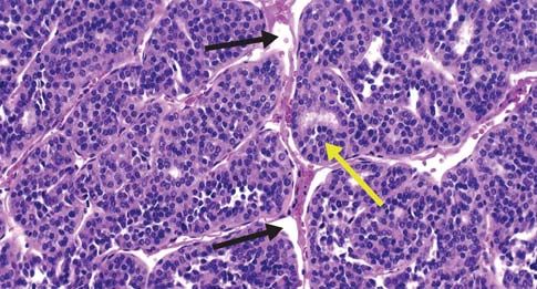
consistent mitotic activity, necrosis, and capsular and
                                                                                vascular invasion that sometimes leads to the formation
                                                                                of peritheliomatous structures. The peritheliomatous
                                                                                structures refer to the insulae surrounding the large
                                                                                blood vessels, which have been spared from necrosis.
                                                                                     Preoperative fine-needle aspiration biopsy (FNAB)
                                                                                can be helpful in planning treatment options and
                                                                                patient management. Pietribiasi et al33 reviewed 6 cases
                                                                                of ITC with preoperative FNAB. They assessed cytolog-
                                                                                ic features and compared them with the final histologic
Fig 2. — Insular thyroid carcinoma, showing the nesting patterns of the         specimen. They consistently found high cellularity,
follicles (yellow arrow) and the artifactually created clefts (black arrows).   necrotic background, low-grade atypia, trabeculae
                                                                                and/or clusters, microfollicles, cytoplasmic vacuoles
described a tumor characterized by a distinct nesting                           containing thyroglobulin (TG), and nuclear inclusions.
pattern, formation of small follicular lumina leading to                        These features were not uniform. This is consistent with
a cribriform configuration, small size and uniformity of                        the histologic heterogeneity seen in these tumors. The
the tumor cells, necrosis, and a focally peritheliomatous                       authors concluded that FNAB could provide a sugges-
pattern of growth (tumor cells around blood vessels,                            tive but not definitive preoperative diagnosis of ITC.
with necrosis of tumor cells farther away from vessels).                             Immunohistochemical staining is an invaluable tool
This tumor entity was ignored by most modern                                    in diagnosing and understanding the pathophysiology of
authors17-19 as an inconsequential morphologic variant                          thyroid tumors. It confirms that ITC is of follicular cell
of follicular carcinoma, partially due to geographic                            origin by staining positive for TG and thyroid transcrip-
differences in the frequency of this neoplasm. It was                           tion factor 1 (TTF-1). ATC does not stain positive for
reinterpreted and termed poorly differentiated “insular”                        TG.21 Immunohistochemical staining for p53 mutation
thyroid carcinoma by Carcangiu et al3 in 1984. The                              also can be helpful in determining tumor progression.
term insular was used to describe these tumors                                  WDTC does not usually stain for p53 mutation, whereas
because the cellular appearance was similar to that                             staining varies between 0% and 38% in ITC and is >70%
seen in the insular type of carcinoid tumors. Since                             in ATC.25,27,34 Furthermore, the presence of well-differ-
this revised description in 1984, over 200 cases of ITC                         entiated and/or anaplastic components within ITC has
have been described in the literature.3,20-32                                   been frequently reported.25,34 In one series, concomitant
                                                                                WDTCs were noted in 59% of patients with ITC.25 These
Pathology                                                                       observations lend support to the hypothesis that ITC
The morphologic appearance of this tumor is similar                             represents an intermediate entity in the dedifferentiation
from case to case. Macroscopic features include a solid,                        of WDTC to ATC. Immunohistochemical staining for
grayish-white tumor with multiple foci of necrosis.                             calcitonin, chromogranin, and carcinoembryonic antigen
They usually display an invasive margin, they tend to be                        is negative in ITC. These stains, along with the distinct
more than 4 cm in size, and they can be either single or                        cellular features, can be helpful not only in narrowing
multinodular. The microscopic features as described by                          the differential diagnosis, but also in ruling out entities
Carcangiu et al3 include solid clusters “nests” of tumor                        such as medullary thyroid cancer.
cells containing a variable number of follicles, often
sharply separated by artifactually created clefts. This is                      Other (Large Cell)
the predominant growth pattern. This picture is typical                         This small group of PDTCs represents tumors that have
for carcinoid and pancreatic endocrine tumors, which                            a variable architecture consistent with intermediate
are referred to as insular (Fig 2). Other critical features                     differentiation. Unlike aggressive variants of papillary
are small size and uniformity of tumor cells, variable but                      thyroid carcinoma, these tumors do not retain the usual

              Table 1. — Comparison of Histologic Features of Insular and Other Subtypes of Poorly Differentiated Thyroid Carcinomas

      Insular Carcinomas                                                              Other Subtypes
      Solid clusters (“insulae”) of tumor cells containing a                          Variable architecture (follicular, solid, trabecular, papillary);
      variable number of follicles is the predominant growth pattern                  insular component minor
      Small size and uniformity of tumor cells                                        Larger size of tumor cells with variable cytology
                                                                                      (oncocytic, clear, pap nuclei)
      Variable but consistent mitotic activity                                        Variable but consistent mitotic activity
      Necrosis                                                                        Necrosis
      Capsular and vascular invasion                                                  Capsular and vascular invasion

April 2006, Vol. 13, No. 2                                                                                                             Cancer Control 121
morphologic characteristic formation of papillary carefully searched for, in the background of a WDTC,
structures, thereby making this a poorly differentiated since it could predict a worse prognosis. They also
tumor entity. Histologically, these tumors are similar to reported age, extrathyroidal extension, and vascular
ITC in that they also display increased mitotic activity, invasion as independent poor prognostic factors.
necrosis, and capsular and vascular invasion, and they
stain positive for TG. These tumors are unlike ITC in Treatment
that they can have a variable architecture (follicular, The rarity of this tumor makes it difficult to draw con-
solid, trabecular, papillary) with a minor insular com- clusions from the literature as to the best treatment
ponent.21 Furthermore, the tumor cells are larger and option for PDTC. Surgical management of this entity is
have variable cytology when compared to ITC (Table 1). the principal treatment approach. Most authors agree
Clinically, these patients behave like patients with ITC, that due to the aggressive nature of these tumors, a
with similar overall survival; however, ITC tends to total thyroidectomy is necessary. With over 50% of
recur more frequently in patients.21 PDTCs having regional nodal metastases, central com-
partment with possible modified radical neck dissec-
Clinical Characteristics tion should be considered.36 The use of radioactive
PDTCs are tumors of intermediate biological aggres- iodine (RAI), external-beam radiation therapy (EBRT),
siveness, consistent with their intermediate differentia- or chemotherapy is still controversial.
tion. They account for up to 10% of all thyroid cancers. PDTCs arise from follicular epithelium and thus
This may be inaccurate due to inconsistent definitions have the distinct potential to concentrate iodine. Justin
of PDTC used by different authors. PDTC has a higher et al38 described 5 patients with PDTC, 4 of whom
incidence in Europe than in the United States and the showed postoperative RAI localization and received
male-to-female ratio is greater than 1:2.14,35 therapeutic doses. Three of the 4 patients showed
We recently reviewed our experience and com- extrathyroidal localization, and 1 patient had resolution
pared the clinical characteristics of WDTC, PDTC, and of metastatic disease. Currently the percentage of
ATC.36 Our results were consistent with others in that PDTCs having sufficient RAI concentration to allow
PDTC displayed clinical characteristics of intermediate postoperative RAI therapy is unknown. We recently
aggressive behavior when compared to WDTC and ATC reviewed our experience in treating patients with
(Table 2). Whether the presence of a minor component PDTC. Preliminary results showed that these tumors
of PDTC worsens the prognosis of WDTC is an area of displayed up to 85% radioavidity. Although prospective
further investigation. Van den Brekel et al29 found that evidence for its use and efficacy is not available, most
the insular component within a WDTC was not associ- authors advocate the use of RAI and L-thyroxine
ated with a poor prognosis, but the follow-up was only because these tumors display differentiated epithelial
2 years.29 Ashfaq et al31 found that the insular compo- function with aggressive behavior, with high rates of
nent correlated with older age, and using multivariate regional and distant metastases.
analysis, only age and stage were prognostic. However, The evidence for and against a role for adjuvant
Pilotti et al7 demonstrated the prognostic impact of dif- EBRT in PDTC is exclusively retrospective in nature,
ferentiation on recurrence and death. Decaussin et al37 with varying criteria for patient selection that results in
found that the presence of an insular component was contradictory conclusions. EBRT is given as a local
an independent poor prognostic factor that must be therapy to reduce the risk of local relapse; no improve-
Table 2. — Comparison of Clinical Characteristics of Well-Differentiated, Poorly Differentiated, and Anaplastic Thyroid Carcinomas at a
Median Follow-Up of 43 Months: Memorial Sloan-Kettering Cancer Center Experience
Clinical Factor WDTC PDTC ATC P Value
(n = 15) (n = 12) (n = 15)
Female sex 6 (40%) 9 (75%) 9 (60%) .1
Agement in overall survival has been documented.39 No with a median survival of 4 to 12 months from the time
studies have specifically evaluated the use of EBRT in of diagnosis.44-47 Long-term survivors are so rare that
PDTC. Based on the characteristics of the tumor and the diagnosis is questioned in reports describing 5-year
the patient, EBRT may represent an added treatment survival rates.48 The incidence of ATC has steadily
modality. Patients with unresectable disease, incom- decreased over the past few decades,49 although the
pletely excised tumors, and locoregional recurrences reason for this decline is not completely understood,
might benefit from EBRT. and several factors may be involved. Some authors
Most data for the use of chemotherapy in thyroid suggest that new diagnostic techniques can help
cancer are based on studies performed for ATC. One distinguish previously described cases of ATC from
study examined the in vitro chemosensitivity of prima- lymphoma and medullary thyroid carcinoma.50-52
ry cultures of 5 PDTCs.40 Four tumors were resistant to Other authors have postulated that since ATC is more
doxorubicin, cisplatin, cyclophosphamide, etoposide, common in iodine-deficient areas, the decline could be
and carboplatin. Cells from one tumor were partially due to iodine prophylaxis and improved socioeco-
sensitive to doxorubicin. In vitro chemosensitivity test- nomic status.53,54 A recent Swedish study, however, did
ing may prevent the administration of ineffective not show any change in the incidence of ATC with the
chemotherapy. The use of chemotherapeutic agents for addition of iodine to their food supply.54 ATC can
PDTC is still being investigated. occur concurrently with a variety of thyroid disorders,
Due to the propensity of PDTC to recur and metas- including WDTC.55 Some have suggested that the
tasize, patients with this disease need to be kept under increased surgical resection of the thyroid gland for a
close surveillance. Serial monitoring of TG levels, which variety of conditions may contribute to the decline in
is a key distinction between PDTC and ATC, can be ATC by potentially eliminating the transformation of
helpful in detecting recurrences. Repeat RAI imaging, WDTC to ATC.56 Despite this decline,ATC usually has
ultrasound, computed tomography scans, and magnetic a fatal outcome, which warrants comprehensive under-
resonance imaging are important in detecting and standing and management of this entity.
assessing the extent of disease. More recently the use
of 18F-fluorodeoxyglucose (FDG) positron emission Pathology
tomography (PET) in thyroid cancer has proven to be a The pathogenesis of ATC is not completely understood.
valuable tool. Progressive dedifferentiation of thyroid Whether it arises de novo or from a preexisting WDTC
tumors leads to underexpression of the sodium iodide is an area of controversy. We believe that it is probably
symporter, decreasing the iodine concentrating ability both. The progression of WDTC to ATC has been well
and resulting in false-negative RAI scans. This has documented at a clinical and molecular level with the
occurred in up to 20% of all differentiated metastatic loss of the p53 tumor suppressor gene.45,47,55-58 Further-
thyroid lesions.41 Wang et al42 performed PET scans fol- more, coexistence of WDTC and ATC with zones of
lowing surgery and RAI ablation on 37 patients with transition have been well described. Demeter et al56
WDTC who had negative diagnostic RAI whole body found 76% of ATC had previous or concurrent thyroid
scans during follow-up but an elevated TG. PET scans disorders, with 47% related to WDTC. Some authors
localized occult disease in 71% of these patients, with a have suggested that all ATC contain foci of WDTC and
positive predictive value of 92% and a negative predictive that the inability to detect these foci is due to inade-
value of 93% and changing the clinical management in quate sectioning of the specimen.59-61 Papillary thyroid
19 of the 37 patients. Benign thyroid carcinoma and carcinoma is the most common type of thyroid cancer
WDTC retain FDG poorly, whereas more malignant associated with ATC; biologically aggressive variants
types (PDTC) appear to have a higher uptake of FDG. such as tall cell are more common.62 Foci of PDTC are
Further studies in 125 patients over 41 months found also common in ATC.25 Recent genetic studies have
that the total volume of FDG-avid disease correlated identified the BRAF mutation as the most common
with prognosis and was the strongest single risk factor mutation leading to the formation of papillary thyroid
predicting survival. PET scans serve as an excellent cancer.63-68 Several studies have now shown that some
localization and prognostic study in patients with ATCs may be derived from BRAF-mutated papillary
PDTC, who often have negative RAI scans and elevated thyroid cancer, and targeted expression of BRAF in
TG levels.42 thyroid cells of transgenic mice results in papillary
thyroid cancers that undergo dedifferentiation.69,70 This
strengthens the theory that WDTC may dedifferentiate
Anaplastic Thyroid Cancer to ATC through intermediate forms. Understanding this
progression might help identify valuable prognostic
ATC is the most aggressive and lethal form of thyroid factors that can serve as potential therapeutic targets.
cancer. Fortunately, it accounts for only 1% to 2% of all Grossly, ATCs are unencapsulated, tan-white, fleshy
thyroid tumors.43 ATC portends a dismal prognosis, tumors that infiltrate into the surrounding soft tissues of
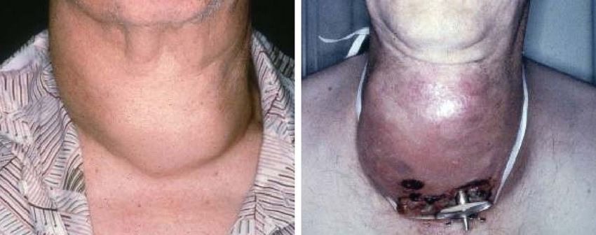
April 2006, Vol. 13, No. 2 Cancer Control 123Fig 3. — Anaplastic thyroid carcinoma, showing the same patient in Fig 2 with rapidly enlarging, fixed mass. Tumor fungation is seen at the tracheostomy site.
the neck. Microscopically, three histologic patterns are lymphoma (ie, Hashimoto’s thyroiditis, female gender).
commonly described: spindle, giant cell, and squamoid. Core biopsies with immunohistochemical staining and
There is no prognostic difference in these patterns. All flow cytometry analysis are valuable in this setting.
three variants have numerous mitotic figures, with large Lymphomas stain for leukocyte-common antigen and do
areas of necrosis, hemorrhage, and vascular invasion.71 not have the marked cellular pleomorphism of ATC.50-52
Unlike PDTC, these tumors often display p53 mutations When considering the diagnosis of medullary thyroid
and do not stain for TG.58 Anaplastic cells typically do cancer, a detailed family history is critical. A genetic
not have thyrotropin receptors, do not transport iodine, workup looking for RET protooncogene mutations
and do not produce TG. might be warranted in this setting. Medullary thyroid
The prognostic impact of the presence of small carcinoma can be distinguished from ATC by staining
foci of ATC in WDTC has not been well studied. Some specifically for calcitonin.
studies have reported improved outcomes in patients Preoperative imaging is helpful in both staging and
with only small foci of ATC; however, others report the treatment planning. A thyroid scan is of no value since
same poor outcome as patients with large, rapidly ATC does not take up RAI. Computed tomography
growing tumors.72,73 As with PDTC, more aggressive scans and magnetic resonance imaging are useful in
therapy may be warranted in patients with WDTC con- defining the local extent of disease and identifying
taining anaplastic foci. distant metastases. PET scans are also useful in detect-
ing distant disease since ATC is highly metabolic.76
Diagnosis
The diagnosis of ATC is usually suspected on clinical Clinical Characteristics
examination and confirmed by FNAB or core biopsy. The peak incidence of ATC occurs in the 6th to 7th
FNAB has been shown to be accurate in 90% of patients decade of life. The mean age at diagnosis is 55 to 65
with ATC.57,74 Core biopsy is useful in narrowing the years. Women comprise 55% to 77% of patients with
differential diagnosis and confirming ATC. Open biop- ATC.45,47,55,77
sy can practically be eliminated; it is indicated only Although some series report incidentally discov-
when clinical suspicion remains high and when FNAB ered ATC in a thyroid nodule, most patients present
and core biopsy are inadequate. Failure to obtain a with a rapidly growing, painful, low anterior neck mass
diagnosis on FNAB or core biopsy may be secondary to that is often firm and fixed to underlying struc-
sampling error or increased areas of necrosis, hemor- tures.44,45,78,79 The mean size of the mass at examination
rhage, or fibrosis. is 8 cm, ranging from 3 cm to 20 cm.47,77 Most patients
ATC has been confused with lymphoma and poorly demonstrate local compressive symptoms including
differentiated medullary thyroid carcinoma. These dysphagia, dysphonia, stridor, dyspnea, and neck pain
tumors, previously classified as small-cell ATC, carry a and tenderness (Fig 3). Regional nodal metastases and
better prognosis and might account for the high sur- vocal cord paralysis are seen in up to 40% and 30%,
vival reported in some ATC series.75 For these reasons, respectively, of the patients with ATC.77 Over 70% of
both lymphoma and poorly differentiated medullary patients with ATC have direct invasion of surrounding
thyroid carcinoma need to be distinguished from ATC. tissues, such as fat, trachea, muscle, esophagus, and lar-
A detailed history may help raise the suspicion of ynx.80 Systemic metastases occur in up to 75% of
124 Cancer Control April 2006, Vol. 13, No. 2patients, with lung being the most common site (80%), reported high long-term survival in patients receiving
followed by bone (6% to 15%) and brain (5% to 13%).44 radiotherapy and chemotherapy after complete gross
Despite the high rate of synchronous metastases, death removal of all tumor. The number of these cases is usu-
is usually related to extensive local disease with ulti- ally small. A recent consensus on the treatment of ATC
mate airway obstruction. suggests that total thyroidectomy is justified if cervical
and mediastinal disease can be resected with limited
Treatment morbidity.85 Resection of vital structures, such as the
Surgery: The role of surgery in ATC, whether it is larynx, pharynx, and esophagus, should be avoided.
removal of all gross disease or palliation, remains con- One of the central issues in the management of
troversial. Patients often present at an advanced stage, ATC is palliation. Palliative management is meant to
making curative surgical resection not feasible. Most prevent death from asphyxiation. Securing a safe air-
studies find that neither the extent of surgery nor the way is a critical component of this effort. Airway man-
completeness of resection has a significant effect on agement may be elective or emergent, depending on
survival.55 Some studies suggest that in a select subset the patient’s presentation. Airway obstruction occurs
of patients with localized disease, survival can be by one of three mechanisms: external compression of
improved by achieving complete resection of all gross the trachea, intraluminal tumor extension, and bilateral
disease.72,81-83 The incidence of regional metastases is vocal cord paralysis. External compression of the tra-
high, and neck dissection should be performed for clin- chea is the most common cause of airway impairment
ically evident disease. De Crevoisier et al84 recently in ATC. Patients with either stridor or rapid tumor
Fig 4. — Algorithm for managing anaplastic thyroid cancer.
April 2006, Vol. 13, No. 2 Cancer Control 125growth should be considered for tracheostomy since bining treatment modalities stems from the failure of
further airway compromise can be expected. Comput- any one individual therapy. EBRT combined with
ed tomography scans should be obtained to determine surgery can improve local control, and chemotherapy
the extent of airway compromise and presence of intra- combined with EBRT can increase the radiosensitivity
luminal tumor. The patient should be taken to the of ATC.45,47,81,83,100 Controversy persists over the timing
operating room for secure airway management prior to of chemoradiation in relation to surgery. Several
performing the tracheostomy. Preoperative sedation authors believe that administering EBRT postoperative-
should be avoided and the patient should be intubated ly provides a theoretical advantage by allowing radia-
under direct vision or fiberoptically. Once the airway tion to treat a smaller tumor burden.45,47 Others have
is secured, the tracheotomy can be performed under used the effects of radiation and chemotherapy pre-
more controlled conditions.86 An extended-length tra- operatively to allow for the potential resection of the
cheostomy tube is often necessary. In an emergent tumor.78,82 A recent study suggested that primary
setting, a cricothyrotomy can be useful. A prophylactic chemoradiation followed by surgery had a positive
tracheostomy is often difficult to perform and does not impact on short-term survival.101 Despite the variable
improve survival. It can be associated with increased sequence of treatment, multimodality regimens cur-
morbidity due to healing problems and tumor funga- rently offer the best hope for patients with ATC. Fig 4
tion, and it might delay radiotherapy. A comprehensive represents our management of patients with ATC.
discussion with the patient and family members is nec-
essary to address the patient’s needs appropriately. Future Directions
Radiotherapy: Achieving local control is impor- ATC remains one of the deadliest of human malignancies.
tant since death from ATC is usually a consequence of Novel treatment strategies are necessary if we are to
uncontrolled local disease. The indications for EBRT make any progress in treating ATC. Promising future
range from providing palliation to improving survival. directives include tumor suppressor gene therapy, induc-
It is used either alone or in combination with surgery tion of cell cycle arrest, and selective inhibition of certain
and/or chemotherapy. Although ATC is relatively proteins, thus inducing apoptosis. Blagosklonny et al102
radioresistant, some studies have shown palliative local showed that adenovirus-mediated p53 tumor suppressor
control in 68% to 80% of patients.83,87-90 The timing, gene therapy increased the in vitro chemosensitivity of
dose, and mode of delivery of EBRT remain controver- ATC to doxorubicin. Nagayama et al103 showed similar
sial. Some investigators have tried hyperfractionated results in vivo. Studies using bone morphogenetic pro-
radiotherapy to keep up with the rapid doubling times tein (BMP-7) and bovine seminal ribonuclease have
of ATC.78,88-92 The efficacy of EBRT needs to be bal- shown efficacy in treating ATC in vitro and in vivo.104,105
anced with its toxicity. Reported complications Several ongoing clinical trials are using vascular and
include pharyngoesophagitis, tracheitis, and myelopa- growth factor-targeted therapies. Agents such as imatinib
thy.89,92 Patients undergoing EBRT spend a significant and combretastatin A4 phosphate are currently being
portion of their remaining life coping with the related used on protocol. More information regarding these and
morbidity. The effect of EBRT is limited, and most other trials can be found at www.clinicaltrials.gov. These
patients progress and ultimately die of their disease. In studies hold promise for improved outcomes and help
select cases, EBRT in combination with surgery and/or guide investigators in the search for better treatment
chemotherapy can improve short-term survival and strategies for patients with ATC.
provide some palliation.
Chemotherapy: Chemotherapy plays an impor-
tant role in the management of ATC since the majority Conclusions
of patients present with or develop distant metastases.
Most series studying the effects of chemotherapeutic Although PDTC and ATC make up a rare group of
agents on ATC have been unsuccessful in altering the tumors, they account for a significant portion of the
fatal outcome of this disease.49 Doxorubicin is the morbidity and mortality associated with thyroid cancer.
most frequently used drug. Monotherapy with doxoru- It is important for clinicians to be able to recognize and
bicin demonstrated a response rate of approximately differentiate these entities. Both of these tumors are
20% with no evidence of a complete response.93,94 more aggressive than WDTC. They have distinctive
Combination therapy with cisplatin or bleomycin clinicopathologic features, and recognition of these
demonstrated little improvement in the clinical histologic variants is important for appropriate man-
response.40,95,96 The recent addition of paclitaxel agement of these tumors. Surgery remains the mainstay
showed some improvement in response but did not of treatment for PDTC, whereas treatment for ATC is
alter the fatal outcome.97-99 The main limitation of all not so clear. Multimodality therapy is usually required
the combination therapies was drug toxicity. to treat both of these rare tumors. Current investiga-
Multimodality Therapy: The rationale of com- tions hold promise for better therapies in the future.
126 Cancer Control April 2006, Vol. 13, No. 2References tures in tall cell papillary carcinoma and insular thyroid carcinoma. Laryn-
goscope. 1997;107:254-259.
1. Shaha AR. Implications of prognostic factors and risk groups in the
30. Rodriguez JM, Parrilla P, Moreno A, et al. Insular carcinoma: an
management of differentiated thyroid cancer. Laryngoscope. 2004;114:393-
infrequent subtype of thyroid cancer. J Am Coll Surg. 1998;187:503-508.
402.
31. Ashfaq R, Vuitch F, Delgado R, et al. Papillary and follicular thyroid
2. Sakamoto A., Kasai n, Sugano H. Poorly differentiated carcinoma of
carcinomas with an insular component. Cancer. 1994;73:416-423.
the thyroid. A clinicopathologic entity for a high-risk group of papillary and
32. Sasaki A, Daa T, Kashima K, et al. Insular component as a risk fac-
follicular carcinomas. Cancer. 1983;52:1849-1855.
tor of thyroid carcinoma. Pathol Int. 1996;46:939-946.
3. Carcangiu ML, Zampi G, Rosai J, et al. Poorly differentiated (“insular”)
33. Pietribiasi F, Sapino A, Papotti M, et al. Cytologic features of poorly
thyroid carcinoma: a reinterpretation of Langhans’ “wuchernde Struma.” Am
differentiated “insular” carcinoma of the thyroid, as revealed by fine-needle
J Surg Pathol. 1984;8:655-668.
aspiration biopsy. Am J Clin Pathol. 1990;94:687-692.
4. Nishida T, Katayama S, Tsujimoto M, et al. Clinicopathological sig-
34. Pilotti S, Collini P, Del Bo R, et al. A novel panel of antibodies that
nificance of poorly differentiated thyroid carcinoma. Am J Surg Pathol.
segregates immunocytochemically poorly differentiated carcinoma from
1999;23:205-211.
undifferentiated carcinoma of the thyroid gland. Am J Surg Pathol. 1994;18:
5. Sobrinho-Simoes M, Sambade C, Fonseca E, et al. Poorly differen-
1054-1064.
tiated carcinomas of the thyroid gland: a review of the clinicopathologic fea-
35. Sywak M, Pasieka JL, Ogilvie T, et al. A review of thyroid cancer with
tures of a series of 28 cases of a heterogeneous, clinically aggressive group
intermediate differentiation. J Surg Oncol. 2004;86:44-54.
of thyroid tumors. Int J Surg Pathol. 2002;10:123-131.
36. Wreesmann VB, Ghossein RA, Patel SG, et al. Genome-wide
6. Carcangiu ML, Bianchi S. Diffuse sclerosing variant of papillary thy-
appraisal of thyroid cancer progression. Am J Pathol. 2002;161:1549-1556.
roid carcinoma: clinicopathologic study of 15 cases. Am J Surg Pathol.
37. Decaussin M, Bernard MH, Adeleine P, et al. Thyroid carcinomas with
1989;13:1041-1049.
distant metastases: a review of 111 cases with emphasis on the prognostic
7. Pilotti S, Collini P, Manzari A, et al. Poorly differentiated forms of pap-
significance of an insular component. Am J Surg Pathol. 2002;26: 1007-
illary thyroid carcinoma: distinctive entities or morphological patterns?
1015.
Semin Diagn Pathol. 1995;12:249-255.
38. Justin EP, Seabold JE, Robinson RA, et al. Insular carcinoma: a dis-
8. Sobrinho-Simoes M, Nesland JM, Johannessen JV. Columnar-cell
tinct thyroid carcinoma with associated iodine-131 localization. J Nucl Med.
carcinoma. Another variant of poorly differentiated carcinoma of the thyroid. 1991;32:1358-1363.
Am J Clin Pathol. 1988;89:264-267.
39. Brierley JD, Tsang RW. External-beam radiation therapy in the treat-
9. Akslen LA, LiVolsi VA. Poorly differentiated thyroid carcinoma: it is ment of differentiated thyroid cancer. Semin Surg Oncol. 1999;16:42-49.
important. Am J Surg Pathol. 2000;24:310-313.
40. Asakawa H, Kobayashi T, Komoike Y, et al. Chemosensitivity of
10. Nikiforov YE, Erickson LA, Nikiforova MN, et al. Solid variant of papil- anaplastic thyroid carcinoma and poorly differentiated thyroid carcinoma.
lary thyroid carcinoma: incidence, clinical-pathologic characteristics, molecu- Anticancer Res. 1997;17:2757-2762.
lar analysis, and biologic behavior. Am J Surg Pathol. 2001;25:1478-1484. 41. Wang W, Macapinlac H, Larson SM, et al. [18F]-2-fluoro-2-deoxy-D-
11. Nikiforov Y, Gnepp DR. Pediatric thyroid cancer after the Chernobyl glucose positron emission tomography localizes residual thyroid cancer in
disaster: pathomorphologic study of 84 cases (1991-1992) from the Repub- patients with negative diagnostic (131)I whole body scans and elevated
lic of Belarus. Cancer. 1994;74:748-666. serum thyroglobulin levels. J Clin Endocrinol Metab. 1999;84:2291-2302.
12. Nikiforov Y, Gnepp DR, Fagin JA. Thyroid lesions in children and 42. Wang W, Larson SM, Fazzari M, et al. Prognostic value of [18F]
adolescents after the Chernobyl disaster: implications for the study of radi- fluorodeoxyglucose positron emission tomographic scanning in patients with
ation tumorigenesis. J Clin Endocrinol Metab. 1996;81:9-14. thyroid cancer. J Clin Endocrinol Metab. 2000;85:1107-1113.
13. Yunta PJ, Ponce JL, Prieto M, et al. The importance of a tumor cap- 43. Gilliland FD, Hunt WC, Morris DM, et al. Prognostic factors for thy-
sule in columnar cell thyroid carcinoma: a report of two cases and review of roid carcinoma. A population-based study of 15,698 cases from the Sur-
the literature. Thyroid. 1999;9:815-819. veillance, Epidemiology and End Results (SEER) program 1973-1991. Can-
14. Burman KD, Ringel MD, Wartofsky L, et al. Unusual types of thyroid cer. 1997;79:564-573.
neoplasms. Endocrinol Metab Clin North Am. 1996;25:49-68. 44. Nel CJ, van Heerden JA, Goellner JR, et al.. Anaplastic carcinoma of
15. Livolsi VA. Surgical Pathology of the Thyroid. Philadelphia, Pa: the thyroid: a clinicopathologic study of 82 cases. Mayo Clin Proc. 1985;60:
Saunders; 1990. 51-58.
16. Langhans T. Uber die epithelialen formen der malignen struma. Vir- 45. Venkatesh YS, Ordonez NG, Schultz PN, et al. Anaplastic carcino-
chows Arch A Pathol Anat. 1907;189:69-188. ma of the thyroid: a clinicopathologic study of 121 cases. Cancer. 1990;66:
17. Hedinger CE. Thyroid Cancer. Berlin, New York: Springer; 1969. 321-330.
18. Meissner WA, Warren S. Tumors of the Thyroid Gland. Washington, 46. Lo CY, Lam KY, Wan KY, et al. Anaplastic carcinoma of the thyroid.
DC: Armed Forces Institute of Pathology; 1969. Am J Surg. 1999;177:337-339.
19. Woolner LB, Beahrs OH, Black BM, et al. Classification and progno- 47. Tan RK, Finley RK III, Driscoll D, et al. Anaplastic carcinoma of the
sis of thyroid carcinoma. A study of 885 cases observed in a thirty year peri- thyroid: a 24-year experience. Head Neck. 1995;17:41-48.
od. Am J Surg. 1961;102:354-387. 48. Schaefer CJ. Long-term survival in anaplastic thyroid cancer. Md
20. Albareda M, Puig-Domingo M, Wengrowicz S, et al. Clinical forms of Med J. 1988;37:873-874.
presentation and evolution of diffuse sclerosing variant of papillary carcino- 49. Pasieka JL. Anaplastic thyroid cancer. Curr Opin Oncol. 2003;15:
ma and insular variant of follicular carcinoma of the thyroid. Thyroid. 78-83.
1998;8:385-391. 50. Holting T, Moller P, Tschahargane C, et al. Immunohistochemical
21. Papotti M, Botto Micca F, Favero A, et al. Poorly differentiated thyroid reclassification of anaplastic carcinoma reveals small and giant cell lym-
carcinomas with primordial cell component: a group of aggressive lesions phoma. World J Surg. 1990;14:291-295.
sharing insular, trabecular, and solid patterns. Am J Surg Pathol. 1993;17: 51. Tobler A, Maurer R, Hedinger CE, et al. Undifferentiated thyroid
291-301. tumors of diffuse small cell type. Histological and immunohistochemical
22. Pilotti S, Collini P, Mariani L, et al. Insular carcinoma: a distinct de novo evidence for their lymphomatous nature. Virchows Arch A Pathol Anat
entity among follicular carcinomas of the thyroid gland. Am J Surg Pathol. Histopathol. 1984;404:117-126.
1997;21:1466-1473. 52. Myskow MW, Krajewski AS, Dewar AE, et al. The role of immunoper-
23. Hwang TS, Suh JS, Kim YI, et al. Poorly differentiated carcinoma of oxidase techniques on paraffin embedded tissue in determining the histoge-
the thyroid retrospective clinical and morphologic evaluation. J Korean Med nesis of undifferentiated thyroid neoplasms. Clin Endocrinol (Oxf). 1986;24:
Sci. 1990;5:47-52. 335-341.
24. Flynn SD, Forman BH, Stewart AF, et al. Poorly differentiated (“insu- 53. Bakiri F, Djemli FK, Mokrane LA, et al. The relative roles of endem-
lar”) carcinoma of the thyroid gland: an aggressive subset of differentiated ic goiter and socioeconomic development status in the prognosis of thyroid
thyroid neoplasms. Surgery. 1988;104:963-970. carcinoma. Cancer. 1998;82:1146-1153.
25. Lam KY, Lo CY, Chan KW, et al. Insular and anaplastic carcinoma of 54. Pettersson B, Coleman MP, Ron E, et al. Iodine supplementation in
the thyroid: a 45-year comparative study at a single institution and a review Sweden and regional trends in thyroid cancer incidence by histopathologic
of the significance of p53 and p21. Ann Surg. 2000;231:329-338. type. Int J Cancer. 1996;65:13-19.
26. Machens A, Hinze R, Lautenschlager C, et al. Multivariate analysis 55. McIver B, Hay ID, Giuffrida DF, et al. Anaplastic thyroid carcinoma:
of clinicopathologic parameters for the insular subtype of differentiated thy- a 50-year experience at a single institution. Surgery. 2001;130:1028-1034.
roid carcinoma. Arch Surg. 2001;136:941-944. 56. Demeter JG, De Jong SA, Lawrence AM, et al. Anaplastic thyroid
27. Takeuchi Y, Daa T, Kashima K, et al. Mutations of p53 in thyroid car- carcinoma: risk factors and outcome. Surgery. 1991;110:956-963.
cinoma with an insular component. Thyroid. 1999;9:377-381. 57. Ain KB. Anaplastic thyroid carcinoma: a therapeutic challenge.
28. Killeen RM, Barnes L, Watson CG, et al. Poorly differentiated (‘insu- Semin Surg Oncol. 1999;16:64-69.
lar’) thyroid carcinoma: report of two cases and review of the literature. Arch 58. Fagin JA. Molecular genetics of human thyroid neoplasms. Annu
Otolaryngol Head Neck Surg. 1990;116:1082-1086. Rev Med. 1994;45:45-52.
29. van den Brekel MW, Hekkenberg RJ, Asa SL, et al. Prognostic fea- 59. Carcangiu ML, Steeper T, Zampi G, et al. Anaplastic thyroid carci-
April 2006, Vol. 13, No. 2 Cancer Control 127noma: a study of 70 cases. Am J Clin Pathol. 1985;83:135-158. 90. Kim JH, Leeper RD. Treatment of locally advanced thyroid carcino- 60. Nishiyama RH, Dunn EL, Thompson NW. Anaplastic spindle-cell and ma with combination doxorubicin and radiation therapy. Cancer. 1987;60: giant-cell tumors of the thyroid gland. Cancer. 1972;30:113-127. 2372-2375. 61. Ibanez ML, Russell WO, Albores-Saavedra J, et al. Thyroid carcino- 91. Tennvall J, Lundell G, Wahlberg P, et al. Anaplastic thyroid carcino- ma: biologic behavior and mortality. Postmortem findings in 42 cases, ma: three protocols combining doxorubicin, hyperfractionated radiotherapy including 27 in which the disease was fatal. Cancer. 1966;19:1039-1052. and surgery. Br J Cancer. 2002;86:1848-1853. 62. Moreno Egea A, Rodriguez Gonzalez JM, Sola Perez J, et al. Prog- 92. Wong CS, Van Dyk J, Simpson WJ. Myelopathy following hyperfrac- nostic value of the tall cell variety of papillary cancer of the thyroid. Eur J tionated accelerated radiotherapy for anaplastic thyroid carcinoma. Radio- Surg Oncol. 1993;19:517-521. ther Oncol. 1991;20:3-9. 63. Cohen Y, Xing M, Mambo E, et al. BRAF mutation in papillary thyroid 93. Shimaoka K, Schoenfeld DA, DeWys WD, et al. A randomized trial carcinoma. J Natl Cancer Inst. 2003;95:625-627. of doxorubicin versus doxorubicin plus cisplatin in patients with advanced 64. Xing M. BRAF mutation in thyroid cancer. Endocr Relat Cancer. thyroid carcinoma. Cancer. 1985;56:2155-2160. 2005;12:245-262. 94. Ahuja S, Ernst H. Chemotherapy of thyroid carcinoma. J Endocrinol 65. Xing M, Westra WH, Tufano RP, et al. BRAF mutation predicts a Invest. 1987;10:303-310. poorer clinical prognosis for papillary thyroid cancer. J Clin Endocrinol 95. De Besi P, Busnardo B, Toso S, et al. Combined chemotherapy with Metab. 2005;90:6373-6379. Epub 2005 Sep 20. bleomycin, adriamycin, and platinum in advanced thyroid cancer. J 66. Xing M, Vasko V, Tallini G, et al. BRAF T1796A transversion muta- Endocrinol Invest. 1991;14:475-480. tion in various thyroid neoplasms. J Clin Endocrinol Metab. 2004;89:1365- 96. Williams SD, Birch R, Einhorn LH. Phase II evaluation of doxorubicin 1368. plus cisplatin in advanced thyroid cancer: a Southeastern Cancer Study 67. Xu X, Quiros RM, Gattuso P, et al. High prevalence of BRAF gene Group Trial. Cancer Treat Rep. 1986;70:405-407. mutation in papillary thyroid carcinomas and thyroid tumor cell lines. Can- 97. Ain KB, Egorin MJ, DeSimone PA, et al. Treatment of anaplastic thy- cer Res. 2003;63:4561-4567. roid carcinoma with paclitaxel: phase 2 trial using ninety-six-hour infusion. 68. Puxeddu E, Moretti S, Elisei R, et al. BRAF(V599E) mutation is the Collaborative Anaplastic Thyroid Cancer Health Intervention Trials (CATCHIT) leading genetic event in adult sporadic papillary thyroid carcinomas. J Clin Group. Thyroid. 2000;10:587-594. Endocrinol Metab. 2004;89:2414-2420. 98. Yeung SC, Xu G, Pan J, et al. Manumycin enhances the cytotoxic 69. Quiros RM, Ding HG, Gattuso P, et al. Evidence that one subset of effect of paclitaxel on anaplastic thyroid carcinoma cells. Cancer Res. 2000; anaplastic thyroid carcinomas are derived from papillary carcinomas due to 60:650-656. BRAF and p53 mutations. Cancer. 2005;103:2261-2268. 99. Xu G, Pan J, Martin C, et al. Angiogenesis inhibition in the in vivo 70. Knauf JA, Ma X, Smith EP, et al. Targeted expression of BRAFV600E antineoplastic effect of manumycin and paclitaxel against anaplastic thyroid in thyroid cells of transgenic mice results in papillary thyroid cancers that carcinoma. J Clin Endocrinol Metab. 2001;86:1769-1777. undergo dedifferentiation. Cancer Res. 2005;65:4238-4245. 100. Sugino K, Ito K, Mimura T, et al. The important role of operations in 71. Pasieka JL. Anaplastic cancer, lymphoma, and metastases of the the management of anaplastic thyroid carcinoma. Surgery. 2002;131:245- thyroid gland. Surg Oncol Clin N Am. 1998;7:707-720. 248. 72. Kobayashi T, Asakawa H, Umeshita K, et al. Treatment of 37 patients 101. Besic N, Auersperg M, Us-Krasovec M, et al. Effect of primary treat- with anaplastic carcinoma of the thyroid. Head Neck. 1996;18:36-41. ment on survival in anaplastic thyroid carcinoma. Eur J Surg Oncol. 2001; 73. Pacheco-Ojeda LA, Martinez AL, Alvarez M, et al. Anaplastic thyroid 27:260-264. carcinoma in Ecuador: analysis of prognostic factors. Int Surg. 2001;86: 102. Blagosklonny MV, Giannakakou P, Wojtowicz M, et al. Effects of p53- 117-121. expressing adenovirus on the chemosensitivity and differentiation of 74. Us-Krasovec M, Golouh R, Auersperg M, et al. Anaplastic thyroid anaplastic thyroid cancer cells. J Clin Endocrinol Metab. 1998;83:2516- carcinoma in fine needle aspirates. Acta Cytol. 1996;40:953-958. 2522. 75. Wolf BC, Sheahan K, DeCoste D, et al. Immunohistochemical analy- 103. Nagayama Y, Yokoi H, Takeda K, et al. Adenovirus-mediated tumor sis of small cell tumors of the thyroid gland: an Eastern Cooperative Oncol- suppressor p53 gene therapy for anaplastic thyroid carcinoma in vitro and in ogy Group study. Hum Pathol. 1992;23:1252-1261. vivo. J Clin Endocrinol Metab. 2000;85:4081-4086. 76. Poppe K, Lahoutte T, Everaert H, et al. The utility of multimodality 104. Franzen A, Heldin NE. BMP-7-induced cell cycle arrest of anaplas- imaging in anaplastic thyroid carcinoma. Thyroid. 2004;14:981-982. tic thyroid carcinoma cells via p21(CIP1) and p27(KIP1). Biochem Biophys 77. Ain KB. Anaplastic thyroid carcinoma: behavior, biology, and thera- Res Commun. 2001;285:773-781. peutic approaches. Thyroid. 1998;8:715-726. 105. Kotchetkov R, Cinatl J, Krivtchik AA, et al. Selective activity of BS- 78. Tennvall J, Lundell G, Hallquist A, et al. Combined doxorubicin, RNase against anaplastic thyroid cancer. Anticancer Res. 2001;21:1035- hyperfractionated radiotherapy, and surgery in anaplastic thyroid carcinoma: 1042. report on two protocols. The Swedish Anaplastic Thyroid Cancer Group. Cancer. 1994;74:1348-1354. 79. Hadar T, Mor C, Shvero J, et al. Anaplastic carcinoma of the thyroid. Eur J Surg Oncol. 1993;19:511-516. 80. Giuffrida D, Gharib H. Anaplastic thyroid carcinoma: current diag- nosis and treatment. Ann Oncol. 2000;11:1083-1089. 81. Haigh PI, Ituarte PH, Wu HS, et al. Completely resected anaplastic thyroid carcinoma combined with adjuvant chemotherapy and irradiation is associated with prolonged survival. Cancer. 2001;91:2335-2342. 82. Nilsson O, Lindeberg J, Zedenius J, et al. Anaplastic giant cell car- cinoma of the thyroid gland: treatment and survival over a 25-year period. World J Surg. 1998;22:725-730. 83. Junor EJ, Paul J, Reed NS, et al. Anaplastic thyroid carcinoma: 91 patients treated by surgery and radiotherapy. Eur J Surg Oncol. 1992;18: 83-88. 84. De Crevoisier R, Baudin E, Bachelot A, et al. Combined treatment of anaplastic thyroid carcinoma with surgery, chemotherapy, and hyperfraction- ated accelerated external radiotherapy. Int J Radiat Oncol Biol Phys. 2004;60:1137-1143. 85. Thyroid Carcinoma Task Force. AACE/AAES medical/surgical guide- lines for clinical practice: management of thyroid carcinoma. American Association of Clinical Endocrinologists. American College of Endocrinology. Endocr Pract. 2001;7:202-220. 86. Pellitteri PK, McCaffrey TV. Endocrine Surgery of the Head and Neck. Clifton Park, NY: Thomson Delmar Learning; 2003. 87. Levendag PC, De Porre PM, van Putten WL. Anaplastic carcinoma of the thyroid gland treated by radiation therapy. Int J Radiat Oncol Biol Phys. 1993;26:125-128. 88. Simpson WJ. Anaplastic thyroid carcinoma: a new approach. Can J Surg. 1980;23:25-27. 89. Kim JH, Leeper RD. Treatment of anaplastic giant and spindle cell carcinoma of the thyroid gland with combination Adriamycin and radiation therapy: a new approach. Cancer. 1983;52:954-957. 128 Cancer Control April 2006, Vol. 13, No. 2
You can also read