Oncolytic Herpes Simplex Virus Encoding IL12 Controls Triple-Negative Breast Cancer Growth and Metastasis - Frontiers
←
→
Page content transcription
If your browser does not render page correctly, please read the page content below
ORIGINAL RESEARCH
                                                                                                                                           published: 24 March 2020
                                                                                                                                      doi: 10.3389/fonc.2020.00384

                                              Oncolytic Herpes Simplex Virus
                                              Encoding IL12 Controls
                                              Triple-Negative Breast Cancer
                                              Growth and Metastasis
                                              Shanawaz M. Ghouse 1† , Hong-My Nguyen 1† , Praveen K. Bommareddy 2 ,
                                              Kirsten Guz-Montgomery 1 and Dipongkor Saha 1*
                                              1
                                               Department of Immunotherapeutics and Biotechnology, School of Pharmacy, Texas Tech University Health Sciences Center,
                                              Abilene, TX, United States, 2 School of Graduate Studies, Rutgers University, New Brunswick, NJ, United States

                                              Triple-negative breast cancer (TNBC) is a difficult-to-treat disease with high rates of
                                              local recurrence, distant metastasis, and poor overall survival with existing therapies.
                                              Thus, there is an unmet medical need to develop new treatment regimen(s) for TNBC
                                              patients. An oncolytic herpes simplex virus encoding a master anti-tumor cytokine,
                                              interleukin 12, (designated G471-mIL12) selectively kills cancer cells while inducing
                           Edited by:         anti-tumor immunity. G471-mIL12 efficiently infected and killed murine (4T1 and EMT6)
                          Daniel Olive,       and human (HCC1806 and MDA-MB-468) mammary tumor cells in vitro. In vivo in
       Aix Marseille Université, France
                                              the 4T1 syngeneic TNBC model, it significantly reduced primary tumor burden and
                        Reviewed by:
             Luis De La Cruz-Merino,
                                              metastasis, both at early and late stages of tumor development. The virus-induced local
          Hospital Universitario Virgen       and abscopal effects were confirmed by significantly increased infiltration of CD45+
                     Macarena, Spain          leukocytes and CD8+ T cells, and reduction of granulocytic and monocytic MDSCs in
                    Cristian Smerdou,
          University of Navarra, Spain        tumors, both treated and untreated contralateral, and in the spleen. Significant trafficking
                 *Correspondence:             of dendritic cells (DCs) were only observed in spleens of virus-treatment group, indicating
                     Dipongkor Saha           that DCs are primed and activated in the tumor-microenvironment following virotherapy,
          dipongkor.saha@ttuhsc.edu
                                              and trafficked to lymphoid organs for activation of immune cells, such as CD8+
     † These   authors have contributed       T cells. DC priming/activation could be associated with virally enhanced expression
                    equally to this work
                                              of several antigen processing/presentation genes in the tumor microenvironment, as
                   Specialty section:         confirmed by NanoString gene expression analysis. Besides DC activation/priming,
         This article was submitted to        G471-mIL12 treatment led to up-regulation of CD8+ T cell activation markers in the
Cancer Immunity and Immunotherapy,
                                              tumor microenvironment and inhibition of tumor angiogenesis. The anti-tumor effects
               a section of the journal
                 Frontiers in Oncology        of G471-mIL12 treatment were CD8-dependent. These studies illustrate the ability of
       Received: 24 December 2019             G471-mIL12 to immunotherapeutically treat TNBC.
          Accepted: 04 March 2020
                                              Keywords: herpes simplex virus, oncolytic immunotherapy, breast cancer, metastasis, anti-angiogenesis
         Published: 24 March 2020
                            Citation:
             Ghouse SM, Nguyen H-M,
Bommareddy PK, Guz-Montgomery K
                                              INTRODUCTION
 and Saha D (2020) Oncolytic Herpes
Simplex Virus Encoding IL12 Controls
                                              Breast cancer is one of the most prevalent malignancies and the second most common cause
Triple-Negative Breast Cancer Growth          of death among women in the United States (1). Almost 15 to 20% of breast cancer cases are
and Metastasis. Front. Oncol. 10:384.         classified as triple negative breast cancer (TNBC) variants, which lack the expression of estrogen
        doi: 10.3389/fonc.2020.00384          and progesterone receptors, and human epidermal growth factor receptor 2 (HER2) proteins on

Frontiers in Oncology | www.frontiersin.org                                        1                                            March 2020 | Volume 10 | Article 384
Ghouse et al.                                                                                              Oncolytic HSV-IL-12 for TNBC Treatment

tumor cells (2). Tumor recurrence, metastasis to other vital               in myeloid-derived suppressor cells (MDSCs), and inhibition of
organs, and high heterogeneity are considered hallmarks of                 angiogenesis. G471-mIL12 exerted its anti-TNBC effects in a
TNBC variants (3). Since TNBC does not express hormone                     CD8+ T cell-dependent manner. These studies establish G471-
receptors, the current treatment for TNBC mainly relies on                 mIL12 virus as a powerful oncolytic immunotherapeutic agent
surgery and chemotherapy; targeted therapy, such as hormone-               for TNBC.
based treatments or HER2 antagonists, is not a treatment option
(4). Moreover, TNBC initially responds well but often develops             MATERIALS AND METHODS
resistance against chemotherapy (5), which underscores the need
for developing novel therapeutic modalities that efficiently cure          Cells and Viruses
TNBC in patients.                                                          Mouse (4T1 and EMT6) and human (HCC1806 and MDA-
    Oncolytic viruses (OVs) (6), such as oncolytic herpes simplex          MB-468) mammary tumor cells were purchased from American
viruses (OHSVs) are a promising therapeutic strategy for cancer            Type Culture Collection (ATCC) and grown in Dulbecco’s
treatment (7, 8). OHSVs are defined as genetically modified                Modified Eagle Medium (DMEM) (ThermoFisher Scientific)
OVs that preferentially replicate in and kill tumor cells without          supplemented with 10% heat-inactivated fetal bovine serum
harming normal cells (6). They have been established as                    (HyClone), 2 mM L-glutamine (Corning), 1% MEM non-
strong in situ anticancer vaccines that activate antigen presenting        essential amino acids (Gibco), 1% sodium pyruvate (Gibco),
cells (APCs), enhance APC-mediated tumor cell phagocytosis,                and 0.5% penicillin G-streptomycin sulfate-amphotericin B
augment antigen processing and presentation, and prime T                   complex (Corning). Cells were trypsinized with 0.05% trypsin
cell responses (9). OHSVs have been successfully transitioned              supplemented with 0.54 mM EDTA (Corning) for passaging.
into clinical trials against various human cancers, including              Cells were low-passage and confirmed to be mycoplasma-free
melanoma, glioma, pancreatic, and breast cancers (7, 8). In                (LookOut mycoplasma kit, Sigma).
2015, the U.S. Food and Drug Administration (FDA) approved                    G471-mIL12, an oHSV encoding IL-12, was constructed
the first OHSV (designated T-VEC) for the treatment of                     from G471 [containing deletions in α47 and γ34.5 genes and an
advanced melanoma in the United States. T-VEC is a genetically             inactivating insertion of Escherichia Coli LacZ into ICP6 (22)] by
engineered OHSV expressing human granulocyte-macrophage                    insertion of mouse IL-12 cDNA (p35 and p40 units are separated
colony-stimulating factor (hGM-CSF) (10), and is the furthest              by two bovine elastin motifs) into the ICP6 gene (14). G471-
along in the clinic for cancer treatment (10). The safety and              mCherry was described previously (14). Prior to in vitro and
efficacy of T-VEC (as a monotherapy or combination therapy                 in vivo studies, the titers of infectious G471-mIL12 virus were
with paclitaxel) in TNBC patients is under clinical trial evaluation       determined by plaque assay on Vero cells (14).
(8, 11, 12). However, T-VEC has not demonstrated durable
responses in a majority of advanced melanoma patients (10),                Mice
especially those with visceral metastases (13), which raises               Female BALB/c mice (aged 8–9 weeks) were obtained from
questions about its possible long-term efficacy in TNBC patients           the Jackson laboratory (Bar Harbor, ME) and utilized for all
with metastatic disease.                                                   in vivo mouse studies involving the 4T1 mammary tumor cell
    G471-mIL12 (14) is a genetically engineered OHSV that has              line (21). Mice were housed at the Texas Tech University Health
similar genetic modifications to T-VEC (15, 16) but contains an            Sciences Center (TTUHSC) Laboratory Animal Resources
extra safety feature [i.e., ICP6 inactivation that restricts OHSV          Center (LARC)-Abilene under BSL2 conditions. All mouse
replication to cancer cells (16)] and expresses murine Interleukin         procedures were approved by the Institutional Animal Care and
12 (IL-12) (instead of GM-CSF). Upon infection of tumor cells,             Use Committee (IACUC) at the TTUHSC.
G471-mIL12 releases a significant amount of IL12 (14), a master
regulator of antitumor immunity, that enhances activation of               Cell Viability Assay
dendritic cells and T lymphocytes, induces IFN-γ production,               Mouse and human mammary tumor cells were dissociated and
and inhibits angiogenesis (17–19). Previous reports affirm G471-           seeded into 96-well plates (3,000 cells per well for mouse lines
mIL12 as a potent oncolytic viral therapy for glioblastoma (14)            and 10,000 cells per well for human lines), treated with G471-
and malignant peripheral nerve sheath tumors (20).                         mIL12 at the indicated multiplicity of infection (MOI), incubated
    In this study, we have chosen to evaluate the therapeutic              at 37◦ C for up to 72–96 h and CellTiter96 AQueous One Solution
efficacy of G471-mIL12 in a 4T1 tumor model, which is an                   Cell Viability (MTS) Assays (Promega) performed according to
immune-competent, highly tumorigenic, and invasive mouse                   the manufacturer’s instructions. Values for virus-infected cells
mammary carcinoma that can spontaneously metastasize from                  were normalized to those for mock-infected cells (percent cell
the primary tumor in the mammary gland to multiple distant                 viability). The experiments were performed in triplicate and
sites, such as lung (21). In addition, 4T1 serves as a model for           repeated at least 2–4 times. Dose response curves and IC50 values
stage IV of advanced breast cancer in humans. We found that                were calculated using Prism 7 GraphPad software version 7.0e.
G471-mIL12 efficiently infected and eliminated both murine
and human TNBC cells in vitro. In vivo, G471-mIL12 treatment               Tumor Immunotherapy With G471-mIL12
effectively inhibited 4T1 tumor growth, both primary and                   at Early-Stages of Tumor Development
contralateral, and prevented metastasis to the lungs, which                Mice were implanted subcutaneously (s.c.) with 1 × 10⁵ 4T1
were associated with an enhanced APC activation, increased                 tumor cells into the mammary fat pad to generate orthotopic
intratumoral CD8+ T-cell infiltration with subsequent reduction            breast tumors. When tumors were palpable and reached 50-70

Frontiers in Oncology | www.frontiersin.org                            2                                       March 2020 | Volume 10 | Article 384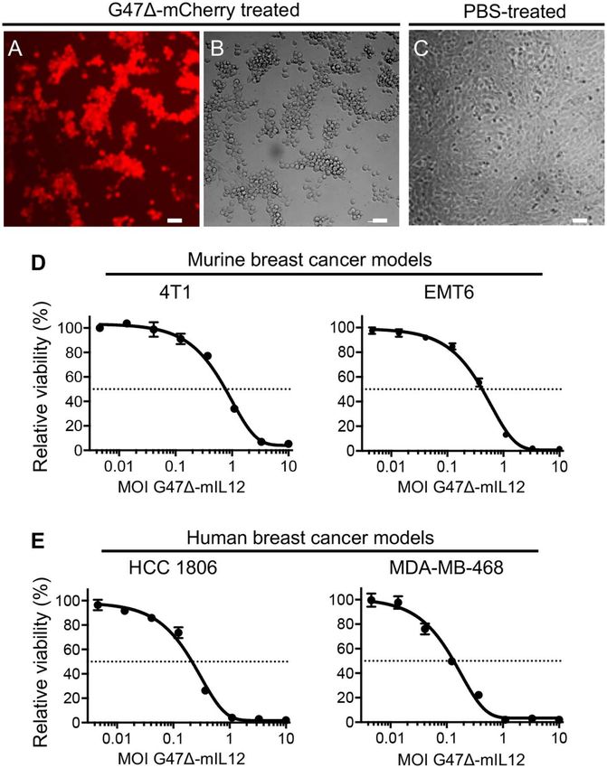
Ghouse et al.                                                                                          Oncolytic HSV-IL-12 for TNBC Treatment

mm3 in tumor volume, mice were randomly divided into groups            Multi-Color Flow Cytometry
and intratumorally (i.t.) treated with G471-mIL12 (in 25 µl PBS)       For multi-color flow cytometric analysis (FACS), mammary
or PBS on days 6, 9, 12, 15, and 18 post-tumor implantations.          tumor tissues were harvested and minced, and single-cell
Tumors were measured at regular intervals with a digital caliper       suspensions prepared by incubation of minced tissues in RPMI
throughout the course of the experiment. The tumor volume              1640 medium containing 10 mg/mL Collagenase (Roche),
was calculated using the following formula: (length x width            0.4mg/mL DNase I (Roche), and 100 µg/mL Trypsin inhibitor
x depth)/2.                                                            (Sigma) for 30 min at 37◦ C. Enzymatic digestion was stopped
                                                                       by adding RPMI containing 10% fetal bovine serum (FBS;
Tumor Immunotherapy With G471-mIL12                                    Corning), triturated, passed through a 70-µm cell strainer,
                                                                       washed twice with PBS, resuspended in FACS buffer, and counted
at Late-Stages of Tumor Development
                                                                       using Vi-CELL XR Cell Viability Analyzer (Beckman Coulter).
Orthotopic 4T1 tumors were established bilaterally in the
                                                                       The samples were pre-incubated with purified anti-CD16/32
right and left axillary mammary fat pads (1 × 10⁵ cells per
                                                                       unconjugated antibody (clone 93) to block Fc receptors prior
mammary pad) on day 0. When tumors reached between
                                                                       to surface staining with fluorochrome-conjugated anti-mouse
100–125 mm3 in volume, tumors located at the right axillary
                                                                       monoclonal antibodies, which include: Brilliant Violet 605 anti-
mammary fat pad were intratumorally injected with PBS
                                                                       mouse CD45 (clone 30-F11), Alexa Fluor 700 anti-mouse CD3
or G471-mIL12 on days 10, 13, 16, and 19, while the
                                                                       (clone 17A2), PerCP/Cyanine5.5 anti-mouse CD4 (clone GK1.5),
tumors at the left axillary mammary fat pad remained
                                                                       PE/Cy7 anti-mouse CD8a (clone 53-6.7), PE anti-mouse CD86
untreated and served as contralateral tumors. Tumor volumes
                                                                       (clone GL-1), PerCP/Cyanine5.5 anti-mouse F4/80 (clone BM8),
(treated and untreated) were periodically measured by caliper,
                                                                       APC/Cyanine7 anti-mouse CD11c (clone N418), Alexa Fluor
and mice were followed for survival until they become
                                                                       700 anti-mouse/human CD11b (clone M1/70), Brilliant Violet
moribund or tumors reached to their burden limit, i.e., 1.5 cm
                                                                       570 anti-mouse Ly6G (clone 1A8), PE/Cy7 anti-mouse Ly6C
in size.
                                                                       (clone HK1.4), APC anti-mouse CD274 (clone 10F.9G2), and
                                                                       Brilliant Violet 650 anti-mouse I-A/I-E (clone M5/114.15.2),
Metastatic Tumor Study                                                 as well as appropriate isotype control antibodies, as described
4T1 tumor cells (1 × 10⁵ ) were implanted s.c. into the mammary        (24, 26). All antibodies were purchased from Biolegend.
fat pad and treated i.t. with G471-mIL12 or PBS as described           Fixable Viability Dye eFluor 506 (eBioscience) was used to
above in “Tumor immunotherapy with G471-mIL12 at early-                stain dead cells as per manufacturer instructions. Intracellular
stages of tumor development” section. Mice were sacrificed on          FOXP3 staining was performed using PE-conjugated anti-mouse
day 21 post-tumor implantation, and lungs were isolated and            FOXP3 antibody (clone MF-14, Biolegend) following the FOXP3
fixed in Bouin’s fixative solution (23). Twenty-four hours post-       intracellular staining protocol (eBioscience). For multi-color
fixation, Bouin’s solution was replaced by 70% ethanol and the         FACS staining of spleens and tumor-draining lymph nodes, single
number of lung surface metastatic nodules were counted with            cell suspensions from these organs were prepared as described
a Nikon Stereo Microscope with Plan APO 1x WD70 objective              (14) and stained as above. Fluorescent minus one (FMO)
(23). Images for lung surface metastatic nodules were captured         controls were included for each color-conjugated anti-mouse
by Motic FEIN OPTIC SMZ-168 stereo microscope connected                antibody, e.g., fluorescent minus F4/80 means staining cells
with a ToupTek digital camera.                                         with all colors except PerCP/Cyanine5.5 anti-mouse F4/80 (24).
                                                                       UltraComp eBeads (eBioscience) were used to prepare single-
Immune Cell Depletion Studies                                          color compensation controls for each fluorescently conjugated
4T1 tumor cells (1 × 10⁵ ) were implanted s.c. into the mammary        antibody according to manufacturer instructions (26). Single-cell
fat pad and treated with G471-mIL12 or PBS injected i.t. on            suspensions from harvested tissues were used to prepare a single-
days 8, 11, 14, and 17 post-tumor implantations. For CD8+              color compensation control for fixable viability dye eFluor 506.
cell depletion, mice were injected intraperitoneally (i.p.) with       Data were acquired on BD Fortessa and analyzed with FlowJo
anti-CD8a antibodies (clone 2.43, 10 mg/kg, BioXCell) (24)             software version 10.6.1 (Tree Star). Scientific personnel involved
or isotype control rat IgG2b antibodies specific to keyhole            in acquiring and gating the data was blinded to the treatments.
limpet hemocyanin (clone LTF-2, 10 mg/kg, BioXCell) (24) on
days−4 and−1 prior to tumor implantation and on days 4,                Indirect Immunofluorescence Staining
8, 12, and 16 post-tumor implantation (25). Three groups of            Mammary tumor tissues were harvested, snap-freezed in Tissue-
mice were included in CD8+ immune cell depletion studies:              Tek O.C.T. compound (Sakura), 5 µm cryostat sections prepared,
group 1 received PBS + IgG2b, group 2 received G471-                   fixed in methanol for 10 min at −20◦ C, sections dried at −20◦ C
mIL12 + IgG2b, and group 3 received G471-mIL12 + anti-                 for 30 min followed by drying by hair dryer for 5 min at room
CD8a. The concurrent tumor load study was performed as                 temperature, and rehydrated in DPBS for 5 min. Rehydrated
described above in this methods section. Mice were monitored           sections were blocked by incubation with 1% bovine serum
for ill health and euthanized before becoming moribund. Tumor          albumin (BSA) diluted in DPBS for 30 min at room temperature.
free survival curves were generated for the day each mouse             Sections were then incubated overnight with a purified rat anti-
reached its tumor burden limits, i.e., a maximum diameter              mouse CD31 antibody (clone MEC 13.3, BD Pharmingen; 1:50
of 15 mm.                                                              dilution in 0.1% BSA/DPBS) at 4◦ C in a humidified chamber.

Frontiers in Oncology | www.frontiersin.org                        3                                       March 2020 | Volume 10 | Article 384
Ghouse et al.                                                                                            Oncolytic HSV-IL-12 for TNBC Treatment

Following three washes in DPBS+0.1% Tween 20 (5 min each),             days of infection (28). It was previously demonstrated that
sections were incubated with goat anti-Rat IgG (H+L) cross-            that G471-mIL12 (OHSV with IL12 expression) efficiently
adsorbed secondary antibody conjugated with alexa fluor 488            infects and kills syngeneic mouse brain tumor cells (14, 29)
(Invitrogen; 1:200 dilution in 0.1% BSA/DPBS) for 45 min at            while releasing IL-12 in culture supernatants (14). Here, we
room temperature protected from light, followed by 3 times             tested the entry and sensitivity of 4T1 mouse mammary
wash with DPBS+0.1% Tween 20, and nuclear counterstained               carcinoma cells to G471-mCherry (an OHSV with fluorescent
with Fluoro-Gel II with DAPI (Electron Microscopy Sciences).           reporter mCherry expression) and G471-mIL12, respectively.
Sections (3–5 random fields/tumor section, n = 4 or 5                  G471-mCherry efficiently enters into 4T1 cells (Figure 1A)
mice/group) were imaged at 20x magnification with a Nikon              and induces cytopathic effects 24 hours after virus treatment
fluorescent microscope. ImageJ software (NIH) was used to              (Figure 1B), not seen in untreated 4T1 cells (Figure 1C). G471-
quantify the CD31+ areas. Scientific personnel involved in             mIL12 efficiently kills 4T1 murine TNBC cells (Figure 1D)
acquiring fluorescent images and ImageJ analysis was blinded to        with an IC50 = MOI ∼ 0.8, which is comparable to our
the treatments.                                                        previous cytotoxicity studies in syngeneic mouse glioblastoma
                                                                       (GBM) models (14, 29). The cytotoxic activity of G471-
Nano String Gene Expression Analysis                                   mIL12 (IC50 ∼ 0.5) was confirmed in a second murine
BALB/c mice were bilaterally implanted with 4T1 tumor cells            breast cancer model, EMT6 (Figure 1D). Similar to murine
(1 × 10⁵ ) in left and right axillary mammary fat pads. Tumors         TNBC cells, the cytotoxic activity of G471-mIL12 treatment
(∼125 mm3 ) in the right fat pad were treated with PBS or G471-        is also efficient in human TNBC cells with IC50s of 0.15-
mIL12 on indicated days, whereas tumors in the left mammary            0.25 (Figure 1E). These studies show that G471-mIL12 can
pads were left untreated. Tumor tissues were harvested at              exhibit oncolytic effect both in mouse and human TNBC
indicated time points and gene expression analysis performed           cells in vitro.
using the NanoString PanCancer Immune Profiling Panel as
previously described (27). In brief, 100 ng of total RNA per
sample was mixed with a 3′ -biotinylated capture probe and a           G471-mIL12 Treatment Controls TNBC
5′ -reporter probe tagged with a fluorescent barcode from the          Growth and Inhibits Metastasis
custom gene expression code set. Probes and target transcripts         To test the therapeutic effects of G471-mIL12 on tumor burden
were hybridized at 65◦ C for 16 h. Hybridized samples were             and metastasis, 4T1 TNBC cells (1 × 10⁵ ) were implanted
run on the prep station platform as recommended by the                 subcutaneously into the mammary fat pad of BALB/c mice.
manufacturer’s protocol. The samples were scanned at maximum           When tumors reached ∼70 mm3 in size, mice were treated
scan resolution on the nCounter Digital Analyzer. Data were            with intratumoral injections of PBS or G471-mIL12 (2 x 10⁶
processed using nSolver Analysis Software and the nCounter             PFU) on days 6, 9, 12, 15, and 18 (see schema in Figure 2A).
Advanced Analysis module. For gene expression analysis, data           G471-mIL12 therapy significantly controlled the tumor growth
were normalized using the geometric mean of housekeeping               compared to controls (P = 0.0003 at day 10, P ≤ 0.0001 at
genes selected by the GeNorm algorithm. Gene expression                day 13, P ≤ 0.0001 at day 16, P = 0.0045 at day 18, P =
signatures were analyzed using Nsolver advanced analysis               0.0082 at day 21; Figure 2B). Because metastasis is associated
software (4.0) according to the manufacturer’s guidelines. For         with a poor survival outcome in patients with TNBC (30), we
heatmap generation, normalized data were scaled, and average           ought to determine whether G471-mIL12 treatment can inhibit
linkage performed using cluster 3.0, and heat maps were                metastasis. Three days after the last treatment (i.e., day 21), lungs
generated using JavaTree.                                              were fixed in Bouin’s fixative (23), and the number of metastatic
                                                                       nodules counted with a stereomicroscope (23). We observed
Statistical Analysis                                                   that G471-mIL12 oncolytic virus therapy significantly inhibited
All statistical analysis was done with Prism 7 GraphPad software       the metastatic ability of 4T1 tumor cells, as demonstrated by
version 7.0e. To compare tumor growth kinetics, unpaired 2-            an almost 3-fold reduction in surface metastatic colonies in
tailed student t-test was performed on mean tumor volumes at           lungs (P = 0.0003 vs. PBS control; Figures 2C,D). Overall, these
indicated time points. For comparison of immune cells infiltrate       studies illustrate the ability of G471-mIL12 virus therapy to
data, unpaired student t-test was applied. Survival data were          effectively control primary and metastatic diseases associated
analyzed by Kaplan-Meier survival curves and comparisons were          with TNBC.
performed by Log Rank test. P values of < 0.05 were considered             To determine anti-cancer effects of oncolytic virotherapy
statistically significant.                                             at late stages of tumor development, 4T1 tumor cells were
                                                                       implanted bilaterally in the mammary fat pad. When
RESULTS                                                                tumors reached ∼100–125 mm3 in size, tumors on the
                                                                       right axillary mammary fat pad were treated with PBS or
G471-mIL12 Efficiently Kills Murine and                                G471-mIL12 on indicate days, while the tumors at the
Human Breast Cancer Cells                                              left axillary mammary fat pad remained untreated and
In vitro, the G471 virus (our base OHSV with no IL-12                  served as contralateral tumors (Figure 2E). We observed
expression) efficiently kills human breast cancer cells with no        that G471-mIL12 treatment effectively and significantly
observable cytotoxicity in normal breast cells even after 5            controlled the growth of both treated and untreated

Frontiers in Oncology | www.frontiersin.org                        4                                        March 2020 | Volume 10 | Article 384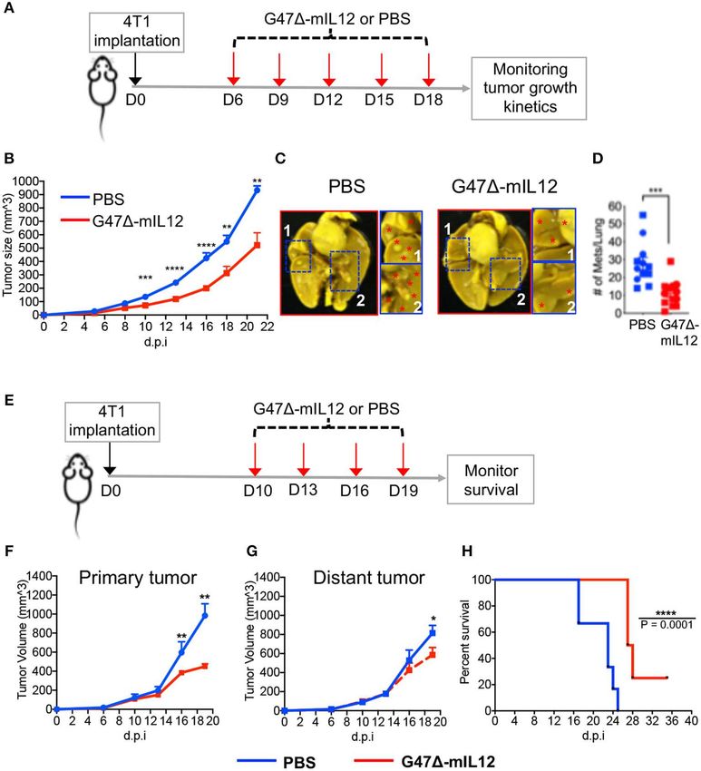
Ghouse et al.                                                                                                               Oncolytic HSV-IL-12 for TNBC Treatment

 FIGURE 1 | (A–D) G471-mIL12 efficiently infects and kills murine breast cancer[[Inline Image]] cells. (A–C) 4T1 murine TNBC cells treated with G471-mCherry (MOI
 = 1.0) or PBS and imaged at 24 hours post-treatment. mCherry red fluorescence image (in A) shows virus infection and phase contrast image (in B) shows round
 cytopathic cells following virus treatment. PBS treated 4T1 tumor cells were served as controls (in C). (D) Dose-response curves of G471-mIL12 in 4T1 (left panel)
 and EMT6 (right panel) murine breast cancer models at 3 and 4 days post-treatment, respectively, as measured by MTS assay (Promega). (E) G471-mIL12 efficiently
 kills human TNBC cells. Dose-response curve of G471-mIL12 in HCC1806 (left panel) and MDA-MB-468 (right panel) TNBC cells at 4 days post-treatment, as
 measured by MTS assay. Mean ± SEM. Each graph represents an average of 2–4 experiments performed in triplicate.

tumors at late-stages of tumor development (Figures 2F,G),                           bilaterally in right and left axillary mammary fat pads of BALB/c
leading to significant extension of survival with 25% mice                           mice. Due to the sample’s scarcity for downstream applications,
surviving long-term compared to PBS treatment group                                  inoculated tumors were allowed to grow between 100 and 125
(Figure 2H).                                                                         mm3 . Tumors located at the right axillary mammary fat pad were
                                                                                     treated with intratumoral injections of PBS or G471-mIL12
G471-mIL12 Induces Local and Abscopal                                                on days 10, 13, 16, and 19, whereas tumors at the left axillary
Immune Effects                                                                       mammary fat pad remained untreated. To evaluate virus-
To understand the role of different immune cell populations                          induced abscopal effects and immune responses, treated tumors,
contributing to G471-mIL12-mediated control of tumor                                 contralateral tumors, and the spleens were harvested on day
growth and metastasis, orthotopic 4T1 tumors were established                        21 and subjected to multicolor flow cytometry for immune cell

Frontiers in Oncology | www.frontiersin.org                                      5                                              March 2020 | Volume 10 | Article 384
Ghouse et al. Oncolytic HSV-IL-12 for TNBC Treatment FIGURE 2 | (A-D) G471-mIL12 controls primary TNBC growth and inhibits metastasis in an orthotopic 4T1 mouse mammary carcinoma model. (A) Experimental schema. 1 × 10⁵ viable 4T1 cells in 100 µl PBS were injected into the right axillary mammary fat pad of female BALB/c mice. G471-mIL12 (2 × 10⁶ PFU diluted in 25 µl PBS) or PBS injected intratumorally on day 6 (when tumors reached ∼70 mm3 ), 9, 12, 15, and 18. Tumor volumes were measured every 2–3 days. (B) Tumor growth kinetics. Mean tumor volume of PBS injected tumors was compared to mean tumor volume of G471-mIL12 injected tumors from day 10 to day 21. (C) Representative images of lungs. Mice from experiment 2B were euthanized on day 21 and lungs were fixed in Bouin’s fixative and imaged at 24 hours post-fixation. Red asterisk indicates metastatic nodules at higher magnification in insets. (D) Total number of lung surface metastatic nodules counted with a stereo microscope. Mice from experiment 2B. Data presented by combining two independent experiments (n = 12 mice/group) and each experimental data are indicated by round- and square-shaped symbols. Mean ± SEM. Unpaired Student’s t-test (two-tailed), **P ≤ 0.01, ***P ≤ 0.001, ****P ≤ 0.0001; d.p.i., days post-tumor implantation. (E-H) G471-mIL12 treatment controls growth of treated and untreated contralateral tumors at late-stage of tumor development and extends survival. (E) Experimental schema. Orthotopic 4T1 tumors were established bilaterally in the right and left axillary mammary fat pads (1 × 10⁵ cells per mammary pad) on day 0. When tumors reached between 100 and 125 mm3 in volume, tumors located at the right axillary mammary fat pad intratumorally injected with PBS (n = 6) or G471-mIL12 (106 pfu/injection) (n = 8) on days 10, 13, 16, and 19, and the tumors at the left axillary mammary fat pad remained untreated and served as contralateral tumors. Tumor volumes periodically measured by caliper and mice were followed for survival until they become moribund or until tumors reached to their burden limit, i.e., 1.5 cm in size. (F, G) Tumor growth kinetics of treated and untreated contralateral tumors. Mean tumor volume of PBS treatment group was compared to mean tumor volume of G471-mIL12 treatment group on day 16 and 19 by Unpaired Student’s t-test (two-tailed). *P ≤ 0.05, **P ≤ 0.01. (H) Kaplan-Meier survival curve. Median survival of mice treated with G471-mIL12 (27.5 days; 25% of mice surviving until the end of experiment on day 36) was compared to median survival of mice treated with PBS (23 days; P = 0.0001) by Log Rank test. ****P ≤ 0.0001. d.p.i., days post-tumor implantation. analysis. First, we selected the CD45 surface marker to distinguish G471-mIL12 treatment resulted in following immune cell CD45- tumor cells from CD45+ hematopoietic immune cells by alterations in both local (treated) and contralateral (untreated) adopting a specific gating strategy (Supplementary Figure 1). tumors and spleens that included: (1) significantly increased Frontiers in Oncology | www.frontiersin.org 6 March 2020 | Volume 10 | Article 384
Ghouse et al.                                                                                            Oncolytic HSV-IL-12 for TNBC Treatment

infiltration of CD45+ immune cells in treated and contralateral          presentation that eventually could be responsible for promoting
tumors (P ≤ 0.05 vs PBS; Figure 3A), (2) further characterization        CD8+ T cell responses in virus-injected and non-injected
of CD45+ cells revealing a significant increase in infiltration of       tumor lesions.
CD8+ T lymphocytes, but not CD4+ T cells (except spleen), in
both tumor lesions and spleens (P ≤ 0.05 vs. PBS; Figures 3B,C),         G471-mIL12 Treatment Induces
(3) significantly reduced macrophages and both granulocytic and
                                                                         Anti-angiogenic Effects in vivo
monocytic MDSCs in both tumor lesions (Figures 3D–F), and
                                                                         Interleukin 12 (IL-12) is an anti-angiogenic cytokine (19).
(4) significant reduction of regulatory T cells (CD4+ FoxP3+ )
                                                                         IL-12 elicits its anti-anti-angiogenic effects through release
observed in tumor draining lymph nodes from G471-mIL12
                                                                         of IFN-γ, which activates IFN-inducible protein 10 [IP-10
treated mice (Figure 3G), which suggests G471-mIL12-induced
                                                                         or CXC chemokine ligand (CXCL) 10], a chemokine that
beneficial systemic immune responses. As expected, we have
                                                                         mediates chemotaxis of lymphocytes and angiostatic effects
observed a significantly higher infiltration of CD45+ cells in
                                                                         (19, 35, 36). It was previously demonstrated that G471-
virus-treated tumors as compared to untreated contralateral
                                                                         mIL12 treatment can inhibit angiogenesis (14). In order to
tumors, while no significant differences were observed in
                                                                         determine whether viral expression of IL-12 (i.e., G471-
other immune cell population between virus treated and
                                                                         mIL12) can induce any anti-angiogenic effects in murine
untreated tumors (Supplementary Figure 2). While CD8+
                                                                         TNBC model, 4T1 TNBC cells (1 × 10⁵ ) were implanted
T cells are an important contributor in inducing effector
                                                                         subcutaneously into the mammary fat pad of BALB/c mice,
anti-tumor immunity, MDSCs can inhibit adaptive anti-tumor
                                                                         and treated intratumorally with PBS or G471-mIL12 (2 x
immunity and are an obstacle to cancer immunotherapies (31).
                                                                         106 PFU) (see schema in Figure 2A). Three days after the
Thus, enhanced CD8+ T cell infiltration and reduced MDSC
                                                                         last treatment (i.e., day 21), methanol-fixed cryostat sections
populations in treated and untreated tumor lesions due to virus
                                                                         were subjected to indirect immunofluorescence staining for
treatment clearly show the ability of G471-mIL12 to induce
                                                                         CD31+ tumor vasculatures, as described in ‘Materials and
beneficial anti-tumor immune effects (local and abscopal), which
                                                                         Methods’ section. We observed that G471-mIL12 treatment
could play a critical role in inhibiting primary and metastatic
                                                                         significantly reduced CD31+ tumor vascularity by 2-fold
TNBC (Figure 2).
                                                                         compared to PBS treatment mice (Figures 5A,B). Nano String
                                                                         gene expression analysis also revealed significantly increased
G471-mIL12 Treatment Leads to DC                                         gene expression of anti-angiogenic molecule CXCL-10 (IP-10)
Maturation and T Cell Activation in vivo                                 in 4T1 tumors (P = 0.0049 vs. PBS) (Figure 5C). These studies
OVs have been established as strong in situ anti-cancer                  demonstrate anti-angiogenic properties of the G471-mIL12
vaccines (9, 14, 24, 29, 32) that activate antigen presenting            virus in TNBC.
cells (APCs), augment antigen processing and presentation,
and prime CD8+ T cell responses (9). We observed no                      CD8+ T Cell Depletion Abrogates
significant changes in dendritic cell (DC) population in                 G471-mIL12-Induced Anti-TNBC Efficacy
virus-treated tumor lesions (Figure 4A). However, G471-                  It was previously demonstrated that G471-mIL12 treatment
mIL12 treatment led to significant trafficking of DCs in the             leads to local production of IL-12 (14). Viral release of IL-12 is
spleens of treated mice versus PBS control group (Figure 4A),            accompanied by a marked release of IFN-γ (14), which facilitates
indicating that DCs primed in the tumor microenvironment                 CD8+ T-cell-mediated killing of tumor cells (37). Because there
following virus treatment likely trafficked to lymphoid organs           is an increased intratumoral CD8+ T cell infiltration (Figure 3C)
for antigen presentation to immune cells such as CD8+ T                  with enhanced cytotoxic T cell activation markers (Figure 4C)
cells (Figure 3C). Spleen-localized DCs were overwhelmingly              following G471-mIL12 treatment, we hypothesized that anti-
in a mature APC state based on significant expression of                 TNBC efficacy of G471-mIL12 can be CD8+ T cell-dependent.
the activation marker CD86 in virus-treated group versus                 To address whether the changes seen in tumor infiltrating CD8+
the PBS control group (Figure 4A), which is critical for T               T immune cells are necessary to elicit anti-tumor efficacy of
cell activation (33). Nano String gene expression analysis               G471-mIL12, we performed antibody depletion studies of CD8+
further confirmed upregulation of genes involved in DC                   cells as described (25). CD8+ T cell depletion was confirmed
maturation (e.g., ITGAX, CD40), DC-specific co-stimulatory               by flow cytometric analysis (Supplementary Figure 3). When
signaling (CD83, CD86, BTLA, ICOS, ICOSL) and CD8+ T                     orthotopic mammary tumors reached 80-90 mm3 in volume,
cell activation (CD3e, CD8α, Granzyme B, and IFN-γ ) (34)                mice were treated intratumorally with G471-mIL12 or PBS,
(Figures 4B,C). Gene expression analysis also revealed enhanced          and intraperitoneally with anti-CD8 antibodies or isotype
expression of antigen processing/presentation genes (e.g., H2-           control IgG on indicated days (Figure 5D). The results show
Ab1, H2-Eb1, H2-Aa, H2-k1, H2-T23, H2-D1, H2-DMa, H2-                    that depletion of CD8+ cells abrogated anti-tumor effects of
Q2, H2-Ea-ps) in virus injected tumor lesions (Figure 4D),               G471-mIL12 treatment, as demonstrated by similar tumor
suggesting virus-induced DC priming and activation, and                  growth kinetics and tumor free survival as mock-treated
enhanced antigen presentation. Altogether, these data establish          animals (Figures 5E,F). Overall, these studies demonstrate
the ability of our G471-mIL12 virus to induce efficient in               that G471-mIL12-mediated anti-tumor immune response is
situ vaccine effects in promoting DC maturation and antigen              CD8-dependent.

Frontiers in Oncology | www.frontiersin.org                          7                                       March 2020 | Volume 10 | Article 384
Ghouse et al. Oncolytic HSV-IL-12 for TNBC Treatment
FIGURE 3 | G471-mIL12 induces local and abscopal immune responses. Orthotopic 4T1 tumors were established bilaterally in the right and left axillary mammary fat
pads (1 × 10⁵ cells per mammary pad) on day 0. When tumors reached 100–125 mm3 in volume, tumors located at the right axillary mammary fat pad intratumorally
treated with PBS or G471-mIL12 (106 pfu/injection) on days 10, 13, 16, and 19, and the tumors at the left axillary mammary fat pad remained untreated (n = 4
mice/group). On day 21, primary treated tumors, contralateral tumors, spleens and tumor draining lymph nodes (TDLNs) were harvested and subjected to multicolor
flow cytometry as described in section Materials and Methods. Tumor infiltrating immune cells were gated based on gating strategy as presented in
Supplementary Figure 1. (A–F) Frequencies of live CD45+ cells (A), live CD3e+ CD4+ T cells (B), live CD3e+ CD8a+ T cells (C), live CD45+ F4/80+ macrophages
(D), live polymorphonuclear MDSCs (CD11b+ Ly6C− Ly6G+ ) (E), and mononuclear MDSCs (CD11b+ Ly6G− Ly6C+ ) (F) in treated tumors (panels in upper row),
contralateral tumors (panels in 2nd row), and spleens (panels in 3rd row). (G) Frequencies of live CD45+ cells, live CD3e+ CD4+ T cells, live CD3e+ CD8a+ T cells, and
CD3e+ CD4+ FoxP3+ regulatory T cells (Tregs) in tumor draining lymph nodes (TDLNs; panels in bottom row). Mean ± SEM. Statistically significant differences between
groups are reported as P-values in the figures. Statistical significance was assessed by Student’s t test. *P ≤ 0.05, **P ≤0.01, ***P ≤ 0.001; ns, not significant.
DISCUSSION atezolizumab; anti-PD-1, designated pembrolizumab) can lead to
higher progression-free survival (IMpassion130) (41) and a more
Current treatments for TNBC patients are limited to surgery pathological complete response (pCR) rate (KEYNOTE-522)
and chemotherapy. TNBC patients initially respond well (42), respectively, compared to chemotherapy alone. However,
to chemotherapy but most patients develop resistance at a vast majority of patients (i.e., 79.4% in the intention-to-treat
advanced stages. Moreover, immune checkpoint blockade (ICB) population) treated with the combination (ICB immunotherapy
immunotherapy alone (e.g., anti-PD-1), which is usually + chemotherapy) have experienced disease progression or died
successful in immunologically “hot” tumors (e.g., subsets of (IMpassion130) (41). Thus, there is an unmet medical need to
melanoma) (38, 39), produces a low overall response rate (Ghouse et al. Oncolytic HSV-IL-12 for TNBC Treatment FIGURE 4 | G471-mIL12 treatment leads to DC maturation and T cell activation in vivo. (A) Frequencies of CD45+ CD11b+ CD11C+ DCs in treated, untreated contralateral tumors and spleen (left panel), and mean fluorescent intensity (MFI) of activated DCs (CD11b+ CD11c+ CD86+ ) in spleen (right panel). Same experiment as in Figure 3 (n = 4/group). Mean ± SEM. Statistically significant differences between groups are reported as P-values in the figures. Unpaired Student’s t test (two-tailed), **P
Ghouse et al.                                                                                                                 Oncolytic HSV-IL-12 for TNBC Treatment

 FIGURE 5 | (A-C) G471-mIL12 treatment induces anti-angiogenic effects. (A) Immunofluorescence staining of CD31+ tumor blood vessels after G471-mIL12
 treatment. Same experiment as Figure 2A. Representative images are presented; scale bar = 100 µm. (B) CD31+ positive areas from 3 to 5 random fields/tumor
 section (1 section/mouse; n = 5 for PBS and n= 4 mice/group for G471-mIL12) were measured by ImageJ software and presented as Mean ± SEM. (C)
 G471-mIL12 treatment increases expression of CXCL10 (IP10) in tumor microenvironment. Same as experiment 4B-D. Mean ± SEM. Unpaired Student’s t test
 (two-tailed). *P ≤0.05, **P ≤0.01. (D-F) CD8a+ T cell depletion abrogates G471-mIL12-induced anti-TNBC efficacy. (D) Experimental schema. BALB/c female mice
 were implanted with 4T1 tumor cells (1 × 10⁵ cells) in the mammary fat pad on day 0. When tumors reached approximately 80-90 mm3 in volume (day 8), mice were
 treated intratumorally with G471-mIL12 (1 × 10⁶ pfu / 25 µl in PBS) or PBS on days 8, 11, 14, and 17 (upward red arrows). Anti-CD8a antibody (5 mg/kg) or isotype
 control IgG (5 mg/kg rat IgG) injected IP on day-4 and−1 prior to tumor implantation and on days 4, 8, 12, and 16 post-tumor implantations (downward blue arrows).
 (E) Growth kinetics of tumors in mice treated with PBS/IgG (n = 6), G471-mIL12/IgG (n = 5), or G471-mIL12/Anti-CD8a (n = 6). Mean tumor volume of PBS/IgG
 treated mice was compared to mean tumor volume of G471-mIL12/IgG treated mice on indicated days. Mean ± SEM of all mice presented. Statistical differences
 between groups were compared by Unpaired 2-tailed Student’s t test. *P ≤ 0.05. (F) Kaplan-Meier survival curve. Mice from experiment (E) were followed for ill health
 and the survival curve was generated on the day each mouse reached its tumor burden limit, i.e., a maximum diameter of 15 mm. Median survival of mock (PBS+IgG)
 treated mice (21 days; n = 6) was compared to mice treated with G471-mIL12+IgG (31 days; n = 5, P = 0.0116) and G471-mIL12+anti-CD8a (21 days; n = 6, P =
 0.4070). Median survival of mice treated with G471-mIL12+IgG (31 days) was compared to mice treated with G471-mIL12+anti-CD8a (21 days; P = 0.0369). The
 survival data was analyzed by Log Rank test. *P ≤ 0.05; ns, not significant; s.c., subcutaneous; i.p., intraperitoneal; i.t., intratumoral; d.p.i., days post-tumor
 implantation.

Frontiers in Oncology | www.frontiersin.org                                       10                                              March 2020 | Volume 10 | Article 384
Ghouse et al.                                                                                               Oncolytic HSV-IL-12 for TNBC Treatment

first we tested the ability of G471-mIL12 to infect and kill               among TNBC patients (47, 48). In this study, the quantification
mouse and human TNBC cells in vitro. Then we evaluated the                 of immune infiltrate demonstrated a significant increase in
therapeutic efficacy of G471-mIL12 as a monotherapy in an                  CD8+ T cells in both G471-mIL12-treated and untreated
immunocompetent, syngeneic, and highly metastatic 4T1 mouse                contralateral tumors and spleens (Figure 3C). When CD8+
model of mammary carcinoma. In this model, G471-mIL12                      T cells were depleted, G471-mIL12 treatment efficacy was
monotherapy significantly inhibited TNBC tumor growth and                  abrogated (Figures 5E,F), indicating CD8+ T cells are essential
prevented metastasis, both at early and late-stages of tumor               for the antitumor effects of G471-mIL12. Myeloid-derived
development, and was associated with increased DC maturation               suppressor cells (MDSCs) are immuno-suppressive cells that
and T cell activation, enhanced infiltration of CD8+ T cells               inhibit antitumor immunity (31). One of the striking findings
and reduced infiltration of MDSCs into treated and distant                 in our study was a significant reduction of monocytic and
tumors. The anti-tumor efficacy of G471-mIL12 treatment                    granulocytic MDSCs in both treated and untreated tumors
on primary tumor growth was abrogated in the absence of                    following oncolytic immunovirotherapy (Figures 3D,F). In
CD8+ T cells.                                                              contrast to MDSCs, we did not find any significant treatment
    There has not been any study performed so far testing the              effect on DC population in either treated or contralateral
cytotoxic activity of OHSV in murine TNBC cell lines in vitro,             tumors. However, we observed a significant increase in splenic
other than a study testing viral replication in 4T1 tumor cells            DCs, which were overwhelmingly positive for CD86, an
(43). Here, we observed that both 4T1 and EMT6 murine breast               activation marker for DCs (Figure 4A). It is important to
cancer cells are sensitive to G471-mIL12 treatment with IC50s              note that IL-12 in G471-mIL12 virus may have played a
of MOIs 0.9 and 0.5, respectively, which are similar to the killing        critical role in activating APCs, since IL-12 is a potent MDSC
activity of G471-mIL12 in vitro in syngeneic mouse glioblastoma            modulator and shifts splenic MDSCs (isolated from 4T1-
cells (14, 29). Human cancer cells are typically more permissive,          tumor bearing BALB/c mice) into CD11c+ CD86+ activated
and therefore, should be more sensitive to OHSV treatment than             DCs (17). Nano String gene expression analysis further
mouse cancer cells (22). Indeed, the tumor cell killing efficiency         confirmed DC maturation and activation by demonstrating
of G471-mIL12 is better in human TNBC cells (HCC1806 and                   virally enhanced expression of genes associated with DC
MDA-MB-468), and requires a lesser MOI (IC50=0.15-0.25),                   activation, antigen processing and presentation, and T cell
which is at least 2-6 fold lower than that in mouse breast cancer          activation in G471-mIL12 treatment tumors (Figures 4B,D).
models. Similar to G471-mIL12, other OHSVs with different                  Since G471-mIL12 is an efficient modulator of antigen
genetic backgrounds or modifications also efficiently kills human          processing/presentation, it remains to be determined whether
TNBC cells (44–46).                                                        other APC activators can synergize with the in situ vaccine effects
    A recently published study in a pre-surgical neoadjuvant               of G471-mIL12 treatment and improve the therapeutic outcome
setting shows that an ICP0-deleted OHSV replicates poorly in the           in TNBC.
4T1 model and is not effective at all in controlling the growth of             Recently published clinical studies in TNBC patients
the injected tumors in a subcutaneous 4T1 flank model, despite             demonstrate that combination of chemotherapy with an ICB
predominantly controlling the growth of secondary 4T1 tumors,              results in significantly higher progression-free survival or pCR
which resembles TNBC metastasis (43). Similar to Martin et al.             rate in PD-L1-positive patient population compared to PD-L1-
(43), the present study shows that G471-mIL12 monotherapy                  negative TNBC patients [KEYNOTE-522 (42) and IMpassion130
significantly reduced TNBC metastasis, as demonstrated by                  (41)]. This suggests that PD-L1 expression plays a key role in
a significant reduction of surface metastatic nodules in the               determining the treatment efficacy. An important limitation of
lungs (Figures 2C,D), and significant growth inhibition of                 the work presented here is that the G471-mIL12 monotherapy
non-injected contralateral tumors (Figure 2G). Although we                 did not eliminate 4T1 primary tumors and metastasis, and
found similarity in controlling metastatic tumor burden, Martin            we observed that virus treatment dramatically upregulated
et al. (43) findings contradict other key findings in our study.           PD-L1 expression in 4T1 tumors (Figure 4B). It was previously
For instance, G471-mIL12 efficiently replicated in and killed              demonstrated that ICB treatment improves therapeutic outcome
mouse cancer cells (14) (Figure 1), significantly controlled the           of OHSV therapy in mouse glioblastoma (24) and human
growth of injected tumors (Figures 2B,F), and extended survival            advanced melanoma (49). Thus, future studies involving
(Figure 2H), as opposed to poor viral replication and “no” anti-           combination immunovirotherapy (i.e., G471-mIL12 + ICB) can
tumor efficacy against injected tumors observed by an ICP0-                further augment the therapeutic effects of G471-mIL12 and may
deleted OHSV (43). The contradictory observations can be                   lead to complete eradication of TNBCs.
explained by the fact that G471-mIL12 has an intact ICP0, a                    Because IL-12 is well known for its anti-angiogenic properties
critical immediate-early protein of viral tegument, which is freed         (19), it was our expectation that G471-IL12 treatment would
into the cytosol upon infection to prepare the cell for virus              lead to inhibition of tumor angiogenesis in TNBC model.
replication (16). Intact ICP0 in the G471-mIL12 virus may have             Indeed, we observed a significant reduction of 4T1 tumor
played a role making G471-mIL12 efficacious against TNBC                   vascularity following G471-IL12 treatment compared to
models, both in vitro and in vivo.                                         PBS treatment group (Figures 5A,B). Production of CXCL-
    Tumor microenvironment plays a vital role in the success               10 (IP-10) is inversely correlated with tumor growth and
of oncolytic virus therapy. Tumor infiltrating CD8+ T cells                angiogenesis (50). Here, we noticed a significant upregulation
are associated with reduced recurrence and longer survival                 of CXCL-10 in virus-treated 4T1 tumors (Figure 5C).

Frontiers in Oncology | www.frontiersin.org                           11                                       March 2020 | Volume 10 | Article 384
Ghouse et al. Oncolytic HSV-IL-12 for TNBC Treatment
These findings are similar to what was reported previously AUTHOR CONTRIBUTIONS
with G471-IL12 in a mouse glioblastoma model (14),
such as inhibition of glioblastoma angiogenesis with an SG performed experiments, prepared figures, wrote initial draft,
increased expression of intratumoral CXCL-10 (14). These and edited the manuscript. H-MN performed experiments,
studies also indicate that anti-angiogenic effects of G471- made figures, wrote, and edited the manuscript. PB performed
IL12 can synergize with other anti-angiogenic agents and Nano String experiments/analysis and edited the manuscript.
may improve the therapeutic outcome (to be tested in KG-M helped with the animal experiments and analysed
future studies). data, edited the manuscript. DS performed experiments,
In summary, we show for the first time that an OHSV conceptualization, original draft preparation, preparation
expressing mouse IL-12 (G471-mIL12) virus effectively of figures, review and editing, data analysis, and funding
infects and kills mouse and human breast cancer cell lines. acquisition. All authors agree to be accountable for the content of
Treatment of syngeneic mice bearing 4T1 TNBC tumors with the work.
G471-mIL12 lead to a CD8+ T cell-dependent inhibition
of 4T1 tumor growth, inhibition of tumor angiogenesis, and
prevention of lung metastasis, suggesting local and systemic
FUNDING
anti-cancer effects of G471-mIL12. Finally, G471-mIL12 DS was supported by startup funds from Dodge Jones
treatment also leads to an increase in PD-L1 expression in Foundation-Abilene and TTUHSC-School of Pharmacy.
tumors, suggesting the therapeutic benefit of combining G471-
mIL12 with anti-PD-1/PD-L1 antibodies. Overall, these findings
strongly suggest that G471-mIL12-based immunovirotherapy ACKNOWLEDGMENTS
could be a promising therapeutic approach for
TNBC patients. We thank Drs. Samuel Rabkin and Robert Martuza for providing
OHSVs, and Dr. Rabkin for review of the manuscript. We
DATA AVAILABILITY STATEMENT thank Arani Datta for his technical assistance with the flow
cytometry, and the animal facility personnel for taking care
Publicly available datasets were analyzed in this study, these can of mice.
be found in the NCBI Gene Expression Omnibus (GSE144333).
SUPPLEMENTARY MATERIAL
ETHICS STATEMENT
The Supplementary Material for this article can be found
The animal study was reviewed and approved by Institutional online at: https://www.frontiersin.org/articles/10.3389/fonc.
Animal Care and Use Committee (IACUC) at the TTUHSC. 2020.00384/full#supplementary-material
REFERENCES 8. Mathis JM. Oncolytic virotherapy for breast cancer treatment. Curr Gene
Ther. (2018) 18:192–205. doi: 10.2174/1566523218666180910163805
1. Thakur V, Kutty RV. Recent advances in nanotheranostics for triple 9. Russell SJ, Barber GN. Oncolytic viruses as antigen-agnostic cancer vaccines.
negative breast cancer treatment. J Exp Clin Cancer Res. (2019) 38:430. Cancer Cell. (2018) 33:599–605. doi: 10.1016/j.ccell.2018.03.011
doi: 10.1186/s13046-019-1443-1 10. Andtbacka RH, Kaufman HL, Collichio F, Amatruda T, Senzer N,
2. Diana A, Franzese E, Centonze S, Carlino F, Della Corte CM, Ventriglia J, Chesney J, et al. Talimogene laherparepvec improves durable response
et al. Triple-negative breast cancers: systematic review of the literature on rate in patients with advanced melanoma. J Clin Oncol. (2015) 33:2780–8.
molecular and clinical features with a focus on treatment with innovative doi: 10.1200/JCO.2014.58.3377
drugs. Curr Oncol Rep. (2018) 20:76. doi: 10.1007/s11912-018-0726-6 11. Vikas P, Borcherding N, Zhang W. The clinical promise of immunotherapy
3. Garrido-Castro AC, Lin NU, Polyak K. Insights into molecular classifications in triple-negative breast cancer. Cancer Manag Res. (2018) 10:6823–33.
of triple-negative breast cancer: improving patient selection for treatment. doi: 10.2147/CMAR.S185176
Cancer Discov. (2019) 9:176–98. doi: 10.1158/2159-8290.CD-18-1177 12. Soliman H, Hogue D, Han H, Lee C, Ismail-Khan R, Khong H, et al.
4. Waks AG, Winer EP. Breast cancer treatment: a review. JAMA. (2019) Abstract OT2-07-01: Phase 1/2 trial of the oncolytic virus, talimogene
321:288–300. doi: 10.1001/jama.2018.19323 laherparpvec, in combination with neoadjuvant chemotherapy in stage
5. Echeverria GV, Ge Z, Seth S, Zhang X, Jeter-Jones S, Zhou X, et al. II/III triple negative breast cancer. Cancer Res. (2018) 78(4 Suppl.):1.
Resistance to neoadjuvant chemotherapy in triple-negative breast cancer doi: 10.1158/1538-7445.SABCS17-OT2-07-01
mediated by a reversible drug-tolerant state. Sci Transl Med. (2019) 11:488. 13. Senzer NN, Kaufman HL, Amatruda T, Nemunaitis M, Reid T, Daniels
doi: 10.1158/1538-7445.SABCS18-GS5-05 G, et al. Phase II clinical trial of a granulocyte-macrophage colony-
6. Bommareddy PK, Peters C, Saha D, Rabkin SD, Kaufman stimulating factor-encoding, second-generation oncolytic herpesvirus in
HL. Oncolytic herpes simplex viruses as a paradigm for the patients with unresectable metastatic melanoma. J Clin Oncol. (2009) 27:5763–
treatment of cancer. Ann Rev Cancer Biol. (2018) 2:155–73. 71. doi: 10.1200/JCO.2009.24.3675
doi: 10.1146/annurev-cancerbio-030617-050254 14. Cheema TA, Wakimoto H, Fecci PE, Ning J, Kuroda T, Jeyaretna
7. Harrington K, Freeman DJ, Kelly B, Harper J, Soria J-C. Optimizing oncolytic DS, et al. Multifaceted oncolytic virus therapy for glioblastoma in an
virotherapy in cancer treatment. Nat Rev Drug Dis. (2019) 18:689–706. immunocompetent cancer stem cell model. Proc Natl Acad Sci USA. (2013)
doi: 10.1038/s41573-019-0029-0 110:12006–11. doi: 10.1073/pnas.1307935110
Frontiers in Oncology | www.frontiersin.org 12 March 2020 | Volume 10 | Article 384Ghouse et al. Oncolytic HSV-IL-12 for TNBC Treatment
15. Bommareddy PK, Patel A, Hossain S, Kaufman HL. Talimogene laherparepvec and decreases angiogenesis in dogs with spontaneous cancer. Vet Comp Oncol.
(T-VEC) and other oncolytic viruses for the treatment of melanoma. Am J Clin (2017) 15:1187–205. doi: 10.1111/vco.12255
Dermatol. (2017) 18:1–15. doi: 10.1007/s40257-016-0238-9 36. Angiolillo AL, Sgadari C, Tosato G. A role for the interferon-inducible protein
16. Peters C, Rabkin SD. Designing herpes viruses as oncolytics. Mol Ther 10 in inhibition of angiogenesis by interleukin-12. Ann N Y Acad Sci. (1996)
Oncolytics. (2015) 2:100. doi: 10.1038/mto.2015.100 795:158–67. doi: 10.1111/j.1749-6632.1996.tb52664.x
17. Choi JN, Sun EG, Cho SH. IL-12 enhances immune response by modulation 37. Bhat P, Leggatt G, Waterhouse N, Frazer IH. Interferon-gamma derived from
of myeloid derived suppressor cells in tumor microenvironment. Chonnam cytotoxic lymphocytes directly enhances their motility and cytotoxicity. Cell
Med J. (2019) 55:31–9. doi: 10.4068/cmj.2019.55.1.31 Death Dis. (2017) 8:e2836. doi: 10.1038/cddis.2017.67
18. Berraondo P, Etxeberria I, Ponz-Sarvise M, Melero I. Revisiting interleukin- 38. Hargadon KM, Johnson CE, Williams CJ. Immune checkpoint
12 as a cancer immunotherapy agent. Clin Cancer Res. (2018) 24:2716–8. blockade therapy for cancer: an overview of FDA-approved immune
doi: 10.1158/1078-0432.CCR-18-0381 checkpoint inhibitors. Int Immunopharmacol. (2018) 62:29–39.
19. Del Vecchio M, Bajetta E, Canova S, Lotze MT, Wesa A, Parmiani G, et al. doi: 10.1016/j.intimp.2018.06.001
Interleukin-12: biological properties and clinical application. Clin Cancer Res. 39. Constantinidou A, Alifieris C, Trafalis DT. Targeting programmed
(2007) 13:4677–85. doi: 10.1158/1078-0432.CCR-07-0776 cell death−1 (PD-1) and Ligand (PD-L1): a new era in cancer
20. Antoszczyk S, Spyra M, Mautner VF, Kurtz A, Stemmer-Rachamimov AO, active immunotherapy. Pharmacol Ther. (2019) 194:84–106.
Martuza RL, et al. Treatment of orthotopic malignant peripheral nerve sheath doi: 10.1016/j.pharmthera.2018.09.008
tumors with oncolytic herpes simplex virus. Neuro Oncol. (2014) 16:1057–66. 40. Emens LA. Breast cancer immunotherapy: facts and hopes. Clin Cancer Res.
doi: 10.1093/neuonc/not317 (2018) 24:511–20. doi: 10.1158/1078-0432.CCR-16-3001
21. Aslakson CJ, Miller FR. Selective events in the metastatic process defined 41. Schmid P, Adams S, Rugo HS, Schneeweiss A, Barrios CH, Iwata H,
by analysis of the sequential dissemination of subpopulations of a mouse et al. Atezolizumab and nab-paclitaxel in advanced triple-negative breast
mammary tumor. Cancer Res. (1992) 52:1399–405. cancer. N Engl J Med. (2018) 379:2108–21. doi: 10.1056/NEJMoa18
22. Todo T, Martuza RL, Rabkin SD, Johnson PA. Oncolytic herpes simplex virus 09615
vector with enhanced MHC class I presentation and tumor cell killing. Proc 42. Schmid P, Cortes J, Bergh JCS, Pusztai L, Denkert C, Verma S,
Natl Acad Sci USA. (2001) 98:6396–401. doi: 10.1073/pnas.101136398 et al. KEYNOTE-522: Phase III study of pembrolizumab (pembro) +
23. Wani N, Nasser MW, Ahirwar DK, Zhao H, Miao Z, Shilo K, et al. chemotherapy (chemo) vs placebo + chemo as neoadjuvant therapy
C-X-C motif chemokine 12/C-X-C chemokine receptor type 7 signaling followed by pembro vs placebo as adjuvant therapy for triple-negative
regulates breast cancer growth and metastasis by modulating the tumor breast cancer (TNBC). J Clin Oncol. (2018) 36(15_suppl):TPS602-TPS.
microenvironment. Breast Cancer Res. (2014) 16:R54. doi: 10.1186/bcr3665 doi: 10.1200/JCO.2018.36.15_suppl.TPS602
24. Saha D, Martuza RL, Rabkin SD. Macrophage polarization contributes 43. Martin NT, Roy DG, Workenhe ST, van den Wollenberg DJM, Hoeben RC,
to glioblastoma eradication by combination immunovirotherapy and Mossman KL, et al. Pre-surgical neoadjuvant oncolytic virotherapy confers
immune checkpoint blockade. Cancer Cell. (2017) 32:253–67 e5. protection against rechallenge in a murine model of breast cancer. Sci Rep.
doi: 10.1016/j.ccell.2017.07.006 (2019) 9:1865. doi: 10.1038/s41598-018-38385-7
25. Balogh KN, Templeton DJ, Cross JV. Macrophage migration inhibitory 44. Gholami S, Chen CH, Gao S, Lou E, Fujisawa S, Carson J, et al. Role of MAPK
factor protects cancer cells from immunogenic cell death and impairs in oncolytic herpes viral therapy in triple-negative breast cancer. Cancer Gene
anti-tumor immune responses. PLoS ONE. (2018) 13:e0197702. Ther. (2014) 21:283–9. doi: 10.1038/cgt.2014.28
doi: 10.1371/journal.pone.0197702 45. Chen X, Han J, Chu J, Zhang L, Zhang J, Chen C, et al. A combinational
26. Bommareddy PK, Lowe DB, Kaufman HL, Rabkin SD, Saha D. therapy of EGFR-CAR NK cells and oncolytic herpes simplex virus
Multi-parametric flow cytometry staining procedure for analyzing 1 for breast cancer brain metastases. Oncotarget. (2016) 7:27764–77.
tumor-infiltrating immune cells following oncolytic herpes simplex virus doi: 10.18632/oncotarget.8526
immunotherapy in intracranial glioblastoma. J Biol Methods. (2019) 6:e112. 46. Lee TJ, Nair M, Banasavadi-Siddegowda Y, Liu J, Nallanagulagari T,
doi: 10.14440/jbm.2019.281 Jaime-Ramirez AC, et al. Enhancing therapeutic efficacy of oncolytic
27. Bommareddy PK, Aspromonte S, Zloza A, Rabkin SD, Kaufman HL. herpes simplex virus-1 with integrin beta1 blocking antibody OS2966.
MEK inhibition enhances oncolytic virus immunotherapy through increased Mol Cancer Ther. (2019) 18:1127–36. doi: 10.1158/1535-7163.MCT-1
tumor cell killing and T cell activation. Sci Transl Med. (2018) 10:471. 8-0953
doi: 10.1126/scitranslmed.aau0417 47. Ali HR, Provenzano E, Dawson SJ, Blows FM, Liu B, Shah M, et al. Association
28. Wang J, Hu P, Zeng M, Rabkin SD, Liu R. Oncolytic herpes simplex between CD8+ T-cell infiltration and breast cancer survival in 12,439 patients.
virus treatment of metastatic breast cancer. Int J Oncol. (2012) 40:757–63. Ann Oncol. (2014) 25:1536–43. doi: 10.1093/annonc/mdu191
doi: 10.3892/ijo.2011.1266 48. Li X, Gruosso T, Zuo D, Omeroglu A, Meterissian S, Guiot MC,
29. Saha D, Wakimoto H, Peters CW, Antoszczyk SJ, Rabkin SD, Martuza et al. Infiltration of CD8(+) T cells into tumor cell clusters in triple-
RL. Combinatorial effects of VEGFR kinase inhibitor axitinib and oncolytic negative breast cancer. Proc Natl Acad Sci U S A. (2019) 116:3678–87.
virotherapy in mouse and human glioblastoma stem-like cell models. Clin doi: 10.1073/pnas.1817652116
Cancer Res. (2018) 24:3409–22. doi: 10.1158/1078-0432.CCR-17-1717 49. Ribas A, Dummer R, Puzanov I, VanderWalde A, Andtbacka RHI, Michielin
30. Al-Mahmood S, Sapiezynski J, Garbuzenko OB, Minko T. Metastatic and O, et al. Oncolytic virotherapy promotes intratumoral t cell infiltration
triple-negative breast cancer: challenges and treatment options. Drug Deliv and improves anti-pd-1 immunotherapy. Cell. (2017) 170:1109–19 e10.
Transl Res. (2018) 8:1483–507. doi: 10.1007/s13346-018-0551-3 doi: 10.1016/j.cell.2017.08.027
31. Ostrand-Rosenberg S. Myeloid derived-suppressor cells: their role 50. Liu M, Guo S, Stiles JK. The emerging role of CXCL10 in cancer (Review).
in cancer and obesity. Curr Opin Immunol. (2018) 51:68–75. Oncol Lett. (2011) 2:583–9. doi: 10.3892/ol.2011.300
doi: 10.1016/j.coi.2018.03.007
32. Saha D, Wakimoto H, Rabkin SD. Oncolytic herpes simplex virus Conflict of Interest: PB is an employee of Replimune Inc. The authors declare
interactions with the host immune system. Curr Opin Virol. (2016) 21:26–34. that the research was conducted in the absence of any commercial or financial
doi: 10.1016/j.coviro.2016.07.007 relationships that could be construed as a potential conflict of interest.
33. Thomas IJ, Petrich de Marquesini LG, Ravanan R, Smith RM, Guerder S,
Flavell RA, et al. CD86 has sustained costimulatory effects on CD8 T cells. J Copyright © 2020 Ghouse, Nguyen, Bommareddy, Guz-Montgomery and Saha. This
Immunol. (2007) 179:5936–46. doi: 10.4049/jimmunol.179.9.5936 is an open-access article distributed under the terms of the Creative Commons
34. Cesano A. nCounter((R)) pancancer immune profiling panel (NanoString Attribution License (CC BY). The use, distribution or reproduction in other forums
Technologies, Inc., Seattle, WA). J Immunother Cancer. (2015) 3:42. is permitted, provided the original author(s) and the copyright owner(s) are credited
doi: 10.1186/s40425-015-0088-7 and that the original publication in this journal is cited, in accordance with accepted
35. Cicchelero L, Denies S, Haers H, Vanderperren K, Stock E, Van Brantegem L, academic practice. No use, distribution or reproduction is permitted which does not
et al. Intratumoural interleukin 12 gene therapy stimulates the immune system comply with these terms.
Frontiers in Oncology | www.frontiersin.org 13 March 2020 | Volume 10 | Article 384You can also read