Regulation of Cancer Metastasis by TRAIL/Death Receptor Signaling - MDPI
←
→
Page content transcription
If your browser does not render page correctly, please read the page content below
biomolecules

Review
Regulation of Cancer Metastasis by TRAIL/Death
Receptor Signaling
You-Take Oh and Shi-Yong Sun *

                                          Department of Hematology and Medical Oncology, Emory University School of Medicine and Winship Cancer
                                          Institute, Atlanta, GA 30322, USA; youtake.oh@emory.edu
                                          * Correspondence: ssun@emory.edu; Tel.: +1-404-778-2170

                                          Abstract: Death ligands such as tumor necrosis factor-related apoptosis-inducing ligand (TRAIL;
                                          TNFSF10) and their corresponding death receptors (e.g., DR5) not only initiate apoptosis through
                                          activation of the extrinsic apoptotic pathway but also exert non-apoptotic biological functions such
                                          as regulation of inflammation and cancer metastasis. The involvement of the TRAIL/death receptor
                                          signaling pathway in the regulation of cancer invasion and metastasis is complex as both positive and
                                          negative roles have been reported. The underlying molecular mechanisms are even more complicated.
                                          This review will focus on discussing current knowledge in our understanding of the involvement of
                                          TRAIL/death receptor-mediated signaling in the regulation of cancer cell invasion and metastasis.

                                          Keywords: TRAIL; death receptor; apoptosis; invasion; metastasis; cancer

                                          1. Introduction
                                     Metastasis is a key cause of cancer death. To metastasize to distant organ sites,

                                          cancer cells must detach from the primary tumor sites (local invasion; intravasation),
Citation: Oh, Y.-T.; Sun, S.-Y.           translocate into the circulatory system, survive in the circulation and arrest at a distant
Regulation of Cancer Metastasis by        organ site (extravasation), and finally adapt to the new microenvironment of distant tissues
TRAIL/Death Receptor Signaling.           (micrometastasis formation; metastatic colonization) [1]. Therefore, cell death is a key
Biomolecules 2021, 11, 499. https://
                                          mechanism for preventing the dissemination of metastatic cells. Cell death can occur after
doi.org/10.3390/biom11040499
                                          the detachment of primary tumor cells from the extracellular matrix, in the circulation
                                          through tumor immune surveillance or destruction by mechanical stresses, and after
Academic Editor: Ladislav Anděra
                                          extravasation during the phase of micrometastasis formation in a secondary organ [2]. It
                                          has been estimated that only about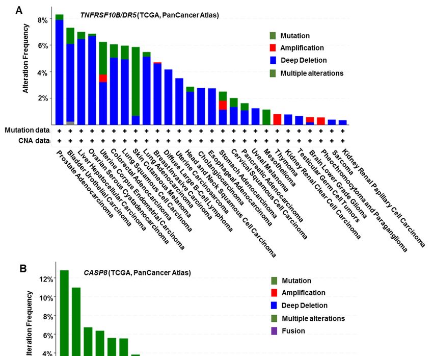
cules 2021, 11, x 2 of 16
2. 499
Biomolecules 2021, 11, TRAIL/Death Receptor-Mediated Apoptotic Signaling Pathway 2 of 16
There are two TRAIL death receptors in humans: death receptor 5 (DR5; also called
TRAIL-R2, TNFRSF10B or Killer/DR5) and death receptor 4 (DR4; also called TRAIL-R1
or TNFRSF10A). Both DR5 and Both
or TNFRSF10A). DR4 DR5shareand
redundant
DR4 share functions
redundantin triggering
functionsapoptosis
in triggering apoptosis
but have some but
distinct
have some distinct biological functions, such as regulation of cancerascell metastasis,
biological functions, such as regulation of cancer cell metastasis,
discussed below. In contrast,
as discussed thereIniscontrast,
below. only one TRAIL
there death
is only onereceptor named
TRAIL death murinenamed murine
receptor
TRAIL death receptor (mDR) in mice [9,10] or mouse homolog of KILLER/DR5
TRAIL death receptor (mDR) in mice [9,10] or mouse homolog of KILLER/DR5 (MK) [11]. (MK) [11].
TRAIL ligationTRAIL
to its functional cell surface death receptors or induction of death receptor
ligation to its functional cell surface death receptors or induction of death receptor
trimerization or aggregation or
trimerization (e.g., via overexpression
aggregation (e.g., via or agonistic antibodies)
overexpression leads antibodies)
or agonistic to re- leads to
cruitment of therecruitment
adaptor protein, Fas-associated
of the adaptor protein,death domain (FADD),
Fas-associated to the cytoplasmic
death domain (FADD), to the cytoplasmic
region of the receptor
region offollowed by recruitment
the receptor followed byofrecruitment
pro-caspase-8 or pro-caspase-10.
of pro-caspase-8 The
or pro-caspase-10. The
formation of this death-inducing signaling complex (DISC) triggers cleavage and activa-
formation of this death-inducing signaling complex (DISC) triggers cleavage and activation
tion of caspase-8
of or caspase-10,
caspase-8 which in turn
or caspase-10, whichactivates
in turndownstream caspase-3,caspase-3,
activates downstream -6, and -7, -6, and -7, and
and eventual apoptosis [12,13]. Therefore, TRAIL/death receptor/FADD/caspase-8
eventual apoptosis [12,13]. Therefore, TRAIL/death receptor/FADD/caspase-8 signal- signaling
ing normally triggers
normally apoptotic
triggerscell death (Figure
apoptotic cell death1A).
(Figure 1A).
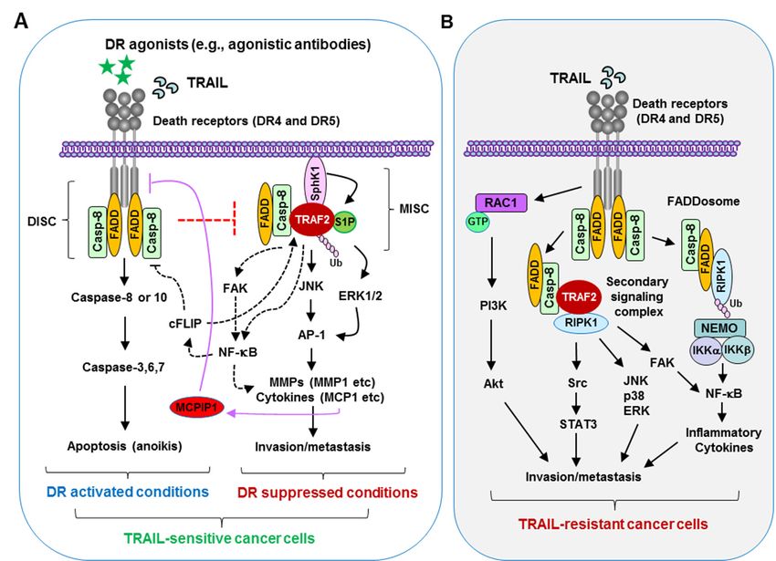
Figurenecrosis
Figure 1. Tumor 1. Tumor necrosis factor-related
factor-related apoptosis-inducing
apoptosis-inducing ligand (TRAIL)/death
ligand (TRAIL)/death receptor signaling
receptor signaling in the regulation of
in the regulation of apoptosis and metastasis of cancer cells. (A) Under normal apoptotic conditions
apoptosis and metastasis of cancer cells. (A) Under normal apoptotic conditions (e.g., in cancer cells sensitive to TRAIL with
(e.g., in cancer cells sensitive to TRAIL with activated death receptor pathway), TRAIL ligation with
activated death receptor pathway), TRAIL ligation with its death receptors (DR4 and DR5) or an agonistic antibody binding
its death receptors (DR4 and DR5) or an agonistic antibody binding to a corresponding death recep-
to a corresponding death receptor (DR4 or DR5) on the surface of cancer cells induces the formation of the death-inducing
tor (DR4 or DR5) on the surface of cancer cells induces the formation of the death-inducing signaling
signaling complex
complex (DISC) involving
(DISC) involving Fas-associated
Fas-associateddeath domain
death (FADD)
domain recruitment
(FADD) of pro-caspase-8
recruitment via its via
of pro-caspase-8 death effector
domain, resulting in caspase-8 or -10 activation followed by cleavage and activation of caspase-3,
its death effector domain, resulting in caspase-8 or -10 activation followed by cleavage and activa- -6, and -7, and eventual
execution oftion
apoptosis or anoikis.
of caspase-3, This
-6, and -7, mechanism
and eventual restricts the of
execution formation
apoptosis of or
theanoikis.
metastasis
Thisand invasionrestricts
mechanism signaling complex
(MISC) andthe formation of
subsequently the metastasis
suppresses and invasion
cell invasion signaling When
and metastasis. complex (MISC) andreceptors
TRAIL/death subsequently sup- or their
are inhibited
presses
functions are cell invasion
compromised, and FADD
available metastasis. When TRAIL/death
and caspase-8 may recruit and receptors areTNF-receptor-associated
stabilize inhibited or their functions factor (TRAF)2
areofcompromised,
with the help S1P, resultingavailable
in enhanced FADD and caspase-8
TRAF2 may recruit
polyubiquitination andand stabilize TNF-receptor-associated
activation, likely through a self-ubiquitination
mechanism.factor (TRAF)2
This will with
further leadthe
to help of S1P,
activation ofresulting in enhanced
ERK/JNK/AP-1 TRAF2
signaling andpolyubiquitination and activa-
NF-κB activation, which activates MMPs
tion, likely through a self-ubiquitination mechanism. This will further lead to activation of
(e.g., MMP1) and enhances the release of inflammatory cytokines (e.g., monocyte chemoattractant protein 1 (MCP1)) that
ERK/JNK/AP-1 signaling and NF-κB activation, which activates MMPs (e.g., MMP1) and enhances
promote invasion and metastasis of cancer cells. MCP1 may induce monocyte chemotactic protein-induced protein-1
the release of inflammatory cytokines (e.g., monocyte chemoattractant protein 1 (MCP1)) that pro-
(MCPIP1) expression,
mote invasion leading to a reduction
and metastasis in DR5
of cancer levels,
cells. MCP1 including
may inducecell surface
monocyteDR5;chemotactic
this favors protein-in-
MISC formation and
metastasis. duced
(B) Inprotein-1
TRAIL-insensitive
(MCPIP1) expression, leading to a reduction in DR5 levels, including cellsignaling
cells, TRAIL treatment will induce the formation of a second surface complex
or FADDosome, resulting in the activation of multiple protein kinases such as NF-κB, JNK, p38, ERK, and Src that are
involved in the positive regulation of invasion and metastasis of cancer cells. In KRAS-mutated cancer cells, TRAIL/DR5
activation can activate the Rac1/PI3K/Akt axis, promoting cell invasion and metastasis. RIPK1, receptor-interacting
serine/threonine-protein kinase 1. DR, death receptor.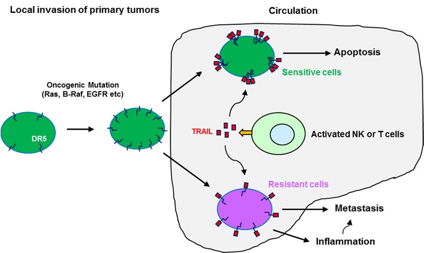
Biomolecules 2021, 11, 499                                                                                            3 of 16

                                  The cellular FLICE-like inhibitory protein (c-FLIP) is a truncated and dominant-
                             negative homolog of caspase-8 and functions to inhibit death receptor-mediated apoptosis
                             by competing with caspase-8 for binding to FADD [14,15]. FLIP, which is expressed as
                             short (FLIPS ) and long (FLIPL ) splice forms, has two death effector domains and can allow
                             the recruitment of caspase-8. The C-terminal region of FLIPL contains a caspase homolog
                             that lacks caspase activity. Formation of the pro-caspase-8/FLIPL heterodimer complex
                             can limit caspase-8 activation. In FLIPS , there is no such homolog of caspase; instead,
                             there is a short region of 20 amino acids responsible for its ubiquitylation and proteasomal
                             degradation. Both forms of FLIP can compete for caspase-8 homodimer complex assembly,
                             and so, when FLIP is heterodimerized with caspase-8, it inhibits caspase-8-dependent cell
                             death [16,17].
                                  Epithelial cells normally undergo detachment-induced apoptosis, termed anoikis, once
                             losing anchorage to the extracellular matrix or when adhesion to the correct substrate is
                             disrupted, primarily by triggering the death receptor-mediated extrinsic apoptotic pathway.
                             In contrast, malignant epithelial cells with metastatic potential resist anoikis and can survive
                             in an anchorage-independent fashion [18,19].

                             3. TRAIL/Death Receptors in Negative Regulation of Cancer Metastasis
                             3.1. Critical Role of TRAIL Produced by Immune Cells in Negative Regulation of Tumor
                             Development and Metastasis
                                   TRAIL is expressed in various tissues, particularly immune cells [20], and plays a
                             critical role in NK or T cell-mediated immune surveillance against tumor development
                             and metastasis [21]. Earlier studies suggested that TRAIL can suppress experimental liver
                             metastasis or contribute to interferon (INFγ)-mediated anti-metastatic effects [22,23]. When
                             neutralizing monoclonal antibody against TRAIL was administered, significantly increased
                             experimental liver metastases of several TRAIL-sensitive tumor cell lines were observed.
                             Such an anti-metastatic effect of TRAIL was not observed in NK cell-depleted mice or
                             INFγ-deficient mice, the latter of which lacked TRAIL in liver NK cells [22]. Consistently,
                             administration of therapeutic doses of interleukin (IL)-12, a powerful inducer of IFNγ
                             production by NK cells and NKT cells, upregulated TRAIL expression in the liver, spleen,
                             and lung NK cells, and suppressed metastases in both the liver and lungs in a TRAIL-
                             dependent fashion [23]. To further demonstrate the important role of TRAIL in suppressing
                             tumor initiation and metastasis, TRAIL gene-targeted mice were generated, which lacked
                             TRAIL in liver and spleen mononuclear cells. These TRAIL gene-targeted mice were more
                             susceptible to experimental and spontaneous tumor metastasis and also more sensitive to
                             the chemical carcinogen methylcholanthrene, further supporting TRAIL as an important
                             natural effector molecule used in the host defense against transformed cells and cancer
                             cell metastasis [24]. These early studies strongly suggest that TRAIL expressed in immune
                             cells, such as NK cells, contributes to the natural suppression of tumor development and
                             metastasis, likely via TRAIL-mediated apoptosis.

                             3.2. TRAIL-Targeted Therapeutic Approaches Against Metastatic Cancers
                                   Based on this rationale, therapeutic strategies against metastatic cancer cells have
                             been explored. In a breast cancer xenograft model, injection of TRAIL-expressing CD34+
                             cells, CD34-TRAIL(+) cells, did not lead to tumor growth inhibition; however, lungs in the
                             tumor-bearing mice were completely free of metastases at 12 days after the last injection
                             of CD34-TRAIL(+) cells, whereas metastases were present in all control mouse lungs [25].
                             In a renal cell carcinoma orthotopic tumor model with aggressive primary tumors and
                             lung metastases detectable by day 7, intra-renal administration of Ad5mTRAIL+CpG,
                             an adenovirus-encoded TRAIL/CpG immunotherapy regimen, on day 7 led to an influx
                             of effector phenotype CD4 and CD8 T cells into the kidney by day 12 and regression of
                             established primary renal tumors. Moreover, systemic immune responses characterized by
                             splenomegaly, elevated serum IgG levels, increased CD4 and CD8 T cell infiltration into the
                             lungs, and elimination of metastatic lung tumors were also detected [26]. A recent study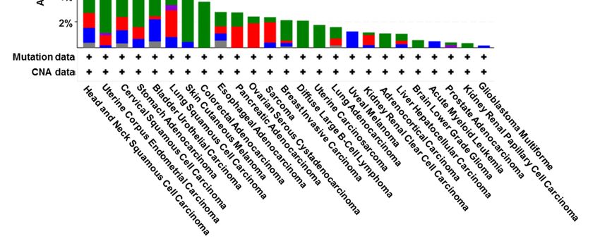
Biomolecules 2021, 11, 499                                                                                          4 of 16

                             reported that TRAIL-modified adipose-derived stem cells (ADSCs) could migrate toward
                             hepatocellular carcinoma (HCC) cells to inhibit tumor growth and the metastasis of im-
                             planted HCC tumors [27]. It was also shown that TRAIL-coated leukocytes effectively killed
                             cancer cells in the circulation, mimicking the activity of NK cells to neutralize circulating
                             tumor cells that enter blood with the potential to form new metastases [28]. This approach
                             was further shown to prevent the spontaneous formation and growth of metastatic tumors
                             in an orthotopic xenograft model of prostate cancer by greatly reducing the number of
                             circulating tumor cells [29]. In agreement, minimal dosing of leukocyte-targeting TRAIL
                             via administration of E-selectin-TRAIL liposomes decreased triple-negative breast cancer
                             metastasis following tumor resection in a preclinical mouse model [30].
                                  It has been shown that tumor cell lines derived from metastatic lymph nodes dis-
                             played a greater TRAIL resistance than cell lines derived from the respective primary oral
                             tumors [31]. Consistently, TRAIL gene therapy alone or in combination with chemotherapy
                             decreased the number of lung metastases from both chemosensitive and chemoresistant
                             breast cancer cell lines. The combination of TRAIL gene therapy and chemotherapy
                             resulted in a further reduction of lung metastatic nodules with minimal toxicity [32].
                             Co-administration of soluble TRAIL with anticancer drugs inhibited liver metastasis of
                             TRAIL-resistant colorectal cancer cells [33]. Another study reported that recombinant
                             TRAIL treatment marginally prevented metastatic spread because liver metastases were
                             detected in 71% of mice, whereas 86% of vehicle-treated mice developed liver metas-
                             tases; however, treatment with APG350, a TRAIL-receptor agonist and potent inducer of
                             apoptosis, inhibited liver metastases by 29% [34]. These studies further document the
                             anti-metastasis function of activating the TRAIL/death receptor pathway. In a model
                             for metastatic colon cancer, tail vein infusion of a tumor-targeted and conditionally repli-
                             cating oncolytic adenovirus vector expressing TRAIL (Ad5/35.IR-E1A/TRAIL) resulted
                             in the elimination of pre-established liver metastases [35]. Administration of a different
                             oncolytic adenovirus encoding TRAIL (P55-HTERT-HRE-TRAIL) also significantly reduced
                             orthotopic breast tumor growth and extended survival in a metastatic model [36]. In
                             another study using telomerase-specific oncolytic adenovirus expressing TRAIL, it was
                             shown that this approach significantly inhibited peritoneal metastasis of gastric cancer
                             and prolonged the survival of mice without treatment-related toxicity, indicating effective
                             suppression of peritoneal dissemination of gastric cancer cells [37]. Similarly, recombi-
                             nant adeno-associated virus-mediated TRAIL gene therapy suppressed liver metastatic
                             tumors [38]. These studies suggest the potential of using these means for gene therapy
                             of metastatic cancer. More recently, the TRAIL-inducing small molecule, ONC201, which
                             is being tested in clinical trials, was shown to effectively inhibit cancer metastasis and
                             promote intratumoral NK cell recruitment, including stimulation of an increase in activated
                             TRAIL-secreting NK cells in the peripheral blood of patients [39].

                             3.3. Critical Role of TRAIL Death Receptor Expression in Negative Regulation of
                             Cancer Metastasis
                                   Early studies using a mouse knockout model suggest that mDR plays a suppressive
                             role in cancer metastasis without affecting primary tumor growth since mDR knockout
                             significantly increased lymph node metastasis of carcinogen-induced skin carcinomas [40]
                             and metastasis of lymphoma cells to the liver and lungs during MYC-driven lymphoma-
                             genesis [41]. In agreement, our study using human cancer cells showed that inhibition
                             of DR5 by knockdown or knockout increased invasion of several human cancer cell lines
                             and significantly increased lung metastasis of cancer cells in a nude mouse carrying sub-
                             cutaneous lung cancer xenografts [42]. Moreover, DR5 agonistic antibody lexatumumab
                             was shown to robustly suppress lymph node or lung metastasis in an orthotopic model
                             of triple-negative breast cancer [43]. Similar results were also generated with another
                             DR5 agonist named MEDI3039 [44]. These preclinical studies strongly suggest that DR5
                             negatively regulates invasion and metastasis of both murine and cancer cells beyond its
                             apoptosis-inducing function.
lines and significantly increased lung metastasis of cancer cells in a nude mouse carrying
subcutaneous lung cancer xenografts [42]. Moreover, DR5 agonistic antibody lexatu-
mumab was shown to robustly suppress lymph node or lung metastasis in an orthotopic
model of triple-negative breast cancer [43]. Similar results were also generated with an-
Biomolecules 2021, 11, 499 other DR5 agonist named MEDI3039 [44]. These preclinical studies strongly suggest5 of 16
that
DR5 negatively regulates invasion and metastasis of both murine and cancer cells beyond
its apoptosis-inducing function.
Reduced
Reduced DR5DR5 expression
expression inin human
human melanoma
melanoma tumor
tumor samples
samples was reported to
was reported to be
be
associated with metastatic lesions [45]. Our own study found that DR5
associated with metastatic lesions [45]. Our own study found that DR5 expression inexpression in pri-
mary
primaryhead
headand
andneck
neckcancer
cancer(HNC)
(HNC)specimens
specimenswithwithmetastasis
metastasisand
andtheir
their matching
matching lymph
lymph
node
node metastasis was significantly reduced in relation to primary tumors without evidence
metastasis was significantly reduced in relation to primary tumors without evidence
of
of metastasis
metastasis [46].
[46]. Intriguingly, inactivating mutations
Intriguingly, inactivating mutations primarily
primarily in the death
in the death domain
domain of of
DR5
DR5 were detected exclusively
were detected exclusively in approximately 12%
in approximately 12% of breast cancer
of breast cancer with
with lymph
lymph node
node
metastasis, but not in breast cancer without metastasis [47]. Mutations in DR5
metastasis, but not in breast cancer without metastasis [47]. Mutations in DR5 might alter might alter
DR5-mediated apoptosis and metastasis, albeit with varied low frequencies
DR5-mediated apoptosis and metastasis, albeit with varied low frequencies in many typesin many types
of
of cancer
cancer in general (Figure
in general 2A). These
(Figure 2A). These findings
findings from
from human
human cancer
cancer tissues
tissues also
also support
support
the role of DR5 in the suppression of cancer metastasis.
the role of DR5 in the suppression of cancer metastasis.
DR5 (TNFRSF10B)
Figure 2. DR5 (TNFRSF10B) and CASP8 gene
and CASP8 gene mutations
mutationsin
indifferent
differentcancer
cancertypes
typeswith
withTCGA
TCGAdata
data
analysis.
analysis. These
Thesedata
datawere
weregenerated
generatedfrom
fromthe
thecBioPortal
cBioPortalwebsite
websiteatathttps://www.cbioportal.org/
https://www.cbioportal.org/
(accessed
(accessed on 16 March
on 16 March 2021).
2021).
In the early stage of HNCs with no lymph node metastasis, we found that higher
expression of caspase-8 alone or in combination with higher DR5 expression significantly
correlated with better disease-free survival and overall survival [46], suggesting an in-
hibitory role of DR5/caspase-8 signaling in the regulation of cancer development and
progression. However, in later stage HNCs with lymph node metastasis, high caspase-8
and DR5 together was significantly associated with poor disease-free and overall sur-
vival [46]. This is also true for HNCs with lymph node metastasis that possess highBiomolecules 2021, 11, 499 6 of 16
FADD expression alone or in combination with high DR5 or caspase-8 expression [48].
These findings suggest that DR5/FADD/caspase-8 signaling may have a role in promot-
ing cancer metastasis in the late stage of cancer (e.g., metastasis). It is possible that the
DR5/FADD/caspase-8 signaling in primary HNCs predominantly contributes to activation
of apoptosis (e.g., anoikis), resulting in preventing metastasis, but in metastatic HNCs that
may have already escaped from apoptosis (i.e., are resistant to anoikis), this signaling may
be converted to pro-metastatic signaling, causing promotion of invasion and metastasis.
4. TRAIL/Death Receptors in Positive Regulation of Cancer Metastasis
Despite the suppressive function of TRAIL/death receptor in the regulation of cancer
cell invasion and metastasis as discussed above, TRAIL has also been reported to strongly
induce the expression of pro-inflammatory cytokines interleukin-8 and monocyte chemoat-
tractant protein 1 (MCP1), to enhance the invasion of apoptosis-resistant pancreatic ductal
adenocarcinoma (PDAC) cells in vitro by upregulation of the urokinase-type plasminogen
activator expression, and to strongly increase the distant metastatic spread of pancreatic
tumors in vivo [49]. Similarly, TRAIL was also shown to induce cell migration and invasion
in apoptosis-resistant cholangiocarcinoma cells [50]. TRAIL, like CD95 ligand, could stimu-
late invasion of colorectal tumor cells and liver metastases in a K-Ras-dependent fashion.
Loss of mutant K-Ras switched TRAIL receptors back into apoptosis mode and abrogated
metastatic potential [51]. It was suggested that levels of soluble TRAIL in human plasma
were approximately 28 pg/mL [52], a concentration at which TRAIL is insufficient for
effective induction of apoptosis in lung cancer, colorectal cancer, and pancreatic cancer [53].
Beyond acquiring resistance to TRAIL, some circulating TRAIL-responsive cancer cells
under such conditions may survive TRAIL-mediated killing and eventually metastasize to
distal sites.
Bioinformatic analyses of patient data from 839 adenocarcinoma (AC) and 356 squa-
mous cell carcinoma (SCC) of lung cancer cases by cBioPortal (genomic analyses) showed
that TRAIL expression leads to differential outcomes of disease-free survival in AC and
SCC. Oncomine datamining (transcript analyses) revealed that TRAIL is upregulated in
167 SCC as compared to 350 AC patients from six data sets. Genomic analyses using cBio-
Portal revealed high rates of KRAS mutation in AC accompanied by a higher incidence of
metastasis and increased amplification of the TRAIL gene in SCC. Bioinformatic analyses of
an additional lung cancer patient database also showed that the risk of disease progression
was significantly increased with high TRAIL expression in AC (461 samples) [54]. Patients
with mesenchymal subtype colorectal cancer have a poor prognosis, in particular, patients
with stroma-rich tumors and aberrant SMAD4 expression. A recent study suggests that
TRAIL produced from SMAD4-deficient colorectal cancer cells induces BMP2 in fibroblasts,
which enhances invasiveness and metastasis of colorectal cancer [55].
There was a study demonstrating that mDR and human DR5 promote K-Ras-driven
cancer progression, invasion, and metastasis [56]; these results are contradictory to their
previous findings using an H-Ras-driven skin carcinogenesis model [40] and to our find-
ings with several human cancer cell lines such as A549, H460, and HCT116, which all
have mutant K-Ras [42]. In this study, cancer cell-restricted genetic ablation of mDR in
autochthonous KRAS-driven models of non-small cell lung cancer (NSCLC) and PDAC
was shown to reduce tumor growth, blunt metastasis, and prolong survival by inhibit-
ing cancer cell-autonomous migration, proliferation, and invasion. Moreover, this study
detected DR5 expression in human PDAC and colorectal cancer tissues and found that
high DR5 expression correlated with invasion of human PDAC into lymph vessels and
with shortened metastasis-free survival of KRAS-mutated colorectal cancer patients [56].
Mechanistically, the study suggested that constitutive signaling from DR5 promotes activa-
tion of a Rac1/PI3K/Akt signaling axis that increases migration and invasion in a cancer
cell-autonomous manner [56].
In a different study using an osteotropic variant of MDA-MB-231 breast cancer cells,
knockdown of DR5 increased the levels of E-cadherin and decreased migration withBiomolecules 2021, 11, 499 7 of 16
strongly impaired ability to form bone metastases in vivo after intracardiac injection. Eval-
uating possible underlying mechanisms revealed a strong downregulation of CXCR4, the
receptor for the chemokine SDF-1 important for homing of cancer cells to the bone. In accor-
dance, cell migration toward SDF-1 was significantly impaired by DR5 knockdown. Con-
versely, overexpression of DR5 upregulated CXCR4 levels and enhanced SDF-1-directed
migration [57]. This study supports the metastasis-promoting function of DR5, albeit
using a single cell line model. In another study, DR5 knockdown in pancreatic cancer cells
decreased local relapses and the number of macroscopic liver metastases after primary
tumor resection in an orthotopic PDAC model. However, the number of micrometastases
was increased. The outgrowth of liver metastases was suggested to be associated with
increased liver inflammation induced by resection of primary PDAC [58]. This study also
suggests a potential role of DR5 in the positive regulation of cancer cell metastasis under
specific conditions.
5. FADD, Caspase-8, and c-FLIP in Regulation of Cancer Metastasis
FADD and caspase-8 are key proteins in transmitting death receptor-mediated apop-
totic signaling, whereas c-FLIP is the key inhibitor of this death signaling. However, there
are critical roles of these proteins in the regulation of cancer cell invasion and metastasis;
some may be largely independent of death receptors [59–61].
5.1. FADD in Regulation of Cancer Metastasis
High FADD expression was associated with an increased rate of lymph node metasta-
sis of HNCs and with a shorter distant metastasis-free interval when lymph node metastases
were present [62]. Consistently, we also detected a significant increase in FADD expression
in primary HNC tumors. Lower FADD expression was significantly associated with better
disease-free survival and overall survival in HNC patients with lymph node metastasis,
although FADD expression did not significantly affect the survival of HNC patients without
lymph node metastasis [48]. In another study with samples from a cohort of Taiwanese
HNC patients with oral squamous cell carcinomas, both FADD gene copy number am-
plification and high protein expression were significantly associated with lymph node
metastasis. Patients with both FADD copy number amplification and high protein expres-
sion had the shortest disease-free survival and overall survival [63]. Focal adhesion kinase
(FAK) is an integrin-associated protein tyrosine kinase that is frequently overexpressed
in advanced human cancers and may support tumor growth and metastasis [64]. Both
FADD and FAK were reported to be overexpressed in human melanoma tissue. In murine
melanoma cell lines, miR-7a, a tumor suppressor that prohibits cell migration and invasion,
downregulated FAK expression. When FADD was overexpressed, miR-7a was inhibited
and subsequently enhanced FAK expression. FADD interference could reduce the rate of
cell migration, which could be rescued by inhibiting miR-7a expression [65]. These studies
suggest the connection between FADD overexpression and potential cancer metastasis.
5.2. Caspase-8 in Regulation of Cancer Metastasis
Although caspase-8 was reported to be associated with FAK and other proteins, re-
sulting in enhancement of cleavage of focal adhesion substrates and subsequent promotion
of cancer cell migration and metastasis under apoptosis-compromised conditions [66],
caspase-8 may primarily function as a negative regulator of cancer metastasis. Dysregula-
tion of caspase-8 was suggested to contribute to apoptotic escape and to associate with ma-
lignancy and metastasis of cancers. The loss of caspase-8 expression occurs very frequently
in neuroendocrine cancers such as neuroblastoma, medulloblastoma, and glioblastoma [67].
The suppression of caspase-8 expression potentiates neuroblastoma metastases, and re-
constitution of caspase-8 in deficient neuroblastoma cells suppressed their metastases [68].
Consistent with these results, the knockdown of caspase-8 expression in triple-negative
MDA-MB-231 breast cancer cells resulted in a significant increase in migration and in-
vasion [69]. Furthermore, the highest number of genetic alterations of caspase-8 hasBiomolecules 2021, 11, 499 8 of 16
been registered in head and neck, uterine, cervical, and gastric cancers involving somatic,
frameshift, and missense mutations [70–72] (Figure 2B). Mutations in pro-caspase-8 identi-
fied from HNCs inhibit caspase activation and apoptotic cell death following stimulation
of TRAIL. In the case of the L105H, S375*, and S386* mutants, it appears possible that the
proteins are constitutively associated with FADD in the absence of death receptor stimula-
tion. Notably, these pro-caspase-8 mutants promoted more rapid cellular migration and
invasion than the wild-type protein [73]. Low expression of caspase-8 or mutations that
block its pro-apoptotic activity may be associated with apoptosis resistance and thereby
contribute to cancer metastasis.
5.3. c-FLIP in Regulation of Cancer Metastasis
In human cancer samples, a high abundance of c-FLIP was found to be significantly
associated with lymph node metastasis in both gastric adenocarcinoma [74] and endome-
trial carcinoma [75]. Consistently, c-FLIP expression was also significantly higher in the
lung metastases than in the primary tumors of osteosarcoma [76]. In a preclinical study,
inhibition of FLIP expression with both chemical inhibitors and siRNA increased anoikis
and reduced distal tumor formation in a mouse model for circulating prostate cancer
metastasis [77], indicating that FLIP inhibition decreases the survival of circulating tumor
cells and thereby decreases metastatic tumor formation in distant organs. Collectively,
these findings suggest that c-FLIP may play a role in the positive regulation of cancer
cell invasion and metastasis. In addition to its anti-apoptotic functions, c-FLIP has been
shown to exert other physiological functions related to cell proliferation and tumorigenesis,
including metastasis [78]. One possible underlying mechanism is that FLIP interacts with
TNF-receptor-associated factors (TRAF) 1 and 2, as well as with the RIP kinases and Raf-1,
resulting in activation of the NF-κB and ERK signaling pathways [79,80]. Activation of
these signaling pathways may be associated with the role of c-FLIP in enhancing cancer
cell invasion and metastasis.
6. Possible Molecular Mechanisms Accounting for Distinct Functions of TRAIL/Death
Receptors in Regulation of Cancer Invasion and Metastasis
It is apparent that TRAIL/death receptor signaling is involved in both negative and
positive regulation of cancer invasion and metastasis. Under apoptosis-compromised
conditions, TRAIL/death receptor has a high likelihood to exert non-apoptotic functions,
including invasion/metastasis-promoting activity.
6.1. Possible Mechanisms Underlying Positive Regulation of Cancer Cell Metastasis by
TRAIL/Death Receptor Signaling
It is now known that non-canonical TRAIL/death receptor signaling exists and results
in the subsequent activation of various kinases including RIPK1, IκB/NF-κB, mitogen-
activated protein kinases (MAPK) p38, JNK, ERK1/2, MAP3K/TAK1, PKC, PI3K/Akt, and
Src in cancer cells, particularly under apoptosis-resistant conditions (see review; [81,82]).
Whether activation of these kinases contributes to cancer cell invasion and metastasis
induced by TRAIL/death receptor is largely undefined. One study identified that the
RIPK1/Src/STAT3 axis mediates TRAIL-dependent migration and invasion of TRAIL-
resistant NSCLC cells [83]. In addition, NF-κB activation may also contribute to TRAIL-
induced migration and invasion of apoptosis-resistant cholangiocarcinoma cells [50]. The
small GTPase Rac1 is an important mediator of migration and invasion. In the study in
which DR5 promoted K-Ras cancer cell invasion and metastasis, it was suggested that
constitutive signaling from DR5 promotes activation of a Rac1/PI3K/Akt signaling axis
that increases migration and invasion [56] (Figure 1B).
In addition to the primary DISC formation, TRAIL also induces the formation of a
secondary signaling complex that retains the DISC components FADD and caspase-8 but
recruits several other factors involved in kinase activation such as RIPK1, TRAF2, and
NEMO/IKKγ. This secondary complex formation requires FADD, as well as caspase-8
activity, and activates NF-κB, MAPKs, JNK, and p38 pathways [84]. Therefore, underBiomolecules 2021, 11, 499 9 of 16
certain conditions such as apoptosis resistance or suppression, activation of these signaling
pathways following the formation of the secondary signaling complex can stimulate the
survival/proliferation, migration/invasion/metastasis, and inflammation in a context-
dependent manner (Figure 1B).
Although inflammation alone is insufficient for causing cancer or metastasis, the
inflammatory response can greatly increase the risk of metastasis through the genera-
tion of pro-inflammatory biomolecules (e.g., TNFα, IL6, IL8, and MCP1), sphingosine-1-
phosphate (S1P), matrix metalloproteinases (MMPs), and angiogenic factors (e.g., VEGF)
at the site of inflammation [85–87]. While exerting apoptosis-inducing activity, TRAIL
can also induce NF-κB-dependent expression of multiple pro-inflammatory cytokines
and chemokines, particularly under apoptosis-compromised conditions [88]. Endogenous
TRAIL/death receptor-mediated caspase-8 and FADD-dependent inflammatory cytokine
(e.g., CCL2/MCP-1) secretion in some TRAIL-resistant cancer cells promotes the accu-
mulation of tumor-supportive immune cells in the cancer microenvironment, thereby
encouraging cancer growth. In this process, induction of the assembly of a similar sec-
ondary caspase-8-FADD-RIPK1 inflammatory complex called the FADDosome that leads
to the activation of NF-κB-dependent signaling is a critical event [88,89] (Figure 1B). Here,
caspase-8 functions as an essential scaffold for the assembly of the complex independent
of its caspase activity [88]. Whether this mechanism contributes to enhanced cancer cell
invasion and metastasis needs further investigation.
TRAF2 is present in the DISC complex and negatively regulates TRAIL-induced
caspase-8 activity [90]. Moreover, it is also a key component in the secondary signaling
complex induced by TRAIL that activates different kinases [84]. High TRAF2 expression is
associated with tumor metastasis and poor survival in gastric cancer [91]. Overexpression
of TRAF2 promotes invasion of pancreatic tumor cells and resistance to CD95-mediated
apoptosis [92]. S1P, which promotes the formation of lung premetastatic niches and lung
metastasis of breast cancer [93], is a lipid cofactor for TRAF2 biological activity [94]. It has
been recently demonstrated that TRAF2 directly interacts with FAK and colocalizes to intra-
cellular cell membranes, promoting NF-κB and cell survival via resistance to anoikis [95].
Hence, it is likely that TRAF2 may play a critical role in the regulation of the survival and
metastasis of TRAIL-resistant cancer cells (Figure 1).
6.2. Possible Mechanisms Accounting for Negative Regulation of Cancer Metastasis by
TRAIL/Death Receptor Signaling
In addition to the positive regulation of cancer cell invasion and metastasis, TRAIL/death
receptor signaling is largely involved in the negative regulation of cancer cell migration,
invasion, and metastasis, as discussed above. Beyond its apoptosis or anoikis-inducing
activity that helps to eliminate circulating metastatic cancer cells to prevent dissemination
of cancer cells, activation of the pathway may also exert non-apoptotic functions in the
suppression of cancer metastasis. Based on our findings [42,96], we suggest that the activa-
tion of DR5 normally favors the formation of the DISC, resulting in induction of apoptosis
or anoikis as well as other potential biological consequences; this will not only lead to
direct killing of detached cancer cells (e.g., via anoikis or TRAIL/DR5-mediated immune
surveillance) but also restrict the formation of another complex named the metastasis
and invasion signaling complex (MISC), eventually resulting in the suppression of cancer
cell invasion and metastasis. When DR5 is inhibited (e.g., by mutation, deficiency, or
reduced expression), cancer cells will become resistant to anoikis/apoptosis or immune
surveillance. Available FADD and caspase-8 may recruit and stabilize TRAF2; this process
will be enhanced by intracellular S1P (e.g., generated by SphK1). TRAF2 will then be polyu-
biquitinated and activated, likely through a self-ubiquitination mechanism, leading to the
activation of ERK1/2 and particularly JNK signaling and subsequent AP-1-dependent
expression and activation of MMPs (e.g., MMP1) and finally, promotion of invasion and
metastasis of cancer cells [42,96] (Figure 1A).
Monocyte chemotactic protein-induced protein-1 (MCPIP1; also called Regnase-1)
encoded by the ZC3H12A gene critically regulates inflammatory responses and immuneBiomolecules 2021, 11, 499 10 of 16
homeostasis primarily [97]. As its name implies, it is an MCP1-induced protein. We
recently found that MCPIP1 and MCP1 negatively regulate DR5 protein levels, including
cell surface DR5 and cancer cell responses to TRAIL, primarily through modulating DUB-
mediated protein autophagic/lysosomal degradation [98]. Given that TRAIL induces
MCP1 expression in TRAIL-resistant cancer cells [88], it is plausible to speculate that
increased MCP1 expression and release is likely to induce MCPIP1 expression; this, in turn,
downregulates the levels of DR5 including cell surface DR5, promoting the formation of
MISC and subsequent invasion and metastasis of cancer cells (Figure 1A). Thus, further
investigation in this direction is warranted.
Oncogenic mutations of Ras and B-Raf frequently occur in many cancer types and are
critical for cell transformation and tumorigenesis. Interestingly, DR5 has been shown to
positively regulate cell invasion and metastasis in KRAS-mutated cancer cells [56] while
suppressing invasion and metastasis in HRAS mutant animal models [40], albeit with an
undefined underlying mechanism. We have shown that both Ras and B-Raf induce DR5
expression by enforced expression of oncogenic Ras (e.g., H-Ras12V or K-Ras12V) or B-Raf
(i.e., V600E) in cells and by analyzing gene expression array data generated from cancer cell
lines and from human cancer tissues. This finding is further validated through knockdown
of endogenous K-Ras or B-Raf (V600E), which reduced the expression of DR5. This DR5
induction occurs primarily through co-activation of ERK/RSK and JNK signaling pathways
and subsequent cooperative effects among the transcriptional factors CHOP, Elk1, and
c-Jun in enhancing DR5 gene transcription. We also showed that the majority of cancer
cell lines highly sensitive to the DR5 agonistic antibody AMG655 have either Ras or B-Raf
mutations [99]. It is possible that RAS- or RAF-mutated cells with elevated DR5 expression
barely survive in circulation and will be eliminated (e.g., via immune surveillance) if
they are sensitive to TRAIL/DR5-mediated apoptosis. A subset of RAS- or RAF-mutated
cells with resistance to TRAIL/DR5-mediated killing may survive in the circulation and
eventually be able to metastasize to distal organs. Under such a scenario, elevated DR5
may favor the formation of metastasis-related signaling complexes such as the FADDosome
Biomolecules 2021, 11, x as illustrated in Figure 1B and the enhancement of inflammation and cancer cell invasion
11 of 16
and metastasis (Figure 3). This proposed connection also needs further investigation.
Figure 3. A schematic illustration of the potential impact of oncogenic Ras and Raf mutation-in-
Figure 3. A schematic illustration of the potential impact of oncogenic Ras and Raf mutation-induced
duced DR5 expression on apoptosis and metastasis of cancer cells. While the majority of Ras or Raf
DR5 expression
mutant cancer on apoptosis
cells and metastasis
with elevated DR5 in the ofcirculation
cancer cells. While
will the majority
be eliminated by of Ras or Raf
activated mutant
immune
cancer
cells cells
such with
as NK elevated
and TDR5 cellsinthat
the circulation will betriggering
produce TRAIL, eliminatedTRAIL/DR5-mediated
by activated immune apoptosis
cells such as
NK and Tligation
through cells that produce
with TRAIL,
cell surface triggering
death receptorsTRAIL/DR5-mediated
(e.g., DR5) of cancer cells,apoptosis through
a subset of ligation
TRAIL/DR5-
resistant
with cells with
cell surface elevated
death DR5(e.g.,
receptors mayDR5)
survive from escaping
of cancer the immune
cells, a subset surveillance in circulation
of TRAIL/DR5-resistant cells with
to eventually
elevated DR5 may formsurvive
metastases;
fromthe elevated
escaping DR5
the here may
immune favor the formation
surveillance of metastasis-related
in circulation to eventually form
signaling complexes such as the FADDosome and enhancement of inflammation and cancer cell
metastases; the elevated DR5 here may favor the formation of metastasis-related signaling complexes
invasion and metastasis.
such as the FADDosome and enhancement of inflammation and cancer cell invasion and metastasis.
7. Summary and Perspectives
TRAIL/death receptor signaling has long been considered an apoptosis-inducing sig-
naling cascade. However, it can also initiate other signaling pathways, unveiling non-
apoptosis-related functions, including stimulation of inflammatory responses and cancer
metastasis under certain circumstances. Hence, death receptor-mediated signaling com-Biomolecules 2021, 11, 499 11 of 16
7. Summary and Perspectives
TRAIL/death receptor signaling has long been considered an apoptosis-inducing
signaling cascade. However, it can also initiate other signaling pathways, unveiling non-
apoptosis-related functions, including stimulation of inflammatory responses and cancer
metastasis under certain circumstances. Hence, death receptor-mediated signaling complex
proteins can serve as an important platform to regulate cancer apoptosis, inflammation,
and metastasis depending on cell types or contexts. Under TRAIL-resistant or apoptosis-
compromising conditions, activation of TRAIL/death receptor signaling may likely favor
the positive regulation of cancer cell invasion and metastasis. Oncogenic Ras proteins such
as K-Ras and H-Ras, in general, have shared redundant biological activities [100]. However,
mDR or DR5 exert opposing effects on the regulation of cancer cell metastasis in murine
tumorigenic models with different RAS mutations [40,56]. The underlying mechanisms
accounting for this difference are largely unknown. Given the fact that there are two TRAIL
death receptors named DR4 and DR5 in humans, whereas there is only one TRAIL death
receptor (mDR) in mice [9,10] and the regulatory mechanisms of mDR expression are
largely unknown in comparison with human DR5 expression, caution needs to be taken
when extending the findings from murine models to the setting of human cancer.
TRAIL/death receptor-targeted therapeutics including recombinant proteins and
agonistic antibodies have been developed for cancer therapy and are currently being tested
in the clinic [12,13,101]. Unfortunately, although some positive trends have been observed
with tolerable safety, no statistically significant anticancer activity has been achieved.
Since the ultimate goal is to apply our knowledge to prevent metastatic dissemination
during cancer therapy, including chemotherapy, targeted therapy, and immunotherapy,
while simultaneously sensitizing cancer cells to die, we need a deep understanding of the
biological functions of TRAIL/death receptor signaling in the regulation of cell death and
metastasis. This will also inform appropriate strategies to design better therapeutics in
the future for effective cancer therapy through targeting this critical signaling pathway
for activating cell death and suppressing cancer cell invasion and metastasis. It is also
important to distinguish TRAIL-sensitive from TRAIL-insensitive or resistant tumors
so that we can apply tailored strategies for the treatment of specific cancer types with
TRAIL/death receptor-targeting regimens while avoiding the stimulation of potential
cancer cell invasion and metastasis.
Since resistance to TRAIL/death receptor-mediated apoptosis is a key factor associated
with enhanced invasion and metastasis of cancer cells, it is critical to consider strategies
aiming to target these cancer cells with resistance to TRAIL/death receptor-initiated apop-
tosis. Fortunately, many types of resistant cancer cells can be sensitized to TRAIL or death
receptor activation-induced apoptosis by various means, particularly some small molecules
that are able to increase death receptor expression and/or decrease the levels of c-FLIP
and/or other anti-apoptotic proteins such as Mcl-1 [102,103]. Considering the heterogeneity
of cancer cells, sensitive cancer cells may contain a subset of resistant cell populations that
can escape TRAIL/death receptor-mediated killing. Therefore, combinatorial approaches
offer a rational and actionable strategy to treat cancer to maximally eliminate cancer cells
through the induction of apoptosis while minimizing the risk of increasing invasion and
metastasis, which is often the reason for treatment failure.
Author Contributions: Y.-T.O. and S.-Y.S. wrote the article. All authors have read and agreed to the
published version of the manuscript.
Funding: This research received no external funding.
Institutional Review Board Statement: Not applicable.
Informed Consent Statement: Not applicable.
Data Availability Statement: Not applicable.Biomolecules 2021, 11, 499 12 of 16
Acknowledgments: We are also grateful to Anthea Hammond in our department for editing
the manuscript.
Conflicts of Interest: The authors declare no conflict of interest.
References
1. Valastyan, S.; Weinberg, R.A. Tumor metastasis: Molecular insights and evolving paradigms. Cell 2011, 147, 275–292. [CrossRef]
[PubMed]
2. Mehlen, P.; Puisieux, A. Metastasis: A question of life or death. Nat. Rev. Cancer 2006, 6, 449–458. [CrossRef]
3. Fidler, I.J. Metastasis: Quantitative analysis of distribution and fate of tumor emboli labeled with 125 I-5-iodo-2’-deoxyuridine. J.
Natl. Cancer Inst. 1970, 45, 773–782.
4. Luzzi, K.J.; MacDonald, I.C.; Schmidt, E.E.; Kerkvliet, N.; Morris, V.L.; Chambers, A.F.; Groom, A.C. Multistep nature of metastatic
inefficiency: Dormancy of solitary cells after successful extravasation and limited survival of early micrometastases. Am. J. Pathol.
1998, 153, 865–873. [CrossRef]
5. Hanahan, D.; Weinberg, R.A. Hallmarks of cancer: The next generation. Cell 2011, 144, 646–674. [CrossRef]
6. Wajant, H. CD95L/FasL and TRAIL in tumour surveillance and cancer therapy. Cancer Treat Res. 2006, 130, 141–165. [PubMed]
7. Wajant, H.; Pfizenmaier, K.; Scheurich, P. TNF-related apoptosis inducing ligand (TRAIL) and its receptors in tumor surveillance
and cancer therapy. Apoptosis 2002, 7, 449–459. [CrossRef]
8. Falschlehner, C.; Schaefer, U.; Walczak, H. Following TRAIL’s path in the immune system. Immunology 2009, 127, 145–154.
[CrossRef]
9. Kelley, S.K.; Ashkenazi, A. Targeting death receptors in cancer with Apo2L/TRAIL. Curr. Opin. Pharmacol. 2004, 4, 333–339.
[CrossRef]
10. van Roosmalen, I.A.; Quax, W.J.; Kruyt, F.A. Two death-inducing human TRAIL receptors to target in cancer: Similar or distinct
regulation and function? Biochem. Pharmacol. 2014, 91, 447–456. [CrossRef]
11. Wu, G.S.; Burns, T.F.; Zhan, Y.; Alnemri, E.S.; El-Deiry, W.S. Molecular cloning and functional analysis of the mouse homologue
of the KILLER/DR5 tumor necrosis factor-related apoptosis-inducing ligand (TRAIL) death receptor. Cancer Res. 1999, 59,
2770–2775.
12. Lemke, J.; von Karstedt, S.; Zinngrebe, J.; Walczak, H. Getting TRAIL back on track for cancer therapy. Cell Death Differ. 2014, 21,
1350–1364. [CrossRef]
13. Lim, B.; Allen, J.E.; Prabhu, V.V.; Talekar, M.K.; Finnberg, N.K.; El-Deiry, W.S. Targeting TRAIL in the treatment of cancer: New
developments. Expert Opin. Ther. Targets 2015, 19, 1171–1185. [CrossRef]
14. Roth, W.; Reed, J.C. FLIP protein and TRAIL-induced apoptosis. Vitam. Horm. 2004, 67, 189–206. [PubMed]
15. Wajant, H. Targeting the FLICE Inhibitory Protein (FLIP) in cancer therapy. Mol. Interv. 2003, 3, 124–127. [CrossRef]
16. Budd, R.C.; Yeh, W.C.; Tschopp, J. cFLIP regulation of lymphocyte activation and development. Nat. Rev. Immunol. 2006, 6,
196–204. [CrossRef] [PubMed]
17. Humphreys, L.; Espona-Fiedler, M.; Longley, D.B. FLIP as a therapeutic target in cancer. FEBS J. 2018, 285, 4104–4123. [CrossRef]
18. Simpson, C.D.; Anyiwe, K.; Schimmer, A.D. Anoikis resistance and tumor metastasis. Cancer Lett. 2008, 272, 177–185. [CrossRef]
19. Chiarugi, P.; Giannoni, E. Anoikis: A necessary death program for anchorage-dependent cells. Biochem. Pharmacol. 2008, 76,
1352–1364. [CrossRef]
20. Daniels, R.A.; Turley, H.; Kimberley, F.C.; Liu, X.S.; Mongkolsapaya, J.; Ch’En, P.; Xu, X.N.; Jin, B.Q.; Pezzella, F.; Screaton, G.R.
Expression of TRAIL and TRAIL receptors in normal and malignant tissues. Cell Res. 2005, 15, 430–438. [CrossRef]
21. Smyth, M.J.; Takeda, K.; Hayakawa, Y.; Peschon, J.J.; van den Brink, M.R.; Yagita, H. Nature’s TRAIL—On a path to cancer
immunotherapy. Immunity 2003, 18, 1–6. [CrossRef]
22. Takeda, K.; Hayakawa, Y.; Smyth, M.J.; Kayagaki, N.; Yamaguchi, N.; Kakuta, S.; Iwakura, Y.; Yagita, H.; Okumura, K. Involvement
of tumor necrosis factor-related apoptosis-inducing ligand in surveillance of tumor metastasis by liver natural killer cells. Nat.
Med. 2001, 7, 94–100. [CrossRef]
23. Smyth, M.J.; Cretney, E.; Takeda, K.; Wiltrout, R.H.; Sedger, L.M.; Kayagaki, N.; Yagita, H.; Okumura, K. Tumor necrosis
factor-related apoptosis-inducing ligand (TRAIL) contributes to interferon gamma-dependent natural killer cell protection from
tumor metastasis. J. Exp. Med. 2001, 193, 661–670. [CrossRef] [PubMed]
24. Cretney, E.; Takeda, K.; Yagita, H.; Glaccum, M.; Peschon, J.J.; Smyth, M.J. Increased susceptibility to tumor initiation and
metastasis in TNF-related apoptosis-inducing ligand-deficient mice. J. Immunol. 2002, 168, 1356–1361. [CrossRef] [PubMed]
25. Rossini, A.; Giussani, M.; Giacomini, A.; Guarnotta, C.; Tagliabue, E.; Balsari, A. Surveillance of spontaneous breast cancer
metastasis by TRAIL-expressing CD34(+) cells in a xenograft model. Breast Cancer Res. Treat. 2012, 136, 457–467. [CrossRef]
26. Norian, L.A.; Kresowik, T.P.; Rosevear, H.M.; James, B.R.; Rosean, T.R.; Lightfoot, A.J.; Kucaba, T.A.; Schwarz, C.; Weydert, C.J.;
Henry, M.D.; et al. Eradication of metastatic renal cell carcinoma after adenovirus-encoded TNF-related apoptosis-inducing
ligand (TRAIL)/CpG immunotherapy. PLoS ONE 2012, 7, e31085. [CrossRef]
27. Liu, Z.; Li, S.; Ma, T.; Zeng, J.; Zhou, X.; Li, H.; Tang, M.; Liu, X.; Li, F.; Jiang, B.; et al. Secreted TRAIL gene-modified adipose-
derived stem cells exhibited potent tumor-suppressive effect in hepatocellular carcinoma cells. Immun. Inflamm. Dis. 2020.
[CrossRef]Biomolecules 2021, 11, 499 13 of 16
28. Mitchell, M.J.; Wayne, E.; Rana, K.; Schaffer, C.B.; King, M.R. TRAIL-coated leukocytes that kill cancer cells in the circulation.
Proc. Natl. Acad. Sci. USA 2014, 111, 930–935. [CrossRef]
29. Wayne, E.C.; Chandrasekaran, S.; Mitchell, M.J.; Chan, M.F.; Lee, R.E.; Schaffer, C.B.; King, M.R. TRAIL-coated leukocytes that
prevent the bloodborne metastasis of prostate cancer. J. Control Release 2016, 223, 215–223. [CrossRef]
30. Jyotsana, N.; Zhang, Z.; Himmel, L.E.; Yu, F.; King, M.R. Minimal dosing of leukocyte targeting TRAIL decreases triple-negative
breast cancer metastasis following tumor resection. Sci. Adv. 2019, 5, eaaw4197. [CrossRef]
31. Vigneswaran, N.; Wu, J.; Nagaraj, N.; Adler-Storthz, K.; Zacharias, W. Differential susceptibility of metastatic and primary oral
cancer cells to TRAIL-induced apoptosis. Int. J. Oncol. 2005, 26, 103–112. [CrossRef] [PubMed]
32. Lin, T.; Zhang, L.; Davis, J.; Gu, J.; Nishizaki, M.; Ji, L.; Roth, J.A.; Xiong, M.; Fang, B. Combination of TRAIL gene therapy and
chemotherapy enhances antitumor and antimetastasis effects in chemosensitive and chemoresistant breast cancers. Mol. Ther.
2003, 8, 441–448. [CrossRef]
33. Ishii, M.; Iwai, M.; Harada, Y.; Kishida, T.; Asada, H.; Shin-Ya, M.; Itoh, Y.; Imanishi, J.; Okanoue, T.; Mazda, O. Soluble TRAIL
gene and actinomycin D synergistically suppressed multiple metastasis of TRAIL-resistant colon cancer in the liver. Cancer Lett.
2007, 245, 134–143. [CrossRef]
34. Legler, K.; Hauser, C.; Egberts, J.H.; Willms, A.; Heneweer, C.; Boretius, S.; Rocken, C.; Gluer, C.C.; Becker, T.; Kluge, M.; et al. The
novel TRAIL-receptor agonist APG350 exerts superior therapeutic activity in pancreatic cancer cells. Cell Death Dis. 2018, 9, 445.
[CrossRef]
35. Sova, P.; Ren, X.W.; Ni, S.; Bernt, K.M.; Mi, J.; Kiviat, N.; Lieber, A. A tumor-targeted and conditionally replicating oncolytic
adenovirus vector expressing TRAIL for treatment of liver metastases. Mol. Ther. 2004, 9, 496–509. [CrossRef] [PubMed]
36. Zhu, W.; Zhang, H.; Shi, Y.; Song, M.; Zhu, B.; Wei, L. Oncolytic adenovirus encoding tumor necrosis factor-related apoptosis
inducing ligand (TRAIL) inhibits the growth and metastasis of triple-negative breast cancer. Cancer Biol. Ther. 2013, 14, 1016–1023.
[CrossRef]
37. Zhou, W.; Dai, S.; Zhu, H.; Song, Z.; Cai, Y.; Lee, J.B.; Li, Z.; Hu, X.; Fang, B.; He, C.; et al. Telomerase-specific oncolytic adenovirus
expressing TRAIL suppresses peritoneal dissemination of gastric cancer. Gene Ther. 2017, 24, 199–207. [CrossRef]
38. Ma, H.; Liu, Y.; Liu, S.; Kung, H.F.; Sun, X.; Zheng, D.; Xu, R. Recombinant adeno-associated virus-mediated TRAIL gene therapy
suppresses liver metastatic tumors. Int. J. Cancer. 2005, 116, 314–321. [CrossRef] [PubMed]
39. Wagner, J.; Kline, C.L.; Zhou, L.; Campbell, K.S.; MacFarlane, A.W.; Olszanski, A.J.; Cai, K.Q.; Hensley, H.H.; Ross, E.A.;
Ralff, M.D.; et al. Dose intensification of TRAIL-inducing ONC201 inhibits metastasis and promotes intratumoral NK cell
recruitment. J. Clin. Investig. 2018, 128, 2325–2338. [CrossRef] [PubMed]
40. Grosse-Wilde, A.; Voloshanenko, O.; Bailey, S.L.; Longton, G.M.; Schaefer, U.; Csernok, A.I.; Schutz, G.; Greiner, E.F.; Kemp, C.J.;
Walczak, H. TRAIL-R deficiency in mice enhances lymph node metastasis without affecting primary tumor development. J. Clin.
Investig. 2008, 118, 100–110. [CrossRef]
41. Finnberg, N.; Klein-Szanto, A.J.; El-Deiry, W.S. TRAIL-R deficiency in mice promotes susceptibility to chronic inflammation and
tumorigenesis. J. Clin. Investig. 2008, 118, 111–123. [CrossRef] [PubMed]
42. Oh, Y.T.; Yue, P.; Wang, D.; Tong, J.S.; Chen, Z.G.; Khuri, F.R.; Sun, S.Y. Suppression of death receptor 5 enhances cancer cell
invasion and metastasis through activation of caspase-8/TRAF2-mediated signaling. Oncotarget 2015, 6, 41324–41338. [CrossRef]
43. Malin, D.; Chen, F.; Schiller, C.; Koblinski, J.; Cryns, V.L. Enhanced metastasis suppression by targeting TRAIL receptor 2 in a
murine model of triple-negative breast cancer. Clin. Cancer Res. 2011, 17, 5005–5015. [CrossRef]
44. Greer, Y.E.; Gilbert, S.F.; Gril, B.; Narwal, R.; Peacock Brooks, D.L.; Tice, D.A.; Steeg, P.S.; Lipkowitz, S. MEDI3039, a novel highly
potent tumor necrosis factor (TNF)-related apoptosis-inducing ligand (TRAIL) receptor 2 agonist, causes regression of orthotopic
tumors and inhibits outgrowth of metastatic triple-negative breast cancer. Breast Cancer Res. 2019, 21, 27. [CrossRef]
45. Zhuang, L.; Lee, C.S.; Scolyer, R.A.; McCarthy, S.W.; Zhang, X.D.; Thompson, J.F.; Screaton, G.; Hersey, P. Progression in melanoma
is associated with decreased expression of death receptors for tumor necrosis factor-related apoptosis-inducing ligand. Hum.
Pathol. 2006, 37, 1286–1294. [CrossRef]
46. Elrod, H.A.; Fan, S.; Muller, S.; Chen, G.Z.; Pan, L.; Tighiouart, M.; Shin, D.M.; Khuri, F.R.; Sun, S.Y. Analysis of death receptor 5
and caspase-8 expression in primary and metastatic head and neck squamous cell carcinoma and their prognostic impact. PLoS
ONE 2010, 5, e12178. [CrossRef]
47. Shin, M.S.; Kim, H.S.; Lee, S.H.; Park, W.S.; Kim, S.Y.; Park, J.Y.; Lee, J.H.; Lee, S.K.; Lee, S.N.; Jung, S.S.; et al. Mutations of tumor
necrosis factor-related apoptosis-inducing ligand receptor 1 (TRAIL-R1) and receptor 2 (TRAIL-R2) genes in metastatic breast
cancers. Cancer Res. 2001, 61, 4942–4946.
48. Fan, S.; Muller, S.; Chen, Z.G.; Pan, L.; Tighiouart, M.; Shin, D.M.; Khuri, F.R.; Sun, S.Y. Prognostic impact of Fas-associated death
domain, a key component in death receptor signaling, is dependent on the presence of lymph node metastasis in head and neck
squamous cell carcinoma. Cancer Biol. Ther. 2013, 14, 365–369. [CrossRef]
49. Trauzold, A.; Siegmund, D.; Schniewind, B.; Sipos, B.; Egberts, J.; Zorenkov, D.; Emme, D.; Roder, C.; Kalthoff, H.; Wajant, H.
TRAIL promotes metastasis of human pancreatic ductal adenocarcinoma. Oncogene 2006, 25, 7434–7439. [CrossRef] [PubMed]
50. Ishimura, N.; Isomoto, H.; Bronk, S.F.; Gores, G.J. Trail induces cell migration and invasion in apoptosis-resistant cholangiocarci-
noma cells. Am. J. Physiol. Gastrointest Liver Physiol. 2006, 290, G129–G136. [CrossRef] [PubMed]You can also read