Antibody targeting tumor-derived soluble NKG2D ligand sMIC reprograms NK cell homeostatic survival and function and enhances melanoma response to ...
←
→
Page content transcription
If your browser does not render page correctly, please read the page content below

Basher et al. Journal of Hematology & Oncology               (2020) 13:74
https://doi.org/10.1186/s13045-020-00896-0

 RESEARCH                                                                                                                                            Open Access

Antibody targeting tumor-derived soluble
NKG2D ligand sMIC reprograms NK cell
homeostatic survival and function and
enhances melanoma response to PDL1
blockade therapy
Fahmin Basher1,9, Payal Dhar2,3, Xin Wang4, Derek A. Wainwright3,5,6, Bin Zhang6,7, Jeffrey Sosman7, Zhe Ji4,8 and
Jennifer D. Wu2,3,6*

  Abstract
  Background: Melanoma patients who have detectable serum soluble NKG2D ligands either at the baseline or post-
  treatment of PD1/PDL1 blockade exhibit poor overall survival. Among families of soluble human NKG2D ligands,
  the soluble human MHC I chain-related molecule (sMIC) was found to be elevated in melanoma patients and
  mostly associated with poor response to PD1/PDL1 blockade therapy.
  Methods: In this study, we aim to investigate whether co-targeting tumor-released sMIC enhances the therapeutic
  outcome of PD1/PDL1 blockade therapy for melanoma. We implanted sMIC-expressing B16F10 melanoma tumors
  into syngeneic host and evaluated therapeutic efficacy of anti-sMIC antibody and anti-PDL1 antibody combination
  therapy in comparison with monotherapy. We analyzed associated effector mechanism. We also assessed sMIC/MIC
  prevalence in metastatic human melanoma tumors.
  Results: We found that the combination therapy of the anti-PDL1 antibody with an antibody targeting sMIC
  significantly improved animal survival as compared to monotherapies and that the effect of combination therapy
  depends significantly on NK cells. We show that combination therapy significantly increased IL-2Rα (CD25) on NK
  cells which sensitizes NK cells to low dose IL-2 for survival. We demonstrate that sMIC negatively reprograms gene
  expression related to NK cell homeostatic survival and proliferation and that antibody clearing sMIC reverses the
  effect of sMIC and reprograms NK cell for survival. We further show that sMIC/MIC is abundantly present in
  metastatic human melanoma tumors.
  (Continued on next page)

* Correspondence: jennifer.wu@northwestern.edu
2
 Department of Urology, Feinberg School of Medicine, Northwestern
University, Chicago, IL 60611, USA
3
 Driskill Graduate Program in Life Science, Feinberg School of Medicine,
Northwestern University, Chicago, IL 60611, USA
Full list of author information is available at the end of the article

                                         © The Author(s). 2020 Open Access This article is licensed under a Creative Commons Attribution 4.0 International License,
                                         which permits use, sharing, adaptation, distribution and reproduction in any medium or format, as long as you give
                                         appropriate credit to the original author(s) and the source, provide a link to the Creative Commons licence, and indicate if
                                         changes were made. The images or other third party material in this article are included in the article's Creative Commons
                                         licence, unless indicated otherwise in a credit line to the material. If material is not included in the article's Creative Commons
                                         licence and your intended use is not permitted by statutory regulation or exceeds the permitted use, you will need to obtain
                                         permission directly from the copyright holder. To view a copy of this licence, visit http://creativecommons.org/licenses/by/4.0/.
                                         The Creative Commons Public Domain Dedication waiver (http://creativecommons.org/publicdomain/zero/1.0/) applies to the
                                         data made available in this article, unless otherwise stated in a credit line to the data.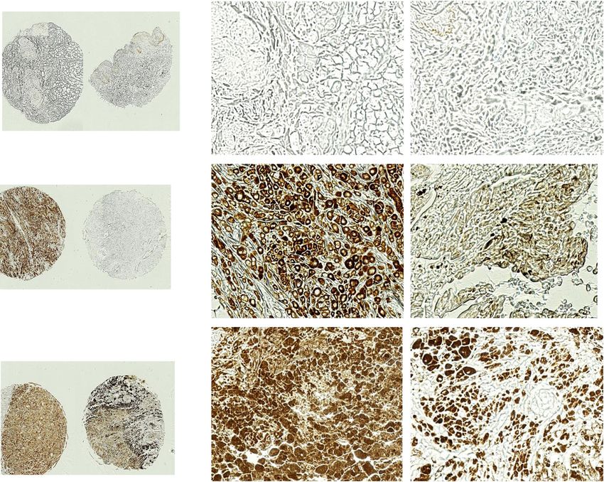
Basher et al. Journal of Hematology & Oncology   (2020) 13:74                                                  Page 2 of 16

 (Continued from previous page)
 Conclusions: Our findings provide a pre-clinical proof-of-concept and a new mechanistic understanding to
 underscore the significance of antibody targeting sMIC to improve therapeutic efficacy of anti-PD1/PDL1 antibody
 for MIC/sMIC+ metastatic melanoma patients.
 Keywords: NKG2D ligands, Soluble MHC I chain-related molecule (sMIC), PD1/PDL1 blockade, Anti-sMIC antibody,
 Melanoma

Introduction                                                    function, impairing CD8 T cell function by destabilizing
Immunotherapy by blocking the axis of the immune                CD3ζ [16], and expanding myeloid-derived suppressive
checkpoint molecule programmed cell death protein               cells (MDSCs) in the tumor microenvironment [17].
(PD1) or its ligand PDL1 has presented remarkable sur-          These mechanistic understandings along with the re-
vival benefit and thus became a frontline treatment for         ported clinical observations prompt us to test the hy-
metastatic or unresectable melanoma [1, 2]. While the           pothesis that co-targeting tumor-derived soluble
survival in advanced melanoma has improved substan-             NKG2D ligands would enhance melanoma tumor re-
tially since FDA approved PD1/PDL1 blockade therapy,            sponse to PD/PDL1 blockade therapy.
objective response rates approach only 50% at best with           We have previously described that clearance of tumor-
checkpoint inhibitors combination therapy [3]. Combin-          derived soluble NKG2D ligands, sMIC, with a monoclo-
ation of anti-PD1 antibody and the antibody to the cyto-        nal antibody (mAb) B10G5 restores NK cell homeostatic
toxic T cell lymphocyte-associated protein 4 (CTLA-4)           renewal, enhances NK cell and antigen-specific CD8 T
slightly increased the response rate compared to anti-          cell function, immobilizes NK and CD8 T cell to the tu-
PD-1 monotherapy, however with significantly increased          mors, and re-modulates tumor microenvironment by
autoimmune toxicity [3]. Therefore, the need to increase        eliminating MDSCs and tumor-associated macrophages
the response rate with new combination therapy without          [17, 18]. In this study, we demonstrate that antibody tar-
increased toxicity is still imperative.                         geting sMIC increases the IL-2 sensing receptor IL-2Rα
   Identifying tumor-derived targetable factors that may        on NK cells, reprograms NK cell homeostatic mainten-
impact patients’ response to PD1/PDL1 blockade would            ance, and enhances the therapeutic response of melan-
rationalize a potential combination therapy to improve          oma tumors to PD1/PDL1 blockade therapy. Our
the clinical outcome. In recent clinical studies, the pres-     current study provides a new mechanistic understanding
ence of circulating soluble NKG2D ligands was shown             to accentuate the significance of co-targeting tumor-
to be negatively correlated with clinical outcome to anti-      derived soluble NKG2D ligand, sMIC, to enhance the
PD1/PDL1 response [4, 5]. In humans, there are two              therapeutic efficacy of PD1/PDL1 checkpoint blockade
major family members of NKG2D ligands, the MHC I-               therapy for melanoma patients.
chain-related molecules MICA and MICB and the viral
HCMV UL16-binding proteins ULBP1-6 family. NKG2D                Materials and methods
ligands are rarely expressed by normal tissues unless           Mice and cell lines
under stress insults, such as infection [6], but induced in     Mice were bred and housed under specific pathogen-free
most tumor cells in part through activation of DNA              conditions in the animal facility of the Medical Univer-
damage response pathway or oxidative stress [7–9]. Al-          sity of South Carolina and Northwestern University in
though the MICA and MICB family molecules are better            accordance with institutional guidelines with approved
characterized and more prevalently expressed than the           IACUC protocols. All mice used in this study were male
ULBP family proteins, these ligands often co-exist in one       rPB-MICB mice on the B6 background as previously de-
tumor type, presumably through host-viral co-                   scribed and thereafter defined as MICB/B6 mice [19].
evolutionary processes [10]. While levels of the NKG2D          sMIC-expressing B16F10-sMICB cell line was developed
ligand MIC have been correlated with survival benefits          by transduction of B16F10 cells (ATCC) with an IRES-
in the early stages of several cancers, the opposite has        GFP retroviral vector containing the construct for re-
been demonstrated with more invasive tumors [11–14].            combinant soluble MICB, as described previously [20].
In most invasive human tumors, NKG2D ligands also               sMIC+ B16F10 cells were selected by puromycin and
exist as a soluble form through proteolytic shedding or         further by flow cytometry sorting for GFP-positive cells.
exosome secretion [15]. Soluble human NKG2D ligands
have been shown to subvert antitumor immunity
through multiple mechanisms, including but not limited          Antibodies, peptides, and tetramers
to, perturbing NK cell homeostatic maintenance and              InVivoMAb anti-mouse PDL1 (clone 10F.9G2) was pur-
                                                                chased from BioXCell. Generation of the anti-MIC mAb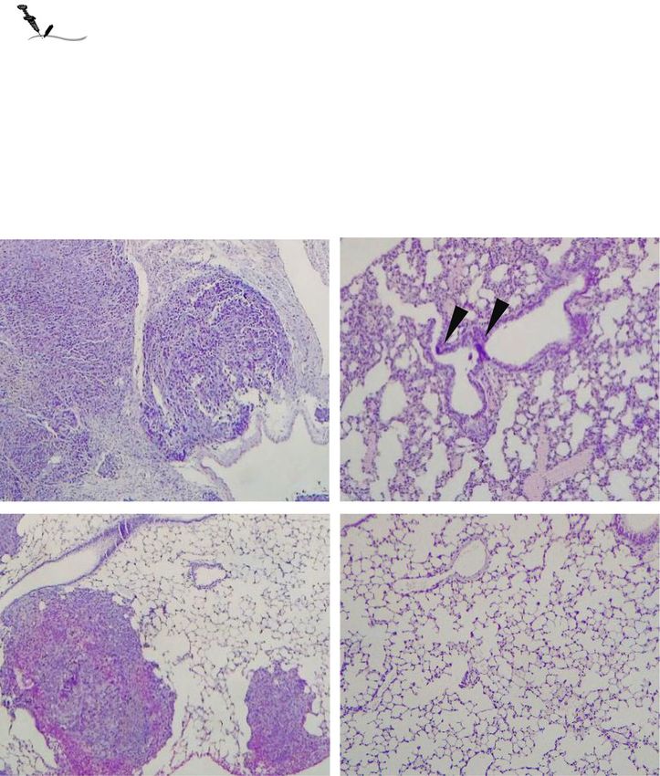
Basher et al. Journal of Hematology & Oncology   (2020) 13:74                                                   Page 3 of 16

B10G5 was previously described [19]. B10G5 is a mouse           at 37°C for 6 h with 50 ng/ml phorbol myristate acetate
IgG1 isotype, recognizing both MICA and MICB. B10G5             (PMA) and 500 ng/ml ionomycin. To assess melanoma
binds to free sMIC but does not block the interaction of        antigen-specific T cell function, single-cell suspension of
sMIC with the receptor NKG2D [18]. B10G5 was pro-               bulked splenocytes or tumor-draining lymph nodes was
duced and purified from the hybridoma culture by BioX-          stimulated with 1 μg/ml of melanoma antigen gp10025-33
Cell (West Lebanon, NH). In vivo NK cell-depleting              peptides overnight. IFNγ production was assayed by
anti-NK1.1 (clone PK136) and CD8 T cell-depleting               intracellular staining with BD IFNγ staining Kits follow-
antibody anti-CD8α (clone 2.43) were purchased from             ing the manufacturer’s instruction.
BioXcell. Peptide gp10025-33 (KVPRNQDWL) was syn-
thesized by GenScript. H-2Db/ gp10025-33 tetramer was           Flow cytometry analysis
produced by NIH Tetramer Core Facility at Emory                 Single-cell suspensions were incubated on ice for 30 min
University.                                                     with a combination of antibodies specific to cell surface
                                                                markers for identification of lymphocyte subsets. These
Tumor inoculation and in vivo experiments                       antibodies are anti-NK1.1 (clone PK136), anti-CD3
For subcutaneous study, B16F10-sMICB cells were im-             (clone 145–2C11), anti-CD8α (clone 53–6.7), anti-
planted subcutaneously into the right flank of cohorts of       NKG2D (clone CX5), anti-CD44 (clone IM7), anti-CD25
syngeneic MICB/B6 male mice (4 × 10⁵ cells/mouse) at            (clone PC61), anti-Gr1 (clone RB6-8C5), and anti-CD11b
ages 8–10 weeks old. When tumor volume reached ap-              (clone ICRF44). All antibodies used for flow cytometry
proximately 75–100 mm3, animals were randomized into            analyses were purchased from Biolegend (San Diego, CA,
four therapy groups (n = 5 to 7 per group): (1) control         USA). Tetramer staining was performed with 2 μg/ml of
mouse IgG (3.0 mg/kg BW), (2) anti-MIC mAb B10G5                PE-labeled H-2Db/ gp10025-33 tetramer at 37 °C for 20 min
(3.0 mg/kg BW), (3) anti-PDL1 mAb (3.0 mg/kg BW), and           and followed by surface marker staining. For intracellular
(4) B10G5 and anti-PDL1 mAb. All antibodies were given          staining, cells were stained with surface markers followed
via I.P. injection every 3 days. For survival studies, tumor    by fixation and permeabilization with BD Perm/Fix kits
volume of 1800 mm3 was defined as survival endpoint. For        and antibodies specific to intracellular molecules. Cells
mechanistic studies, animals were euthanized after 9 days       were analyzed using the BD Fortessa. Data were analyzed
of treatment. After euthanization, the spleens and two in-      using the FlowJo software (Tree Star).
guinal draining lymph nodes (dLN) and tumors were har-
vested. Partial of the tumors were formalin fixed, paraffin     Histological and immunohistochemistry staining
embedded, and sectioned for histology and immunohisto-          Five micrometers of formalin-fixed paraffin-embedded
chemistry staining (IHC). The remaining tumors were             sections were stained with H&E for pathological evalu-
used for single-cell suspension preparation by the method       ation and used for immunohistochemistry (IHC) stain-
of mincing, mechanically processing, and passing through        ing. Mouse tumor sections were also stained with the
a 70-μm filter. Single-cell suspension of splenocytes, dLN,     following: (a) anti-NKp46/NCR1 (rabbit IgG; 1:200;
and tumors was used for ex vivo stimulation and flow cy-        Abcam); (b) anti-CD8 (BD biosciences); (c) anti-arginase
tometry analyses.                                               1 (rabbit IgG; 1:200; Santa Cruz Biotechnology); (d) anti-
  For lung metastasis, B16F10-sMICB cells were injected         CD31 (rabbit IgG; 1:100; Abcam); and (e) anti-Ki67
into the lateral tail vein of syngeneic B6/MICB male            (rabbit IgG, Abcam, 1 μg/ml). Human tissue microarray
mice (2 × 10⁵ cells/mouse) at ages 8–10 weeks old. At           (TMA) sections containing 62 cases of malignant melan-
day 10 post-tumor inoculation at which time point               oma, 21 metastatic malignant melanoma, and other con-
tumor nodules were visible on the surface of the lung by        trol tissues were purchase from US Biomax (Cat.
random examination of three animals, mice were ran-             ME1004g) and were stained with the mouse monoclonal
domized into four therapy groups (n = 5 per group): (1)         anti-MIC antibody D4H3 (Supplement Material and
control mouse IgG (3.0 mg/kg BW), (2) anti-MIC mAb              Methods). Sections were deparaffinized and incubated
B10G5 (3.0 mg/kg BW), (3) anti-PDL1 mAb (3.0 mg/kg              for 10 min in 10 mM citrate buffer (pH 6.0) at 95 °C for
BW), and (4) B10G5 and anti-PDL1 mAb. All antibodies            antigen retrieval. Endogenous peroxidase activity was
were given via I.P. injection every 3 days. Animals were        quenched with 3% hydrogen peroxide. After quenching
euthanized at day 21 following tumor inoculation.               endogenous peroxidase activity and blocking nonspecific
Spleens, inguinal draining lymph nodes, and lungs were          binding, slides were incubated with specific primary
harvested for analyses.                                         antibody overnight at 4 °C followed by subsequent incu-
                                                                bation with the appropriate biotinylated secondary anti-
Ex vivo cytokine re-stimulation assay                           body: goat anti-rabbit IgG or goat anti-mouse (Vector)
For general re-stimulation, single-cell suspensions of          at a 1:1000 dilution for 20 min at 37 °C. Immunoreactive
splenocytes and draining lymph nodes were stimulated            antigens were detected using the Vectastain Elite ABC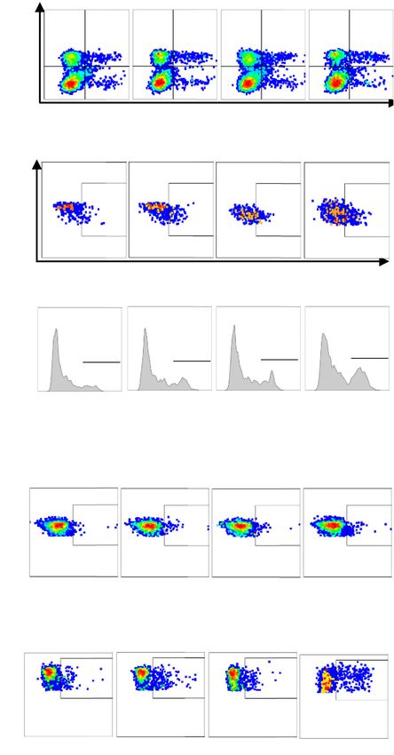
Basher et al. Journal of Hematology & Oncology   (2020) 13:74                                                  Page 4 of 16

Immunoperoxidase Kit and DAB. All slides were coun-             Statistics
terstained with hematoxylin (Vector) and mounted with           All statistical data were expressed as mean ± SEM. Dif-
Permount (Fisher Scientific).                                   ference between means of populations was compared by
                                                                standard Student’s t test using one-way ANOVA. Sur-
                                                                vival was determined via Kaplan-Meier analysis with a
RNAseq and data analyses
                                                                comparison of curves using the Mantel-Haenszel log-
Single-cell suspension from the spleens from SCID mice
                                                                rank test. A P value of 0.05 or less was considered sig-
was prepared as described [20]. After removal of adher-
                                                                nificant. GraphPad Prism software was used for all
ent cells for 2 h in complete media, splenocytes were
                                                                analyses.
cultured in media containing 1000 U/ml IL-2 for 3 days.
NK cells were negatively selected with EasySep™ mouse
NK isolation kits (StemCell Technologies). A 99% purity
                                                                Results
was obtained. Purified NK cells were cultured with puri-
                                                                Targeting sMIC in combination with PD1 blockade
fied recombinant sMIC(B)-His (Sino Biologicals) or
                                                                cooperatively increased survival in mice bearing sMIC-
sMIC(B)-Fc (R&D) for 12 h, with or without the pres-
                                                                positive melanoma tumors
ence of the anti-sMIC mAb B10G5. Total RNA was pre-
                                                                Melanoma patients who have high levels of circulating
pared with RNeasy kit (Qiagen). RNAseq library were
                                                                sMIC responded poorly to PD1/PDL1 blockade therapy
constructed with Illumina TruSeq Stranded mRNA Li-
                                                                [4, 5]. To test whether our animal model recapitulates
brary Prep Kit. Twenty to 32 million of RNAseq reads
                                                                the biology in melanoma patients, we utilized a trans-
were obtained for each sample using single-end 50 bp se-
                                                                plantable B16F10-sMICB syngeneic tumor model as pre-
quencing. Trim Galore (https://www.bioinformatics.bab-
                                                                viously described [19, 20]. Briefly, since mice do not
raham.ac.uk/projects/trim_galore/) was used to validate
                                                                express homologs of the human ligand MICA/B, we uti-
the quality of the reads and to remove ones with low
                                                                lized MICB/B6 male transgenic mice as a syngeneic host
quality by default parameters. STAR program was used
                                                                to prevent potential unwanted immunogenicity against
to align the reads against the mouse reference genome
                                                                human sMICB overexpressed by mouse tumors (Fig. 1a).
mm10 with the transcriptome annotation GTF file from
                                                                The MICB/B6 transgenic mice have an inducible MICB
ENSEMBL (GRCm38.82) via the default parameters [21].
                                                                expression under the control of the male hormone-
FeatureCounts was used to calculate gene expression,
                                                                sensitive promoter probasin and tolerant to sMICB-
represented as transcript per million (TPM) values [22].
                                                                expressing tumors [19]. As shown in Fig. 1b, c, anti-
Only genes showing TPM value greater than 4 in at least
                                                                PDL1 antibody treatment significantly inhibited B16F10
one sample were included in the downstream differential
                                                                tumor growth but had nominal impact on the growth of
gene expression analyses. DAVID online tool (http://da-
                                                                B16F10-sMICB tumors. These data suggest that the
vid.abcc.ncifcrf.gov/) was used for the gene enrichment
                                                                B16F10-sMICB syngeneic tumor model recapitulates the
analysis. Genes showing consistent differential expres-
                                                                biology of sMIC in melanoma patients in the context of
sion (> 2 fold in two biological replicates) upon sMIC
                                                                response to PD1/PDL1 blockade therapy.
treatments as compared to the control sample were se-
                                                                   To test the hypothesis that targeting the soluble
lected for the Gene Ontology (GO) analysis and for the
                                                                NKG2D ligand MIC enhances therapeutic efficacy of
heatmap.
                                                                PD1 blockade, we randomized a cohort of B16F10-
                                                                sMICB tumor-bearing mice into four therapeutic
Quantitative RT-PCR                                             groups (n = 8–10 per group) with control IgGs (cIgG)
Total RNA was prepared as described above. Comple-              in vehicle PBS, single-agent sMICB-targeting mAb
mentary DNA (cDNA) was synthesized using the Super-             B10G5 or anti-PDL1 mAb, or an antibody cocktail of
Script II kit (Invitrogen). A volume of 1 μl of cDNA was        B10G5 and anti-PDL1 mAb. Consistent with our pre-
mixed with Power SYBR Green qPCR SuperMix (Bio-                 vious observation [18], monotherapy with mAb
Rad, USA), and specific primer sets were added to a final       B10G5 significantly inhibited tumor growth and ex-
concentration of 400 nM in 20 μl of reaction mixture.           tended survival as compared to control or anti-PDL1
The reaction was performed on a Bio-Rad CFX96 Touch             monotherapy (Fig. 1d, e). Remarkably, the combin-
Real-Time PCR Detection System. Data were analyzed              ation of anti-PDL1 and B10G5 resulted in a further
using CFX Maestro Software (BioRad). Each sample was            significant inhibition of tumor growth and improved
assayed in triplicates. Target mRNA levels were normal-         survival as compared to monotherapy of B10G5, sug-
ized against mouse GAPDH. Gene expression level in              gesting a cooperative therapeutic effect of targeting
control NK cells was used as a reference for calculating        sMIC and PD1/PDL1 blockade. These results were
expression fold changes. The primers used are listed in         further corroborated in a syngeneic RM9-sMICB
Supplement Table S1.                                            prostate tumor model (Supplement Figure S1).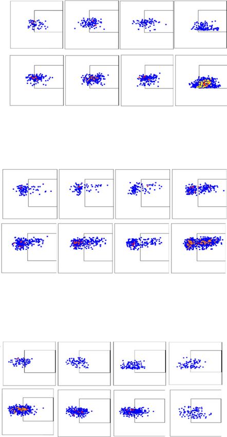
Basher et al. Journal of Hematology & Oncology      (2020) 13:74                                                                  Page 5 of 16

 Fig. 1 sMIC compromises tumor response to anti-PDL1 mAb therapy and that antibody targeting sMIC generates cooperative therapeutic effect
 with anti-PDL1 mAb. a Depict of the therapy. B16F10 or sMICB-expressing B16F10-sMICB cells (4 × 10⁵ cells/mouse) were s.c. injected into
 syngeneic MICB/B6 host. Therapy initiated when tumors reached a volume of 75–100 mm3. Antibody (3 mg/kg) was given i.p. twice weekly till
 designated study endpoint as specified in the “Materials and methods” section. b, c Therapeutic response of B16F10 (b) and B16F10-sMICB (c)
 tumors to anti-PDL1 therapy. Data showed a compromised response of B16F10-sMICB tumors to anti-PDL1 therapy. d B16F10-sMICB tumor
 growth curve in response to sMIC-targeting antibodyB10G5 and anti-PDL1 antibody single-agent therapy and combination therapy. e Kaplan-
 Meier survival curve showing that B10G5 and anti-PDL1 combination therapy significantly prolonged survival of mice bearing B16F10-sMICB
 tumors. Note that tumor volume of 1800 mm3 was designated as survival endpoint. N = 5–7 per group, *p < 0.05; **p < 0.01
Basher et al. Journal of Hematology & Oncology   (2020) 13:74                                                  Page 6 of 16

Combination therapy remodels tumor microenvironment                One of the significant immune-suppressive effects of
to suppress tumor growth                                        soluble NKG2D ligands is systemic downregulation of
We assessed treatment on tumor proliferation by immu-           NKG2D expression on CD8 T cells to diminish the co-
nohistochemistry (IHC) staining of tumor sections with          stimulatory pathway of CD8 T cells [26]. B10G5 single-
ki67. While therapy with anti-PDL1 mAb did not signifi-         agent therapy significantly increased the percentage of
cantly inhibit tumor proliferation, treatment with the          NKG2D+ CD8 T cells in the spleen. Although anti-PDL1
sMIC-targeting mAb B10G5 evidently reduced tumor                mAb single agent therapy did not result in a significant
proliferation as compared to control cIgG treatment             impact on NKG2D expression on CD8 T cells, the com-
(Fig. 2a). The combination therapy of anti-PDL1 mAb             bination of B10G5 and anti-PDL1 mAb significantly in-
and B10G5 resulted in a remarkable inhibition of tumor          creased the percentage of NKG2D+ CD8 T cells
cell proliferation as compared to therapy with single-          compared to B10G5 therapy (Fig. 3c, d). NKG2D is con-
agent or cIgG (Fig. 2a).                                        stitutively expressed by all human CD8 T cells; however,
  Anti-PDL1 antibody single-agent therapy did not sig-          it is only expressed by activated mouse CD8 T cells [27].
nificantly impact intra-tumoral lymphocyte infiltration         Hence, these observations indicate an increased activa-
although a trend of increased density of CD8 T cells was        tion of CD8 T cells in response to therapies. Consist-
observed (Fig. 2a, b). B10G5 single-agent noticeably in-        ently, effector memory CD8 T cells represented by
creased the density of CD8 T and NK cells in tumors.            CD44+ CD8 T cells were significantly enriched with the
Therapy with the combination of B10G5 and anti-PDL1             combination therapy (Fig. 3e, f).
mAb remarkably enriched CD8 and NK cells in tumors                 We further investigated antigen-specific CD8 T cell re-
as compared single-agent therapy (Fig. 2a, b). In some          sponses with monotherapy and combination therapy.
cases, tumors were encased by NK and CD8 T cells (Fig.          Melanoma antigen gp100-specific CD8 T cell popula-
2a). Arginase I+ cells in tumor infiltrates are considered      tions, as represented by H-2Db-restricted gp100-
a hallmark for tumor-promoting macrophages and                  tetramer+ population, were significantly enriched in tu-
myeloid-derived suppressor cells [23–25]. Control IgG-          mors with B10G5, but not with anti-PDL1, single-agent
treated tumors were infiltrated with a high density of ar-      therapy (Fig. 3g, h). Combination therapy of anti-PDL1
ginase I+ cells (Fig. 2a). Single-agent therapy with            and B10G5 significantly enriched gp100-tetramer+ CD8
B10G5, but not anti-PDL1 mAb, reduced the infiltration          T cells as compared to B10G5 therapy single-agent ther-
of arginase I+ cells at a significant level (Fig. 2a). Argi-    apy (Fig. 3g, h). Moreover, the gp100-tetramer+ CD8 T
nase I+ cells were rarely found in the tumors of mice           cells was significantly more responsive to ex vivo gp100
that received combination therapy (Fig. 2a). Consistently,      peptide re-stimulation with B10G5 therapy than control
the number of MDSCs, the major producer of arginase I,          IgG or anti-PDL1 therapy, as measured by intracellular
was significantly reduced in tumors with combination            IFNγ staining (Fig. 3i, j). Combination therapy of B10G5
therapy as compared to single-agent therapy (Fig. 2b). In       and anti-PDL1 further enhanced the responsiveness of
accordance with this observation, tumor vasculature as          gp100-tetramer+ CD8 T cells to gp100 peptide re-
represented by CD31 IHC staining was evidently the              stimulation (Fig. 3i, j). Together, these data demonstrate
combination therapy (Fig. 2a). These data demonstrate           that antibody targeting sMIC in combination with PD-1/
that the anti-PDL1 mAb and B10G5 act cooperatively to           PDL1 blockade cooperatively increase CD8 T cell intrin-
modulate tumor microenvironment to be more active               sic ability to respond to stimulation and augments
immune-primed for anti-tumor responses.                         antigen-specific CD8 T cells in the tumors.

Combination therapy cooperatively augmented tumor               Co-targeting sMIC during anti-PDL1 therapy increases
CD8 T cell intrinsic and melanoma anti-specific responses       CD25 expression on NK cells and augments NK cell
To understand the underlying mechanisms of the com-             homeostatic maintenance
bined therapeutic effect, we first evaluated how each ther-     Soluble NKG2D ligand sMIC negatively affects the
apy modulated the anti-tumor potential of CD8 T cells.          maintenance of peripheral and tumor-infiltrating NK
We first assessed the impact of therapy on intrinsic CD8        cells and downregulates NKG2D surface expression [19].
T cell functional potential by IFNγ production in response      Targeting sMIC with mAb B10G5 has been shown to
to ex vivo re-stimulation with PMA/ionomycin. Single-           overcome the disruption and to restore NK cell homeo-
agent therapy with B10G5 significantly increased the total      static renewal ability [18]. Given that PD1 blockade ther-
number of splenic IFNγ+ CD8 T cells, whereas anti-PDL1          apy was shown to restore NK cell anti-tumor activity
therapy alone did not present such a significant impact         [28], we thus sought to determine the impact of the
(Fig. 3a, b). Combined therapy resulted in further signifi-     combination therapy on NK cells. As shown in Fig. 4a,
cantly increased responsiveness of splenic CD8 T cells as       combination therapy significantly increased NK cell
compared to each monotherapy (Fig. 3a, b).                      numbers in the spleen and dLN in addition to the
Basher et al. Journal of Hematology & Oncology   (2020) 13:74   Page 7 of 16

 Fig. 2 (See legend on next page.)
Basher et al. Journal of Hematology & Oncology      (2020) 13:74                                                                    Page 8 of 16

 (See figure on previous page.)
 Fig. 2 Combined therapy of B10G5 targeting sMIC and anti-PDL1 results in reduced tumor proliferation and a more immune primed tumor
 microenvironment with decreased neovascularization. a Representative micrographs of immunohistochemistry staining (IHC) of subcutaneous
 B16-sMICB tumors demonstrating that combined therapy resulted in reduced tumor cell proliferation as shown by Ki67 staining, increased NK
 and CD8 T cell, and decreased arginase 1+ cells in tumors. Combined therapy also decreased neovascularization within the tumor shown by
 CD31 staining. b Quantitation of NK cell, CD8 T cell, and the major arginase I producer MDSC in representative tumors by flow cytometry
 analyses. Data obtained at day 8 following treatment initiation in an experiment designed to understand therapeutic mechanisms as detailed in
 the text. *p < 0.05 as compared to the control group. **p < 0.05, combination therapy as compared to monotherapy

significant increase in tumors as presented in Fig. 1 sug-                homeostatic maintenance [32]. We sought the under-
gesting an enhanced NK cell homeostatic maintenance.                      lying mechanism associated with the effect of sMIC sup-
NKG2D expression was significantly restored or in-                        pressing NK cell homeostatic maintenance and how sMIC
creased with B10G5 therapy, although only a trend of in-                  targeting can rescue NK cell maintenance. We cultured
crease with anti-PDL1 therapy. Combination therapy                        purified mouse NK cells in the presence of sMIC and
significantly restored surface NKG2D expression as                        sMIC plus anti-sMIC(B10G5) for 12 h and performed
compared to single agent therapies, although no signifi-                  RNAseq and Gene Ontology (GO) analyses focusing on
cant additive or cooperative effect (Fig. 4b, c). NK cell                 the survival and proliferation pathways. As shown in Fig.
intrinsic ability in response to ex vivo stimulation, such                5, exposure to recombinant sMIC downregulated cluster
as PMA/I, was significantly augmented by B10G5 ther-                      of genes positively regulating cell proliferation and survival
apy and further by combination therapy (Fig. 4d, e).                      and upregulated genes that are pro-apoptotic and inhibi-
  Interestingly, combination therapy induced more NK                      tors of cell cycle (Fig. 5a, b). Clearance of sMIC with the
cells to express the IL-2Rα, CD25 (Fig. 4f, g), which                     mAb B10G5 reversed the effect of sMIC (Fig. 5a, b). Not-
most likely couples with the common γ-chain family re-                    ably, expression of IL-2Rα and downstream signaling mol-
ceptors (IL-2/IL-15Rβ and γc/IL-2Rγ) to form the high                     ecule, such as Jak2, were also significantly upregulated
affinity IL-2 receptor to enable NK cells to utilize low-                 with the B10G5 in the presence of sMIC (Fig. 5c). The
dose IL-2 sourced from activated T cells for survival and                 regulation of representative genes in NK cells by sMIC
proliferate [29, 30]. To support this hypothesis, we mea-                 and the ability to be rescued by B10G5 targeting sMIC
sured serum levels of IL-2 before therapy and at day 8 of                 were further confirmed by semi-quantitative RT-PCR (Fig.
therapy. While only neglectable levels of IL-2 were de-                   5d). Collectively, these data support a potential significant
tected in the serum of all animals before therapy, a sig-                 mechanism whereby targeting sMIC by B10G5 antibody
nificant amount of IL-2 was detected in the serum of                      enhances NK cell peripheral maintenance and thus the re-
animals receiving therapy of B10G5 or B10G5 in com-                       sponse to PD1/PDL1 blockade therapy.
bination with anti-PDL1 (Supplement Figure S2). These
data suggest that B10G5 co-targeting sMIC likely re-                      Combination therapy of B10G5 and anti-PDL1 effectively
stores NK homeostatic maintenance and thus NK cell-                       eliminates lung metastasis
mediated anti-tumor immunity through regulating the                       With the proof-of-concept that combination therapy of
availability of IL-2 and sensitivity of IL-2 signaling on                 anti-PDL1 and targeting sMIC significantly inhibits pri-
NK cells, as compared to anti-PDL1 single agent                           mary tumor growth as compared to single-agent therapy,
therapy.                                                                  we further explored the combined therapeutic effect
  We further addressed the significance of NK cells and                   using the experimental melanoma metastasis model. We
CD8 T cells in mediating the cooperative therapeutic ef-                  implanted B16-sMICB tumor cells via lateral tail vein in-
fect of targeting sMIC and anti-PDL1 by depleting NK                      jection into syngeneic male MICB transgenic mice. At
or CD8 T cells during therapy. As presented in Supple-                    day 10 post-inoculation when pulmonary metastases
ment Figure S3, therapeutic effect was significantly com-                 were evident through random necropsy of three ani-
prised by depletion of NK or CD8 T cells, suggesting                      mals, we randomized the remaining animals into four
that both NK and CD8 T cells are required to achieve                      therapeutic groups as described above with cIgG,
the cooperative benefit of the combination therapy.                       B10G5, anti-PDL1 monotherapy, or combination ther-
                                                                          apy (Fig. 6a). Nearly all animals in the control IgG
sMIC and sMIC/B10G5 complex differentially reprogram                      group succumbed to pulmonary metastases at day 21
NK cell survival and proliferation                                        post-tumor inoculation, which was designated as the
One of the significant immune-suppressive effects of                      study endpoint. Histology examination of the lung sec-
tumor-derived sMIC is impaired NK cell function and                       tions revealed that large areas of the lung from animals
homeostatic maintenance [31]. B10G5 targeting sMIC                        of the cIgG treatment group were tumors (Fig. 6b).
has been shown to restore and enhance NK cell                             Treatment       with     anti-PDL1       reduced    lung
Basher et al. Journal of Hematology & Oncology (2020) 13:74 Page 9 of 16 Fig. 3 Combined therapy with B10G5 and anti-PDL1 increases intrinsic functional potential and response to antigen-specific stimulations. B16F10- sMICB cells (4 × 10⁵ cells/mouse) were s.c. injected into syngeneic MICB/B6 host. When tumors reached a volume of 75–100 mm3, animals received i.p injection of 3 mg/kg of respective antibody every 3 days. After three injections (day 9 of therapy), animals were euthanized. Tissues were harvested for therapy-associated mechanistic studies. a, b Combined therapy significantly increases IFNγ-producing splenic CD8 T cells as assessed by ex vivo PMA/ionomycin stimulation. c, d Combined therapy significantly increased the number of splenic NKG2D+ CD8 T cells. e, f Combined therapy significantly increased the population of CD44+CD8 T cells. Data obtained at day 14 following treatment initiation. g, i Representative flow cytometry dot-plots demonstrating that combined therapy significantly increases gp100-tetramer+ CD8 T cell and increased IFNγ expression with gp100 peptide stimulation. Single-cell suspension of splenocytes was stained with melanoma antigen gp100-specific tetramer or evaluated for CD8 T cell IFNγ expression after stimulation with gp100 peptide overnight. h, j Summary data from the experiments presented in g and i respectively. *p < 0.05 as compared to the control group. **p < 0.05, combination therapy as compared to monotherapy
Basher et al. Journal of Hematology & Oncology       (2020) 13:74                                                                  Page 10 of 16

 Fig. 4 Combined therapy cooperatively restores NK cell homeostatic function and increase IL-2Rα expression on NK cells. Data associated
 experimental details are described in Fig. 3 legend. a Total number of NK cells harvested from spleen and two inguinal draining lymph nodes per
 mouse. b, c Monotherapy with B10G5 and/or anti-PDL1 increases intensity of NKG2D expression on NK cells in the draining LN of mice bearing
 B16-sMICB tumors, while combined therapy further increases NKG2D expression. d, e Combined therapy increases IFNγ production after ex vivo
 PMA/ionomycin stimulation of NK cells from the draining LN of mice bearing B16-sMICB tumors. f, g Combined therapy significantly increases
 numbers of NK cells in tumor-draining lymph nodes expressing CD25. *p < 0.05 as compared to the control group. **p < 0.05, combination
 therapy as compared to monotherapy

micrometastasis although did not reach a statistical sig-                   NK cells are known to be of significance in con-
nificance; treatment with B10G5 remarkably reduced                        trolling tumor metastasis [5, 33, 34]. As shown in
the micrometastasis (Fig. 6b, c). Remarkably, microme-                    Fig. 6d–f, total number of NK cells, CD25+ NK cells,
tastases were rarely found in lungs from animals that                     and NK cell intrinsic responsiveness to PMA/I
received the combination therapy of anti-PDL1 mAb                         stimulation as presented by intracellular IFNγ stain-
and B10G5 (Fig. 6b, c).                                                   ing in the draining LN was significantly increased
Basher et al. Journal of Hematology & Oncology       (2020) 13:74                                                                   Page 11 of 16

 Fig. 5 sMIC stimulation impairs the expression of genes related to NK cell homeostatic maintenance. Negatively selected mouse splenic NK cells
 were stimulated with recombinant MICB in the absence or presence of sMIC-clearing mAb B10G5 for 6 h. Total RNA was isolated for RNAseq
 analyses. R1 and R2 are replicates with two different forms of recombinant sMICB, sMICB-His tagged and sMIC-Fc tagged respectively. a Gene
 related to survival pathway that are significantly modulated in NK cells by sMIC and rescued with the sMIC-clearing mAb B10G5. b Gene related
 to proliferation pathway that is significantly modulated in NK cells by sMIC and rescued with the sMIC-clearing mAb B10G5. c Modification of
 genes in IL-2 signaling pathways by sMIC. d Validation of changes in the expression of representative genes as shown in a–c by qRT-PCR
Basher et al. Journal of Hematology & Oncology         (2020) 13:74                                                                       Page 12 of 16

 Fig. 6 Combined therapy with B10G5 and anti-PDL1 significantly decreases established B16-sMIC+ metastases. a Depiction of treatment scheme.
 B6/MICB mice received i.v. injection of 4 × 10⁵ B16F10-sMICB cells. At day 10, i.p. treatment was initiated with (1) control mIgG, (2) B10G5, (3) anti-
 PDL1, and (4) a combination of B10G5 and anti-PDL1. Antibodies were given by i.p. injection every 3 days in 200 μl sterile PBS. b Representative
 H&E sections of lungs of mice with metastatic B16-sMICB tumors. c Quantitation of micrometastasis in one lung section of all lobes. e, f Enhanced
 NK cell numbers, CD25 expression, and IFNγ production in draining LNs (two inguinal) of animals receiving combined treatment. Data obtained
 at day 21 of the experiment. *p < 0.05 as compared to the control group. Arrows show micrometastasis. **p < 0.05, combination therapy as
 compared to monotherapy

in response to B10G5 therapy and further                                      with significantly enhanced NK cell immunity, at-
significantly increased with combination therapy.                             tributing to enhanced peripheral NK cell homeo-
These data demonstrate that the reduction of lung                             static maintenance and NK cell intrinsic functional
metastasis with combination therapy is associated                             potential.
Basher et al. Journal of Hematology & Oncology    (2020) 13:74                                                               Page 13 of 16

Prevalence of MIC/sMIC in metastatic melanoma lesions                  Discussion
We evaluated the prevalence of MIC and sMIC in                         With syngeneic subcutaneous and metastatic tumor
metastatic melanoma lesions using commercial                           models, we demonstrate in this study significantly en-
source tissue microarrays (TMA), which composed                        hanced and cooperative therapeutic effects against mel-
of various stages of malignant melanoma tumors                         anoma with the combination of the sMIC-clearing
with different origins, lymph node (LN) metastatic                     antibody B105 and PDL1/PD1 pathway blockade. We
lesions, benign tumors, and normal skin tissues                        have shown that combination therapy with B10G5 and
(Supplement Figure S4a). Immunohistochemistry                          anti-PDL1 resulted in significantly more inhibition of
staining with a MIC-specific monoclonal antibody                       primary tumor growth and higher degree of clearance of
D4H3 demonstrated a high frequency of MIC/sMIC                         lung metastases than respective monotherapy. We dem-
presence in melanoma tumors, with the highest fre-                     onstrated that the enhanced therapeutic effect with com-
quency in LN metastatic lesions (Fig. 7 and Supple-                    bination therapy is associated with augmented NK and
ment Figure S4b). Amongst the 21 LN metastatic                         CD8 T cell activation and anti-tumor potential. We also
cases on the TMA, two cases have no evident tu-                        demonstrated that combination therapy cooperatively
mors, 17 of 19 cases had tumors with strong MIC/                       reduces expression of immunosuppressive arginase I in
sMIC reactivity. These data re-enforce the transla-                    the tumor microenvironment and inhibits tumor angio-
tion potential of the antibody B10G5 in targeting                      genesis. Interestingly, we demonstrate that expression of
sMIC/MIC for metastatic melanoma patients.                             the receptor to enable NK cell to sensitize low dose IL-

 Fig. 7 Representative MIC/sMIC abundance in metastatic melanoma tumors. The presence of MIC/sMIC was detected on TMA with a MIC (A/B)-
 specific antibody D4H3. Left panel, low magnification of TMA staining images. Right, higher magnification of TMA staining images
Basher et al. Journal of Hematology & Oncology   (2020) 13:74                                                 Page 14 of 16

2, the IL-2Ra, on NK cells was upregulated with B10G5           previously shown that clearing sMIC with B10G5, a non-
therapy and further synergistically upregulated with the        blocking anti-sMIC antibody, can restore NK cell
combination therapy. With RNAseq analyses, we dem-              homeostatic maintenance and function [18], alleviate the
onstrated that clearance of sMIC with B10G5 rescues             immune-suppressive tumor microenvironment by elim-
the impairment of NK cell survival and proliferation            inating arginase I+ MDSCs and tumor-associated macro-
pathway caused by sMIC. Finally, we demonstrate the             phages [17], stabilize CD3ζ expression on CD8 T cells
abundance or prevalence of MIC/sMIC in metastatic               [28], and enhance CD28-NKG2D dual co-stimulation to
melanoma tumors. Given recent clinical findings of an           antigen-specific CD8 T cells [38]. These profound thera-
association between high levels of circulating NKG2D            peutic effects elicited by clearing sMIC could potentiate
ligand sMIC and reduced response to PD1 blockade                the CD8 T cell response, in particular the tumor
therapy in melanoma patients [4], our current study of-         antigen-specific CD8 T cell response, and thus coopera-
fers a viable new combination therapy to improve the re-        tively enhance the response to PDL1/PD1 blockade ther-
sponse of melanoma patients to PD1/PDL1 inhibitors.             apy. Of note, the B16-sMICB tumor cell line in this
   Our study demonstrates that targeting sMIC increases         study is PDL1+; thus, the cooperative therapeutic effect
the IL-2 sensing receptor IL-2Rα on NK cells in vivo            of B10G5 and anti-PDL1 demonstrated is sound. Inter-
and that sMIC/antibody complex reprograms NK cell               estingly, patients bearing tumors that are initially PDL1
for homeostatic survival in vitro, although targeting           negative still clinically responded to PD-1 blockade ther-
sMIC enhances IL-2Ra expression on NK cells warrants            apy [39–43]. Given that we have previously shown that
further investigations. The increased IL-2Ra expression         clearing sMIC with B10G5 can induce the release of
may be important for the cross-talk of NK cells and the         IFNγ [18, 28, 44], a significant regulator of PDL1 expres-
adaptive immune response in the response to PD1/PDL1            sion, one might speculate the combination of B10G5
blockade therapy. Clinical correlative studies have re-         and PD1 blockade may also be effective for tumors ini-
vealed the significance of NK cell in association with re-      tially lacking PDL1 expression. Indeed, we show that
sponse to PD1/PDL1 blockade therapy. In metastatic              clearance of sMIC rescues NK cell survival and function,
melanoma patients, the expression of CD25 on NK cells           presumably in part attributing to enhanced sensitivity to
has been associated with clinical response to anti-PD1          IL-2 released by activated antigen-specific CD8 T cells.
therapy [35], presumably in part due to increased NK            Other studies have also presented that blocking sMIC
cell sensitivity to IL-2 and thus increased survival and        release in preclinical models enhances NK cell function
function. A higher density or frequency of peritumoral          [45]. Considering that NK cells are the major IFNγ pro-
NK cells was found to be associated with response to            ducers in active immune responses, the interaction or
anti-PD1 therapy in metastatic melanoma patients [36,           inter-dependence of NK cells and CD8 T cells through
37]. A concurrent upregulation of NK cell activity re-          effector cytokine, such as IFNγ and IL-2, may account
lated genes and MHC I in tumors that responded to               for one of the mechanisms mediating the synergistic ef-
PD1/PDL1 blockade therapy in melanoma patients [36].            fect of the combination therapy. Based on published
A clustering of NK cells and stimulatory dendritic cells        studies and our current data, we propose an innate-
(DCs) was in tumors of melanoma patients who                    adaptive cross-talk model that confers the cooperative
responded to anti-PD1 therapy and had prolonged sur-            therapeutic effect of targeting sMIC and PD1/PDL1
vival [37]. In preclinical melanoma models, it was found        blockade (Supplement Figure S5). How NKG2D signal-
that NK cells, not T cells, are required for sustaining         ing and blocking PD1 signaling synergistically enhance
stimulatory DC in the tumors [37]. Together, these stud-        NK cell peripheral maintenance and function as we have
ies underscore the significance of NK cells in mediating        shown warrants a further investigation.
tumor response to PD1/PDL1 blockade therapy.                       Checkpoint inhibitors, particularly PD1 pathway
   Monotherapy with B10G5 to clear sMIC was effica-             blockade, have been approved by the FDA for a number
cious in controlling tumor growth and eliminating lung          of indications, including advanced melanoma, head and
metastasis. However, combination therapy presented a            neck cancer, renal cell carcinoma, non-small cell lung
cooperative and significantly enhanced effect. sMIC has         cancer (NSCLC), urothelial carcinoma, and metastatic
been to be highly immune-suppressive via perturbing             Merkel-cell carcinoma, due to enhanced survival benefits
NK cell peripheral maintenance and function [19],               compared to traditional chemotherapy. However,
impairing TCR/CD3 signaling by caspase-dependent                complete clinical responses are still limited to a small
destabilization of CD3ζ [28], and facilitating the expan-       percentage of patients. The anti-PD1 antibody nivolu-
sion of MDSCs and tumor-associated macrophages [17].            mab only demonstrated survival benefit in advanced
These immune-suppressive effects can directly and in-           melanoma patients without the BRAF V600 mutation,
directly impair CD8 T cell activation, and thus negatively      with a 72.9% survival rate at 1 year and increase in me-
impact the response to PD1 blockade therapy. We have            dian progression-free survival by nearly 3 months as a
Basher et al. Journal of Hematology & Oncology             (2020) 13:74                                                                            Page 15 of 16

second-line therapy in patients that progressed after re-                       Funding
ceiving ipilimumab and/or BRAF inhibitor therapy [46].                          This work was supported by NIH/NCI grant 1R01CA208246 and
                                                                                1R01CA204021 (to J. D. Wu). The funding was used to support all study
Overall, PD1/PDL1 blockade only elicited cumulative re-                         design, data analyses, and manuscript constructions.
sponse rates of 31% in patients with melanoma, 19% in pa-
tients with NSCLC, 25% in patients with renal cell                              Availability of data and materials
                                                                                All data generated or analyzed during this study are included in this
carcinoma (RCC), and 13.3% in patients with head and                            published article [and its supplementary information files].
neck cancers [47–50]. PD1/PDL1 therapy currently is also
in phase III clinical trials for the indications of BRAF                        Ethics approval and consent to participate
V600-mutated melanoma [51], RCC [18, 30], head and                              All animal studies were approved by the Institutional Animal Care and Use
                                                                                Committee (IACUC) of the Medical University of South Carolina and the
neck squamous cell carcinoma (HNSCC) [31, 52], naso-                            IACUC committee of Northwestern University. Human TMAs were purchased
pharyngeal cancer [53], esophageal carcinoma [54], meso-                        from the commercial vendor, US BIOMAX.
thelioma [55], hepatocellular carcinoma [48], breast
                                                                                Consent for publication
cancer [20], and multiple myeloma [4]. Elevated levels of                       Not applicable.
sMIC have been reported in almost all of these indications
in association with reduced anti-tumor immunity or poor                         Competing interests
                                                                                J. Wu is the inventor of the B10G5 antibody and has international patent
disease prognosis via common immune-suppressive path-                           applications. All other authors declare no conflict of interest.
ways [19, 56–59]. Our current study further accentuates
the potential of enhancing the efficacy of PD1/PDL1 ther-                       Author details
                                                                                1
                                                                                 Department of Microbiology and Immunology, Medical University of South
apy in these malignant indications by targeting sMIC.                           Carolina, Charleston, SC 29425, USA. 2Department of Urology, Feinberg
                                                                                School of Medicine, Northwestern University, Chicago, IL 60611, USA. 3Driskill
Conclusion                                                                      Graduate Program in Life Science, Feinberg School of Medicine,
                                                                                Northwestern University, Chicago, IL 60611, USA. 4Department of
Clinical studies in melanoma patients demonstrated that                         Pharmacology, Feinberg School of Medicine, Northwestern University,
the presence of soluble NKG2D ligand sMIC negatively                            Chicago, IL 60611, USA. 5Department of Neurological Surgery, Feinberg
impact the outcome of immune checkpoint blockade                                School of Medicine, Northwestern University, Chicago, IL 60611, USA.
                                                                                6
                                                                                 Department of Microbiology and Immunology, Feinberg School of
therapy. Our findings provide a pre-clinical proof-of-                          Medicine, Northwestern University, Chicago, IL 60611, USA. 7Division of
concept with novel mechanisms and translational rele-                           Hematology and Oncology, Feinberg School of Medicine, Northwestern
vance for a new avenue of antibody targeting sMIC to                            University, Chicago, IL 60611, USA. 8Department of Biomedical Engineering,
                                                                                McCormick School of Engineering, Northwestern University, Evanston, IL
enhance PD1/PDL1 immune checkpoint blockade ther-                               60628, USA. 9Current address: Department of Medicine, Miller School of
apy for metastatic melanoma patients.                                           Medicine, University of Miami, Miami, FL, USA.

                                                                                Received: 2 March 2020 Accepted: 8 May 2020
Supplementary information
Supplementary information accompanies this paper at https://doi.org/10.
1186/s13045-020-00896-0.                                                        References
                                                                                1. Topalian SL, et al. Safety, activity, and immune correlates of anti-PD-1
 Additional file 1. Supplement Material and Methods. Table S1, Figures S1-S5.       antibody in cancer. N Engl J Med. 2012;366:2443–54.
                                                                                2. Topalian SL, Taube JM, Anders RA, Pardoll DM. Mechanism-driven
                                                                                    biomarkers to guide immune checkpoint blockade in cancer therapy. Nat
Abbreviations                                                                       Rev Cancer. 2016;16:275–87.
MIC: MHC I chain-related molecule; PD-1: Programmed cell death protein 1;       3. Luther C, Swami U, Zhang J, Milhem M, Zakharia Y. Advanced stage
IHC: Immunohistochemistry; LN: Lymph node; dLN: Tumor-draining LN;                  melanoma therapies: detailing the present and exploring the future. Crit Rev
NSCLC: Non-small cell lung cancer; TMA: Tissue microarray                           Oncol Hematol. 2019;133:99–111.
                                                                                4. Maccalli C, et al. Soluble NKG2D ligands are biomarkers associated with the
                                                                                    clinical outcome to immune checkpoint blockade therapy of metastatic
Acknowledgements
                                                                                    melanoma patients. Oncoimmunology. 2017;6:e1323618.
We sincerely thank Dr. Ju Wu for his assistance in performing the
                                                                                5. Lopez-Soto A, Gonzalez S, Galluzzi L. Soluble NKG2D ligands limit the
immunohistochemistry staining.
                                                                                    efficacy of immune checkpoint blockade. Oncoimmunology. 2017;6:
                                                                                    e1346766.
Authors’ contributions                                                          6. Raulet DH, Gasser S, Gowen BG, Deng W, Jung H. Regulation of ligands for
Conception and design: Jennifer Wu and Fahmin Basher. Acquisition of data:          the NKG2D activating receptor. Annu Rev Immunol. 2013;31:413–41.
Fahmin Basher, Payal Dhar, and Jennifer Wu. Analysis and interpretation of      7. Gasser S, Raulet DH. The DNA damage response arouses the immune
data: Fahmin Basher, Jennifer Wu, Xin Wang, and Zhe Ji. Writing and review          system. Cancer Res. 2006;66:3959–62.
of manuscript: Fahmin Basher, Jennifer Wu, Derek Wainwright, Bin Zhang,         8. Gasser S, Orsulic S, Brown EJ, Raulet DH. The DNA damage pathway
Jeffrey Sosman, and Zhe Ji. Study Supervision: Jennifer Wu. The author(s)           regulates innate immune system ligands of the NKG2D receptor. Nature.
read and approved the final manuscript.                                             2005;436:1186–90.
                                                                                9. Soriani A, et al. Reactive oxygen species- and DNA damage response-
Authors’ information                                                                dependent NK cell activating ligand upregulation occurs at transcriptional
Jennifer Wu’s laboratory has been studying how tumors edit immune response          levels and requires the transcriptional factor E2F1. J Immunol. 2014;193:950–60.
through the NKG2D ligand MIC/NKG2D pathway for over 15 years, with the          10. Kasahara M, Sutoh Y. Comparative genomics of the NKG2D ligand gene
optimal goal to target this pathway to enhance host endogenous response for         family. Immunol Rev. 2015;267:72–87.
cancer immunotherapy. This current study describes one of the mechanisms of     11. Watson NF, et al. Expression of the stress-related MHC class I chain-related
action by targeting tumor-secreted soluble NKG2D ligand sMIC.                       protein MICA is an indicator of good prognosis in colorectal cancer
Basher et al. Journal of Hematology & Oncology (2020) 13:74 Page 16 of 16
patients. International journal of cancer Journal international du cancer. 2006; 37. Barry KC, et al. A natural killer-dendritic cell axis defines checkpoint therapy-
118:1445–52. responsive tumor microenvironments. Nat Med. 2018;24:1178–91.
12. de Kruijf EM, et al. NKG2D ligand tumor expression and association with 38. Zhang J, et al. Antibody targeting tumor-derived soluble NKG2D ligand
clinical outcome in early breast cancer patients: an observational study. BMC sMIC provides dual co-stimulation of CD8 T cells and enables sMIC(+)
Cancer. 2012;12:24. tumors respond to PD1/PD-L1 blockade therapy. J Immunother Cancer.
13. McGilvray RW, et al. ULBP2 and RAET1E NKG2D ligands are independent 2019;7:223.
predictors of poor prognosis in ovarian cancer patients. International journal 39. Yearley JH, et al. PD-L2 expression in human tumors: relevance to anti-PD-1
of cancer Journal international du cancer. 2010;127:1412–20. therapy in cancer. Clin Cancer Res. 2017;23:3158–67.
14. Madjd Z, et al. Upregulation of MICA on high-grade invasive operable 40. Larkin J, Hodi FS, Wolchok JD. Combined nivolumab and ipilimumab or
breast carcinoma. Cancer Immun. 2007;7:17. monotherapy in untreated melanoma. N Engl J Med. 2015;373:1270–1.
15. Baragano Raneros A, Suarez-Alvarez B, Lopez-Larrea C. Secretory pathways 41. Ribas A, Hu-Lieskovan S. What does PD-L1 positive or negative mean? J Exp
generating immunosuppressive NKG2D ligands: new targets for therapeutic Med. 2016;213:2835–40.
intervention. Oncoimmunology. 2014;3:e28497. 42. Wolchok JD, et al. Nivolumab plus ipilimumab in advanced melanoma. N
16. Hanaoka N, et al. NKG2D initiates caspase-mediated CD3zeta degradation Engl J Med. 2013;369:122–33.
and lymphocyte receptor impairments associated with human cancer and 43. Postow MA, et al. Nivolumab and ipilimumab versus ipilimumab in
autoimmune disease. J Immunol. 2010;185:5732–42. untreated melanoma. N Engl J Med. 2015;372:2006–17.
17. Xiao, G., et al. Soluble NKG2D ligand promotes MDSC expansion and skews 44. Basher F, Jeng EK, Wong H, Wu J. Cooperative therapeutic anti-tumor effect
macrophage to the alternatively activated phenotype. Journal of of IL-15 agonist ALT-803 and co-targeting soluble NKG2D ligand sMIC.
Hematology & Oncology 8(2015). Oncotarget. 2015.
18. Lu S, et al. Nonblocking monoclonal antibody targeting soluble MIC 45. Ferrari de Andrade L, et al. Antibody-mediated inhibition of MICA and MICB
revamps endogenous innate and adaptive antitumor responses and shedding promotes NK cell-driven tumor immunity. Science. 2018;359:1537–
eliminates primary and metastatic tumors. Clin Cancer Res. 2015;21:4819–30. 42.
19. Liu G, et al. Perturbation of NK cell peripheral homeostasis accelerates 46. Robert C, et al. Nivolumab in previously untreated melanoma without BRAF
prostate carcinoma metastasis. J Clin Invest. 2013;123:4410–22. mutation. N Engl J Med. 2015;372:320–30.
20. Basher F, Jeng EK, Wong H, Wu J. Cooperative therapeutic anti-tumor effect 47. Topalian SL, et al. Survival, durable tumor remission, and long-term safety in
of IL-15 agonist ALT-803 and co-targeting soluble NKG2D ligand sMIC. patients with advanced melanoma receiving nivolumab. J Clin Oncol. 2014;
Oncotarget. 2016;7:814–30. 32:1020–30.
21. Dobin A, et al. STAR: ultrafast universal RNA-seq aligner. Bioinformatics. 2013; 48. Borghaei H, et al. Nivolumab versus docetaxel in advanced nonsquamous
29:15–21. non-small-cell lung cancer. N Engl J Med. 2015;373:1627–39.
22. Liao Y. Smyth, G.K. & Shi, W. featureCounts: an efficient general purpose 49. Motzer RJ, et al. Nivolumab versus everolimus in advanced renal-cell
program for assigning sequence reads to genomic features. Bioinformatics. carcinoma. N Engl J Med. 2015;373:1803–13.
2014;30:923–30. 50. Ferris RL, et al. Nivolumab for recurrent squamous-cell carcinoma of the
23. Gabrilovich DI, Nagaraj S. Myeloid-derived-suppressor cells as regulators of head and neck. N Engl J Med. 2016;375:1856–67.
the immune system. Nat Rev Immunol. 2009;9:162–74. 51. Lundholm M, et al. Prostate tumor-derived exosomes down-regulate
NKG2D expression on natural killer cells and CD8+ T cells: mechanism of
24. Patil MD, Bhaumik J, Babykutty S, Banerjee UC, Fukumura D. Arginine
immune evasion. PLoS One. 2014;9:e108925.
dependence of tumor cells: targeting a chink in cancer’s armor. Oncogene.
52. Salih HR, Holdenrieder S, Steinle A. Soluble NKG2D ligands: prevalence,
2016;35:4957–72.
release, and functional impact. Front Biosci. 2008;13:3448–56.
25. Mondanelli G, Ugel S, Grohmann U, Bronte V. The immune regulation in
53. Ahmad I, Sansom OJ, Leung HY. The role of murine models of prostate
cancer by the amino acid metabolizing enzymes ARG and IDO. Curr Opin
cancer in drug target discovery and validation. Expert Opin Drug Discovery.
Pharmacol. 2017;35:30–9.
2009;4:879–88.
26. Groh V, Wu J, Yee C, Spies T. Tumour-derived soluble MIC ligands impair
54. Zheng X, et al. Clonal deletion of simian virus 40 large T antigen-specific T
expression of NKG2D and T-cell activation. Nature. 2002;419:734–8.
cells in the transgenic adenocarcinoma of mouse prostate mice: an
27. Raulet DH. Roles of the NKG2D immunoreceptor and its ligands. Nat Rev
important role for clonal deletion in shaping the repertoire of T cells
Immunol. 2003;3:781–90.
specific for antigens overexpressed in solid tumors. J Immunol. 2002;169:
28. Zhang J, et al. Antibody-mediated neutralization of soluble MIC significantly
4761–9.
enhances CTLA4 blockade therapy. Sci Adv. 2017;3:e1602133.
55. Holmes MA, Li P, Petersdorf EW, Strong RK. Structural studies of allelic
29. Lee SH, Fragoso MF, Biron CA. Cutting edge: a novel mechanism bridging diversity of the MHC class I homolog MIC-B, a stress-inducible ligand for the
innate and adaptive immunity: IL-12 induction of CD25 to form high-affinity activating immunoreceptor NKG2D. J Immunol. 2002;169:1395–400.
IL-2 receptors on NK cells. J Immunol. 2012;189:2712–6. 56. Holdenrieder S, et al. Soluble MICA in malignant diseases. International
30. Clausen J, et al. Functional significance of the activation-associated journal of cancer Journal international du cancer. 2006;118:684–7.
receptors CD25 and CD69 on human NK-cells and NK-like T-cells. 57. Holdenrieder S, et al. Soluble MICB in malignant diseases: analysis of
Immunobiology. 2003;207:85–93. diagnostic significance and correlation with soluble MICA. Cancer
31. Alspach E, Lussier DM, Schreiber RD. Interferon gamma and its important immunology, immunotherapy : CII. 2006;55:1584–9.
roles in promoting and inhibiting spontaneous and therapeutic cancer 58. Wu JD, et al. Prevalent expression of the immunostimulatory MHC class I
immunity. Cold Spring Harb Perspect Biol. 2018. chain-related molecule is counteracted by shedding in prostate cancer. J
32. Lu S, et al. Non-blocking monoclonal antibody targeting soluble MIC Clin Invest. 2004;114:560–8.
revamps endogenous innate and adaptive anti-tumor responses and 59. Jinushi M, et al. MHC class I chain-related protein A antibodies and
eliminates primary and metastatic tumors. Clin Cancer Res. 2015. shedding are associated with the progression of multiple myeloma. Proc
33. Takeda K, et al. IFN-gamma production by lung NK cells is critical for the Natl Acad Sci U S A. 2008;105:1285–90.
natural resistance to pulmonary metastasis of B16 melanoma in mice. J
Leukoc Biol. 2011;90:777–85.
34. Grundy MA, Zhang T, Sentman CL. NK cells rapidly remove B16F10 tumor Publisher’s Note
cells in a perforin and interferon-gamma independent manner in vivo. Springer Nature remains neutral with regard to jurisdictional claims in
Cancer immunology, immunotherapy : CII. 2007;56:1153–61. published maps and institutional affiliations.
35. Kasanen, H., et al. Age-associated changes in the immune system may
influence the response to anti-PD1 therapy in metastatic melanoma
patients. Cancer immunology, immunotherapy : CII (2020).
36. Lee H, et al. Integrated molecular and immunophenotypic analysis of NK
cells in anti-PD-1 treated metastatic melanoma patients. Oncoimmunology.
2019;8:e1537581.You can also read