INTEGRIN ACTIVATION IS AN ESSENTIAL COMPONENT OF SARS COV 2 INFECTION
←
→
Page content transcription
If your browser does not render page correctly, please read the page content below
www.nature.com/scientificreports
OPEN Integrin activation is an essential
component of SARS‑CoV‑2
infection
Peter Simons1, Derek A. Rinaldi1, Virginie Bondu2, Alison M. Kell2,4, Steven Bradfute3,4,
Diane S. Lidke1,5 & Tione Buranda1,4*
SARS-CoV-2 infection depends on binding its spike (S) protein to angiotensin-converting enzyme 2
(ACE2). The S protein expresses an RGD motif, suggesting that integrins may be co-receptors. Here,
we UV-inactivated SARS-CoV-2 and fluorescently labeled the envelope membrane with octadecyl
rhodamine B (R18) to explore the role of integrin activation in mediating cell entry and productive
infection. We used flow cytometry and confocal microscopy to show that SARS-CoV-2R18 particles
engage basal-state integrins. Furthermore, we demonstrate that Mn2+, which induces integrin
extension, enhances cell entry of SARS-CoV-2R18. We also show that one class of integrin antagonist,
which binds to the αI MIDAS site and stabilizes the inactive, closed conformation, selectively inhibits
the engagement of SARS-CoV-2R18 with basal state integrins, but is ineffective against Mn2+-activated
integrins. RGD-integrin antagonists inhibited SARS-CoV-2R18 binding regardless of integrin activation
status. Integrins transmit signals bidirectionally: ’inside-out’ signaling primes the ligand-binding
function of integrins via a talin-dependent mechanism, and ’outside-in’ signaling occurs downstream
of integrin binding to macromolecular ligands. Outside-in signaling is mediated by Gα13. Using
cell-permeable peptide inhibitors of talin and Gα13 binding to the cytoplasmic tail of an integrin’s β
subunit, we demonstrate that talin-mediated signaling is essential for productive infection.
Severe acute respiratory syndrome coronavirus 2 (SARS-CoV-2) is a novel virus in the Betacoronavirus genus
that causes coronavirus disease 2019 (COVID-19)1. SARS-CoV-2 was first reported in Wuhan, China, and cur-
andemic2,3. SARS-CoV-2 presents similar characteristics with the original SARS-CoV
rently persists as a global p
in genome structure, tissue tropism, and viral pathogenesis. However, SARS-CoV-2 is more transmissible than
SARS-CoV.
Cellular entry of coronaviruses depends on binding of the viral spike (S) protein to a specific cellular receptor,
the angiotensin-converting enzyme 2 (ACE2)4,5, and subsequent S protein priming by cellular protease activity
such as Transmembrane Serine Protease 2 (TMPRSS2)6. Interestingly, ACE2 expression across different human
tissues7 revealed low expression of ACE2 in the lungs compared to elevated expression in the kidney and heart8,9.
Nevertheless, studies have shown that type I and II interferons (IFNs) secreted during viral infection upregulate
the transcription and expression of ACE210,11. Unlike its predecessor, SARS-Cov-2 expresses a novel K403R spike
protein substitution encoding an Arginine-Glycine-Aspartic acid (RGD) m otif12, introducing the potential for
interacting with RGD-binding integrins, as likely mediators for viral cell entry and enhanced p athogenicity13.
ACE2 contains two integrin-binding domains: an RGD motif at position 204–206 and the sequence RKKKNKAR
in the cytoplasmic tail at its C-terminus14. Also, ACE2 binds integrin β1 in the failing human heart14. Correlated
increased expressions of β115 and ACE2 have been reported16,17. Others have shown that ACE2 interacts in cis
with integrin β1 in a manner that enhances RGD-mediated cell adhesion18.
Integrins are heterodimeric transmembrane adhesion protein receptors composed of α and β subunits
whose activation is tightly regulated and b idirectional19. Integrins can exist in three states characterized by their
structural conformation and affinity for their ligands (Fig. 1A). The inactive, bent-closed state (BCS) with a
closed headpiece has a low affinity for extracellular matrix (ECM) ligands. The bent structure inhibits the recep-
tors from inappropriate signaling due to random binding to extracellular matrix proteins. Integrins exhibit an
1
Department of Pathology, University of New Mexico School of Medicine, Albuquerque, NM 87131,
USA. 2Molecular Genetics and Microbiology, University of New Mexico School of Medicine, Albuquerque,
NM 87131, USA. 3Department of Internal Medicine, University of New Mexico School of Medicine, Albuquerque,
NM 87131, USA. 4Center for Infectious Diseases and Immunity, University of New Mexico School of Medicine,
Albuquerque, NM 87131, USA. 5Comprehensive Cancer Center, University of New Mexico Health Sciences Center,
Albuquerque, NM 87131, USA. *email: tburanda@salud.unm.edu
Scientific Reports | (2021) 11:20398 | https://doi.org/10.1038/s41598-021-99893-7 1
Vol.:(0123456789)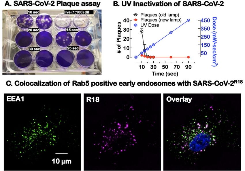
www.nature.com/scientificreports/
Figure 1. Integrin conformational states antagonist targets and SARS-CoV-2 binding. (A) Integrin States:
First, the inactive, bent-closed state (BCS), with a closed headpiece and low affinity for extracellular matrix
(ECM) ligands. The bent structure inhibits the receptors from inappropriate signaling due to random binding to
extracellular matrix proteins. In the BCS form, binding to large ligands is likely limited. Second, when primed,
integrins exhibit an extended-closed state (ECS) with a closed headpiece and higher ligand binding affinity than
BCS. Third, active and extended-open state (EOS) with an open headpiece and maximum affinity for ECM
ligands. Integrin Affinity Regulation: Mn2+ binding to the MIDAS site at the αI and βI domain integrin induces
integrin extension. α2β1 integrin antagonist BTT 3033 binds to the α-I domain, and stabilizes the BCS. GLP0187
blocks binding to the RGD ligand-binding domain. EOS binding to a macromolecular ligand or ECM generates
a force (F) transmitted through the integrin β subunit. (B) Model of Sars-CoV-2 virion structure (https://www.
scientificamerican.com/interactive/inside-the-coronavirus/). SARS-CoV-2 are spherical or ovoid particles of
sizes that span the range of 60–140 nm. The SARS-CoV-2 virion consists of a lipid bilayer envelope membrane
covering a large nucleoprotein (N)-encapsidated, positive-sense RNA genome. The lipid envelope is decorated
with three transmembrane proteins consisting of trimeric spike proteins (S) that project above the lipid bilayer
membrane and relatively small membrane (M) and envelope (E) p roteins78,79. S proteins bind with high-
affinity (1–50 nM) to the angiotensin-converting enzyme 2 (ACE2) for productive infection80. (C) Cartoon
4
alignment of the receptor-binding domain (RBD) and RGD sequence on the trimeric spike protein, which
favors engagement of activated integrin, adapted from ref.25 The illustrations were generated using M icrosoft®
PowerPoint Version 16.51 (21071101).
extended-closed state (ECS) with a closed headpiece and higher ligand binding affinity than BCS when primed.
Active and extended-open state (EOS) presents an open headpiece and maximum affinity for ECM ligands20.
Integrin function involves coordination with cytoskeletal components whose functions regulate cell adhesion
and migration21,22. Changes in integrin conformation can elicit cell-signaling events that increase ligand affin-
ity/avidity, promote cytoskeletal rearrangement, and enable virus internalization. Ligand binding to integrins
is mediated by divalent-cations bound at the Metal Ion Dependent Adhesion Site (MIDAS) domain on top of
either the αI domain, in I domain-containing integrins, or the βI domain in non-αI integrins23. Physiologically,
1 mM Ca2+ and 1 mM Mg2+ in body fluid stabilize the BCS conformation. Under non-physiological conditions,
1 mM Mn2+ initiates and stabilizes ECS conformation even in the presence of C a2+.
Many viruses use integrin-mediated endocytosis pathways for cell entry5,24. A recent bioinformatics-driven
study predicted a model that placed integrins in a central ligating role, whereby SARS-CoV-2 could engage mul-
tiple receptors and form a multicomponent receptor complex and functional signaling p latform25. Interestingly,
25
ACE2 also has a similar MIDAS m otif . Still, it has not yet been established whether the ACE2 MIDAS domain
has a potential role in creating synergy overlap between the ligand-binding profiles and regulation of ACE2 and
integrins25. Several in vitro studies have established experimental evidence in support of cognate binding interac-
tions between SARS-CoV-2 spike proteins, integrin β126,27 and integrin β312,28. In addition, the transmembrane
glycoprotein neuropilin 1 (NRP1), which is abundantly expressed in the olfactory epithelium and promotes the
endocytosis of activated α5β1 integrin29–34, has been recently identified as a receptor for SARS-CoV-2 i nfection34,35.
In this study, we took a mechanistic approach to examine the role of integrins as effectors of SARS-CoV-2
cell entry and productive infection. First, we tested whether inducing a BCS to ECS integrin conformational
change with Mn2+24,36 enhanced cell binding and entry of fluorescently tagged UV-inactivated SARS-CoV-2R18.
Conversely, we used integrin extension or RGD-binding inhibitors to determine the inhibitors’ effect on cellular
entry. Integrins signal bidirectionally via "inside-out” and “outside-in" signaling22,36–41. Inside-out signaling is
initiated by intracellular signaling upstream of talin, and other adaptor proteins binding to the integrin β-subunit
cytoplasmic tail (β-CT), which causes integrin extension (ECS) and concomitant increases in high-affinity ligand
binding21,22. Integrin engagement with macromolecular ligands stimulates the transient exchange of talin for
Gα13’s occupancy of the β-CT42,43 which initiates integrin outside-in signaling. In the context of viral infection,
integrin outside-in signaling induces cell spreading, retraction, and internalization of integrin-associated ligands.
We used cell-permeable inhibitors of integrin outside-in and inside-out signaling42 to test the role of canonical
Scientific Reports | (2021) 11:20398 | https://doi.org/10.1038/s41598-021-99893-7 2
Vol:.(1234567890)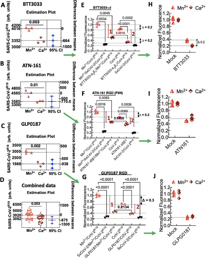
www.nature.com/scientificreports/
Figure 2. Characterization of UV-inactivated virus for Sars-Cov-2 studies. (A) Duplicate plaque assays of
supernatants of Sars-CoV-2 exposed to increasing doses of 254 nm radiation and then tested for viability. The
live virus completely lysed the cells at 1:100 dilution relative to UV exposed virions. (B) Graph shows UV dose–
response, leading to a significant decrease in plaque-forming units at different doses. For our experiments, a
90 s (450 mW s/cm2) UV dose was used to inactivate the virus before removal from the BSL-3 laboratory. (C)
Confocal microscopy imaging of cells after incubation with SARS-CoV-2R18 (magenta) for 15 min, then fixed
and labeled for early endosome marker, early endosome antigen 1 or EEA1 (green), an effector protein for Rab5,
and nuclei (Hoechst 33,258, blue). SARS-CoV-2R18 vesicles are trafficked to the perinuclear region, and a subset
is co-localized with EEA1. Images are maximum projections and have been brightness and contrast-enhanced.
integrin signaling during cell entry of SARS-CoV-2R18 and infectious SARS-CoV-2. Taken together, our results
demonstrate that integrins play a significant role in the infectivity of SARS-CoV-2.
Results
Integrin extension promotes SARS‑CoV‑2R18 cell entry. To facilitate studies of SARS-CoV-2 host-
cell entry outside of BSL-3 containment, we generated UV-inactivated virus particles. Under our experimental
conditions, a minimum UV dose of 100 mW s/cm2 was sufficient to completely inactivate 1 07 virions/ml distrib-
uted in 500 µl samples of a twelve well plate (Fig. 2A,B). UV-inactivated virus samples were fluorescently labeled
with a lipophilic lipid probe, octadecyl rhodamine B (R18), intercalating the envelope m embrane44. Labeled
samples were purified and characterized as we have described previously for the Sin Nombre v irus45.
To assess the ability of SARS-CoV-2R18 to enter cells, we used confocal microscopy to image the relative distri-
bution of SARS-CoV-2R18 and EEA1, an effector protein for Rab5 positive early endosomes. Adherent cells were
incubated on a coverslip with SARS-CoV-2R18 for 15 min, washed, fixed, and immunolabeled for EEA1 (Fig. 2C).
Internalized SARS-CoV-2R18 was frequently found in EEA1 positive early endosomes and perinuclear space,
demonstrating that the SARS-CoV-2R18 internalizes and traffics as one might expect29–34. Together, these results
show that UV-inactivated SARS-CoV-2R18 is a valuable probe for investigating SARS-CoV-2 entry mechanisms.
We hypothesized that activating integrins by M n2+24,36, which induces integrin extension and higher ligand
affinity, would provide a favorable spatial orientation of the RGD-binding motifs to facilitate SARS-CoV-2R18
binding (Fig. 1A). Therefore, we measured initial rates (< 10 min binding time) of binding in activated cells
(Mn2+/Ca2+) relative to resting (1 mM C a2+ only). SNV-CoV-2R18 bound to the M n2+ activated samples at 3 times
the rate of untreated cells (not shown). However, at equilibrium (> 20 min incubation), the cell occupancy of
SARS-CoV-2R18 was only ~ 20% higher in Mn2+-treated samples compared to untreated samples. As expected,
the gap increased when the virus was used as a limiting r eagent46. We used estimation p lots47, to assess the
precision of the results from multiple experiments of the integrin function assay that measured the relative
binding of SARS-CoV-2R18 to Mn2+-activated cells relative to quiescent cells on different days (Fig. 3A–D). The
mean difference between the binding to Mn2+-activated and resting cells was conserved across different samples
when we used at least 5000 SARS-CoV-2R18/cell. However, when we used a lower stoichiometric ratio, e.g., 3000
SARS-CoV-2R18/cell, the gap between the site occupancy of Mn2+ activated and resting cells increased to 30%,
as discussed below.
To further investigate the role of integrins in SARS-CoV-2R18 entry into Vero E6 cells, we used high binding
affinity integrin antagonists: (1) BTT 3033, a selective antagonist (EC50 = 130 nM) of integrin α2β1 that binds to
Scientific Reports | (2021) 11:20398 | https://doi.org/10.1038/s41598-021-99893-7 3
Vol.:(0123456789)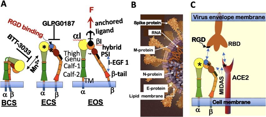
www.nature.com/scientificreports/
Figure 3. Flow cytometry assays of binding inhibition of SARS-CoV-2R18 show specificity of αI allosteric antagonist BTT
3033, RGD fibronectin synergy domain (F9N) ATN-161, and broad-spectrum RGD antagonist, GLPG0187. Vero E6
suspension cells in 40 µl volumes (1000 cells/µl) were first incubated with 10 µM integrin antagonists or 5× unlabeled Sars-
CoV-2 (CoV-2) in ± Mn2+ media for 20 min at 37 °C. Sars-CoV-2R18 was then added to the tubes and incubated for another
20 min. The samples were centrifuged and resuspended in 95 µl HHB buffer and analyzed on a flow cytometer. (A–D) Raw
data panels of SARS-CoV-2R18 binding to M n2+ activated and resting cells. Each data symbol in the left panels shows triplicate
measurements of a single functional test experiment measuring the effect of 1 mM M n2+ on equilibrium binding of SARS-
CoV-2R18 to Vero E6 cells. The right panels show differences between the means of M n2+-replete samples and M n2+-free
samples, and the left panels show 95% confidence intervals (CI). Green arrows are used to link the analysis results of the
same experiment. Combined data show negative controls of multiple experiments (n = 6) testing different inhibitors. As
demonstrated for GLP, samples treated with the virus as a limiting reagent yielded lower signals for M n2+ and C a2+ as shown
for GLP). (E) Effect of BTT 3033 on SARS-CoV-2R18 binding to cells. Red vertical bars 1 and 2 denote the difference between
mock- and inhibitor-treated samples for M n2+-replete and M n2+-free samples. (top) ∆ = 0.2, refers to the relative difference
in fluorescence intensity due to SARS-CoV-2R18 in Mn2+-replete samples and M n2+ free samples; (bottom) ∆ = 0.2, refers
to the fractional difference inhibition of SARS-CoV-2R18 binding by BTT 3033 in M n2+-replete cells and Mn-free samples,
indicating that BTT 3033 is a selective inhibitor of quiescent integrins. (F) Effect of ATN-160 on SARS-CoV-2R18 binding to
cells indicating that ATN-160 is agnostic of integrin activation status. (G) Effect of GLP0187 on SARS-CoV-2R18 binding to
cells. The gap between M n2+ activated cells, and quiescent is shown to be higher ∆ = 0.3 when the virus was used as a limiting
reagent < 5000 SARS-CoV-2R18 /cell as indicated by a lower signal in the raw data (C,D). (H–J) Specific binding of SARS-CoV-
2R18 to cells determined by subtraction of non-specific binding represented by 5xCov-2 data points. The data are normalized to
mock-treated cells (no inhibitors). Data are representative of at least 3 separate measurements for each inhibitor. The p-values
between samples are indicated on the horizontal bar. The figure was produced in GraphPad Prism version 9.2.0.
Scientific Reports | (2021) 11:20398 | https://doi.org/10.1038/s41598-021-99893-7 4
Vol:.(1234567890)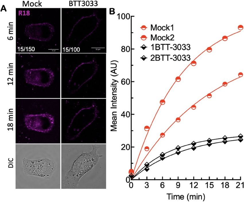
www.nature.com/scientificreports/
Figure 4. Stabilization of bent closed conformation with aI MIDAS domain binding integrin antagonist
inhibits intracellular trafficking of SARS-CoV-2R18. (A) Live cell imaging of SARS-CoV-2R18 (magenta) binding
and endocytosis shows perinuclear localization of SARS-CoV-2R18 vesicles. At the same time, the virus is seen
to accumulate at the plasma membrane when α2β1 integrins are inhibited by 10 µM BTT 3033. Fluorescence
images represent maximum projections of five confocal z slices. Mock and BTT 3033 treated samples are shown
with different lookup tables (LUT) since binding in treated cells was lower than untreated cells. LUT lower/
upper values are presented in the lower-left corner of 6-min timepoint images. Scale bars, 10 µm. (B) Traces
of absolute intensity values of virus binding over time. Two representative cells for each condition are plotted
from data acquired on the same day to compare intensity values directly. Data were fit to a non-linear regression
function with arbitrary constants for appearance purposes. Imaging data were repeated at least three separate
times.
a site close to the α2I MIDAS domain and stabilizes the integrin bent conformation state (BCS)48, (2) ATN-161,
a non-RGD peptide49 derived from the synergy region of fibronectin50, known to exhibit specific antagonism
for α5β1 and αIIbβ3 and also recently shown to inhibit SARS-CoV-2 infectivity27, and (3) GLPG0187, a high-
affinity, broad-spectrum ( EC50 < 10 nM) integrin receptor antagonist of RGD integrins α5β1, αvβ3, αvβ5, αvβ1,
αvβ651. We used a titrated, fivefold excess of unlabeled SARS-CoV-2 relative to fluorescent SARS-CoV-2R18 as a
control for competitive inhibition of SARS-CoV-2R18 binding. Paired samples of cell suspensions in Mn2+-replete
and Mn2+-free media were treated with the above integrin antagonists. Total viral binding was normalized to
Mn2+-treated samples for each experimental condition. The graphs show that M n2+ treatment increased SARS-
CoV-2R18 occupancy of cells by ~ 20% compared to M n2+-free conditions (Fig. 3E,F). As noted above, using
significantly fewer SARS-CoV-2R18 than 5000 increases the gap between binding to the M n2+-activated and
resting as indicated for the GLPG0187 sample (Fig. 3G). We also show in a subsequent experiment that rais-
ing SARS-CoV-2R18 to a stoichiometric excess limited the site occupancy gap to 20% (Supplemental Figure 1).
The positive control for inhibition (5xCov-2 in data graphs) blocked 80% of SARS-CoV-2R18 and equally
inhibited Mn2+ -treated and untreated samples. Reasoning that the residual signal of 5× Cov-2 treated samples
was due to non-specific binding to the cell membrane, we subtracted the fluorescence of cells blocked with
5xCov-2 and then normalized the data to mock-treated cells. Finally, we compared the relative efficacy of the
inhibitors in Mn2+-replete and -free conditions of the normalized data (Fig. 3H–J). The fraction of Mn2+-activated
integrins (20%) were refractory to BTT 3033 treatment (Fig. 3H). BTT 3033 selectively binds to the BCS integrin
structure48 and does not bind to M n2+ activated integrins. In contrast, ATN-161 and GLPG0187 were agnostic to
2+
Mn treated cells, as the same baseline was achieved for either condition (Fig. 3I,J). Overall, GLPG0187 (Fig. 3I)
appeared to be a better competitive inhibitor of SARS-CoV-2R18 compared to ATN-161 (Fig. 3J). The difference
for the latter was potentially due to ATN-161’s overall specificity for integrin α5β1. Thus, the expression level of
α5β1, in Vero E6 cells, relative to other integrins with which α5β1 would compete for SARS-CoV-2R18 engagement
governed its apparent efficacy. Also, ATN-161 is known to exhibit U-shaped dose–response characteristics49 thus,
presenting a need to identify an optimally active-dose by titration of ATN-16127 which is beyond the scope of
our present study. The mechanistic specificity of integrin inhibition by these antagonists regarding SARS-CoV-2
uptake strongly supports the idea that (1) integrin RGD engagement is an essential co-factor for cell entry and
(2) integrin extension is required for cell entry based on BTT 3033’s mechanism of action.
Inhibition of integrin activation or binding to SARS‑CoV‑2R18 blocks intracellular traffick‑
ing. We then used live-cell confocal microscopy to visualize Vero E6 cell entry and trafficking of SARS-CoV-
2R18 in DMSO (mock)- and BTT 3033-treated cells (Fig. 4A, B). Most of the cells treated with GLPG0187 were
de-adhered from the plate and were thus not suitable for imaging. The loss of cells with GLPG0187 was likely due
to the loss of integrin-mediated adhesion by the broad-spectrum inhibitor. Cells were imaged at 3-min intervals
for 21 min after the addition of ~ 107 SARS-CoV-2R18 particles. In DMSO treated cells (Mock in Fig. 4), SARS-
Scientific Reports | (2021) 11:20398 | https://doi.org/10.1038/s41598-021-99893-7 5
Vol.:(0123456789)www.nature.com/scientificreports/
Figure 5. Inhibition of integrin activation blocks cell entry of SARS-CoV-2R18, suggesting integrin-mediated ▸
signaling is required for productive infection. (A) Aligned sequences of β1 and β3-integrin cytoplasmic tails
(β-CT). The NPxY motif tyrosine residues (shown in brown) and the Ser and Thr residues (shown in purple)
are important phosphorylation sites required for exchanging adaptor proteins. Srk family kinase-mediated
phosphorylation of the NPxY motifs inhibits the binding of talin while promoting the association of inhibitor
proteins such as DOK-1. Interaction zones between β-CT and adaptor proteins are denoted by associated
horizontal lines. Functional roles of the proteins are indicated in parenthesis. For a detailed description, see
refs.41,54. The membrane-permeable peptides mP6 and mP13 were based on the integrin β3 cytoplasmic tail.
(B) Model of outside-inside-out signaling for integrin-mediated cell entry. Hypothetical SARS-CoV-2 binding
to integrin β1 initiates Gα13 binding to the β1 cytoplasmic tail, which stimulates outside-in signaling in the
absence of a known receptor-stimulated GPCR mediated inside-out signaling. mP6 is a specific inhibitor of
Gα13 binding to the β1 cytoplasmic tail. The illustrations were generated using Microsoft® PowerPoint Version
16.51 (21071101). (C) Relative fluorescence readings of suspension Vero E6 cells after 30 min incubation with
SARS-CoV-2R18 in vehicle- and 100 µM mP6 treated cells. (D) Live cell imaging of SARS-CoV-2R18 (magenta)
binding and endocytosis shows cell membrane and perinuclear localization of SARS-CoV-2R18 vesicles. At the
same time, the virus is seen to remain at the plasma membrane in cells treated with 50 µM mP6. LUT ranges are
shown in the bottom left corner of 6-min timepoint images. Scale bars, 10 µm. (E) Traces of absolute intensity
values of virus binding over time. Two representative cells for each condition are plotted from data acquired
on the same day to compare intensity values directly. For comparison, mock-treated cell data are the same as
in Fig. 4. Data were fit to a non-linear regression function with arbitrary constants for appearance purposes.
(F) Inhibition of SARS-CoV-2 productive infection. Suspension Vero E6 cells were preincubated with 250 µM
mP6 and 250 µM mP13 for 30 min and followed by infection with 0.01 MOI SARS-CoV-2 for an additional
60 min incubation. Cells were washed twice, transferred to a 12 well plate for 48 h, and assayed for viral RNA by
RT-qPCR. Comparisons were performed using Ordinary one-way ANOVA, with Tukey’s multiple comparison
test using GraphPad Prism version 9.2.0.
CoV-2R18 particles were visible at cell membranes within 3 min, subsequently developed punctate features at the
cell periphery, and trafficked to the perinuclear space. The rate of cell entry (time to perinuclear space ~ 10 min)
was comparable to infectious virions52. For the BTT 3033-treated cells, early peripheral membrane localization
of SARS-CoV-2R18 showed significant diminution of discernable puncta. It did not undergo retrograde traffic
towards the perinuclear region within the timeframe of the experiment. The relative amount of virus binding to
the surface was also reduced with BTT 3033 treatment (Fig. 4A,B), consistent with reduced binding observed by
flow cytometry measurements (Fig. 2).
Blocking of integrin signaling significantly inhibits productive infection of cells by
SARS‑CoV‑2. Integrin activation is a complex and well-regulated spatiotemporal process involving the syn-
chronized assembly and disassembly of multiple signaling elements at the integrin’s β-cytoplasmic tail (β-CT)53–
55
. Various groups have described a network of up to 156 interacting components that comprise the integrin
adhesome56–59. Some adhesome components relevant to our study are shown in (Fig. 5A). Most β-CTs contain
conserved sequences needed for integrin activity, such as the two β chain NPxY/F sequences, which are sites of
competitive binding by adaptor proteins that regulate integrin activation and d eactivation41,53, including sort-
60–63
ing signals for clathrin-mediated endocytosis . The phosphorylatable tyrosine (Y) residues of NPxY motifs
are key regulatory sites of integrin activation on the β-CT. For example, N780PIY783 and N792PKY795 in β1 and
N744PLY747 and N756ITY759 in β3 are motifs phosphorylated by Src family kinases (SFK) that may positively or
negatively regulate interactions with phosphotyrosine-binding (PTB) domain-containing proteins. During the
early stage of integrin activation, inhibitory proteins are displaced from the β-CT in exchange for integrin activa-
tors, ending with the recruitment of talin to the integrin tail38. However, the early wave of talin-mediated inside-
out signaling is transiently terminated to allow Gα13, the effector of outside-in signaling, to bind to the conserved
ExE motif (where x denotes any residue for specific integrins, e.g., EEE for β3-CT and EKE for β1-CT Fig. 5A),
which overlaps the talin binding domain42.
Integrin binding to macromolecular ligands, such as SARS-CoV-2, facilitates Gα13-mediated outside-in sign-
aling. Transmission of the tensile force through the integrin to talin stabilizes high-affinity integrin binding (in
the EOS) to the ECM promotes the ’second wave’ of inside-out signaling (Fig. 5B). The sequential mechanism of
inside-out and outside-in signaling was previously established in part by the use of two myristoylated peptides,
mP6 (Myr-FEEERA-OH), derived from the Gα13-binding domain and mP13 (Myr-KFEEERARAKWDT-OH)
mimicking the β3-CT’s talin binding d omain42. It is worth noting that the previous mP6 and mP13 related study
by Shen et al.42 established that the minimal sequence of EEERA does not interact with talin and is a specific
inhibitor of Gα13 association with the β-CT and had no effect on talin-dependent inside-out signaling, or the late
phase of outside-in signaling associated with the second wave of talin binding. However, mP13 affects all phases
of integrin signaling42. To investigate the relationship between the integrin signaling events and SARS-CoV-2
engagement and cell entry, we treated cells with mP6 peptide, which inhibited cell entry of SARS-CoV-2R18 in
flow cytometry and microscopy experiments (Fig. 5C–E). Similarly, mP13 inhibited cell entry in flow cytometry
experiments (data not shown). The results for mP6 treated cells suggest that SARS-CoV-2 engagement initiates
a Gα13-mediated outside-in integrin activation without a known receptor stimulus which is consistent with the
idea that SARS-CoV-2 binding induces integrin activation64, as we previously demonstrated for the Sin Nombre
virus24.
Scientific Reports | (2021) 11:20398 | https://doi.org/10.1038/s41598-021-99893-7 6
Vol:.(1234567890)www.nature.com/scientificreports/
Because mP6 and mP13 are membrane-permeable peptides, they were suitable for infectivity experiments
while obviating the need to expose cells to DMSO for extended periods. We, therefore, tested the efficacy of mP6
and mP13 at inhibiting cell entry and productive infection in Vero E6 cells with a 0.01 multiplicity of infection
(MOI) of SARS-CoV-2. For the productive infection assay, infected cells were plated at confluency (500,000
cells/well in a 12 well plate) to minimize cell growth for 48 h post-infection. We used RT-qPCR to measure viral
nucleocapsid RNA in the suspended cells or intact cell monolayers at 48 h post-infection, respectively. At 48 h
post-infection, inhibition of productive infection by mP13 was significant relative to mock-treated cells, whereas
the effect of mP6 was insignificant (Fig. 5F). The failure of mP6 to inhibit productive infection is consistent with
the notion that viral r eplication52,65 perturbs C
a2+ homeostasis within the infected c ells66 and thus dispenses with
Gα13 activity in favor of talin-induced outside in-signaling42.
Scientific Reports | (2021) 11:20398 | https://doi.org/10.1038/s41598-021-99893-7 7
Vol.:(0123456789)www.nature.com/scientificreports/
Discussion
This study provides mechanistic evidence for the functionality of extracellular ligand-binding domains of inte-
grin β1 and cytoplasmic tails of integrins in general25,28, which offer possible molecular links between ACE2 and
integrins. We show that M n2+, which induces integrin extension and high-affinity ligand binding, enhances the
cell entry of SARS-CoV-2R18. The increased virus binding and entry is consistent with the notion that integrin
affinity and/or extension are essential for cell entry. In support of integrin-dependent endocytosis as a pathway of
SARS-CoV-2R18 internalization, we used broad-spectrum RGD antagonists such as GLPG0187, which inhibited
cell entry regardless of integrin activation status. Our study also suggested integrin specificity. BTT 3033, an
αI allosteric antagonist that binds to the bent closed conformation of integrin β1 and stabilizes it, supports the
possibility of integrin-dependent endocytosis of SARS-CoV-2R18 upon receptor binding. In a different frame-
work, our data also show that SARS-CoV-2R18 can bind to low affinity and presumptively bent-conformation
integrins23, however, in BTT 3033 treated cells, cell entry by SARS-CoV-2R18 is inhibited because integrin activa-
tion post- SARS-CoV-2R18 engagement is prevented. Thus, our data contextualize integrin extension as the "sine
qua non of integrin cell adhesion function,"23 which in turn is an essential condition for integrin-mediated cell
entry by SARS-CoV-2.
Focal adhesion kinase (FAK)67 is a well-established component of the adhesome that potentially bridges
the signaling gap between integrin signaling turnover68 and ACE2. FAK is a tyrosine kinase known to direct
the recruitment of talin to integrin β1-enriched nascent a dhesions60,61. In ailing heart tissues, ACE2 binds inte-
grin α5β1 in an RGD-independent manner. It is known to regulate FAK mediated cell adhesion and integrin
signaling18, which terminates with endosomal t rafficking30 (of virion-bearing integrins). The binding of macro-
molecular RGD ligands to resting integrins elicits ligand-induced integrin a ctivation64. We hypothesize that in our
in vitro experiments, SARS-CoV-2 binding to inactive integrins triggers a series of spatiotemporally-regulated
recruitment of adhesome components, including Gα13 and talin, to the β-CT. Our data show that talin interaction
with integrin β-CTs, which causes integrin extension, is indispensable for productive infection (Fig. 5). Talin
binding to the β-CT generates the requisite inside-out signal that increases the affinity of the integrin ectodomain
for SARS-CoV-2 binding, which in turn increases viral load. Cell entry of SARS-CoV-2 is clathrin-dependent69.
Endocytosis of integrins is clathrin-dependent and -independent70 and involves adaptor proteins such as Dab2
and Numb71,72 attached to the β-CTs NPxY/NxxY motifs (Fig. 5A). Alternatively, some integrin α-subunits harbor
a common endocytosis motif (Yxxϕ) recognized by the clathrin adaptor protein 2 (AP2)68.
Mészáros et al.25 have used bioinformatics to predict the existence of short amino acid sequences (~ 3–10 resi-
dues): short linear motifs (SLiMs), such as NPxY/Nxxy, Yxxϕ in the cytoplasmic tails of ACE2 and integrins that
mediate endocytosis and autophagy. Some of their theoretical predictions have been validated by experimental
studies. First, Kliche et al.28 confirmed the existence of SLiMs. They extended their findings to establish a potential
connection between ACE2 and integrin β3 cytoplasmic tail interactions with scaffolding and adaptor proteins
linked to endocytosis and autophagy. Second, SLiM sequences known to bind and activate the transmembrane
glycoprotein neuropilin 1 (NRP1) were identified as potential mediators of SARS-CoV-2 endocytosis25. Interest-
ingly, NRP1, which is abundantly expressed in the olfactory epithelium, is now declared as an effector for SARS-
CoV-2 infection34,35. NRP1 localizes at adhesion sites and promotes fibronectin-bound, activated α5β1 integrin
endocytosis, and directs the cargo to the perinuclear c ytoplasm29–34. Studies have shown that the endocytosis of
active and inactive integrins to EEA1-containing early endosomes follows distinct mechanisms involving dif-
ferent adaptor proteins. The inactive integrin is promptly recycled back to the plasma membrane via an ARF6‐
and EEA1‐positive compartment in a Rab4 -dependent m anner31. We observed that in BTT 3033-treated cells
replete with inactive β1 integrins, SARS-CoV-2R18 remained membrane-bound, whereas untreated cells displayed
internalization and perinuclear localization of SARS-CoV-2R18. This is consistent with the known trafficking of
ligand-bearing integrins, including those directed by NRP1, to the perinuclear s pace29,30,32.
Our study has some limitations. Integrin activation is often initiated by other receptors such as G-protein cou-
pled receptors (GPCRs), growth factors, and other i ntegrins38. Future studies will explore the effect of receptor-
mediated inside-out signaling, modeled under inflammatory conditions of COVID-19. In addition, the criteria
for selecting specific integrins as co-factors of SARS-CoV-2 infectivity are not known and thus worthy of future
investigation. Finally, the study is based on the USA-WA1/2020 SARS-CoV-2 strain. Our present study lays the
groundwork for examining the activity of the various emergent SARS-CoV-2 variants.
Although several integrins types12,25–28 are believed to be co-receptors of SARS-CoV-2 infectivity, our study
suggests inhibitor specificity for integrin β1. This is consistent with known factors: (1) correlated increased expres-
sions of β115 and ACE2 in relevant tissues16,17, (2) cytoplasmic tail in cis interactions between ACE2 and integrin
β114, and (3) synergy between ACE2 and integrin β1 signaling that promotes RGD mediated cell adhesion18.
To optimize integrin engagement, our cell-binding assays and primary infection assays were carried out in
suspension such that ACE2 and integrins were not segregated by cell p olarization73,74. However, our micros-
copy studies on adherent cells agreed with the flow cytometry results. Thus, our study represents an initial step
toward establishing a mechanistic role for SARS-CoV-2-mediated integrin activation required for cell entry and
productive infection.
Materials and methods
Materials. USA-WA1/2020 SARS-CoV-2 strain was obtained from BEI Resources (NIAID, NIH). Integ-
rin inhibitors, BTT3033, a selective inhibitor of α2β1, ATN-161 an integrin α5β1 antagonist27, and GLPG0187 a
broad-spectrum integrin inhibitor, were purchased as powders from Tocris Bioscience. The EEA1 rabbit mono-
clonal antibody (clone C45B10) was from Cell Signaling Technologies (CAT# 3288S). Alexa fluor 647 conjugated
F(ab’)2 fragment goat anti-rabbit IgG was from Invitrogen (CAT# A21246). In addition, myristoylated peptides;
mP6 (Myr-FEEERA-OH) and mP13 (Myr-KFEEERARAKWDT-OH) were custom synthesized at Vivitide.
Scientific Reports | (2021) 11:20398 | https://doi.org/10.1038/s41598-021-99893-7 8
Vol:.(1234567890)www.nature.com/scientificreports/
Cell culture. African green monkey kidney cells (Vero E6, ATCC) were maintained in DMEM media from
Sigma CAT# D5796. All media contained 10% heat-inactivated fetal bovine serum (FBS), 100 U/ml penicillin,
100 μg/ml streptomycin, and 2 mM l-glutamine and were kept at 37 °C in a CO2 water-jacketed incubator of 5%
CO2 and 95% air (Forma Scientific, Marietta, OH, USA).
UV inactivation and fluorescent labeling of the envelope membrane of SARS‑CoV‑2 with octa‑
decyl rhodamine (R18). USA-WA1/2020 SARS-CoV-2 strain (from BEI Resources, NIAID, NIH) was cul-
tured in Vero E6 cells in a biosafety level 3 (BSL-3) containment under a protocol approved by the University of
New Mexico’s Institutional Biosafety Committee or IBC (Public Health Service registration number C20041018-
0267). First, live SARS-CoV-2 were harvested at peak titers of 107 plaque-forming units/mL (PFU/ml). Next,
SARS-CoV-2 was UV inactivated using 254 nm (≈ 5 mW/cm2) U.V. irradiation of a TS-254R Spectroline UV
Transilluminator (Spectronics Corp., Westbury, NY) following a similar protocol for inactivating pathogenic
orthohantaviruses45,75. Briefly, Vero E6 cells were inoculated with SARS-CoV-2 and maintained at 37 °C for
2–4 days. At 70–75% cell death (due to viral cytopathic effect), the supernatant was harvested and subjected to
light centrifugation (1000 rpm, 10 min) to remove cellular debris. For UV inactivation, supernatants were added
to a 12 well plate at 500 µl aliquot/well. Then UV -irradiated at 3.8 cm above the sample for 0, 10,15, 20, 25, 30,
60, and 90 s and then tested for viability by a 3-day plaque assay as described e lsewhere76,77. The titration of UV
irradiation times was used to establish a minimal UV dose for complete inactivation. After UV treatment, the
500 µl fractions were pooled into 15 mL tubes stored in a − 80 °C freezer pending the results of a plaque assay.
Under our experimental conditions, we established that a minimum UV irradiation interval of 25 s was required
for the complete inactivation of SARS-CoV-2. A 90 s UV dose was approved by the IBC for removal of inacti-
vated SARS-CoV-2 out of the BSL-3 lab after it was established that the virus particles were capable of specific
binding to Vero E6 cells.
Crude UV-inactivated SARS-CoV-2 samples were purified by floating 10 ml of SARS-CoV-2 supernatant on
a density gradient comprising 2 ml volumes of 1.2 g/ml and 1.0 g/ml CsCl in PBS media in 14 × 89-mm Beckman
polyallomer tubes. The samples were centrifuged for 1.5 h at 4 °C using a Beckman SW41Ti rotor at 30,000 pm.
A band was collected at the interface and purified by centrifugation in HHB using 100 kDa cutoff M icrocon®
Centrifugal Filter. The purified SARS-CoV-2 samples were stored in 1.0 ml aliquots at − 80 °C. SARS-CoV-2
particles were fluorescently labeled and calibrated according to the same protocol used for the Sin Nombre virus
(SNV)45. The final volume for each labeled batch preparation was limited to 500 µl. The number of SARS-CoV-
2R18 particles in each sample preparation was estimated from absorption measurements using the following
equation: # of moles of R18 (derived from sample absorbance) × Avogadro’s number (6.02 × 10²³ molecules
mole−1)/estimated average number of R18 molecules per virion (10,000)45. The yield of SARS-CoV-2R18 particles
was typically in the 108/µl range. Batch samples were stored in 20 µl aliquots at − 80 °C.
Flow cytometry binding assays of SARS‑CoV‑2R18 to vero E6 cells. For flow cytometry assays,
cells were cultured in T25 or T75 flasks to 80% confluence. Cells were then treated with 0.25% trypsin and
transferred to minimum essential medium (MEM) media. Cell counts and viability were performed using a Life
Technologies Countess II FL Automated cell counter (Thermofisher Scientific). Test suspension cell samples
were transferred to microfuge tubes in 40 µl-aliquots (1000 cells/µl). SARS-CoV-2R18 was added to tubes at 5000
SARS-CoV-2R18/cell and incubated using a shaker at 500 rpm for 20 min at 37 °C. For blocking assays, cells were
incubated with 5 × unlabeled SARS-CoV-2 or 10 µM integrin inhibitors for 20 min before the addition of SARS-
CoV-2R18. Samples were centrifuged at 3,000 rpm; the pellet was resuspended in HHB buffer (30 mM HEPES,
110 mM NaCl, 10 mM KCl, 1 mM MgCl2⋅6H2O, and 10 mM glucose, pH 7.4) buffer and read on an Accuri flow
cytometer. For kinetic assays, Vero E6 suspension cells in 40 µl volumes (1000 cells/µl) were placed in ± Mn2+
media in duplicate microfuge tubes at 37 °C. Sars-CoV-2R18 was then added (5000 virions/cell) to the tubes and
incubated for 1, 3, 5, 7, 9 min. At each time point, the tubes were quenched in an ice bath, then samples were
centrifuged and resuspended in 95 µl HHB buffer and analyzed on a flow cytometer.
Live cell confocal microscopy. Imaging was performed using a Leica TCS SP8 Laser Scanning Confocal
Microscope with a 63 × water objective and a Bioptechs objective heater to maintain cells at physiological tem-
perature (~ 36–37 °C). Vero E6 cells were plated in eight-well Lab-Tek (Nunc) chambers at a density of 30,000
cells per well 24 h before imaging. Cells were imaged in Tyrode’s buffer (135 mM NaCl, 10 mM KCl, 0.4 mM
MgCl2, 1 mM CaCl2 20 mM glucose, 0.1% BSA, 10 mM HEPES, pH 7.2). For integrin inhibition, cells were
treated with 10 μM BTT3033-α2β1 or 50 μM MP6 in Tyrode’s buffer for 30 min before imaging. ~ 1 × 10⁹ SARS-
CoV-2R18 particles were added per well, and z-stacks (300 nm thickness) were acquired every 3 min for 21 min
to visualize viral cell entry. R18 was excited using 561 nm light, isolated from the white light source. R18 emis-
sion and differential interference contrast (DIC) transmitted light were captured with Leica Hybrid detectors
(HyD) in a spectral window of 571–636 nm (for R18 emission). Analysis of the accumulation of SARS-CoV-2R18
particles in Vero E6 cells was completed using Matlab. Briefly, regions of interest (ROI) were created around the
cell membrane, and the mean SARS-CoV-2R18 intensity was measured within the cell mask at each time point.
Immunofluorescence. Vero E6 cells were plated on 18-mm coverslips overnight in a 6 well plate at a den-
sity of 100,000 cells/well. Cells were exposed to ~ 1 × 10⁹ SARS-CoV-2R18 particles/well for 15 min at 37 °C, in
the presence or absence of 10 μM BTT 3033. Cells were then washed in phosphate-buffered saline (PBS) and
fixed using 4% paraformaldehyde (PFA) in PBS for 15 min at room temperature. Cells were extensively washed
with 10 mM Tris (pH 7.4) and PBS and permeabilized with 0.1% Triton. Cells were labeled with anti-EEA1 pri-
mary antibody and anti-rabbit Alexa Fluor 647 secondary. Nuclei were stained with Hoechst 33258. Cells were
Scientific Reports | (2021) 11:20398 | https://doi.org/10.1038/s41598-021-99893-7 9
Vol.:(0123456789)www.nature.com/scientificreports/
mounted on microscope slides using Prolong Diamond Antifade Mountant (Invitrogen, CAT#P33970). Samples
were imaged using a Leica TCS SP8 Laser Scanning Confocal Microscope with a 63× oil objective.
Infection inhibition. Vero E6 cells grown at 80% confluency were trypsinized and divided into microfuge
tubes aliquots of 1.5 × 10⁶cells in 750 µl media containing 250 µM mP6, 250 µM mP13, 10 µM BTT 3033, DMSO,
and media only. Samples were shaken at 500 rpm for 30 min at 37 °C. After transfer to a BSL-3 laboratory, 0.01
MOI of SARS CoV-2 (lot #P3: 1.2 × 10⁷ pfu) and then incubated for 60 min while shaking. Tubes were spun
down (1000 rpm for 3 min), resuspended in fresh media, and spun down again. The cells were then resuspended
in 300 µl of media and transferred to a 12 well plate in 100 µl aliquots (500,000 virions/well) for triplicate meas-
urements. An additional 400 µl were added to each well for a final volume of 500 µl. The plate was transferred
to an incubator for 48 h to allow the virus to replicate. The cells were then washed with 1xPBS before RNA was
extracted with TRIzol™ (Thermofisher, #15596026) according to the manufacturer’s protocol:
(https://assets.thermofisher.com/TFS-Assets/LSG/manuals/trizol_reagent.pdf).
The RNA was quantified with a Nanodrop and total cellular cDNA transcribed with the Applied Biosystems™
High-Capacity cDNA Reverse Transcription Kit (Fisher Scientific #43-688-14).
RT-qPCR was performed with the TaqMan Fast Advance Master Mix (Fisher Scientific #4444963) and the
following primers ordered from Integrated DNA Technologies: Forward: 5′-CCCTGTGGGTTTTACACTTAA-
3′, Reverse: 5′-ACGATTG TGCATCAGC TGA -3′, and probe: 5′-[FAM] CCGTCTGCGG TATGTG
GAA
AGGTT
ATGG [BHQ1]-3′. The RT-qPCR was performed using an Applied Biosystems QuantStudio 5 instrument.
Statistical analysis. SARS-CoV-2R18 binding was expressed as the mean channel fluorescence (MCF) out-
put of the flow cytometer. For different batch preparations of SARS-CoV-2R18, we first tested functional bind-
ing of SARS-CoV-2R18 to cells by comparing the MCF readings of 1 mM Mn2+- activated cells and resting cells
after correcting for autofluorescence. Then, assuming sampling from a Gaussian distribution, the two groups
were compared using estimation p lots47 with unpaired two-tailed t-tests to check for consistency between batch
preparations of SARS-CoV-2R18. For integrin activation inhibitor tests involving three or more groups, compari-
sons were performed using Ordinary one-way ANOVA with Tukey’s multiple comparison test. Data analysis was
done with GraphPad Prism software version 9.2.0. Statistical significance was defined as p < 0.05.
Received: 23 July 2021; Accepted: 30 September 2021
References
1. WHO https://www.who.int/emergencies/diseases/novel-coronavirus-2019/technical-guidance/naming-the-coronavirus-disea
se-(covid-2019)-and-the-virus-that-causes-it (2020).
2. Zhou, F. et al. Clinical course and risk factors for mortality of adult inpatients with COVID-19 in Wuhan, China: A retrospective
cohort study. Lancet 395, 1054–1062 (2020).
3. Chen, N. et al. Epidemiological and clinical characteristics of 99 cases of 2019 novel coronavirus pneumonia in Wuhan, China: A
descriptive study. Lancet 395, 507–513 (2020).
4. Wrapp, D. et al. Cryo-EM structure of the 2019-nCoV spike in the prefusion conformation. Science 367, 1260–1263 (2020).
5. Stewart, P. L. & Nemerow, G. R. Cell integrins: Commonly used receptors for diverse viral pathogens. Trends Microbiol. 15, 500–507
(2007).
6. Hoffmann, M. et al. SARS-CoV-2 cell entry depends on ACE2 and TMPRSS2 and is blocked by a clinically proven protease inhibi-
tor. Cell 181, 271–280.e278 (2020).
7. Sungnak, W. et al. SARS-CoV-2 entry factors are highly expressed in nasal epithelial cells together with innate immune genes. Nat.
Med. 26, 681–687 (2020).
8. Li, M. Y., Li, L., Zhang, Y. & Wang, X. S. Expression of the SARS-CoV-2 cell receptor gene ACE2 in a wide variety of human tissues.
Infect. Dis. Poverty 9, 45 (2020).
9. Hikmet, F. et al. The protein expression profile of ACE2 in human tissues. Mol. Syst. Biol. 16, e9610 (2020).
10. Harrison, A. G., Lin, T. & Wang, P. Mechanisms of SARS-CoV-2 transmission and pathogenesis. Trends Immunol. 41, 1100–1115
(2020).
11. Ziegler, C. G. K. et al. SARS-CoV-2 receptor ACE2 is an interferon-stimulated gene in human airway epithelial cells and is detected
in specific cell subsets across tissues. Cell 181, 1016–1035.e1019 (2020).
12. Nader, D., Fletcher, N., Curley, G. F. & Kerrigan, S. W. SARS-CoV-2 uses major endothelial integrin alphavbeta3 to cause vascular
dysregulation in-vitro during COVID-19. PLoS ONE 16, e0253347 (2021).
13. Makowski, L., Olson-Sidford, W. & Weisel, J. W. Biological and clinical consequences of integrin binding via a rogue RGD motif
in the SARS CoV-2 spike protein. Viruses 13, 146 (2021).
14. Lin, Q., Keller, R. S., Weaver, B. & Zisman, L. S. Interaction of ACE2 and integrin beta1 in failing human heart. Biochem. Biophys.
Acta. 1689, 175–178 (2004).
15. Sun, M. et al. Temporal response and localization of integrins beta1 and beta3 in the heart after myocardial infarction: Regulation
by cytokines. Circulation 107, 1046–1052 (2003).
16. Krishnamurthy, P., Subramanian, V., Singh, M. & Singh, K. Deficiency of beta1 integrins results in increased myocardial dysfunc-
tion after myocardial infarction. Heart 92, 1309–1315 (2006).
17. Kuba, K., Imai, Y., Ohto-Nakanishi, T. & Penninger, J. M. Trilogy of ACE2: A peptidase in the renin-angiotensin system, a SARS
receptor, and a partner for amino acid transporters. Pharmacol. Ther. 128, 119–128 (2010).
18. Clarke, N. E., Fisher, M. J., Porter, K. E., Lambert, D. W. & Turner, A. J. Angiotensin converting enzyme (ACE) and ACE2 bind
integrins and ACE2 regulates integrin signalling. PLoS ONE 7, e34747 (2012).
19. Luo, B. H. & Springer, T. A. Integrin structures and conformational signaling. Curr. Opin. Cell Biol. 18, 579–586 (2006).
20. Luo, B. H., Carman, C. V. & Springer, T. A. Structural basis of integrin regulation and signaling. Annu. Rev. Immunol. 25, 619–647
(2007).
Scientific Reports | (2021) 11:20398 | https://doi.org/10.1038/s41598-021-99893-7 10
Vol:.(1234567890)www.nature.com/scientificreports/
21. Nordenfelt, P., Elliott, H. L. & Springer, T. A. Coordinated integrin activation by actin-dependent force during T-cell migration.
Nat. Commun. 7, 13119 (2016).
22. Schurpf, T. & Springer, T. A. Regulation of integrin affinity on cell surfaces. EMBO J. 30, 4712–4727 (2011).
23. Moore, T. I., Aaron, J., Chew, T. L. & Springer, T. A. Measuring integrin conformational change on the cell surface with super-
resolution microscopy. Cell Rep. 22, 1903–1912 (2018).
24. Bondu, V. et al. Low-affinity binding in cis to P2Y2R mediates force-dependent integrin activation during hantavirus infection.
Mol. Biol. Cell 28, 2887–2903 (2017).
25. Mészáros, B. et al. Short linear motif candidates in the cell entry system used by SARS-CoV-2 and their potential therapeutic
implications. Sci. Signal. 14, eabd0334 (2021).
26. Park, E. J. et al. The spike glycoprotein of SARS-CoV-2 binds to beta1 integrins expressed on the surface of lung epithelial cells.
Viruses 13, 645 (2021).
27. Beddingfield, B. J. et al. The integrin binding peptide, ATN-161, as a novel therapy for SARS-CoV-2 infection. JACC Basic Transl.
Sci. 6, 1–8 (2021).
28. Kliche, J., Kuss, H., Ali, M. A. & Ivarsson, Y. Cytoplasmic short linear motifs in ACE2 and integrin β3 link SARS-CoV-2 host cell
receptors to mediators of endocytosis and autophagy. Sci. Signal. 14, eabf1117 (2021).
29. Valdembri, D. et al. Neuropilin-1/GIPC1 signaling regulates alpha5beta1 integrin traffic and function in endothelial cells. PLoS
Biol. 7, e25 (2009).
30. Mana, G., Valdembri, D. & Serini, G. Conformationally active integrin endocytosis and traffic: Why, where, when and how?.
Biochem. Soc. Trans. 48, 83–93 (2020).
31. Arjonen, A., Alanko, J., Veltel, S. & Ivaska, J. Distinct recycling of active and inactive beta1 integrins. Traffic 13, 610–625 (2012).
32. De Franceschi, N., Hamidi, H., Alanko, J., Sahgal, P. & Ivaska, J. Integrin traffic—The update. J. Cell Sci. 128, 839–852 (2015).
33. Lobert, V. H. & Stenmark, H. The ESCRT machinery mediates polarization of fibroblasts through regulation of myosin light chain.
J. Cell Sci. 125, 29–36 (2012).
34. Cantuti-Castelvetri, L. et al. Neuropilin-1 facilitates SARS-CoV-2 cell entry and infectivity. Science 370, 856–860 (2020).
35. Daly, J. L. et al. Neuropilin-1 is a host factor for SARS-CoV-2 infection. Science 370, 861–865 (2020).
36. Ye, F., Kim, C. & Ginsberg, M. H. Reconstruction of integrin activation. Blood 119, 26–33 (2012).
37. Banno, A. & Ginsberg, M. H. Integrin activation. Biochem. Soc. Trans. 36, 229–234 (2008).
38. Kim, C., Ye, F. & Ginsberg, M. H. Regulation of integrin activation. Annu. Rev. Cell Dev. Biol. 27, 321–345 (2011).
39. Zhu, J., Zhu, J. & Springer, T. A. Complete integrin headpiece opening in eight steps. J. Cell Biol. 201, 1053–1068 (2013).
40. Shen, B., Delaney, M. K. & Du, X. Inside-out, outside-in, and inside-outside-in: G protein signaling in integrin-mediated cell
adhesion, spreading, and retraction. Curr. Opin. Cell Biol. 24, 600–606 (2012).
41. Gahmberg, C. G. et al. Regulation of integrin activity and signalling. Biochem. Biophys. Acta. 1790, 431–444 (2009).
42. Shen, B. et al. A directional switch of integrin signalling and a new anti-thrombotic strategy. Nature 503, 131–135 (2013).
43. Gong, H. et al. G protein subunit Galpha13 binds to integrin alphaIIbbeta3 and mediates integrin “outside-in” signaling. Science
327, 340–343 (2010).
44. Buranda, T., Wu, Y., Perez, D., Chigaev, A. & Sklar, L. A. Real-time partitioning of octadecyl rhodamine B into bead-supported
lipid bilayer membranes revealing quantitative differences in saturable binding sites in DOPC and 1:1:1 DOPC/SM/cholesterol
membranes. J. Phys. Chem. 114, 1336–1349 (2010).
45. Buranda, T. et al. Recognition of decay accelerating factor and alpha(v)beta(3) by inactivated hantaviruses: Toward the develop-
ment of high-throughput screening flow cytometry assays. Anal. Biochem. 402, 151–160 (2010).
46. Benson, S. W. Foundations of Chemical Kinetics (McGraw-Hill, 1960).
47. Ho, J., Tumkaya, T., Aryal, S., Choi, H. & Claridge-Chang, A. Moving beyond P values: Data analysis with estimation graphics.
Nat. Methods 16, 565–566 (2019).
48. Nissinen, L. et al. Novel alpha2beta1 integrin inhibitors reveal that integrin binding to collagen under shear stress conditions does
not require receptor preactivation. J. Biol. Chem. 287, 44694–44702 (2012).
49. Donate, F. et al. Pharmacology of the novel antiangiogenic peptide ATN-161 (Ac-PHSCN-NH2): Observation of a U-shaped
dose-response curve in several preclinical models of angiogenesis and tumor growth. Clin. Cancer Res. 14, 2137–2144 (2008).
50. Benito-Jardon, M. et al. The fibronectin synergy site re-enforces cell adhesion and mediates a crosstalk between integrin classes.
Elife 6, e22264 (2017).
51. Cirkel, G. A. et al. A dose escalating phase I study of GLPG0187, a broad spectrum integrin receptor antagonist, in adult patients
with progressive high-grade glioma and other advanced solid malignancies. Investig. New Drugs 34, 184–192 (2016).
52. Bar-On, Y. M., Flamholz, A., Phillips, R. & Milo, R. SARS-CoV-2 (COVID-19) by the numbers. Elife 9, e57309 (2020).
53. Goult, B. T., Yan, J. & Schwartz, M. A. Talin as a mechanosensitive signaling hub. J. Cell Biol. 217, 3776–3784 (2018).
54. Morse, E. M., Brahme, N. N. & Calderwood, D. A. Integrin cytoplasmic tail interactions. Biochemistry 53, 810–820 (2014).
55. Anthis, N. J. & Campbell, I. D. The tail of integrin activation. Trends Biochem. Sci. 36, 191–198 (2011).
56. Zaidel-Bar, R. & Geiger, B. The switchable integrin adhesome. J. Cell Sci. 123, 1385–1388 (2010).
57. Geiger, T. & Zaidel-Bar, R. Opening the floodgates: Proteomics and the integrin adhesome. Curr. Opin. Cell Biol. 24, 562–568
(2012).
58. Ye, F., Lagarrigue, F. & Ginsberg, M. H. SnapShot: Talin and the modular nature of the integrin adhesome. Cell 156, 1340.e1341
(2014).
59. Horton, E. R. et al. The integrin adhesome network at a glance. J. Cell Sci. 129, 4159–4163 (2016).
60. Lawson, C. et al. FAK promotes recruitment of talin to nascent adhesions to control cell motility. J. Cell Biol. 196, 223–232 (2012).
61. Nader, G. P., Ezratty, E. J. & Gundersen, G. G. FAK, talin and PIPKIgamma regulate endocytosed integrin activation to polarize
focal adhesion assembly. Nat. Cell Biol. 18, 491–503 (2016).
62. Ohno, H. et al. Interaction of tyrosine-based sorting signals with clathrin-associated proteins. Science 269, 1872–1875 (1995).
63. Maginnis, M. S. et al. NPXY motifs in the beta1 integrin cytoplasmic tail are required for functional reovirus entry. J. Virol. 82,
3181–3191 (2008).
64. Du, X. P. et al. Ligands “activate” integrin alpha IIb beta 3 (platelet GPIIb-IIIa). Cell 65, 409–416 (1991).
65. Ogando, N. S. et al. SARS-coronavirus-2 replication in Vero E6 cells: Replication kinetics, rapid adaptation and cytopathology. J.
Gen. Virol. 101, 925–940 (2020).
66. Zhou, Y., Frey, T. K. & Yang, J. J. Viral calciomics: Interplays between C a2+ and virus. Cell Calcium 46, 1–17 (2009).
67. Mitra, S. K., Hanson, D. A. & Schlaepfer, D. D. Focal adhesion kinase: In command and control of cell motility. Nat. Rev. Mol. Cell
Biol. 6, 56–68 (2005).
68. Moreno-Layseca, P., Icha, J., Hamidi, H. & Ivaska, J. Integrin trafficking in cells and tissues. Nat. Cell Biol. 21, 122–132 (2019).
69. Bayati, A., Kumar, R., Francis, V. & McPherson, P. S. SARS-CoV-2 infects cells after viral entry via clathrin-mediated endocytosis.
J. Biol. Chem. 296, 100306 (2021).
70. Ezratty, E. J., Bertaux, C., Marcantonio, E. E. & Gundersen, G. G. Clathrin mediates integrin endocytosis for focal adhesion disas-
sembly in migrating cells. J. Cell Biol. 187, 733–747 (2009).
71. Nishimura, T. & Kaibuchi, K. Numb controls integrin endocytosis for directional cell migration with aPKC and PAR-3. Dev. Cell
13, 15–28 (2007).
Scientific Reports | (2021) 11:20398 | https://doi.org/10.1038/s41598-021-99893-7 11
Vol.:(0123456789)You can also read