Virology, Epidemiology, Pathogenesis, and Control of COVID-19 - COVID-19 Database
←
→
Page content transcription
If your browser does not render page correctly, please read the page content below
viruses
Review
Virology, Epidemiology, Pathogenesis, and Control
of COVID-19
Yuefei Jin 1 , Haiyan Yang 1 , Wangquan Ji 1 , Weidong Wu 2 , Shuaiyin Chen 1 , Weiguo Zhang 1,3
and Guangcai Duan 1, *
 1    Department of Epidemiology, College of Public Health, Zhengzhou University, Zhengzhou 450001, China;
      jyf201907@zzu.edu.cn (Y.J.); yhy@zzu.edu.cn (H.Y.); jwq1995zzu@163.com (W.J.); sychen@zzu.edu.cn (S.C.);
      wzhang033@icloud.com (W.Z.)
 2    School of Public Health, Xinxiang Medical University, Xinxiang 453003, China; wdwu2013@126.com
 3    Department of Immunology, Duke University Medical Center, Durham, NC 27710, USA
 *    Correspondence: gcduan@zzu.edu.cn; Tel.: +86-0371-67781405

 Received: 7 March 2020; Accepted: 26 March 2020; Published: 27 March 2020

 Abstract: The outbreak of emerging severe acute respiratory syndrome coronavirus 2 (SARS-CoV-2)
 disease (COVID-19) in China has been brought to global attention and declared a pandemic by the
 World Health Organization (WHO) on March 11, 2020. Scientific advancements since the pandemic of
 severe acute respiratory syndrome (SARS) in 2002~2003 and Middle East respiratory syndrome (MERS)
 in 2012 have accelerated our understanding of the epidemiology and pathogenesis of SARS-CoV-2
 and the development of therapeutics to treat viral infection. As no specific therapeutics and vaccines
 are available for disease control, the epidemic of COVID-19 is posing a great threat for global public
 health. To provide a comprehensive summary to public health authorities and potential readers
 worldwide, we detail the present understanding of COVID-19 and introduce the current state of
 development of measures in this review.

 Keywords: SARS-CoV-2; COVID-19; epidemiology; pathogenesis; therapeutics

1. Introduction
     At the end of 2019, a cluster of pneumonia patients with an unidentified cause emerged in Wuhan,
Hubei Province, China [1]. Since then, outbreaks and sporadic human infections have resulted in
more than 80,000 laboratory confirmed cases (update on March 23, 2020) across mainland China.
Through the analysis of sequence, this unidentified pneumonia was considered to be caused by a
novel coronavirus (CoV) named 2019-nCoV [2]. Subsequently, the World Health Organization (WHO)
announced a standard format of Coronavirus Disease-2019 (COVID-19), according to its nomenclature,
for this novel coronavirus pneumonia on February 11, 2020 [3]. On the same day, the International
Committee on Taxonomy of Viruses (ICTV) named this novel coronavirus as SARS-CoV-2 [4]. So far,
the SARS-CoV-2 infection is still spreading, and this virus poses a serious threat to public health,
though joint prevention and quarantine mechanisms in almost all provinces of mainland China have
been confirmed to be enacted. Due to a lack of specific antiviral treatments and pressure of clinical
treatment, thousands of severe cases have died every day worldwide. In this review, we discuss the
virology, clinical and molecular epidemiology, diagnosis, pathogenesis, and potential therapeutics for
treatment of this infection.

Viruses 2020, 12, 372; doi:10.3390/v12040372                                      www.mdpi.com/journal/viruses
Viruses 2020, 12, 372                                                                                 2 of 17

2. Virology

2.1. Origin, Classification, and Genome
     At the end of 2019, COVID-19 emerged in several local hospitals of Wuhan, Hubei Province,
China (Figure 1). Based on clinical manifestations, blood tests, and chest radiographs, this disease was
diagnosed as virus-induced pneumonia by clinicians. Initial epidemiological investigation suggested
that a majority of suspected cases were associated with their presence (exposure) in a local Huanan
seafood market. Notably, not just seafood, but many kinds of live wild animals were available for
sale in this market all year round before it was forced to close on January 1, 2020. As expected,
SARS-CoV-2 was isolated in environmental samples of the Huanan Seafood Market by China Center for
Disease Control and Prevention (CDC), implying the origin of the outbreak. However, such a decisive
conclusion was disputed because the earliest case had had no reported link connection to the mentioned
market [5]. In addition, it was found that at least two different strains of SARS-CoV-2 had occurred
a few months earlier before COVID-19 was officially reported [6]. A recently phyloepidemiologic
analysis suggests that SARS-CoV-2 at the Huanan Seafood Market could have been imported from other
places [7]. To date, it remains inconsistent with regard to the origin of SARS-CoV-2, and epidemiologic
and etiologic investigations are being conducted by Chinese health authorities.

      Figure 1. Geographic location of Wuhan, Hubei Province in China. Hubei Province is located in the
      central area of China, and the provincial capital is Wuhan.

     SARS-CoV-2 was first isolated in the bronchoalveolar lavage fluid (BALF) of three COVID-19
patients from Wuhan Jinyintan Hospital on December 30, 2019 [2]. After sequence and evolutionary
tree analysis, SARS-CoV-2 was considered as a member of β-CoVs [2,8]. The CoVs family is
a class of enveloped, positive-sense single-stranded RNA viruses having an extensive range of
natural roots. These viruses can cause respiratory, enteric, hepatic, and neurologic diseases [9,10].
The CoVs are genotypically and serologically divided into four subfamilies: α, β, γ, and δ-CoVs.
Human CoV infections are caused by α- and β-CoVs [9,10]. SARS coronavirus (SARS-CoV) and
MERS coronavirus (MERS-CoV) are members of β-CoVs [9]. Genome-wide phylogenetic analysis
indicates that SARS-CoV-2 shares 79.5% and 50% sequence identity to SARS-CoV and MERS-CoV,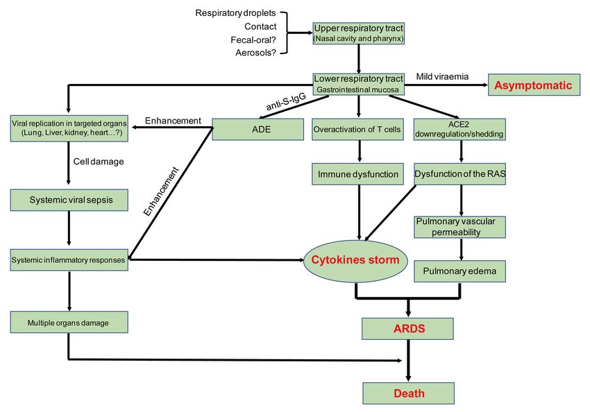
Viruses 2020, 12, 372                                                                                      3 of 17

respectively [2,8,11]. However, there is 94.6% sequence identity between the seven conserved replicase
domains in ORF1ab of SARS-CoV-2 and SARS-CoV [8], and less than 90% sequence identity between
those of SARS-CoV-2 and other β-CoVs [2], implying that SARS-CoV-2 belongs to the lineage B
(Sarbecovirus) of β-CoVs [12].
     As shown in Figure 2A, similar to other β-CoVs, the SARS-CoV-2 virion with a genome size of
29.9 kb [13] possesses a nucleocapsid composed of genomic RNA and phosphorylated nucleocapsid (N)
protein. The nucleocapsid is buried inside phospholipid bilayers and covered by two different types of
spike proteins: the spike glycoprotein trimmer (S) that exists in all CoVs, and the hemagglutinin-esterase
(HE) only shared among some CoVs. The membrane (M) protein and the envelope (E) protein are
located among the S proteins in the viral envelope [12]. The SARS-CoV-2 genome has 50 and 30 terminal
sequences (265 nt at the 50 terminal and 229 nt at the 30 terminal region), which is typical of β-CoVs,
with a gene order 50 -replicase open reading frame (ORF) 1ab-S-envelope(E)-membrane(M)-N-30
(Figure 2B). The predicted S, ORF3a, E, M, and N genes of SARS-CoV-2 are 3822, 828, 228, 669,
and 1260 nt in length, respectively. Similar to SARS-CoV, SARS-CoV-2 carries a predicted ORF8 gene
(366 nt in length) located between the M and N ORF genes [12].

      Figure 2. β-coronavirus particle and genome [9] (A) The β-coronavirus particle. β-coronavirus is an
      enveloped, nonsegmented, positive-sense single-stranded RNA virus genome in a size ranging from
      29.9 kb. The virion has a nucleocapsid composed of genomic RNA and phosphorylated nucleocapsid
      (N) protein, which is buried inside phospholipid bilayers and covered by the spike glycoprotein
      trimmer (S). The membrane (M) protein hemagglutinin-esterase (HE) and the envelope (E) protein are
      located among the S proteins in the virus envelope. (B) 50 and 30 terminal sequences of the SARS-CoV-2
      genome. The gene order is 50 -replicase ORF1ab-S-envelope(E)-membrane(M)-N-30 . ORF3ab, ORF6,
      ORF7ab, ORF8, ORF9ab, and ORF10 are located at the predicted positions shown in the picture. 1a, 1b,
      3a, 3b, 6, 7a, 7b, 8, 9a, 9b, 10 in the picture represent different ORF genes.

2.2. Physicochemical Properties
     The virus particle has a diameter of 60~100 nm and appears round or oval [14]. Most of the
knowledge about the physicochemical properties of CoVs comes from SARS-CoV and MERS-CoV.
SARS-CoV-2 can be inactivated by UV or heated at 56 ◦ C 30 min, and also sensitive to most disinfectants
such as diethyl ether, 75% ethanol, chlorine, peracetic acid, and chloroform [14]. It has been reported
that SARS-CoV-2 was more stable on plastic and stainless steel than on copper and cardboard,
and viable virus was detected up to 72 h after application to these surfaces. On cardboard, the half-life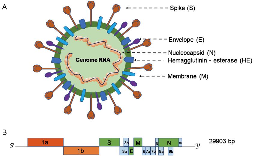
Viruses 2020, 12, 372                                                                              4 of 17

of SARS-CoV-2 was longer than that of SARS-CoV and the longest viability of both viruses was on
stainless steel and plastic [15].

2.3. Receptor Interactions and Cell Entry
      Human angiotensin-converting enzyme 2 (ACE2) is a functional receptor hijacked by SARS-CoV-2
for cell entry, similar to SARS-CoV [8,16]. ACE2 is a type I membrane protein expressed in lung,
heart, kidney, and intestine mainly associated with cardiovascular diseases [17]. The full-length ACE2
consists of an N-terminal peptidase domain (PD) and a C terminal Collectrin-like domain (CLD) that
ends with a single transmembrane helix and an~40-residue intracellular segment [17]. In addition
to cleavage of angiotensin (Ang) I to produce Ang-(1-9), ACE2 also provides a direct binding site for
the S proteins of CoVs [17]. The S protein of CoVs exists in a metastable pre-fusion conformation
that undergoes a dramatic structural rearrangement to fuse the viral membrane with the host cell
membrane [10]. This process is triggered by the S1 subunit and a host–cell receptor binding, which
destabilizes the pre-fusion trimer, resulting in the S1 subunit shedding and the S2 subunit transition to
a highly stable post-fusion conformation [10]. To engage a host–cell receptor, the receptor-binding
domain (RBD) of S1 undergoes hinge-like conformational movements that transiently hide or expose
the determinants of receptor binding [18]. In order to figure out the potential of SARS-CoV-2 to
infect humans, the receptor-binding domain (RBD) of its S protein, which is in contact with ACE2,
was analyzed. The biophysical and structural evidence suggests that SARS-CoV-2 S protein likely
binds to human ACE2 with 10–20 fold higher affinity than SARS-CoV [19]. Another structural evidence
suggests that the ACE2-B0AT1 complex can bind two S proteins simultaneously [20].

2.4. Evolutionary Insights into the Ecology of SARS-CoV-2
     All human CoVs may be of zoonotic origin, and bats are most likely the natural hosts for all
presently known CoVs [21]. During the SARS pandemic in 2002 and 2003, the first hints pointed to a
zoonotic origin of the SARS-CoV, with civets as the suspected natural source of human infection [22].
Genetically diversified SARS-like CoVs were then found in Chinese Rhinolophid bats, and two novel
bat CoVs from Chinese horseshoe bats (family: Rhinolophidae) in Yunnan Province, China are reported
to be very closely related to SARS-CoV, implying that Chinese horseshoe bats are natural host of
SARS-CoV [23,24]. Regarding SARS-CoV-2, it showed a high sequence identity to some bat CoVs such
as BatCoV RaTG13 (96% nt identity to SARS-CoV-2) previously detected in Rhinolophusaffinis from
Yunnan Province, indicating a bat origin of SARS-CoV-2 [12,23].
     Generally, bat habitats are far from human activity areas, and the virus was probably transmitted
to humans by another animal host. Bat SARS like-CoVs cannot directly infect humans unless they
undergo mutation or recombination in animal hosts [22]. For example, animal hosts of SARS-CoV
and MERS-CoV are the civet and camel (Figure 3) before transmission to humans. Regarding the
intermediate animal host of SARS-CoV-2, it has been reported that the sequence identity between
pangolin origin CoVs and SARS-CoV-2 is 99%, indicating that SARS-CoV-2 may be of pangolin
origin [25]. Many studies in China are tracking other potential animal hosts of SARS-CoV-2, which is
of great significance for the prevention and control of COVID-19.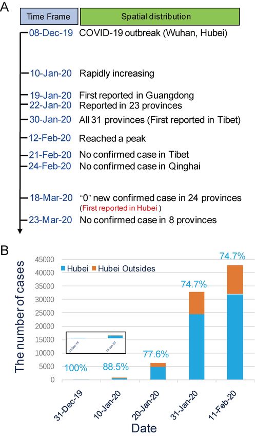
Viruses 2020, 12, 372                                                                                     5 of 17

      Figure 3. Ecology of emerging coronaviruses SARS-CoV, MERS-CoV, and SARS-CoV-2 are all bat
      origin coronaviruses, which cause human infections after circulation in animal hosts of civet, camel,
      and pangolin.

2.5. Genomic Variation
      The initial 10 genomic sequences of SARS-CoV-2 obtained from the nine COVID-19 patients were
extremely similar, exhibiting more than 99.98% sequence identity, implying that not much variation
has taken place [8,11]. A recent study indicates that 120 substitution sites were evenly distributed
in eight coding regions, without evident recombination events [7]. However, Tang et al. found that
SARS-CoV-2 had evolved into two major types of L and S, based on analyses of 103 genomes. Due to
severe selective pressure on the L type, the L type might be more aggressive and spread more quickly,
while the S type might remain milder due to relatively weaker selective pressure [26]. Due to the
unstable nature of RNA viruses, the continuous surveillance of SARS-CoV-2 from humans or animals
is extremely important for disease control.

3. Epidemiology

3.1. Source of Infection
     Currently, COVID-19 patients are the main source of infection, and severe patients are considered
to be more contagious than mild ones. Asymptomatically infected persons or patients in incubation
who show no signs or symptoms of respiratory infection proven to shed infectious virus, may also be
potential sources of infection [27]. Additionally, samples taken from patients recovered from COVID-19
continuously show a positive RT-PCR test [28], which has never been seen in the history of human
infectious diseases. In other words, asymptomatically infected persons and patients in incubation or
recovered from COVID-19 may pose serious challenges for disease prevention and control.

3.2. Spectrum of Infection
      COVID-19 has been considered as a type of self-limiting infectious disease, and most cases with
mild symptoms can recover in 1–2 weeks. SARS-CoV-2 infection can cause five different outcomes:
asymptomatically infected persons (1.2%); mild to medium cases (80.9%); severe cases (13.8%);
critical case (4.7%); and death (2.3% in all reported cases) [29]. The latest study indicates that the
Viruses 2020, 12, 372 6 of 17
proportion of asymptomatic infection in children under 10-years old is as high as 15.8% [30]. Therefore,
the proportion of asymptomatic infection should be further uncovered in the future.
3.3. Clinical Features
In the initial 41 patients [5], fever (98%), cough (76%), and myalgia or fatigue (44%) were the
most common symptoms. Less common symptoms were sputum production (28%), headache (8%),
hemoptysis (5%), and diarrhea (3%). More than half of patients developed dyspnea. The average
incubation period and basic reproduction number (R0) were estimated to be 5.2 d (95% CI: 4.1–7.0) and
2.2 (95% CI, 1.4–3.9), respectively [1,29]. Blood test showed normal or reduced (25%) white blood cell
count and lymphopenia (65%) [5]. A total of 98% of patients had bilateral involvement under chest
CT. Typical findings of chest CT images of ICU patients on admission were bilateral multiple lobular
and subsegmental areas of consolidation. The representative chest CT findings of non-ICU patients
showed bilateral ground-glass opacity and subsegmental areas of consolidation [2,5]. Analysis of 1324
laboratory confirmed cases showed that fever (87.9%) and cough (67.7%) were still the most common
symptoms, while diarrhea is uncommon. Lymphopenia was observed in 82.1% of patients admitted to
ICU [31].
3.4. Epidemiological Characteristics in Mainland China
Spatiotemporal distribution. The initial outbreak (8 December 2020) only occurred in Wuhan
and its surroundings inHubei Province before an imported case was first reported in Guangdong
Province on January 19, 2020 [1,29]. As of January 30, 2020, when the first imported case in Tibet
Province was reported, COVID-19 had spread to all 31 provinces in mainland China (Figure 4A).
Until 11 February2020, 44,672 cases were reported in all 31 provinces of mainland China (74.7% in
Hubei). Among them, 0.2% (100% in Hubei), 1.7% (88.5% in Hubei), 13.8% (77.6% in Hubei),
and 73.1% (74.7% in Hubei) of cases were onset before 31 December 2019, 10 January 2020, 20 January
2020, and 31 January 2020, respectively (Figure 4B). The number of reported cases rose rapidly after
10 January 2020, and reached a peak on 12 February 2020 [32]. Through the analysis of 1688 healthcare
confirmed cases with severe symptoms, there were 1080 cases in Wuhan, accounting for 64.0% of the
total incidence, 394 cases (23.3%) in Hubei except Wuhan, and 214 cases (12.7%) nationwide, except
for Hubei [29]. Tibet and Qinghai Provinces have had no confirmed cases since 21 February 2020 and
24 February 2020, respectively (Figure 4A) [33]. On 18 March 2020, “0” new confirmed cases was first
reported in Hubei Province, and a total of 24 provinces in mainland China had consecutively reported
“0” new confirmed cases. Until 23 March 2020, 81,773 cases (427 imported cases from abroad) were
cumulative reported in 31 provinces of mainland China, and the number of new confirmed cases have
mainly come from abroad and eight provinces have had no confirmed cases (Figure 4A) [34].
Population distribution. China CDC data [29] showed that patients were mainly concentrated
at the age of 30–79, accounting for 89.8%, 88.6%, and 86.6% of confirmed cases in Wuhan, Hubei,
and mainland China, respectively. The gender ratio (male/female) of confirmed cases in Wuhan, Hubei,
and mainland China was 0.99:1, 1.04:1, and 1.06:1, respectively. The proportion of infected healthcare
workers and farmers was 2.09% and 22%, respectively [29].
Case-fatality rate. The total case-fatality rate was 2.3% of 44,672 confirmed cases, while the total
case-fatality rate in Hubei and its surroundings was 2.9% and 0.4%, respectively [29]. In contrast,
the total case-fatality rate of SARS and MERS was 9.6% [35] and 34%, respectively [36]. In all
COVID-19 patients over 80-years old, the case-fatality rate wasas high as 14.8%. The case-fatality rate
of males and femaleswas2.8% and 1.7%, respectively [29]. Patients with underlying basic disorders
showed poor prognosis. The case-fatality rate of cases without basic disorders was as low as 0.9%,
while the case-fatality rate of cases with cardiovascular disease, diabetes, chronic respiratory disease,
hypertension, and cancer was10.5%, 7.3%, 6.3%, 6.0%, and 5.6%, respectively [29]. Notably, critical
cases had the highest case-fatality rate of 49% [29]. As for healthcare workers, the case-fatality rate was
approximately 0.17% of 3019 cases [29].Viruses 2020, 12, 372 7 of 17
Figure 4. Spatiotemporal distribution of COVID-19. (A) The spread and decline of COVID-19 in
mainland China over time. The time point (red words) of “0” new confirmed case first reported
in Hubei Province was 18 March, 2020. (B) Distribution of cases with different onset times before
11 February 2020.
3.5. Other Regions
According to the WHO data updated on March 23, 2020 [37], 190countriesor areas have reported
332,218 laboratory confirmed cases including 14,510 deaths. The total case-fatality rate of global cases
outside China is 4.5%. More attention should be paid to Italy, Spain, the USA, Germany, France,
and Iran with more severe outbreaks [37]. The top five countries with the highest cumulative confirmed
cases in the world are China (24.6%), Italy (17.8%), USA (9.5%), Spain (8.6%), and Germany (7.5%).
Higher case-fatality rates were found in Italy (9.3%), Iran (7.8%), and Spain (6.0%) [37].
3.6. Routes of Transmission
Currently, respiratory droplets and contact transmission are considered to be the main transmission
routes. Recent reports indicate that SARS-CoV-2 can be detected in the urine and stool of laboratory
confirmed patients, implying a risk of fecal–oral transmission [14]. However, it is not yet certain that
the consumption of virus-contaminated foods will cause infection and transmission. There is still
no evidence that SARS-CoV-2 can be transmitted through aerosols or from mother to baby during
pregnancy or childbirth.
3.7. Herd Susceptibility
As an emerging infectious disease, the population of all races and ages is generally susceptible.
In mainland China, 30~65-year-old persons account for 71.45% and children under 10-years-old account
for 0.35% [31]. Elderly people and persons with underlying basic disorders such as asthma, diabetes,Viruses 2020, 12, 372 8 of 17
cardiovascular diseases, and cancer may be more susceptible to SARS-CoV-2 [38,39]. Smoking and
obesity are also susceptible factors [40,41].
3.8. High-Risk Population
Persons who are in close contact with patients or subclinically symptomatic infected persons are
part of the high-risk population. High infection risk is also considered in healthcare workers and the
family members of patients [42].
4. Diagnosis
4.1. Nucleic Acid Test
Viral diagnostics is one important part of our armamentarium against COVID-19. After initial
outbreak, diagnostic tests based on the detection of the viral sequence by RT-PCR or next generation
sequencing platforms soon became available. Subsequently, many biotechnology companies have
successfully developed nucleic acid detection kits, and the China Food and Drug Administration
(CFDA) has urgently approved a batch of fluorescent quantitative kits and sequencing systems [43].
The main concern related to the nucleic acid test is false negatives. To solve the problem of low detection
efficiency, some improved rapid viral nucleic acid diagnostic tests have been invented. Particularly,
a nucleic acid test paper, which can be used for the rapid detection of SARS-CoV-2 with the naked eye
observation within three minutes, has been successfully developed [44].
4.2. Serologic Diagnosis
It has been shown that patients with SARS-CoV-2 infection possess acute serological responses [8].
Combined with immunochromatography, colloidal gold, and other technologies, relevant detection
reagents have been developed rapidly [45,46].
4.3. CRISPR/Cas13 System
The Cas13-based SHERLOCK (specific high-sensitivity enzymatic reporter unlocking) platform
has been widely used to detect Zika virus (ZIKV) and dengue virus (DENV) in patient samples at
concentrations as low as 1 copy per microliter [47]. Recently, Zhang et al. released a CRISPR/Cas13-based
SHERLOCK technology to detect SARS-CoV-2. However, this CRISPR/Cas13 system remains to be
verified because it has not been tested on clinical samples from COVID-19 patients.
4.4. Imaging Technology
Chest radiograph or CT is an important tool for COVID-19 diagnosis in clinical practice.
The majority of COVID-19 cases have similar features on CT images including bilateral distribution of
patchy shadows and ground glass opacity [48]. The great value of using the deep learning machine to
extract radiological graphical features for COVID-19 diagnosis has been introduced [49]. Artificial
Intelligence (AI) can accurately interpret the CT images of the suspected cases of the new crown
within 20 s, and the accuracy rate of the analysis results reached 96%, greatly improving the diagnostic
efficiency. This technique is already being used in clinical practice.
5. Pathogenesis
5.1. Virus Entry and Spread
SARS-CoV-2 is transmitted predominantly via respiratory droplet, contact, and potential in
fecal-oral [14]. Primary viral replication is presumed to occur in mucosal epithelium of upper
respiratory tract (nasal cavity and pharynx), with further multiplication in lower respiratory tract
and gastrointestinal mucosa [50], giving rise to a mild viremia. Few infections are controlled at this
point and remain asymptomatic. Some patients have also exhibited non-respiratory symptoms such asViruses 2020, 12, 372 9 of 17
acute liver and heart injury, kidney failure, diarrhea [5,51–53], implying multiple organ involvement.
ACE2 is broadly expressed in nasal mucosa, bronchus, lung, heart, esophagus, kidney, stomach,
bladder, and ileum, and these human organs are all vulnerable to SARS-CoV-2 [40]. Recently, potential
pathogenicity of the SARS-CoV-2 to testicular tissues hasalso been proposed by clinicians, implying
fertility concerns in young patients [54]. The postulated pathogenesis of SARS-CoV-2 infection is
graphed in Figure 5.
Figure 5. Postulated pathogenesis of SARS-CoV-2 infection. Antibody-dependent enhancement (ADE);
ACE2: angiotensin-converting enzyme 2; RAS: renin-angiotensin system; ARDS: acute respiratory
distress syndrome. Red words represent the important turning points in SARS-CoV-2 infection.
5.2. Pathological Findings
The first report [55] of pathological findings from a severe COVID-19 showed pulmonary
bilateral diffuse alveolar damage with cellular fibromyxoid exudates. The right lung showed evident
desquamation of pneumocytes and hyaline membrane formation, indicating acute respiratory distress
syndrome. The left lung tissue displayed pulmonary edema with hyaline membrane formation,
suggestive of early-phase acute respiratory distress syndrome (ARDS). Interstitial mononuclear
inflammatory infiltrates, dominated by lymphocytes, could be observed in both lungs. Multinucleated
syncytial cells with atypical enlarged pneumocytes characterized by large nuclei, amphophilic granular
cytoplasm, and prominent nucleoli were identified in the intra-alveolar spaces, indicating viral
cytopathic-like changes. These pulmonary pathological findings extremely resemble those seen in
SARS [56] and MERS [57]. Moderate microvascular steatosis and mild lobular and portal activity were
observed in liver biopsy specimens, which might be caused by either SARS-CoV-2 infection or drug use.
In addition, only a few interstitial mononuclear inflammatory infiltrates were found in the heart tissue,
which means that SARS-CoV-2 might not directly impair the heart [55]. Massive mucus secretion in
both lungs was found in death cases with COVID-19, which was different from SARS and MERS [58].Viruses 2020, 12, 372 10 of 17
5.3. Acute Respiratory Distress Syndrome (ARDS)
ARDS is a life-threatening lung condition that prevents enough oxygen from getting to the
lungs and into the circulation, accounting for mortality of most respiratory disorders and acute lung
injury [59]. In fatal cases of human SARS-CoV, MERS-CoV, and SARS-CoV-2 infections, individuals
exhibit severe respiratory distress requiring mechanical ventilation, and the histopathology findings
also support ARDS [55–57]. Previous studies have found that genetic susceptibility, and inflammatory
cytokines were closely related to the occurrence of ARDS. More than 40 candidate genes including
ACE2, interleukin 10 (IL-10), tumor necrosis factor (TNF), and vascular endothelial growth factor
(VEGF) among others have been considered to be associated with the development or outcome of
ARDS [60]. Increased levels of plasma IL-6 and IL-8 were also demonstrated to be related to adverse
outcomes of ARDS [59]. The above biomarkers suggest both a molecular explanation for the severe
ARDS and a possible treatment for ARDS followingSARS-CoV-2 infection.
5.4. Cytokine Storm
Clinical findings showed exuberant inflammatory responses during SARS-CoV-2 infection,
further resulting in uncontrolled pulmonary inflammation, likely a leading cause of case fatality.
Rapid viral replication and cellular damage, virus-induced ACE2 downregulation and shedding,
and antibody dependent enhancement (ADE) are responsible for aggressive inflammation caused by
SARS-CoV-2, as concluded in a recently published review article [61]. SARS-CoV-2 hijacks the same
entry receptor, ACE2, as SARS-CoV for infection, suggesting the likelihood of the same population of
cells being targeted and infected [62]. The initial onset of rapid viral replication may cause massive
epithelial and endothelial cell death and vascular leakage, triggering the production of exuberant
pro-inflammatory cytokines and chemokines [63]. Loss of pulmonary ACE2 function has been
proposed to be related to acute lung injury [64] because ACE2 downregulation and shedding can lead
to dysfunction of the renin-angiotensin system (RAS), and further enhance inflammation and cause
vascular permeability. For SARS-CoV, one confusing issueis that only a few patients, particularly
those who produce neutralizing antibodies early, experience persistent inflammation, ARDS, and even
sudden death, while most patients survive the inflammatory responses and clear the virus [61].
The above phenomenon also exists in SARS-CoV-2 infection. A possible underlying mechanism
of antibody-dependent enhancement (ADE) has been proposed recently [61]. ADE, a well-known
virology phenomenon, has been confirmed in multiple viral infections [65]. ADE can promote viral
cellular uptake of infectious virus–antibody complexes following their interaction with Fc receptors
(FcR), FcγR, or other receptors, resulting in enhanced infection of target cells [65]. The interaction of
FcγR with the virus-anti-S protein-neutralizing antibodies (anti-S-IgG) complex may facilitate both
inflammatory responses and persistent viral replication in the lungs of patients [61].
5.5. Immune Dysfunction
Peripheral CD4 and CD8 T cells showed reduction and hyperactivation in a severe patient.
High concentrations of proinflammatory CD4 T cells and cytotoxic granules CD8 T cells were also
determined, suggesting antiviral immune responses and overactivation of T cells [55]. Additionally,
several studies have reported that lymphopenia is a common feature of COVID-19 [2,5], suggestive of
a critical factor accounting for severity and mortality.
6. Potential Therapeutics
Currently, there are no specificantiviral drugs or vaccines for the control of SARS-CoV-2.
Symptomatic treatment strategies are recommended for clinical practice [14]. Here, we summarize
potential therapeutics available for the treatment of SARS-CoV-2.Viruses 2020, 12, 372 11 of 17
6.1. Type I IFNs
Type I IFNs are antiviral cytokines that induce a large range of proteins that can impair viral
replication in targeted cells. Previous studies have reported that IFN-β was superior against SARS-CoV
compared to IFN-α [66]. Synergistic effects of leukocytic IFN-α with ribavirin [67] and IFN-β with
ribavirin [68] against SARS-CoV were demonstrated in vitro.
6.2. Potential Antiviral Compounds
Ribavirin. During the outbreak of SARS in Hong Kong, ribavirin was broadly used for patients
with or without concomitant use of steroids [69]. Ribavirin and IFN-β could synergistically inhibit
SARS-associated CoV replication in vitro [68]. Due to adverse reactions, the proper dose of ribavirin in
clinical application should be given carefully.
Lopinavir/ritonavir. The combination of lopinavir/ritonavir is widely used in the treatment of HIV
infection. It has been reported that the use of lopinavir/ritonavir with ribavirin has a good therapeutic
effect in SARS [70] and MERS [71]. Lopinavir/ritonavir has been recommended for clinical treatment
for COVID-19.
Remdesivir. Remdesivir (RDV) was previously reported to restrain SARS-CoV in vivo [72], and the
antiviral protection of RDV and IFN-β was found to be superior to that of lopinavir/ritonavir-IFN-β
against MERS-CoV in vitro and in vivo. In addition, remdesivir was used in the treatment of the
first COVID-19 patient in the United States [27] and was shown to have antiviral activity against
SARS-CoV-2 in vitro [73]. However, its effectiveness and safety have not been verified in clinical
trials yet.
Nelfinavir. Nelfinavir is a selective inhibitor of HIV protease, which has been shown to have a
strong inhibition of SARS-CoV [74] implying a possible therapeutic for COVID-19.
Arbidol. Arbidol, a broad-spectrum antiviral compound, is able to block viral fusion against
influenza viruses. In addition, arbidol and its derivative, arbidolmesylate, have been reported to have
antiviral activity against SARS-CoV in vitro [75]. The antiviral activity of arbidol against SARS-CoV-2
has been confirmed in vitro and recommended for clinical treatment [14].
Chloroquine. Chloroquine has many interesting biochemical properties including antiviral
effect [76]. It has been found to be a potent inhibitor of SARS-CoV through interfering with ACE2 [74].
Chloroquine can effectively inhibit SARS-CoV-2 in vitro [73], and is recommended for the clinical
control of viral replication [14].
6.3. Convalescent Plasma
Recently, convalescent plasma has been widely recommended to be used for COVID-19 [77],
but the effect of convalescent plasma cannot be discerned from the effects of patient comorbidities,
stage of illness, or effect of other treatments.
6.4. Protective Monoclonal Antibody
It has been reported that the monoclonal antibody (mAb) can efficiently neutralize SARS-CoV and
inhibit syncytia formation between cells expressing the S protein and those expressing the SARS-CoV
receptor ACE2 [78]. However, mAbs can only recognize a single epitope, and the anti-infective effect
may be limited. In addition, the development of mAbs requires a certain period of time, which is
difficult to achieve in clinical application in a short time.
6.5. Others
Based on the virology of SARS-CoV-2, blocking the binding of S protein to ACE2 is important
for the treatment of virus infection. ACE2 is an important component of the renin-angiotensin
system (RAS). RAS inhibitors, ACEI and AT1R, may be potential therapeutic tools for COVID-19.
Additionally, intravenous transplantation of ACE2-mesenchymal stem cells (MSCs), blocking of FcRViruses 2020, 12, 372 12 of 17
with immunoglobulin (IVIG), and systemic anti-inflammatory drugs to reduce cytokine storm are also
potential therapeutic strategies for severe COVID-19 [61,79]. Traditional Chinese medicines have also
been found to have potential anti-SARS-CoV-2 activity [80].
7. Vaccine Development
Vaccination probably offers the best option for COVID-19 control. Epitopes, mRNA,
and S protein-RBD structure-based vaccines have been widely proposed and started [81,82].
Rapid reconstruction of SARS-CoV-2 using a synthetic genomics platform has been reported, and this
technical advance is helpful for vaccine development [83]. Human ACE2 transgenic mouse and
rhesus monkey models of COVID-19 have been well established for vaccine development [84],
and someSARS-CoV-2 vaccines are already under clinical trial [85,86].
8. Conclusions and Perspective
In summary, SARS-CoV-2 is an emerging human CoV, and appears similar to previous SARS and
MERS outbreaks. Bats are likely an important reservoir for SARS-CoV-2, and the current knowledge
does not support the Huanan Seafood Market as the only source of infection. The main mode of
transmission of SARS-CoV is through inhalation of respiratory droplets and indirect or direct contact,
and infection has been estimated to have mean incubation period of 5.2 days and a R0 of 2.2. The most
common factors behind COVID-19 mortality are older age and concomitant disease. The limited
understanding about the pathogenesis of SARS-CoV-2 infection indicates that COVID-19 and SARS
have similar pathogenesis, and also have differences such as massive mucus secretion in both lungs of
critical patient. There are still no specific antiviral treatments or vaccines available. The most urgent
task at the moment is to develop more interventions that will eventually allow the effective control of
this arising severe viral infection.
The pandemic potential of human CoVs remains a great threat for global public health. However,
human beings have not gained enough experience in previous battles with SARS and MERS. After the
outbreak of COVID-19 in China, SARS-CoV-2 has received worldwide attention as an important
pathogen in respiratory tract infection. Worldwide, over 330,000 people have fallen ill within four
months. To lock down the spread of COVID-19, the Chinese government adopted an unprecedented
containment strategy in Wuhan, the source of infection, and all other provinces or areas also declared an
emergency response. However, in light of the current data, the ability of the virus to spread is beyond
the current estimates. To provide a comprehensive summary to public health authorities and potential
readers worldwide, we detailed the present understanding of COVID-19 and introduced the current
state of development of measures in this review. The results of the present study are also valuable for
informing further studies and health policies in preparation for COVID-19 outbreaks worldwide.
Author Contributions: Conceptualization, Y.J. and G.D.; Methodology, Y.J.; Software, Y.J. and W.J.; Validation,
H.Y., S.C., and W.W.; Formal analysis, Y.J.; Investigation, Y.J., W.J., and S.C.; Resources, Y.J. and S.C.; Data curation,
Y.J.; Writing—original draft preparation, Y.J., W.W., H.Y., G.D. and W.Z.; Writing—review and editing, Y.J.,
W.W., W.Z., S.C., and G.D.; Visualization, Y.J.; Supervision, G.D.; Project administration, G.D. and Y.J.; Funding
acquisition, Y.J. and G.D. All authors have read and agreed to the published version of the manuscript.
Funding: This work was funded by the National Science and Technology Specific Projects (NO.2018ZX10301407);
the National Natural Science Foundation of China (No.81172740 and No.81573205); and the project funded by the
China Postdoctoral Science Foundation (No.2019M662543).
Conflicts of Interest: The authors declare no conflict of interest. The funders had no role in the design of the
study; in the collection, analyses, or interpretation of data; in the writing of the manuscript, or in the decision to
publish the results.
References
1. Li, Q.; Guan, X.; Wu, P.; Wang, X.; Zhou, L.; Tong, Y.; Ren, R.; Leung, K.S.M.; Lau, E.H.Y.; Wong, J.Y.; et al.
Early Transmission Dynamics in Wuhan, China, of Novel Coronavirus–Infected Pneumonia. N. Engl. J. Med.
2020. [CrossRef] [PubMed]Viruses 2020, 12, 372 13 of 17
2. Zhu, N.; Zhang, D.; Wang, W.; Li, X.; Yang, B.; Song, J.; Zhao, X.; Huang, B.; Shi, W.; Lu, R.; et al. A Novel
Coronavirus from Patients with Pneumonia in China, 2019. N. Engl. J. Med. 2020. [CrossRef] [PubMed]
3. World Health Organization Press Conference. The World Health Organization (WHO) Has Officially
Named the Disease Caused by the Novel Coronavirus as COVID-19. Available online: https://www.who.int/
emergencies/diseases/novel-coronavirus-2019 (accessed on 11 February 2020).
4. Gorbalenya, A.E.; Baker, S.C.; Baric, R.S.; de Groot, R.J.; Drosten, C.; Gulyaeva, A.A.; Haagmans, B.L.;
Lauber, C.; Leontovich, A.M.; Neuman, B.W.; et al. Severe acute respiratory syndrome-related coronavirus:
The species and its viruses—A statement of the Coronavirus Study Group. bioRxiv 2020. [CrossRef]
5. Huang, C.; Wang, Y.; Li, X.; Ren, L.; Zhao, J.; Hu, Y.; Zhang, L.; Fan, G.; Xu, J.; Gu, X.; et al. Clinical features
of patients infected with 2019 novel coronavirus in Wuhan, China. Lancet (Lond. Engl.) 2020, 395, 497–506.
[CrossRef]
6. Xiong, C.; Jiang, L.; Chen, Y.; Jiang, Q. Evolution and variation of 2019-novel coronavirus. bioRxiv 2020.
[CrossRef]
7. Yu, W.; Tang, G.; Zhang, L.; Corlett, R.T. Decoding evolution and transmissions of novel pneumonia
coronavirus using the whole genomic data. ChinaXiv 2020. [CrossRef]
8. Zhou, P.; Yang, X.L.; Wang, X.G.; Hu, B.; Zhang, L.; Zhang, W.; Si, H.R.; Zhu, Y.; Li, B.; Huang, C.L.; et al.
A pneumonia outbreak associated with a new coronavirus of probable bat origin. Nature 2020, 1–4. [CrossRef]
9. Weiss, S.R.; Leibowitz, J.L. Coronavirus pathogenesis. Adv. Virus Res. 2011, 81, 85–164. [CrossRef]
10. De Wilde, A.H.; Snijder, E.J.; Kikkert, M.; van Hemert, M.J. Host Factors in Coronavirus Replication.
In Roles of Host Gene and Non-Coding RNA Expression in Virus Infection; Tripp, R.A., Tompkins, S.M., Eds.;
Springer International Publishing: Cham, Switzerland, 2018; pp. 1–42. [CrossRef]
11. Lu, R.; Zhao, X.; Li, J.; Niu, P.; Yang, B.; Wu, H.; Wang, W.; Song, H.; Huang, B.; Zhu, N.; et al.
Genomic characterisation and epidemiology of 2019 novel coronavirus: Implications for virus origins
and receptor binding. Lancet (Lond. Engl.) 2020, 395, 565–574. [CrossRef]
12. Wu, F.; Zhao, S.; Yu, B.; Chen, Y.-M.; Wang, W.; Hu, Y.; Song, Z.-G.; Tao, Z.-W.; Tian, J.-H.; Pei, Y.-Y.; et al.
Complete genome characterisation of a novel coronavirus associated with severe human respiratory disease
in Wuhan, China. bioRxiv 2020. [CrossRef]
13. National Microbiology Data Center. Available online: http://nmdc.cn/coronavirus (accessed on
26 March 2020).
14. General Office of National Health Commission; General Office of National Administration of Traditional
Chinese Medicine. Diagnostic and treatment protocol for Novel Coronavirus Pneumonia; (Trial version 6).
Available online: http://www.nhc.gov.cn/yzygj/s7653p/202002/8334a8326dd94d329df351d7da8aefc2.shtml:
(accessed on 20 February 2020).
15. Van Doremalen, N.; Bushmaker, T.; Morris, D.H.; Holbrook, M.G.; Gamble, A.; Williamson, B.N.; Tamin, A.;
Harcourt, J.L.; Thornburg, N.J.; Gerber, S.I.; et al. Aerosol and Surface Stability of SARS-CoV-2 as Compared
with SARS-CoV-1. N. Engl. J. Med. 2020. [CrossRef] [PubMed]
16. Li, W.; Moore, M.J.; Vasilieva, N.; Sui, J.; Wong, S.K.; Berne, M.A.; Somasundaran, M.; Sullivan, J.L.;
Luzuriaga, K.; Greenough, T.C.; et al. Angiotensin-converting enzyme 2 is a functional receptor for the SARS
coronavirus. Nature 2003, 426, 450–454. [CrossRef] [PubMed]
17. Donoghue, M.; Hsieh, F.; Baronas, E.; Godbout, K.; Gosselin, M.; Stagliano, N.; Donovan, M.; Woolf, B.;
Robison, K.; Jeyaseelan, R.; et al. A novel angiotensin-converting enzyme-related carboxypeptidase (ACE2)
converts angiotensin I to angiotensin 1-9. Circ. Res. 2000, 87, E1–E9. [CrossRef] [PubMed]
18. Li, F. Structure, Function, and Evolution of Coronavirus Spike Proteins. Annu. Rev. Virol. 2016, 3, 237–261.
[CrossRef]
19. Wrapp, D.; Wang, N.; Corbett, K.S.; Goldsmith, J.A.; Hsieh, C.-L.; Abiona, O.; Graham, B.S.; McLellan, J.S.
Cryo-EM Structure of the 2019-nCoV Spike in the Prefusion Conformation. bioRxiv 2020. [CrossRef]
20. Zhou, Q.; Yan, R.; Zhang, Y.; Li, Y.; Xia, L. Structure of dimeric full-length human ACE2 in complex with
B0AT1. bioRxiv 2020. [CrossRef]
21. Vijaykrishna, D.; Smith, G.J.; Zhang, J.X.; Peiris, J.S.; Chen, H.; Guan, Y. Evolutionary insights into the ecology
of coronaviruses. J. Virol. 2007, 81, 4012–4020. [CrossRef]
22. Corman, V.M.; Muth, D.; Niemeyer, D.; Drosten, C. Hosts and Sources of Endemic Human Coronaviruses.
Adv. Virus Res. 2018, 100, 163–188. [CrossRef]Viruses 2020, 12, 372 14 of 17
23. Ge, X.-Y.; Li, J.-L.; Yang, X.-L.; Chmura, A.A.; Zhu, G.; Epstein, J.H.; Mazet, J.K.; Hu, B.; Zhang, W.; Peng, C.;
et al. Isolation and characterization of a bat SARS-like coronavirus that uses the ACE2 receptor. Nature 2013,
503, 535–538. [CrossRef] [PubMed]
24. Lau, S.K.P.; Woo, P.C.Y.; Li, K.S.M.; Huang, Y.; Tsoi, H.-W.; Wong, B.H.L.; Wong, S.S.Y.; Leung, S.-Y.;
Chan, K.-H.; Yuen, K.-Y. Severe acute respiratory syndrome coronavirus-like virus in Chinese horseshoe bats.
Proc. Natl. Acad. Sci. USA 2005, 102, 14040–14045. [CrossRef]
25. Lam, T.T.-Y.; Shum, M.H.-H.; Zhu, H.-C.; Tong, Y.-G.; Ni, X.-B.; Liao, Y.-S.; Wei, W.; Cheung, W.Y.-M.; Li, W.-J.;
Li, L.-F.; et al. Identification of 2019-nCoV related coronaviruses in Malayan pangolins in southern China.
bioRxiv 2020. [CrossRef]
26. Tang, X.; Wu, C.; Li, X.; Song, Y.; Yao, X.; Wu, X.; Duan, Y.; Zhang, H.; Wang, Y.; Qian, Z.; et al. On the origin
and continuing evolution of SARS-CoV-2. Natl. Sci. Rev. 2020. [CrossRef]
27. Hoehl, S.; Berger, A.; Kortenbusch, M.; Cinatl, J.; Bojkova, D.; Rabenau, H.; Behrens, P.; Böddinghaus, B.;
Götsch, U.; Naujoks, F.; et al. Evidence of SARS-CoV-2 Infection in Returning Travelers from Wuhan, China.
N. Engl. J. Med. 2020. [CrossRef] [PubMed]
28. Lan, L.; Xu, D.; Ye, G.; Xia, C.; Wang, S.; Li, Y.; Xu, H. Positive RT-PCR Test Results in Patients Recovered
From COVID-19. JAMA 2020. [CrossRef] [PubMed]
29. Novel Coronavirus Pneumonia Emergency Response Epidemiology Team. The epidemiological
characteristics of an outbreak of 2019 novel coronavirus diseases (COVID-19) in China.
Zhonghualiuxingbingxuezazhi 2020, 41, 145–151. [CrossRef]
30. Lu, X.; Zhang, L.; Du, H.; Zhang, J.; Li, Y.Y.; Qu, J.; Zhang, W.; Wang, Y.; Bao, S.; Li, Y.; et al. SARS-CoV-2
Infection in Children. N. Engl. J. Med. 2020. [CrossRef]
31. Yang, Y.; Lu, Q.; Liu, M.; Wang, Y.; Zhang, A.; Jalali, N.; Dean, N.; Longini, I.; Halloran, M.E.; Xu, B.;
et al. Epidemiological and clinical features of the 2019 novel coronavirus outbreak in China. medRxiv 2020.
[CrossRef]
32. National Health Commission of People’s Republic of China. An Update of Novel Coronavirus
Pneumonia Outbreak as of 24:00 on February 12. Available online: http://www.nhc.gov.cn/xcs/yqtb/
202002/26fb16805f024382bff1de80c918368f.shtml (accessed on 13 February 2020).
33. National Health Commission of People’s Republic of China. An Update of Novel Coronavirus
Pneumonia Outbreak as of 24:00 on February 25. Available online: http://www.nhc.gov.cn/xcs/yqtb/
202002/741ce06130284a77bfbf699483c0fb60.shtml (accessed on 26 February 2020).
34. National Health Commission of People’s Republic of China. An Update of Novel Coronavirus
Pneumonia Outbreak as of 24:00 on March 23. Available online: http://www.nhc.gov.cn/xcs/yqtb/202003/
e6c12d0c2cf04474944187f4088dc021.shtml (accessed on 24 March 2020).
35. World Health Organization. Severe Acute Respiratory Syndrome (SARS). Available online: https://www.who.
int/csr/sars/en/ (accessed on 24 March 2020).
36. World Health Organization. Middle East Respiratory Syndrome Coronavirus (MERS-CoV). Available online:
https://www.who.int/emergencies/mers-cov/en/ (accessed on 23 March 2020).
37. World Health Organization. Coronavirus Disease (COVID-2019) Situation Reports. Update on 24:00 of March
23. Available online: https://www.who.int/emergencies/diseases/novel-coronavirus-2019/situation-reports/
(accessed on 23 March 2020).
38. World Health Organization. Novel Coronavirus (2019-nCoV) Advice for the Public: Myth Buster.
Available online: https://www.who.int/emergencies/diseases/novel-coronavirus-2019/advice-for-public/
myth-busters (accessed on 23 March 2020).
39. Liang, W.; Guan, W.; Chen, R.; Wang, W.; Li, J.; Xu, K.; Li, C.; Ai, Q.; Lu, W.; Liang, H.; et al. Cancer patients
in SARS-CoV-2 infection: A nationwide analysis in China. LancetOncol. 2020. [CrossRef]
40. Zou, X.; Chen, K.; Zou, J.; Han, P.; Hao, J.; Han, Z. The single-cell RNA-seq data analysis on the receptor
ACE2 expression reveals the potential risk of different human organs vulnerable to Wuhan 2019-nCoV
infection. Front. Med. 2020, 1–8. [CrossRef]
41. Jia, X.; Yin, C.; Lu, S.; Chen, Y.; Liu, Q.; Bai, J.; Lu, Y. Two Things about COVID-19 Might Need Attention.
Preprints 2020. [CrossRef]
42. National Health Commission; Ministry of Human Resources and Social Security; Ministry of Finance. Measures
to Improve Working Conditions of and Care for Physical and Mental Health of Healthcare Workers. Available online:
http://www.gov.cn/xinwen/2020-02/11/content_5477476.htm (accessed on 21 February 2020).Viruses 2020, 12, 372 15 of 17
43. China Food and Drug Administration. China Food and Drug Administration Emergency Approval of New
Coronavirus Nucleic Acid Detection Reagents. Available online: http://www.nmpa.gov.cn/WS04/CL2056/
374264.html (accessed on 23 February 2020).
44. Daily, H. Academician Zhang Gaiping’s Team has Achieved a Number of Staged Results on Novel Coronavirus.
Available online: https://baijiahao.baidu.com/s?id=1658653081676286071&wfr=spider&for=pc: (accessed on
16 February 2020).
45. Nankai University News Network. Nankai University Team has Developed a Rapid Antibody Detection Kit for
Novel Coronavirus Within 15 Minutes. Available online: http://news.nankai.edu.cn/ywsd/system/2020/02/15/
030037569.shtml: (accessed on 15 February 2020).
46. Xiamen University News Network. Xiamen University and Shenzhen Third Hospital Successfully Developed
a Novel Coronavirus Antibody Detection kit, Which Can Improve the Clinical Diagnosis. Available online:
http://www.most.gov.cn/dfkj/fj/zxdt/202002/t20200224_151881.htm: (accessed on 24 February 2020).
47. Myhrvold, C.; Freije, C.A.; Gootenberg, J.S.; Abudayyeh, O.O.; Metsky, H.C.; Durbin, A.F.; Kellner, M.J.;
Tan, A.L.; Paul, L.M.; Parham, L.A. Field-deployable viral diagnostics using CRISPR-Cas13. Science 2018,
360, 444–448. [CrossRef] [PubMed]
48. Kanne, J.P. Chest CT Findings in 2019 Novel Coronavirus (2019-nCoV) Infections from Wuhan, China:
Key Points for the Radiologist. Radiology 2020, 200241. [CrossRef] [PubMed]
49. Wang, S.; Kang, B.; Ma, J.; Zeng, X.; Xiao, M.; Guo, J.; Cai, M.; Yang, J.; Li, Y.; Meng, X.; et al. A deep learning
algorithm using CT images to screen for Corona Virus Disease (COVID-19). medRxiv 2020. [CrossRef]
50. Xiao, F.; Tang, M.; Zheng, X.; Li, C.; He, J.; Hong, Z.; Huang, S.; Zhang, Z.; Lin, X.; Fang, Z.; et al. Evidence
for gastrointestinal infection of SARS-CoV-2. medRxiv 2020. [CrossRef] [PubMed]
51. Cheng, Y.; Luo, R.; Wang, K.; Zhang, M.; Wang, Z.; Dong, L.; Li, J.; Yao, Y.; Ge, S.; Xu, G. Kidney impairment
is associated with in-hospital death of COVID-19 patients. medRxiv 2020. [CrossRef]
52. Guan, G.W.; Gao, L.; Wang, J.W.; Wen, X.J.; Mao, T.H.; Peng, S.W.; Zhang, T.; Chen, X.M.; Lu, F.M. Exploring
the mechanism of liver enzyme abnormalities in patients with novel coronavirus-infected pneumonia. Chin. J.
Hepatol. 2020, 28, E002. [CrossRef]
53. Wang, D.; Hu, B.; Hu, C.; Zhu, F.; Liu, X.; Zhang, J.; Wang, B.; Xiang, H.; Cheng, Z.; Xiong, Y.; et al. Clinical
Characteristics of 138 Hospitalized Patients With 2019 Novel Coronavirus-Infected Pneumonia in Wuhan,
China. JAMA 2020. [CrossRef]
54. Fan, C.; Li, K.; Ding, Y.; Lu, W.L.; Wang, J. ACE2 Expression in Kidney and Testis May Cause Kidney and
Testis Damage After 2019-nCoV Infection. medRxiv 2020. [CrossRef]
55. Xu, Z.; Shi, L.; Wang, Y.; Zhang, J.; Huang, L.; Zhang, C.; Liu, S.; Zhao, P.; Liu, H.; Zhu, L.; et al. Pathological
findings of COVID-19 associated with acuterespiratory distress syndrome. Lancet Respir. Med. 2020.
[CrossRef]
56. Ding, Y.; Wang, H.; Shen, H.; Li, Z.; Geng, J.; Han, H.; Cai, J.; Li, X.; Kang, W.; Weng, D.; et al. The clinical
pathology of severe acute respiratory syndrome (SARS): A report from China. J. Pathol. 2003, 200, 282–289.
[CrossRef]
57. Ng, D.L.; Al Hosani, F.; Keating, M.K.; Gerber, S.I.; Jones, T.L.; Metcalfe, M.G.; Tong, S.; Tao, Y.; Alami, N.N.;
Haynes, L.M.; et al. Clinicopathologic, Immunohistochemical, and Ultrastructural Findings of a Fatal Case
of Middle East Respiratory Syndrome Coronavirus Infection in the United Arab Emirates, April 2014. Am. J.
Pathol. 2016, 186, 652–658. [CrossRef] [PubMed]
58. Liu, Q.; Qu, G.; Wang, Y.; Liu, P.; Zhu, Y.; Fei, G.; Ren, L.; Zhou, Y.; Liu, L. Anatomy of a COVID-19 Death
Corpse System. J. Forensic Med. 2020, 36, 21–23. [CrossRef]
59. Thompson, B.T.; Chambers, R.C.; Liu, K.D. Acute Respiratory Distress Syndrome. N. Engl. J. Med. 2017,
377, 562–572. [CrossRef] [PubMed]
60. Meyer, N.J.; Christie, J.D. Genetic heterogeneity and risk of acute respiratory distress syndrome. Semin. Respir.
Crit. Care Med. 2013, 34, 459–474. [CrossRef]
61. Fu, Y.; Cheng, Y.; Wu, Y. Understanding SARS-CoV-2-Mediated Inflammatory Responses: From Mechanisms
to Potential Therapeutic Tools. Virol. Sin. 2020. [CrossRef]
62. Gu, J.; Gong, E.; Zhang, B.; Zheng, J.; Gao, Z.; Zhong, Y.; Zou, W.; Zhan, J.; Wang, S.; Xie, Z.; et al. Multiple
organ infection and the pathogenesis of SARS. J. Exp. Med. 2005, 202, 415–424. [CrossRef]
63. Yang, M. Cell Pyroptosis, a Potential Pathogenic Mechanism of 2019-nCoV Infection. SSRN 2020. [CrossRef]Viruses 2020, 12, 372 16 of 17
64. Imai, Y.; Kuba, K.; Penninger, J.M. The discovery of angiotensin-converting enzyme 2 and its role in acute
lung injury in mice. Exp. Physiol. 2008, 93, 543–548. [CrossRef]
65. Takada, A.; Kawaoka, Y. Antibody-dependent enhancement of viral infection: Molecular mechanisms and
in vivo implications. Rev. Med. Virol. 2003, 13, 387–398. [CrossRef]
66. Scagnolari, C.; Vicenzi, E.; Bellomi, F.; Stillitano, M.G.; Pinna, D.; Poli, G.; Clementi, M.; Dianzani, F.;
Antonelli, G. Increased sensitivity of SARS-coronavirus to a combination of human type I and type II
interferons. Antivir. Ther. 2004, 9, 1003–1011.
67. Chen, F.; Chan, K.H.; Jiang, Y.; Kao, R.Y.; Lu, H.T.; Fan, K.W.; Cheng, V.C.; Tsui, W.H.; Hung, I.F.; Lee, T.S.;
et al. In vitro susceptibility of 10 clinical isolates of SARS coronavirus to selected antiviral compounds.
J. Clin. Virol. Off. Publ. Pan. Am. Soc. Clin. Virol. 2004, 31, 69–75. [CrossRef]
68. Morgenstern, B.; Michaelis, M.; Baer, P.C.; Doerr, H.W.; Cinatl, J., Jr. Ribavirin and interferon-beta
synergistically inhibit SARS-associated coronavirus replication in animal and human cell lines.
Biochem. Biophys. Res. Commun. 2005, 326, 905–908. [CrossRef]
69. Wenzel, R.P.; Edmond, M.B. Managing SARS amidst uncertainty. N. Engl. J. Med. 2003, 348, 1947–1948.
[CrossRef] [PubMed]
70. Chu, C.M.; Cheng, V.C.; Hung, I.F.; Wong, M.M.; Chan, K.H.; Chan, K.S.; Kao, R.Y.; Poon, L.L.; Wong, C.L.;
Guan, Y.; et al. Role of lopinavir/ritonavir in the treatment of SARS: Initial virological and clinical findings.
Thorax 2004, 59, 252–256. [CrossRef]
71. Kim, U.J.; Won, E.J.; Kee, S.J.; Jung, S.I.; Jang, H.C. Combination therapy with lopinavir/ritonavir, ribavirin
and interferon-alpha for Middle East respiratory syndrome. Antivir. Ther. 2016, 21, 455–459. [CrossRef]
72. Agostini, M.L.; Andres, E.L.; Sims, A.C.; Graham, R.L.; Sheahan, T.P.; Lu, X.; Smith, E.C.; Case, J.B.; Feng, J.Y.;
Jordan, R.; et al. Coronavirus Susceptibility to the Antiviral Remdesivir (GS-5734) Is Mediated by the Viral
Polymerase and the Proofreading Exoribonuclease. MBIO 2018, 9. [CrossRef]
73. Wang, M.; Cao, R.; Zhang, L.; Yang, X.; Liu, J.; Xu, M.; Shi, Z.; Hu, Z.; Zhong, W.; Xiao, G. Remdesivir and
chloroquine effectively inhibit the recently emerged novel coronavirus (2019-nCoV) in vitro. Cell Res. 2020.
[CrossRef] [PubMed]
74. Yamamoto, N.; Yang, R.; Yoshinaka, Y.; Amari, S.; Nakano, T.; Cinatl, J.; Rabenau, H.; Doerr, H.W.;
Hunsmann, G.; Otaka, A.; et al. HIV protease inhibitor nelfinavir inhibits replication of SARS-associated
coronavirus. Biochem. Biophys. Res. Commun. 2004, 318, 719–725. [CrossRef] [PubMed]
75. Khamitov, R.A.; Loginova, S.; Shchukina, V.N.; Borisevich, S.V.; Maksimov, V.A.; Shuster, A.M. Antiviral
activity of arbidol and its derivatives against the pathogen of severe acute respiratory syndrome in the cell
cultures. Vopr. Virusol. 2008, 53, 9–13.
76. Savarino, A.; Boelaert, J.R.; Cassone, A.; Majori, G.; Cauda, R. Effects of chloroquine on viral infections:
An old drug against today’s diseases? Lancet Infect. Dis. 2003, 3, 722–727. [CrossRef]
77. Li, H.; Wang, Y.M.; Xu, J.Y.; Cao, B. Potential antiviral therapeutics for 2019 Novel Coronavirus. Chin. J.
Tuberc. Respir. Dis. 2020, 43, E002. [CrossRef]
78. Duan, J.; Yan, X.; Guo, X.; Cao, W.; Han, W.; Qi, C.; Feng, J.; Yang, D.; Gao, G.; Jin, G. A human SARS-CoV
neutralizing antibody against epitope on S2 protein. Biochem. Biophys. Res. Commun. 2005, 333, 186–193.
[CrossRef]
79. Leng, Z.; Zhu, R.; Hou, W.; Feng, Y.; Liu, H.; Jin, R.; Jin, K.; Zhao, R.C. Transplantation of ACE2- mesenchymal
stem cells improves the outcome of patients with COVID-19 pneumonia. Aging Dis. 2020, 11, 216–228.
80. Zhang, D.; Wu, K.; Zhang, X.; Deng, S.; Peng, B. In silico screening of Chinese herbal medicines with the
potential to directly inhibit 2019 novel coronavirus. J. Integr. Med. 2020. [CrossRef] [PubMed]
81. Sun, C.; Chen, L.; Yang, J.; Luo, C.; Zhang, Y.; Li, J.; Yang, J.; Zhang, J.; Xie, L. SARS-CoV-2 and SARS-CoV
Spike-RBD Structure and Receptor Binding Comparison and Potential Implications on Neutralizing Antibody
and Vaccine Development. bioRxiv 2020. [CrossRef]
82. Abdelmageed, M.I.; Abdelmoneim, A.H.; Mustafa, M.I.; Elfadol, N.M.; Murshed, N.S.; Shantier, S.W.;
Makhawi, A.M. Design of multi epitope-based peptide vaccine against E protein of human 2019-nCoV:
An immunoinformatics approach. bioRxiv 2020. [CrossRef]
83. NhuThao, T.T.; Labroussaa, F.; Ebert, N.; V’kovski, P.; Stalder, H.; Portmann, J.; Kelly, J.; Steiner, S.;
Holwerda, M.; Kratzel, A.; et al. Rapid reconstruction of SARS-CoV-2 using a synthetic genomics platform.
bioRxiv 2020. [CrossRef]Viruses 2020, 12, 372 17 of 17
84. Bao, L.; Deng, W.; Huang, B.; Gao, H.; Liu, J.; Ren, L.; Wei, Q.; Yu, P.; Xu, Y.; Qi, F.; et al. The Pathogenicity of
SARS-CoV-2 in hACE2 Transgenic Mice. bioRxiv 2020. [CrossRef]
85. Ministry of National Defense of the People’s Republic of China. The Military Successfully Developed a
Recombinant SARS-CoV-2 Vaccine. Available online: http://www.mod.gov.cn/topnews/2020-03/17/content_
4862066.htm: (accessed on 17 March 2020).
86. American Moderna Vaccine enters Clinical Trial. Available online: https://www.modernatx.com/modernas-
work-potential-vaccine-against-covid-19 (accessed on 23 March 2020).
© 2020 by the authors. Licensee MDPI, Basel, Switzerland. This article is an open access
article distributed under the terms and conditions of the Creative Commons Attribution
(CC BY) license (http://creativecommons.org/licenses/by/4.0/).You can also read