Structural Insights into Ankyrin Repeat-Containing Proteins and Their Influence in Ubiquitylation - MDPI
←
→
Page content transcription
If your browser does not render page correctly, please read the page content below
International Journal of
               Molecular Sciences

Review
Structural Insights into Ankyrin Repeat-Containing Proteins
and Their Influence in Ubiquitylation
Emma I. Kane and Donald E. Spratt *

                                           Gustaf H. Carlson School of Chemistry and Biochemistry, Clark University, 950 Main St.,
                                           Worcester, MA 01610, USA; emkane@clarku.edu
                                           * Correspondence: dspratt@clarku.edu; Tel.: +1-508-793-7536

                                           Abstract: Ankyrin repeat (AR) domains are considered the most abundant repeat motif found in
                                           eukaryotic proteins. AR domains are predominantly known to mediate specific protein–protein
                                           interactions (PPIs) without necessarily recognizing specific primary sequences, nor requiring strict
                                           conformity within its own primary sequence. This promiscuity allows for one AR domain to recognize
                                           and bind to a variety of intracellular substrates, suggesting that AR-containing proteins may be
                                           involved in a wide array of functions. Many AR-containing proteins serve a critical role in biological
                                           processes including the ubiquitylation signaling pathway (USP). There is also strong evidence that
                                           AR-containing protein malfunction are associated with several neurological diseases and disorders.
                                           In this review, the structure and mechanism of key AR-containing proteins are discussed to suggest
                                           and/or identify how each protein utilizes their AR domains to support ubiquitylation and the
                                           cascading pathways that follow upon substrate modification.

                                           Keywords: ankyrin repeat; ubiquitin; ubiquitylation; E3 ubiquitin ligases; deubiquitylase; cancer
                                           development


Citation: Kane, E.I.; Spratt, D.E.
                                           1. Introduction
Structural Insights into Ankyrin                Many proteins have evolved through gene duplication and recombination events to
Repeat-Containing Proteins and             produce repetitive motifs in their primary sequences. These non-overlapping repeat regions,
Their Influence in Ubiquitylation.         commonly referred to as tandem repeats, provide a high amount of sequence conservation
Int. J. Mol. Sci. 2021, 22, 609.           that are generally thought to prevent deleterious residue substitutions that cause alterations
https://doi.org/10.3390/ijms22020609       to the global fold of the domain. Another tangible benefit of tandem repeats can provide
                                           multiple binding sites for various intracellular proteins that can play an important role
Received: 21 December 2020                 in protein structural integrity. Tandemly occurring repeats within the primary sequence
Accepted: 7 January 2021                   display specific characteristics in their three-dimensional structure, forming an integrated
Published: 9 January 2021
                                           assembly which allows for tandem repeat domain characterization [1]. The classification
                                           of tandem repeats is based upon the formation and specific localization of secondary
Publisher’s Note: MDPI stays neu-
                                           structural elements such as α-helical bundles, β-hairpin loops, β-sheets and propellers, and
tral with regard to jurisdictional clai-
                                           horse shoe shapes [2–4]. Although identification of tandem repeats relies heavily on these
ms in published maps and institutio-
                                           characteristics, the size and diversity can greatly vary. Intriguingly, tandem repeat domains
nal affiliations.
                                           occur in 14% of all eukaryotic proteins and are three times as likely to occur in eukaryotic
                                           than prokaryotic proteins [5]. Four abundant classes of repeat domains exist that vary in
                                           their elongated structures that facilitate protein–protein interactions that include ankyrin
Copyright: © 2021 by the authors. Li-      repeats (AR), leucine rich repeats (LRR), armadillo repeats (ARM), and tetratricopeptide
censee MDPI, Basel, Switzerland.           repeats (TPR) [6]. Each repeat domain acts as a scaffold for substrate proteins and their
This article is an open access article     selectivity is dependent on the subtle differences in primary sequence within each repeated
distributed under the terms and con-       domain. It is unclear which residues are critical for the scaffolding structure and which are
ditions of the Creative Commons At-        required for the overall function of the domain [7]. While protein–protein interactions are
tribution (CC BY) license (https://        regulated through specific amino acid sequences or structural characteristics, variations
creativecommons.org/licenses/by/           within the surface exposed residues in AR domains enable specific protein binding [6].
4.0/).

Int. J. Mol. Sci. 2021, 22, 609. https://doi.org/10.3390/ijms22020609                                             https://www.mdpi.com/journal/ijms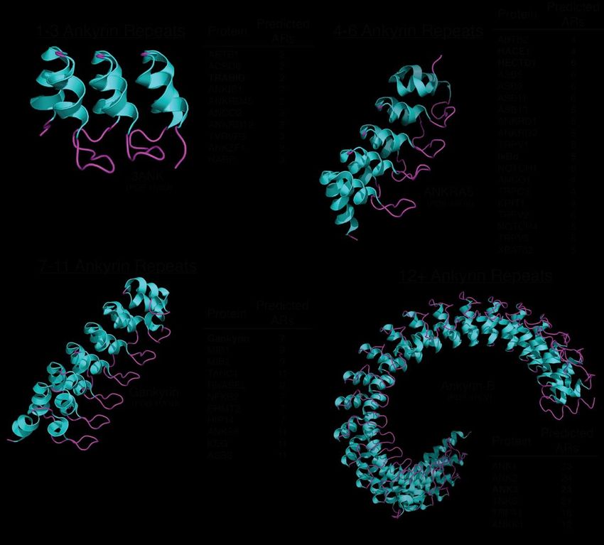
Int. J. Mol. Sci. 2021, 22, 609                                                                                            2 of 13

                                  1.1. Ankyrin-Repeat Containing Proteins
                                        AR domains were first discovered as a repeating sequence in Saccharomyces cerevisiae
                                  cell cycle regulators Swi6, cell division control protein 10 (Cdc10) and Notch in Drosophila
                                  melanogaster [8]. This ~33-amino acid long repeat subsequently was named after the
                                  cytoskeletal protein ANKYRIN, a 206 kDa protein that contains 24 tandem repeats [9].
                                  Since its initial discovery, AR domains have been observed to be present in many eukary-
                                  otic proteins, making this domain potentially the most abundant repeat domain in the
                                  eukaryotic proteome [7]. To date, there are over 367,000 predicted AR domains found
                                  within 68,471 nonredundant proteins annotated in the SMART database [10,11]. With such
                                  prevalence of AR-containing proteins in eukaryotes coupled with AR domains acting as
                                  scaffolds to facilitate protein–protein interactions in the cell, it is speculated the AR domain
                                  originated through evolutionary pressure events to provide the necessary function of
                                  facilitating the variety of signaling pathways eukaryotic organisms use to regulate cellular
                                  homeostasis [6].
                                        Comparing AR-containing proteins has proven to be difficult as each protein has
                                  acquired various characteristics through multiple evolutionary events. This is largely due
                                  to conservation within AR domains relying on various residue types rather than requiring
                                  highly conserved residues at specific sites. Given that specific residue types influence the
                                  protein’s secondary structure coupled with AR domains not relying on specific residue
                                  conservation, it suggests the AR domain is defined primarily on its 3D structural fold rather
                                  than its functional support for AR-containing proteins. For example, highly conserved
                                  regions of AR domains exist between each repeat, whereas variation of hydrophobic
                                  residues can occur while maintaining the structural integrity of the domain [12]. Conserved
                                  motifs that influence α-helical and β-loop folds have recently been identified in AR domains
                                  has allowed for better AR domain identification and prediction from the primary sequence
                                  of a protein.
                                        In comparison to naturally occurring AR domains, there is little deviation from its
                                  typical helix–loop–helix–β–hairpin/loop structure, which is supported through conser-
                                  vation of residue type. Inter-domain interactions tend to be short distances, which aids
                                  in the linear solenoid packing the AR domain fold rather than a typical globular shape.
                                  Both hydrophobic interactions through non-polar regions of the inter and intra α-helices as
                                  well as hydrogen bonding through polar residues found near the N-terminus define and
                                  stabilize the AR domain’s structural integrity [6,13] (Figure 1A).
                                        While the hydrophobic interaction between residues Pro5 and His7 influences the
                                  domain’s L-shape, His7 also interacts with residue Thr4 through hydrogen bonding to
                                  define the 90◦ fold with each additional repeat [6]. The AR domain also requires nonpolar
                                  residues on surface α-helices, specifically at residues 6, 8, 9, 10, 17, 20–22, to limit solvent
                                  accessibility [12], although some variability in these polar residues of N- and C-terminal
                                  AR domains has been observed. Recent insights in AR domain conservation has identified
                                  specific residues and motifs frequently occupying the primary sequence of an AR domain.
                                  This includes the identification of a G-X-TLPHLA motif and two conserved glycine residues
                                  that allow for antiparallel helix termination and β-hairpin loop formation. Throughout
                                  the ~33 amino acids, probability of conserved residues to occur within eukaryotic AR-
                                  containing proteins remains relatively high, with an increase in conservation within the
                                  mid-region (Figure 1B).
                                        While tandem repeats are likely to avoid detrimental mutations deriving from evolu-
                                  tionary events, the AR domain has unique characteristics to its mutation potential. Addition
                                  or substitutions within the primary sequence are tolerated and reported to occur in 9% of
                                  natural AR-containing proteins, while the structural integrity of an AR domain is sensitive
                                  to any residue deletions [14]. The plasticity of the AR domains remains intriguing, con-
                                  sidering additions within the primary sequence can range from a single residue upwards
                                  to a separate folded domain as observed in TRABID [15]. It is noteworthy such additions
                                  typically reside within the loop region preceding each β-hairpin [6]. Observations in intact
                                  AR domains with deletions, on the other hand, tend to be within the antiparallel α-helices
Int. J. Mol. Sci. 2021, 22, 609 3 of 13
to cause helical shortening. This is evident in the cyclin-dependent kinase inhibitor (CKI)
INK4 family of proteins that contain a shortened inner helix brought on by deletions within
the second AR [6]. This variety of insertion size can cause the improper annotation of AR
domain evaluation through various databases due to the misinterpretation of the primary
sequence, thus resulting in the presence of AR domains that may go unnoticed.
To better understand AR domain folding, researchers have recently synthesized
generic two, three and four AR domain protein constructs. These sequences were derived
from approximately 4000 AR domain-containing protein sequences while ensuring general
conservation within the identified AR motifs [7]. Their studies support the notion that the
conservation levels of residues dictate the AR domain structure and function. For example,
the β-hairpin/loop region and short α-helix, which typically acts as the protein recognition
site, remains semiconserved in comparison to the convex surface residues being highly
conserved [7]. The innate flexibility of the AR domain recognition site allows for a multitude
of binding partners, whereas the highly conserved regions act as the structural backbone to
serve as a scaffold in support of the recognition site. Further evaluation utilizing generic
AR constructs will aid in the elucidation of the unique mechanisms used by AR domain-
containing proteins to recognize and bind substrates in the cell, as well as potential features
of this binding pocket amongst other AR-containing proteins in similar protein families.
ol. Sci. 2021, 22, x FOR PEER REVIEW 3 of 14
This also opens the possibility of engineering new AR domain proteins with varying
function based upon the novel discoveries of protein recognition and interactions that AR
domain proteins partake in that could prove useful in drug development.
Figure 1. Structural conservation within ankyrin repeat domains. (A) Residues Thr4, Pro5 and His7 play a critical role in
Figure
providing both the1.90Structural conservation
◦ and L-shape formationwithin
throughankyrin repeat
hydrogen domains.
bonding. The(A) Residuessolenoid
elongated Thr4, Pro5 andof an AR domain
shape
His7 play a critical role in providing both the 90 and L-shape formation through
is predominantly dictated by these three residues while hydrophobic interactions in the core of the AR hydrogen bond-
are required to
ing. The elongated solenoid shape of an AR domain is predominantly dictated by these three resi-
stabilize the domain’s 3D fold. (B) The hidden Markov model (HMM) profile of the AR-containing proteins for N-terminal,
dues while hydrophobic interactions in the core of the AR are required to stabilize the domain’s
internal and C-terminal repeats were analyzed to highlight the occurrence of residues in identified AR domain families. The
3D fold. (B) The hidden Markov model (HMM) profile of the AR-containing proteins for N-termi-
classic G-X-TPLHLA
nal, internalmotif
andwas readily identified
C-terminal to have
repeats were a strong
analyzed probability
to highlight theofoccurrence
occurring inofthese AR-containing
residues in iden- proteins,
whereas intified N- and
both AR domainC-terminal
families.repeats wereG-X-TPLHLA
The classic observed to contain only
motif was portions
readily of thistomotif.
identified have aWhile
strongconservation
within AR domains
probabilityis more prevalent
of occurring inwithin
these AR-containing C-terminal
the internal andproteins, repeats,
whereas the N-terminal
in both AR still retains
N- and C-terminal re- similar AR
peats were observed to contain only portions of this motif. While conservation within AR domains
domain characteristics.
is more prevalent within the internal and C-terminal repeats, the N-terminal AR still retains simi-
lar AR domain characteristics.
1.2. Ubiquitylation Signaling Pathway and Ubiquitin Chain Formation
While the hydrophobic interactionsignaling
The ubiquitylation between pathway
residues Pro5
(USP)and His7 influences
involves the
a highly regulated enzyme
domain’s L-shape, His7that
cascade also interacts
results with
in the residue
covalent Thr4 through
attachment hydrogen
of ubiquitin on bonding to protein [16].
to a substrate
First,
define the 90° fold theeach
with ubiquitin activating
additional repeat enzyme
[6]. The AR(E1)domain
activates
alsoubiquitin
requires in the presence of ATP,
nonpolar
residues on surface α-helices, specifically at residues 6, 8, 9, 10, 17, 20–22, to limit solvent (E2) enzyme
allowing for the subsequent transfer of ubiquitin to the ubiquitin conjugating
viaalthough
accessibility [12], a thioester
somebond. This ininturn
variability theseinitiates the formation
polar residues of an
of N- and E2-E3 ubiquitin ligase
C-terminal
AR domains has been observed. Recent insights in AR domain conservation has identified
specific residues and motifs frequently occupying the primary sequence of an AR domain.
This includes the identification of a G-X-TLPHLA motif and two conserved glycine resi-
dues that allow for antiparallel helix termination and β-hairpin loop formation. Through-Int. J. Mol. Sci. 2021, 22, 609 4 of 13
complex that directs the transfer of ubiquitin onto a substrate protein to form a stable
isopeptide bond between the C-terminus of ubiquitin and the ε-amine of a lysine residue
on the substrate [16]. This post-translational modification is critical for regulating a wide
array of biological processes including intracellular protein localization, the DNA damage
response, protein activation or inactivation, innate immune response and 26S proteasomal
degradation [17].
The human genome encodes for two E1 enzymes, 37 E2 enzymes, and hundreds
E3 ligases allowing for exquisite specificity in substrate recognition for ubiquitin attach-
ment [18]. There are three subfamilies of E3 ubiquitin ligases that are classified based
on their structural and functional similarities—the really interesting new gene (RING),
homologous to E6AP C-terminus (HECT), and RING-between-RING (RBR) E3 ubiquitin
ligases [17]. The RING E3 ligases are the largest most well-studied subfamily with over
600 E3 ligases currently annotated in the human genome. The RING E3 ubiquitin ligases
use their Zn2+ -binding RING domain as a scaffold that engages and properly orients
the E2~ubiquitin complex for efficient transfer of ubiquitin onto a substrate protein [19].
The HECT E3 ubiquitin ligases family is comprised of 28 members that all have a highly
conserved bi-lobal HECT domain at their C-termini that contains an absolutely required
cysteine residue for accepting ubiquitin from an E2 and the subsequent transfer on to its
substrates [17]. HECT E3 ubiquitin ligases use their N-terminal protein–protein interaction
domains for to carefully recognize and recruit proteins for ubiquitylation [17]. There are
14 identified members of the RBR E3 ubiquitin ligase subfamily that each contain a RING1-
IBR-RING2 domain that employ a hybrid RING/HECT mechanism to attach ubiquitin on
to its substrates [20]. Similar to the RING E3 ligases, the RBRs use their RING1 domain
to recruit the E2~ubiquitin complex, which in turn is able to transfer ubiquitin onto its
conserved catalytic cystine residue within the RING2 domain (aka Required for catalysis;
Rcat) prior to covalently tagging its substrates with ubiquitin [21,22].
The specific attachment site(s) of ubiquitin on a substrate protein is decided by the E2-
E3 pair [23], and depending on the ubiquitin chain type, the fate of the ubiquitin tagged pro-
tein is decided. For example, the attachment of a single ubiquitin (i.e., monoubiquitylation)
can signal for various biological processes such as endocytosis, while ubiquitin chains (i.e.,
polyubiquitylation) connected by identical (homotypic) or varying (heterotypic/branched)
linkers influence a myriad of signaling pathways yet can remain highly specific with each
additional attachment. K48-linked polyubiquitin chain formation is the most well studied
and understood modification that targets the substrate protein for degradation by the 26S
proteasome [24,25]. Alternative polyubiquitin chains with different linkages have also been
shown to regulate cellular processes. These include K6, K27, K29 and K63 polyubiquity-
lation to initiate the DNA damage response [26]; M1 and K11 polyubiquitin to activate
the NF-κβ signaling pathway [26]; and various branched polyubiquitylation chains (i.e.,
K11/K48, K29/K48, K48/K63) have been observed to target proteins for proteolysis [27].
The complexity and permutations of ubiquitin chain types can include the modification of
ubiquitin residues such as lysine acetylation that can repress K6 and K48 polyubiquitin
chain formation [25], and serine/threonine phosphorylation that can act as an activator of
the RBR E3 ubiquitin ligase parkin [28].
2. Ankyrin Repeats and the USP—Working Together to Regulate Intracellular Processes
With the prevalence of AR domains in eukaryotic proteins (Figure 2) coupled with
all of the reported intracellular processes regulated by ubiquitin, it not surprising that
there are some important proteins and enzymes that take part at the poorly understood
intersection of AR domains and ubiquitylation. This review aims to highlight the functional
roles of some prominent AR domain containing proteins and what is currently known and
unknown on their ubiquitin biology.ol. Sci. 2021, 22, x FOR PEER REVIEW 6 of 14
Int. J. Mol. Sci. 2021, 22, 609 5 of 13
Figure 2. Examples of AR domain-containing proteins based on AR repeat number. AR domains can contain a wide variety
Figure 2. Examples of AR domain-containing proteins based on AR repeat number. AR domains
of repeats, can
but contain
predominately
a wide contain four
variety of to six. but
repeats, With each repeat, the
predominately 90◦ and
contain L-shape
four to six. formation
With eachbecomes more prevalent
repeat, the
resulting in90
a 3D
and L-shape formation becomes more prevalent resulting in a 3D revolving twist. In general, complexity of
revolving twist. In general, with each additional AR repeat, there is a correlation between
the AR-containing
with eachprotein’s biological
additional role in
AR repeat, cellular
there processes that
is a correlation it regulates.
between This of
complexity maythebeAR-containing
due to the increased number
in substrate binding pockets available that could be recognized by the substrate. The AR domain-containing
protein’s biological role in cellular processes that it regulates. This may be due to the increased proteins
number in substrate binding pockets
discussed in this review are highlighted in bold. available that could be recognized by the substrate. The AR
domain-containing proteins discussed in this review are highlighted in bold.
2.1. HACE1 and HECTD1: Ankyrin Repeat Containing HECT E3 Ubiquitin Ligases
2.1. HACE1 and HECTD1: Ankyrin Repeat Containing HECT E3 Ubiquitin Ligases
Many HECT E3 ubiquitin ligases have been identified to have dual functions through
Many HECT E3 ubiquitin
their ligases havewith
specific interactions beencellular
identified to have
proteins dual
that is functions
completely through
independent of their
their specific interactions withactivities.
ubiquitylating cellular proteins that is completely
A prime example independent
is HECT domain of their
and ankyrin repeat containing
ubiquitylating activities. A prime example is HECT domain and ankyrin repeat contain-
E3 ubiquitin protein ligase 1 (HACE1), which was first identified in 2004 as a tumor
ing E3 ubiquitinsuppressor
protein ligase
caused1 (HACE1), which was
by chromosomal first identified
translocation in 2004
that leads as a tumor
to sporadic Wilms tumor with
suppressor caused by chromosomal translocation that leads to sporadic Wilms
decreased HACE1 expression [29]. Apart from containing a highly conserved tumor C-terminal
with decreased HECT
HACE1 expression
domain [29].for
required Apart from containing
ubiquitylation, HACE1 a highly conserved
was also C-ter- of the HECT
the first member
minal HECT domain required
E3 ubiquitin for ubiquitylation,
ligases to contain six HACE1
predictedwasARalso the first
domains at member of the (Figure 3A).
its N-terminus
HECT E3 ubiquitin ligases to contain six predicted AR domains at its N-terminus (Figure
3A).Mol. Sci. 2021, 22, x FOR PEER REVIEW 7 of 14
Int. J. Mol. Sci. 2021, 22, 609 6 of 13
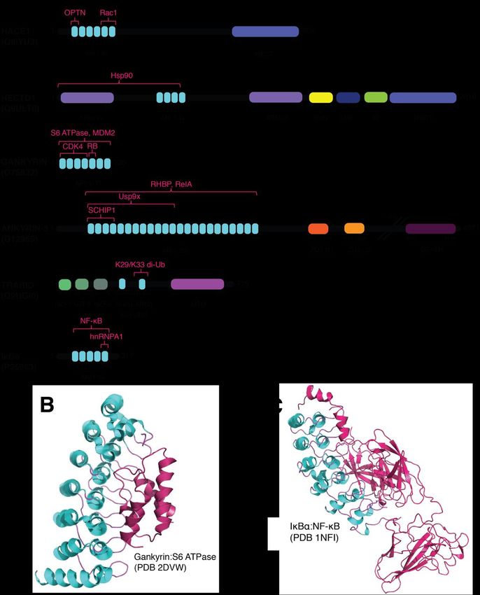
Figure architecture
Figure 3. Domain 3. Domain architecture of AR domain-containing
of AR domain-containing proteins andproteins and AR domain-complex
AR domain-complex formation.for- (A) Putative
mation. (A) Putative domain architecture of AR-containing proteins discussed in this
domain architecture of AR-containing proteins discussed in this review. While each protein has a variety of domains review.
While each protein has a variety of domains that facilitate protein–protein interactions, the AR
that facilitate protein–protein interactions, the AR domains are highlighted for their role in mediating the ubiquitylation
domains are highlighted for their role in mediating the ubiquitylation signaling pathway. Domain
signaling pathway. Domain abbreviations used include: AR, ankyrin repeat; HECT, homologous to E6AP C-terminus;
abbreviations used include: AR, ankyrin repeat; HECT, homologous to E6AP C-terminus; ARM,
ARM, armadillo repeat-containing
armadillo repeat-containingdomain; SUN,SUN,
domain; SAD1/UNC
SAD1/UNC domain;
domain; MIB, mind-bomb
MIB, mind-bombdomain;
domain;H, H,helical bundle;
ZU5, ZU5 domain;
helical bundle; ZU5, ZU5 domain; DEATH, death domain; NZF, Npl4 zinc finger domain; OUT, Identified
DEATH, death domain; NZF, Npl4 zinc finger domain; OUT, ovarian tumor domain.
substrates and sites oftumor
ovarian interaction
domain. are highlighted in magenta.
Identified substrates andSubstrate abbreviations
sites of interaction areused included:
highlighted in OPTN, optineurin [30];
magenta.
Substrate
Rac1, Ras-related abbreviations
C3 botulinum toxinused included:
substrate OPTN,
1 [31]; optineurin
Hsp90, heat shock[30];protein
Rac1, Ras-related
90 [32]; MDM2,C3 botulinum
mouse double minute
toxinCDK4,
2 homolog [33]; substrate 1 [31]; Hsp90, heat
cyclin-dependent shock
kinase protein
4 [34]; 90 [32]; MDM2,protein
RB, retinoblastoma mouse double minute
[35]; RHBP, 2 homolog
rhodnius heme-binding
protein [36]; [33];
RelA,CDK4, cyclin-dependent
transcription factor p65 kinase 4 [34];ubiquitin
[37]; Usp9x, RB, retinoblastoma protein 9[35];
specific peptidase RHBP,[38];
X-Linked rhodnius
proteosomal subunit
heme-binding
S6 ATPase [39]; protein [36];interacting
SCHIP1, schwannomin RelA, transcription factor
protein 1 [40]; p65 [37];
di-Ub, Usp9x, ubiquitin
di-ubiquitin specificheterogeneous
[41]; hnRNPA1, peptidase nuclear
9 X-Linked [38]; proteosomal subunit S6 ATPase [39]; SCHIP1, schwannomin interacting protein 1
ribonucleoprotein A1 [42]; NF-κβ [43]. (B) Structure of AR-containing protein gankyrin in complex with S6 ATPase (PDB
[40]; di-Ub, di-ubiquitin [41]; hnRNPA1, heterogeneous nuclear ribonucleoprotein A1 [42]; NF-κβ
2DVW; [39]). Gankyrin utilizes the internal b-hairpin loops to bind onto the C-terminal a-helix of S6 ATPase. (C) Structure
[43]. (B) Structure of AR-containing protein gankyrin in complex with S6 ATPase (PDB 2DVW;
of AR-containing
[39]).protein
Gankyrin IκBα in complex
utilizes with NF-κB
the internal (PDB
b-hairpin 1NFI;
loops to [43]).
bind onto the C-terminal a-helix of S6
ATPase. (C) Structure of AR-containing protein IκBα in complex with NF-κB (PDB 1NFI; [43]).Int. J. Mol. Sci. 2021, 22, 609 7 of 13
Besides being genetically linked to Wilms tumor formation, HACE1 has also been
reported to play a role in the onset of liver, lymphoma, osteosarcoma, breast and colorectal
cancers [44–47]. Studies have also revealed that HACE1 mediates Golgi membrane forma-
tion during cell division through the ubiquitylation of Rab proteins for proteolysis [48].
Various N-terminal putative protein interaction domains have been annotated for each
HECT E3 ubiquitin ligase family member, which gave rise to the sub-classification of this
family. This variety is predicted to be involved in substrate recognition and recruitment [17].
For instance, HACE1 has been reported to interact with a recently identified substrate
optineurin (OPTN) through its AR domains (Figure 3A) [30]. The HACE1-dependent
ubiquitylation of OPTN resulted in K27 and K48-specific polyubiquitin chains were ob-
served to be covalently attached onto OPTN at K193 that can then promote the formation
of an autophagy receptor complex with p62/SQSTM1. While K48 polyubiquitin chains
are ideally utilized to signal for proteasomal degradation, K27 polyubiquitin chains can
also be recognized by the proteasome but are rarely recognized and cleaved by deubiqui-
tylases [49]. It is suggested that HACE1-dependent ubiquitylation of OTPN is required
to encourage this complex formation, which in turn accelerates the elimination process of
p62 leading to suppression of lung carcinoma cell growth [30].
HACE1 also acts as an adaptor protein that is critical in cardiac protection during
hemodynamic stress. For example, HACE1 facilitates the transfer of ubiquitylated proteins
p62 and microtubule-associated proteins 1A/1B light chain 3B (LC3) for autophagic degra-
dation. This is accomplished through recognizing and binding to these modified proteins
via its AR domain, independent of its typical E3 ligase activity [50]. HACE1 also possesses
antioxidative stress response capabilities; it was previously reported that HACE1 promotes
the activation of nuclear factor erythroid 2-related factor 2 (NRF2), a key regulator of the
antioxidation stress response to mediate redox homeostasis through activation of antiox-
idative genes [51]. Decreased HACE1 expression levels were found to severely alter the
expression levels and stability of NRF2, inhibiting antioxidative stress response precursors
which eliminate oxidative triggers such as mutant Huntington protein [51,52]. Patients with
Huntington’s disease (HD) have also been observed to have decreased HACE1 levels in the
striatum, the region of the brain where HD initially manifests [51,52]. These cumulative
results suggest that HACE1 can serve as an early stage neurodegenerative disease target.
Improving our understanding on the mechanisms used by HACE1 to bind to its substrates
through its AR domains is paramount as it remains unclear how exactly HACE1 utilizes
its AR domain to modify these essential proteins. Further evaluation into this prominent
domain within HACE1 can also potentially identify abnormalities in substrate recognition
and binding the AR domain typically takes on, which can lead towards the development
of neurodegenerative disease treatment.
Another intriguing yet poorly understood member of the HECT E3 ubiquitin ligase
family is HECTD1. HECTD1 was first classified as a HECT E3 ubiquitin ligase in 2007 upon
the discovery of its role in head mesenchyme development and neural tube closure [53].
Similar to HACE1, HECTD1 also contains AR domains in its N-terminus (Figure 3A).
HECTD1 is also predicted through sequence similarity to have two ARMs that flank four
AR domains, a SADL/UNC1 (SUN) domain, a mind-bomb (MIB) domain and a helical
bundle that are located N-terminal to its HECT domain [54–56].
HECTD1 has been reported to play an important role in embryonic development by
regulating cranial mesenchyme cellular behavior [53]. Researchers have shown that the
N-terminal region of HECTD1 (ARM1 and the N-terminal AR domains; a.a. 1-551) was
shown to interact with heat shock protein 90 (Hsp90) to influence cranial mesenchymal
cell behavior (Figure 3A). Hsp90 is secreted from the cell to increase cellular motility,
however, when HECTD1 targets Hsp90 via K63 polyubiquitin chain, this changes Hsp90’s
intracellular localization which in turn inhibits its ability to be expressed extracellularly [32].
Interestingly, the boundaries for the predicted AR domains in HECTD1 should end at
residue 612, where the third repeat ends at residue 491, whereas the fourth repeat starts
at residue 579. This larger gap between AR domains 3 and 4 of HECTD1 is atypical, thusInt. J. Mol. Sci. 2021, 22, 609 8 of 13
it can be speculated that either another folded domain or an unrecognized AR sequence
may exist within this gap that might be required for Hsp90 recognition and binding. With
HECTD1 also containing its first ARM repeat between residues 8-254, it is possible that
Hsp90 could be recognized through a different domain. Further studies are needed to
clarify the mechanism for HECTD1-dependent recruitment of Hsp90.
HECTD1 plays an intricate role in regulating epithelial-mesenchymal transition (EMT)
through ubiquitylation. EMT is an integral process that allows for plasticity in a phenotype
switch in epithelial cells to mesenchymal cells [57]. The activation of EMT is essential
during development, but it can also be triggered in various cancers through influencing
cell-cell adhesion and metastasis [58]. HECTD1 contains eight nuclear localization se-
quences (NLS) and four nuclear export signals (NES), giving rise to its presence within the
nucleus despite typical cytoplasmic localization; this localization is specifically mediated
through exportin 1 (XPO1) [59]. With this ability to translocate from the cytosol to the
nucleus, HECTD1 is able to regulate zinc finger protein SNAIL1 (SNAIL) expression levels.
Since SNAIL increases mesenchymal characteristics in epithelial cells, HECTD1 controls
SNAIL through ubiquitylation to targeting it SNAIL for cytoplasmic translocation and its
subsequent proteasomal degradation. This translocation causes the repression of the tumor
suppressor protein E-cadherin and helps to maintain proper EMT during development [60].
E-cadherin colocalizes to the cell membrane and encourages cell-cell adhesion which is
believed to initiate tumor metastasis in its absence [61]. SNAIL regulates E-cadherin lev-
els through binding at specific E-box sites on its promotor to inhibit expression [60,62].
Another EMT regulator protein, actin cross-linking factor 7 (ACF7), has been shown to
interact with Rac1 to stabilize membrane protrusions while simultaneously increasing
motility [63]. HECTD1 was also identified to regulate EMT through targeting ACF7 for pro-
teasomal degradation. A negative correlation was observed between the expression levels
of both HECTD1 and ACF7 in breast cancer cells that further encouraged metastasis and
cytotoxin drug resistance due to the inability to readily target ACF7 for degradation [63].
Since histone methyltransferase interacts with SNAIL through its AR domain to regulate
EMT [64], as well as ACF7 having multiple protein–protein interaction domains [65], it can
be speculated that HECTD1 regulates EMT by its AR domains to recognize and bind to
both SNAIL and ACF7 to target these proteins for degradation.
Beyond EMT, HECTD1 has also been shown to play an essential in regulating Iκβα,
a key regulator of the NF-kB transcription factor. Specifically, HECTD1 mediates Iκβα
K48 polyubiquitylation through its direct interaction with the ribosomal protein subunit
3 (Rsp3). HECTD1 forms a ternary complex with latexin (LAX), Iκβα and ribosomal
protein subunit 3 (Rps3); this triggers Iκβα ubiquitylation and upregulates the NF-κβ
stimulated inflammatory stress response [66]. Interestingly, only Rsp3 detachment from
the complex attenuates LEX/HECTD1 interaction [66]. Overall, the formation of the
Rsp3/HECTD1 complex encourages ubiquitylation of Iκβα and LAX can competitively
bind to the Rsp3 binding site to inhibit ubiquitylation. Further biochemical and biophysical
analysis are needed to further clarify how HECTD1 uses its AR domain for substrate
recognition and subsequent protein degradation by the proteasome.
2.2. Usp9x and TRABID: Deubiquitylases Facilitating Ub Cleavage through Internal and External
AR Domains
Deubiquitylation involves the enzymatic removal of ubiquitin or ubiquitin chains
from a substrate protein, and arguably plays the most critical role in regulating ubiqui-
tylated substrate fate [67]. While the human genome encodes for ~90 deubiquitylases
(DUBs) [67,68], their biological relevance beyond deubiquitylation is still unclear. Recent
studies indicate that DUBs may be an ideal drug target [69] through enhancement or
inhibition of substrate binding [70].
Ubiquitin specific peptidase 9 X-linked (Usp9x) is a prominently known for acting
as the DUB to protect SMURF1 and RNF115 from proteolysis [71,72], and there is emerg-
ing evidence that Usp9x can also interact with other AR-containing proteins to regulate
intracellular events. For instance, Usp9x has been observed to interact with the cytoskele-Int. J. Mol. Sci. 2021, 22, 609 9 of 13
tal protein ankyrin-3 (ANK3) to promote spine morphogenesis [38,73]. Studies have
shown that the disruption of Usp9x and ANK3 complex formation results in deficient
synaptic structural maintenance that can contribute to neurodevelopmental disorders [38].
Usp9x/ANK3 complex formation is stimulated by TFG-β signaling leading to the phos-
phorylation of Usp9x, which in turn increases Usp9x binding affinity to the AR repeats of
ANK3 [73] (Figure 3A). Interestingly, abnormal expression of Usp9x and ANK3 have also
be correlated with neurodevelopmental and neurodegenerative and psychiatric disorders
such as autism-spectrum disorder, Parkinson’s disease, Alzheimer’s disease, epilepsy, and
bipolar disorder [73–76]. These cumulative reports suggest that AR domain-containing
proteins are clinically relevant in various diseases and the USP through E3 ubiquitin ligase
protection. Further investigation should be directed towards how other DUBs utilize their
own or external AR domains to facilitate USP.
Intriguingly, TRAF-binding domain (TRABID) was found to contain two AR domains
between a ubiquitin binding domain, named AnkUBD [41], which remained hidden due to
the elusive characteristics of its AR domain (Figure 3A). TRABID has been suggested to
play a role in the Wnt signaling pathway by using its AnkUBD domain to properly orient
heterotypic polyubiquitin chains to specifically cleave K29 and K33 polyubiquitin linkages
(Figure 3A) [15]. TRABID has also been shown to regulate the expression levels of the
transcriptional regulator Twist1 in hepatocellular carcinoma (HCC). TRABID is responsible
for the specific cleavage of K63-linked polyubiquitin chains from Twist1 resulting in
Twist1 being able to form a complex with beta-transducin repeats-containing protein
(β-TrCP) and the subsequent K48-specific polyubiquitylation of Twist1 for proteasomal
degradation [77]. With the emerging evidence that DUBs and AR-containing proteins could
serve as novel drug targets, it is imperative that increased attention be directed towards
improving our knowledge on how AR proteins are involved in protein–protein interactions
and intracellular targeting.
2.3. Gankyrin: An Oncogenic AR Domain-Containing Protein
Gankyrin is a seven AR domain-containing liver oncoprotein that is involved in a
myriad of cellular processes including cell cycle progression, liver regeneration, protein
translocation and enzymatic regulation (Figure 3A) [78–80]. When comparing the primary
sequences of gankyrin’s seven AR repeats there is significant conservation within the AR
repeats especially between the first six AR repeats (Figure 3A,B). Previous studies have
shown that the overexpression of gankyrin is linked to the onset of various malignancies
making it a potential oncogenic biomarker [78,81]. It has also been suggested that gankyrin
competes for cyclin-dependent kinase 4 (CDK4) binding with INK4a in order to regulate
transcription factor e2f expression (Figure 3A) [80,82].
Importantly, gankyrin plays a critical role in the encouragement of mouse double
minute 2 (MDM2), a RING E3 ubiquitin ligase, to ubiquitylate p53 for cytoplasmic localiza-
tion and degradation [83]. Specifically, gankyrin utilizes its AR domain to bind to MDM2 to
facilitate the MDM2-dependent ubiquitylation of p53 (Figure 3A) [83]. Residue deletions
within the gankyrin AR domain abolished gankyrin/MDM2 interactions but did not affect
MDM2/p53 interactions, suggesting that gankyrin associates with MDM2 away from the
p53-MDM2 binding site. Evaluating AR-containing proteins in comparison to gankyrin
can reveal potential ubiquitylating mediating functions.
Gankyrin also has been demonstrated to be involved in EMT through its regulation of
downstream cytokines interleukin 6 (IL-6) and transforming growth factor beta (TGF-β)
that induce the EMT phenotype [84]. In contrast to HECTD1, increased gankyrin expression
levels was observed to cause decreased E-cadherin expression in non-small cell lung cancer
(NSCLC) cells that overexpress gankyrin [84].
The mechanisms used by gankyrin to regulate transcription factor levels is currently
unclear and need further examination. Likewise, a detailed comparison of gankyrin to
a similar AR domain-containing protein that regulates nuclear factor-kappa-β (NF-κβ)
expression levels is warranted. For instance, the Iκβα is a five AR-containing regulatoryInt. J. Mol. Sci. 2021, 22, 609 10 of 13
protein with capabilities of regulating NF-κβ expression levels (Figure 3A,C). Once Iκβα is
modified via ubiquitylation and phosphorylation, it is released from the p50/RelA (p65)
complex to allow for nuclear localization and NF-κβ activation, followed by ubiquitin-
mediated degradation [78,85]. Similarly, gankyrin was discovered to regulate NF-κβ
through its AR domain (Figure 3A) [42]. Gankyrin was observed to bind to and inhibit
the NF-kβ/RelA complex activity by preventing NF-kβ nuclear localization [78]. Similar
methods should be evaluated when identifying other AR domain-containing proteins
that play similar roles in various signaling pathways to further characterize and better
understand the significance of the unique AR domain.
3. AR Domain-Containing Proteins with Unknown Ubiquitylation Mechanisms—
What Is Next?
AR domain-containing proteins are ubiquitous and play essential roles in numerous
biological processes that can also influence the onset of various diseases and disorders.
Many AR domain-containing proteins have been identified in numerous occasions to
support the USP and reverse the process of deubiquitylation through sequence alignment.
Being essential scaffolds to mediate protein–protein interactions, it is intriguing that this
domain is not functionally driven but rather dependent on the structural characteristics
of the AR domain. Understanding how these AR-domain containing proteins and their
role in ubiquitin signaling on the molecular level is a major challenge in our present
understanding of their function(s) and activity. Expanded biochemical and biophysical
examinations on the novel mechanisms used for AR-domain protein recruitment are
needed. By experimentally identifying and validating potential substrates and interactors
for these AR-domain proteins, we will be able to improve our knowledge of how these
proteins work and will be able to better assess if these proteins could serve as novel drug
targets or as biomarkers for disease.
Author Contributions: Conceptualization, E.I.K. and D.E.S.; writing—original draft preparation,
review and editing, E.I.K. and D.E.S.; supervision, D.E.S. All authors have read and agreed to the
published version of the manuscript.
Funding: This research was supported by the National Institutes of Health, R15GM126432.
Data Availability Statement: All figures and discussed literature are found in the main text of
this review.
Conflicts of Interest: The authors declare no conflict of interest.
References
1. Andrade, M.A.; Perez-Iratxeta, C.; Ponting, C.P. Protein repeats: Structures, functions, and evolution. J. Struct. Biol. 2001, 134,
117–131.
2. Yoder, M.D.; Lietzke, S.E.; Jurnak, F. Unusual structural features in the parallel beta-helix in pectate lyases. Structure 1993, 1,
241–251.
3. Kobe, B.; Kajava, A.V. The leucine-rich repeat as a protein recognition motif. Curr. Opin. Struct. Biol. 2001, 11, 725–732.
4. Groves, M.R.; Barford, D. Topological characteristics of helical repeat proteins. Curr. Opin. Struct. Biol. 1999, 9, 383–389. [PubMed]
5. Pellegrini, M. Tandem Repeats in Proteins: Prediction Algorithms and Biological Role. Front. Bioeng. Biotechnol. 2015, 3, 143.
6. Mosavi, L.K.; Cammett, T.J.; Desrosiers, D.C.; Peng, Z.Y. The ankyrin repeat as molecular architecture for protein recognition.
Protein Sci. 2004, 13, 1435–1448. [PubMed]
7. Mosavi, L.K.; Minor, D.L., Jr.; Peng, Z.Y. Consensus-derived structural determinants of the ankyrin repeat motif. Proc. Natl. Acad.
Sci. USA 2002, 99, 16029–16034. [PubMed]
8. Breeden, L.; Nasmyth, K. Similarity between cell-cycle genes of budding yeast and fission yeast and the Notch gene of Drosophila.
Nature 1987, 329, 651–654.
9. Lux, S.E.; John, K.M.; Bennett, V. Analysis of cDNA for human erythrocyte ankyrin indicates a repeated structure with homology
to tissue-differentiation and cell-cycle control proteins. Nature 1990, 344, 36–42.
10. Letunic, I.; Doerks, T.; Bork, P. SMART: Recent updates, new developments and status in 2015. Nucleic Acids Res. 2015, 43,
D257–D260.
11. Letunic, I.; Bork, P. 20 years of the SMART protein domain annotation resource. Nucleic Acids Res. 2018, 46, D493–D496. [PubMed]Int. J. Mol. Sci. 2021, 22, 609 11 of 13
12. Parra, R.G.; Espada, R.; Verstraete, N.; Ferreiro, D.U. Structural and Energetic Characterization of the Ankyrin Repeat Protein
Family. PLoS Comput. Biol. 2015, 11, e1004659.
13. Li, J.; Mahajan, A.; Tsai, M.D. Ankyrin repeat: A unique motif mediating protein-protein interactions. Biochemistry 2006, 45,
15168–15178. [PubMed]
14. Galpern, E.A.; Freiberger, M.I.; Ferreiro, D.U. Large Ankyrin repeat proteins are formed with similar and energetically favorable
units. PLoS ONE 2020, 15, e0233865.
15. Kristariyanto, Y.A.; Abdul Rehman, S.A.; Campbell, D.G.; Morrice, N.A.; Johnson, C.; Toth, R.; Kulathu, Y. K29-selective ubiquitin
binding domain reveals structural basis of specificity and heterotypic nature of k29 polyubiquitin. Mol. Cell 2015, 58, 83–94.
16. Hershko, A.; Ciechanover, A. The ubiquitin system. Annu. Rev. Biochem. 1998, 67, 425–479.
17. Wang, Y.; Argiles-Castillo, D.; Kane, E.I.; Zhou, A.; Spratt, D.E. HECT E3 ubiquitin ligases—Emerging insights into their biological
roles and disease relevance. J. Cell Sci. 2020, 133, jcs228072.
18. Komander, D. The emerging complexity of protein ubiquitination. Biochem. Soc. Trans. 2009, 37, 937–953.
19. Buetow, L.; Huang, D.T. Structural insights into the catalysis and regulation of E3 ubiquitin ligases. Nat. Rev. Mol. Cell Biol. 2016,
17, 626–642.
20. Dove, K.K.; Klevit, R.E. RING-between-RING E3 Ligases: Emerging Themes amid the Variations. J. Mol. Biol. 2017, 429,
3363–3375.
21. Spratt, D.E.; Walden, H.; Shaw, G.S. RBR E3 ubiquitin ligases: New structures, new insights, new questions. Biochem. J. 2014, 458,
421–437. [CrossRef] [PubMed]
22. Wenzel, D.M.; Lissounov, A.; Brzovic, P.S.; Klevit, R.E. UBCH7 reactivity profile reveals parkin and HHARI to be RING/HECT
hybrids. Nature 2011, 474, 105–108. [CrossRef] [PubMed]
23. David, Y.; Ternette, N.; Edelmann, M.J.; Ziv, T.; Gayer, B.; Sertchook, R.; Dadon, Y.; Kessler, B.M.; Navon, A. E3 ligases determine
ubiquitination site and conjugate type by enforcing specificity on E2 enzymes. J. Biol. Chem. 2011, 286, 44104–44115. [CrossRef]
[PubMed]
24. Komander, D.; Rape, M. The ubiquitin code. Annu. Rev. Biochem. 2012, 81, 203–229. [CrossRef] [PubMed]
25. Swatek, K.N.; Komander, D. Ubiquitin modifications. Cell Res. 2016, 26, 399–422. [CrossRef] [PubMed]
26. Akutsu, M.; Dikic, I.; Bremm, A. Ubiquitin chain diversity at a glance. J. Cell Sci. 2016, 129, 875–880. [CrossRef] [PubMed]
27. Kane, E.I.; Spratt, D.E. New Discoveries on the Roles of “Other” HECT E3 Ubiquitin Ligases in Disease Development. IntechOpen
2020. Available online: https://www.intechopen.com/books/ubiquitin-proteasome-pathway/new-discoveries-on-the-roles-
of-other-hect-e3-ubiquitin-ligases-in-disease-development (accessed on 1 December 2020). [CrossRef]
28. Zhuang, N.; Li, L.; Chen, S.; Wang, T. PINK1-dependent phosphorylation of PINK1 and Parkin is essential for mitochondrial
quality control. Cell Death Dis. 2016, 7, e2501. [CrossRef]
29. Anglesio, M.S.; Evdokimova, V.; Melnyk, N.; Zhang, L.; Fernandez, C.V.; Grundy, P.E.; Leach, S.; Marra, M.A.; Brooks-Wilson,
A.R.; Penninger, J.; et al. Differential expression of a novel ankyrin containing E3 ubiquitin-protein ligase, Hace1, in sporadic
Wilms’ tumor versus normal kidney. Hum. Mol. Genet. 2004, 13, 2061–2074. [CrossRef]
30. Liu, Z.; Chen, P.; Gao, H.; Gu, Y.; Yang, J.; Peng, H.; Xu, X.; Wang, H.; Yang, M.; Liu, X.; et al. Ubiquitylation of autophagy receptor
Optineurin by HACE1 activates selective autophagy for tumor suppression. Cancer Cell 2014, 26, 106–120. [CrossRef]
31. Andrio, E.; Lotte, R.; Hamaoui, D.; Cherfils, J.; Doye, A.; Daugaard, M.; Sorensen, P.H.; Bost, F.; Ruimy, R.; Mettouchi, A.; et al.
Identification of cancer-associated missense mutations in hace1 that impair cell growth control and Rac1 ubiquitylation. Sci. Rep.
2017, 7, 44779. [CrossRef] [PubMed]
32. Sarkar, A.A.; Zohn, I.E. Hectd1 regulates intracellular localization and secretion of Hsp90 to control cellular behavior of the
cranial mesenchyme. J. Cell Biol. 2012, 196, 789–800. [CrossRef] [PubMed]
33. Higashitsuji, H.; Liu, Y.; Mayer, R.J.; Fujita, J. The oncoprotein gankyrin negatively regulates both p53 and RB by enhancing
proteasomal degradation. Cell Cycle 2005, 4, 1335–1337. [CrossRef] [PubMed]
34. Li, J.; Tsai, M.D. Novel insights into the INK4-CDK4/6-Rb pathway: Counter action of gankyrin against INK4 proteins regulates
the CDK4-mediated phosphorylation of Rb. Biochemistry 2002, 41, 3977–3983. [CrossRef]
35. Higashitsuji, H.; Itoh, K.; Nagao, T.; Dawson, S.; Nonoguchi, K.; Kido, T.; Mayer, R.J.; Arii, S.; Fujita, J. Reduced stability of
retinoblastoma protein by gankyrin, an oncogenic ankyrin-repeat protein overexpressed in hepatomas. Nat. Med. 2000, 6, 96–99.
[CrossRef]
36. Lopez, C.; Metral, S.; Eladari, D.; Drevensek, S.; Gane, P.; Chambrey, R.; Bennett, V.; Cartron, J.P.; Le Van Kim, C.; Colin, Y. The
ammonium transporter RhBG: Requirement of a tyrosine-based signal and ankyrin-G for basolateral targeting and membrane
anchorage in polarized kidney epithelial cells. J. Biol. Chem. 2005, 280, 8221–8228. [CrossRef]
37. Huang, Y.C.; Hsiao, Y.C.; Chen, Y.J.; Wei, Y.Y.; Lai, T.H.; Tang, C.H. Stromal cell-derived factor-1 enhances motility and integrin
up-regulation through CXCR4, ERK and NF-kappaB-dependent pathway in human lung cancer cells. Biochem. Pharmacol. 2007,
74, 1702–1712. [CrossRef]
38. Yoon, S.; Parnell, E.; Kasherman, M.; Forrest, M.P.; Myczek, K.; Premarathne, S.; Sanchez Vega, M.C.; Piper, M.; Burne, T.H.J.;
Jolly, L.A.; et al. Usp9X Controls Ankyrin-Repeat Domain Protein Homeostasis during Dendritic Spine Development. Neuron
2020, 105, 506–521. [CrossRef]
39. Nakamura, Y.; Nakano, K.; Umehara, T.; Kimura, M.; Hayashizaki, Y.; Tanaka, A.; Horikoshi, M.; Padmanabhan, B.; Yokoyama, S.
Structure of the oncoprotein gankyrin in complex with S6 ATPase of the 26S proteasome. Structure 2007, 15, 179–189. [CrossRef]Int. J. Mol. Sci. 2021, 22, 609 12 of 13
40. Papandreou, M.J.; Vacher, H.; Fache, M.P.; Klingler, E.; Rueda-Boroni, F.; Ferracci, G.; Debarnot, C.; Piperoglou, C.;
Garcia del Cano, G.; Goutebroze, L.; et al. CK2-regulated schwannomin-interacting protein IQCJ-SCHIP-1 association with
AnkG contributes to the maintenance of the axon initial segment. J. Neurochem. 2015, 134, 527–537. [CrossRef]
41. Licchesi, J.D.; Mieszczanek, J.; Mevissen, T.E.; Rutherford, T.J.; Akutsu, M.; Virdee, S.; El Oualid, F.; Chin, J.W.; Ovaa, H.;
Bienz, M.; et al. An ankyrin-repeat ubiquitin-binding domain determines TRABID’s specificity for atypical ubiquitin chains. Nat.
Struct. Mol. Biol. 2011, 19, 62–71. [CrossRef] [PubMed]
42. Hay, D.C.; Kemp, G.D.; Dargemont, C.; Hay, R.T. Interaction between hnRNPA1 and IkappaBalpha is required for maximal
activation of NF-kappaB-dependent transcription. Mol. Cell Biol. 2001, 21, 3482–3490. [CrossRef] [PubMed]
43. Jacobs, M.D.; Harrison, S.C. Structure of an IkappaBalpha/NF-kappaB complex. Cell 1998, 95, 749–758. [CrossRef]
44. El-Naggar, A.M.; Clarkson, P.W.; Negri, G.L.; Turgu, B.; Zhang, F.; Anglesio, M.S.; Sorensen, P.H. HACE1 is a potential tumor
suppressor in osteosarcoma. Cell Death Dis. 2019, 10, 21. [CrossRef] [PubMed]
45. Li, J.C.; Chang, X.; Chen, Y.; Li, X.Z.; Zhang, X.L.; Yang, S.M.; Hu, C.J.; Zhang, H. Loss of the Tumor Suppressor HACE1 Contributes
to Cancer Progression. Curr. Drug Targets 2019, 20, 1018–1028. [CrossRef]
46. Yu, Z.; Li, Y.; Han, T.; Liu, Z. Demethylation of the HACE1 gene promoter inhibits the proliferation of human liver cancer cells.
Oncol. Lett. 2019, 17, 4361–4368. [CrossRef]
47. Zhou, Z.; Zhang, H.S.; Zhang, Z.G.; Sun, H.L.; Liu, H.Y.; Gou, X.M.; Yu, X.Y.; Huang, Y.H. Loss of HACE1 promotes colorectal
cancer cell migration via upregulation of YAP1. J. Cell Physiol. 2019, 234, 9663–9672. [CrossRef]
48. Tang, D.; Xiang, Y.; De Renzis, S.; Rink, J.; Zheng, G.; Zerial, M.; Wang, Y. The ubiquitin ligase HACE1 regulates Golgi membrane
dynamics during the cell cycle. Nat. Commun. 2011, 2, 501. [CrossRef]
49. Castaneda, C.A.; Dixon, E.K.; Walker, O.; Chaturvedi, A.; Nakasone, M.A.; Curtis, J.E.; Reed, M.R.; Krueger, S.; Cropp, T.A.;
Fushman, D. Linkage via K27 Bestows Ubiquitin Chains with Unique Properties among Polyubiquitins. Structure 2016, 24,
423–436. [CrossRef]
50. Zhang, L.; Chen, X.; Sharma, P.; Moon, M.; Sheftel, A.D.; Dawood, F.; Nghiem, M.P.; Wu, J.; Li, R.K.; Gramolini, A.O.; et al.
HACE1-dependent protein degradation provides cardiac protection in response to haemodynamic stress. Nat. Commun. 2014, 5,
3430. [CrossRef]
51. Rotblat, B.; Southwell, A.L.; Ehrnhoefer, D.E.; Skotte, N.H.; Metzler, M.; Franciosi, S.; Leprivier, G.; Somasekharan, S.P.;
Barokas, A.; Deng, Y.; et al. HACE1 reduces oxidative stress and mutant Huntingtin toxicity by promoting the NRF2 response.
Proc. Natl. Acad. Sci. USA 2014, 111, 3032–3037. [CrossRef] [PubMed]
52. Roze, E.; Cahill, E.; Martin, E.; Bonnet, C.; Vanhoutte, P.; Betuing, S.; Caboche, J. Huntington’s Disease and Striatal Signaling.
Front. Neuroanat. 2011, 5, 55. [CrossRef] [PubMed]
53. Zohn, I.E.; Anderson, K.V.; Niswander, L. The Hectd1 ubiquitin ligase is required for development of the head mesenchyme and
neural tube closure. Dev. Biol. 2007, 306, 208–221. [CrossRef] [PubMed]
54. UniProt, C. UniProt: A worldwide hub of protein knowledge. Nucleic Acids Res. 2019, 47, D506–D515.
55. Mitchell, A.L.; Attwood, T.K.; Babbitt, P.C.; Blum, M.; Bork, P.; Bridge, A.; Brown, S.D.; Chang, H.Y.; El-Gebali, S.; Fraser, M.I.; et al.
InterPro in 2019: Improving coverage, classification and access to protein sequence annotations. Nucleic Acids Res. 2019, 47,
D351–D360. [CrossRef]
56. Helander, S.; Montecchio, M.; Lemak, A.; Fares, C.; Almlof, J.; Yi, Y.; Yee, A.; Arrowsmith, C.; DhePaganon, S.; Sunnerhagen, M.
Basic Tilted Helix Bundle—A new protein fold in human FKBP25/FKBP3 and HectD1. Biochem. Biophys. Res. Commun. 2014, 447,
26–31. [CrossRef]
57. Kalluri, R.; Weinberg, R.A. The basics of epithelial-mesenchymal transition. J. Clin. Investig. 2009, 119, 1420–1428. [CrossRef]
58. Lamouille, S.; Xu, J.; Derynck, R. Molecular mechanisms of epithelial-mesenchymal transition. Nat. Rev. Mol. Cell Biol. 2014, 15,
178–196. [CrossRef]
59. Wang, X.; De Geyter, C.; Jia, Z.; Peng, Y.; Zhang, H. HECTD1 regulates the expression of SNAIL: Implications for epithelialmes-
enchymal transition. Int. J. Oncol. 2020, 56, 1186–1198. [CrossRef]
60. Wang, Y.; Shi, J.; Chai, K.; Ying, X.; Zhou, B.P. The Role of Snail in EMT and Tumorigenesis. Curr. Cancer Drug Targets 2013, 13,
963–972. [CrossRef]
61. Petrova, Y.I.; Schecterson, L.; Gumbiner, B.M. Roles for E-cadherin cell surface regulation in cancer. Mol. Biol. Cell 2016, 27,
3233–3244. [CrossRef] [PubMed]
62. Batlle, E.; Sancho, E.; Franci, C.; Dominguez, D.; Monfar, M.; Baulida, J.; Garcia De Herreros, A. The transcription factor snail is a
repressor of E-cadherin gene expression in epithelial tumour cells. Nat. Cell Biol. 2000, 2, 84–89. [CrossRef] [PubMed]
63. Duhamel, S.; Goyette, M.A.; Thibault, M.P.; Filion, D.; Gaboury, L.; Cote, J.F. The E3 Ubiquitin Ligase HectD1 Suppresses EMT
and Metastasis by Targeting the +TIP ACF7 for Degradation. Cell Rep. 2018, 22, 1016–1030. [CrossRef] [PubMed]
64. Dong, C.; Wu, Y.; Yao, J.; Wang, Y.; Yu, Y.; Rychahou, P.G.; Evers, B.M.; Zhou, B.P. G9a interacts with Snail and is critical for
Snail-mediated E-cadherin repression in human breast cancer. J. Clin. Investig. 2012, 122, 1469–1486. [CrossRef] [PubMed]
65. Antolik, C.; Catino, D.H.; O’Neill, A.M.; Resneck, W.G.; Ursitti, J.A.; Bloch, R.J. The actin binding domain of ACF7 binds directly
to the tetratricopeptide repeat domains of rapsyn. Neuroscience 2007, 145, 56–65. [CrossRef]
66. Li, Y.; Huang, B.; Yang, H.; Kan, S.; Yao, Y.; Liu, X.; Pu, S.; He, G.; Khan, T.M.; Qi, G.; et al. Latexin deficiency in mice up-regulates
inflammation and aggravates colitis through HECTD1/Rps3/NF-kappaB pathway. Sci. Rep. 2020, 10, 9868. [CrossRef]Int. J. Mol. Sci. 2021, 22, 609 13 of 13
67. Clague, M.J.; Barsukov, I.; Coulson, J.M.; Liu, H.; Rigden, D.J.; Urbe, S. Deubiquitylases from genes to organism. Physiol. Rev.
2013, 93, 1289–1315. [CrossRef]
68. Nijman, S.M.; Luna-Vargas, M.P.; Velds, A.; Brummelkamp, T.R.; Dirac, A.M.; Sixma, T.K.; Bernards, R. A genomic and functional
inventory of deubiquitinating enzymes. Cell 2005, 123, 773–786. [CrossRef]
69. Pfoh, R.; Lacdao, I.K.; Saridakis, V. Deubiquitinases and the new therapeutic opportunities offered to cancer. Endocr. Relat. Cancer
2015, 22, T35–T54. [CrossRef]
70. Davis, M.I.; Simeonov, A. Ubiquitin-Specific Proteases as Druggable Targets. Drug Target Rev. 2015, 2, 60–64.
71. Xie, Y.; Avello, M.; Schirle, M.; McWhinnie, E.; Feng, Y.; Bric-Furlong, E.; Wilson, C.; Nathans, R.; Zhang, J.; Kirschner, M.W.; et al.
Deubiquitinase FAM/USP9X interacts with the E3 ubiquitin ligase SMURF1 protein and protects it from ligase activity-dependent
self-degradation. J. Biol. Chem. 2013, 288, 2976–2985. [CrossRef] [PubMed]
72. Lu, Q.; Lu, D.; Shao, Z.M.; Li, D.Q. Deubiquitinase ubiquitin-specific protease 9X regulates the stability and function of
E3 ubiquitin ligase ring finger protein 115 in breast cancer cells. Cancer Sci. 2019, 110, 1268–1278. [CrossRef] [PubMed]
73. Yoon, S.; Parnell, E.; Penzes, P. TGF-beta-Induced Phosphorylation of Usp9X Stabilizes Ankyrin-G and Regulates Dendritic Spine
Development and Maintenance. Cell Rep. 2020, 31, 107685. [CrossRef] [PubMed]
74. Iqbal, Z.; Vandeweyer, G.; Van Der Voet, M.; Waryah, A.M.; Zahoor, M.Y.; Besseling, J.A.; Roca, L.T.; Vulto-van Silfhout, A.T.;
Nijhof, B.; Kramer, J.M.; et al. Homozygous and heterozygous disruptions of ANK3: At the crossroads of neurodevelopmental
and psychiatric disorders. Hum. Mol. Genet. 2013, 22, 1960–1970. [CrossRef] [PubMed]
75. Sanders, S.J.; Murtha, M.T.; Gupta, A.R.; Murdoch, J.D.; Raubeson, M.J.; Willsey, A.J.; Ercan-Sencicek, A.G.; DiLullo, N.M.;
Parikshak, N.N.; Stein, J.L.; et al. De novo mutations revealed by whole-exome sequencing are strongly associated with autism.
Nature 2012, 485, 237–241. [CrossRef] [PubMed]
76. Murtaza, M.; Jolly, L.A.; Gecz, J.; Wood, S.A. La FAM fatale: USP9X in development and disease. Cell Mol. Life Sci. 2015, 72,
2075–2089. [CrossRef]
77. Zhu, Y.; Qu, C.; Hong, X.; Jia, Y.; Lin, M.; Luo, Y.; Lin, F.; Xie, X.; Xie, X.; Huang, J.; et al. Trabid inhibits hepatocellular carcinoma
growth and metastasis by cleaving RNF8-induced K63 ubiquitination of Twist1. Cell Death Differ. 2019, 26, 306–320. [CrossRef]
78. Zamani, P.; Matbou Riahi, M.; Momtazi-Borojeni, A.A.; Jamialahmadi, K. Gankyrin: A novel promising therapeutic target for
hepatocellular carcinoma. Artif. Cells Nanomed. Biotechnol. 2018, 46, 1301–1313. [CrossRef]
79. Higashitsuji, H.; Higashitsuji, H.; Liu, Y.; Masuda, T.; Fujita, T.; Abdel-Aziz, H.I.; Kongkham, S.; Dawson, S.; John Mayer, R.;
Itoh, Y.; et al. The oncoprotein gankyrin interacts with RelA and suppresses NF-kappaB activity. Biochem. Biophys. Res. Commun.
2007, 363, 879–884. [CrossRef]
80. Dawson, S.; Apcher, S.; Mee, M.; Higashitsuji, H.; Baker, R.; Uhle, S.; Dubiel, W.; Fujita, J.; Mayer, R.J. Gankyrin is an ankyrin-
repeat oncoprotein that interacts with CDK4 kinase and the S6 ATPase of the 26 S proteasome. J. Biol. Chem. 2002, 277, 10893–10902.
[CrossRef]
81. Shan, Y.F.; Zhou, W.P.; Fu, X.Y.; Yan, H.X.; Yang, W.; Liu, S.Q.; Cao, H.F.; Kang, B.; Wu, M.C.; Wang, H.Y. The role of p28GANK in
rat oval cells activation and proliferation. Liver Int. 2006, 26, 240–247. [CrossRef] [PubMed]
82. Lozano, G.; Zambetti, G.P. Gankyrin: An intriguing name for a novel regulator of p53 and RB. Cancer Cell 2005, 8, 3–4. [CrossRef]
[PubMed]
83. Higashitsuji, H.; Higashitsuji, H.; Itoh, K.; Sakurai, T.; Nagao, T.; Sumitomo, Y.; Masuda, T.; Dawson, S.; Shimada, Y.;
Mayer, R.J.; et al. The oncoprotein gankyrin binds to MDM2/HDM2, enhancing ubiquitylation and degradation of p53. Cancer
Cell 2005, 8, 75–87. [CrossRef] [PubMed]
84. Wang, W.P.; Sun, Y.; Lu, Q.; Zhao, J.B.; Wang, X.J.; Chen, Z.; Ni, Y.F.; Wang, J.Z.; Han, Y.; Zhang, Z.P.; et al. Gankyrin
promotes epithelial-mesenchymal transition and metastasis in NSCLC through forming a closed circle with IL-6/ STAT3 and
TGF-beta/SMAD3 signaling pathway. Oncotarget 2017, 8, 5909–5923. [CrossRef]
85. Sahu, I.; Sangith, N.; Ramteke, M.; Gadre, R.; Venkatraman, P. A novel role for the proteasomal chaperone PSMD9 and
hnRNPA1 in enhancing IkappaBalpha degradation and NF-kappaB activation—Functional relevance of predicted PDZ domain-
motif interaction. FEBS J. 2014, 281, 2688–2709. [CrossRef]You can also read


