Further Characterization of Intrastriatal Lipopolysaccharide Model of Parkinson's Disease in C57BL/6 Mice - MDPI
←
→
Page content transcription
If your browser does not render page correctly, please read the page content below
International Journal of
               Molecular Sciences

Article
Further Characterization of Intrastriatal Lipopolysaccharide
Model of Parkinson’s Disease in C57BL/6 Mice
Isaac Deng 1 , Frances Corrigan 2 , Sanjay Garg 1 , Xin-Fu Zhou 1 and Larisa Bobrovskaya 1, *

                                          1   Health and Biomedical Innovation, Clinical and Health Sciences, University of South Australia,
                                              Adelaide 5000, Australia; denbi002@mymail.unisa.edu.au (I.D.); Sanjay.Garg@unisa.edu.au (S.G.);
                                              Xin-Fu.Zhou@unisa.edu.au (X.-F.Z.)
                                          2   Medical Sciences, University of Adelaide, Adelaide 5000, Australia; Frances.Corrigan@adelaide.edu.au
                                          *   Correspondence: Larisa.Bobrovskaya@unisa.edu.au

                                          Abstract: Parkinson’s disease (PD) is the most common movement disorder, characterized by pro-
                                          gressive degeneration of the nigrostriatal pathway, which consists of dopaminergic cell bodies in
                                          substantia nigra and their neuronal projections to the striatum. Moreover, PD is associated with an
                                          array of non-motor symptoms such as olfactory dysfunction, gastrointestinal dysfunction, impaired
                                          regulation of the sleep-wake cycle, anxiety, depression, and cognitive impairment. Inflammation
                                          and concomitant oxidative stress are crucial in the pathogenesis of PD. Thus, this study aimed to
                                          model PD via intrastriatal injection of the inflammagen lipopolysaccharide (LPS)to investigate if
                                          the lesion causes olfactory and motor impairments, inflammation, oxidative stress, and alteration in
                                          synaptic proteins in the olfactory bulb, striatum, and colon. Ten µg of LPS was injected unilaterally
                                          into the striatum of 27 male C57BL/6 mice, and behavioural assessment was conducted at 4 and 8

                                   weeks post-treatment, followed by tissue collection. Intrastriatal LPS induced motor impairment
Citation: Deng, I.; Corrigan, F.; Garg,   in C57BL/6 mice at 8 weeks post-treatment evidenced by reduced latency time in the rotarod test.
S.; Zhou, X.-F.; Bobrovskaya, L.          LPS also induced inflammation in the striatum characterized by increased expression of microglial
Further Characterization of               marker Iba-1 and astrocytic marker GFAP, with degeneration of dopaminergic neuronal fibres (re-
Intrastriatal Lipopolysaccharide          duced tyrosine hydroxylase immunoreactivity), and reduction of synaptic proteins and DJ-1 protein.
Model of Parkinson’s Disease in           Additionally, intrastriatal LPS induced inflammation, oxidative stress and alterations in synaptic
C57BL/6 Mice. Int. J. Mol. Sci. 2021,     proteins within the olfactory bulb, although this did not induce a significant impairment in olfactory
22, 7380. https://doi.org/10.3390/        function. Intrastriatal LPS induced mild inflammatory changes in the distal colon, accompanied
ijms22147380
                                          by increased protein expression of 3-nitrotyrosine-modified proteins. This model recapitulated the
                                          major features of PD such as motor impairment and degeneration of dopaminergic neuronal fibres in
Academic Editor: Michael Ugrumov
                                          the striatum, as well as some pathological changes in the olfactory bulb and colon; thus, this model
                                          could be suitable for understanding clinical PD and testing neuroprotective strategies.
Received: 20 May 2021
Accepted: 2 July 2021
Published: 9 July 2021
                                          Keywords: Parkinson’s disease; intrastriatal; inflammation; olfactory bulb; colon

Publisher’s Note: MDPI stays neutral
with regard to jurisdictional claims in
published maps and institutional affil-   1. Introduction
iations.                                        Parkinson’s disease (PD) is the most common movement disorder, characterized by
                                          core motor symptoms such as resting tremors, rigidity, bradykinesia, and postural insta-
                                          bility [1,2]. These manifestations are predominately a result of progressive degeneration
                                          in the nigrostriatal pathway, which consists of dopaminergic cell bodies in the substantia
Copyright: © 2021 by the authors.         nigra (SN), and their neuronal projections to the striatum [1,2]. PD is also associated with
Licensee MDPI, Basel, Switzerland.        non-motor symptoms such as olfactory dysfunction, gastrointestinal dysfunction, impaired
This article is an open access article    regulation of the sleep-wake cycle, anxiety, depression, and cognitive impairment; however,
distributed under the terms and           the pathological processes responsible for these complications are not well understood [1,3].
conditions of the Creative Commons              Another major neuropathological hallmark of PD is the presence of Lewy bodies/Lewy
Attribution (CC BY) license (https://     neurites in the surviving dopaminergic neurons, consisting of abnormal aggregates of α-
creativecommons.org/licenses/by/          synuclein protein [4]. α-Synuclein protein plays a major role in synaptic function, and
4.0/).

Int. J. Mol. Sci. 2021, 22, 7380. https://doi.org/10.3390/ijms22147380                                          https://www.mdpi.com/journal/ijms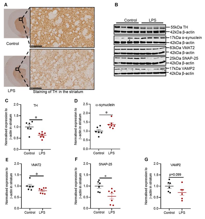
Int. J. Mol. Sci. 2021, 22, 7380                                                                                            2 of 20

                                   alterations in this protein lead to synaptic dysfunction [4–7]. The α-synuclein protein inter-
                                   acts with vesicle-associated membrane protein 2 (VAMP2) to promote the assembly of the
                                   SNARE complex. The SNARE complex is formed by VAMP2, syntaxin and synaptosomal
                                   associated protein 25 (SNAP-25), which regulates docking and fusion of synaptic vesicles
                                   at the presynaptic membrane [8,9]. Moreover, vesicular monoamine transporter 2 (VMAT2)
                                   is critical for the assembly of cytosolic monoamines into synaptic vesicles. It has been
                                   reported that VMAT2 levels are reduced in the striatum and SN of PD patients [10–13].
                                   Other proteins contributing to synaptic dysfunction in PD include deglycase DJ-1 (DJ-1),
                                   Parkin and PTEN induced putative kinase 1 (PINK1) [5,14]. DJ-1, Parkin and PINK1 are
                                   associated with hereditary PD and they are also important for mitochondrial function and
                                   protection against oxidative stress [5]. Thus, we aimed to investigate some of these proteins
                                   in this study.
                                         Neurotoxin and genetic-based models have been invaluable in understanding the
                                   aetiology of PD, and have illustrated the importance of inflammation and oxidative stress
                                   in the disease process [15]. Microglia cells are the prominent mediators of neuroinflamma-
                                   tion, and indeed, activated microglia cells have been identified in the SN of post-mortem
                                   brain samples of PD patients [16]. Moreover, astrogliosis is evident in the SN of PD pa-
                                   tients [17,18]. Inflammatory cytokines and free radicals produced by activated microglia
                                   and astrocytes under pathological conditions can induce the expression of enzymes in-
                                   volved in the secretion of potent free radicals such as nitric oxide and superoxide [17,18].
                                   The aforementioned free radicals can react together to form peroxynitrite, which is involved
                                   in the nitration of free tyrosine residues on proteins and can be assessed by examining
                                   the expression of 3-nitrotyrosine (3-NT)-modified proteins [19,20]. Neuroinflammation
                                   can also affect the levels of brain-derived neurotrophic factor (BDNF), a protein essential
                                   for differentiation, proliferation, survival, and synaptic plasticity in the CNS. Indeed, it
                                   has been reported that BDNF mRNA and protein are reduced in the SN and serum of PD
                                   patients compared to healthy controls [21,22].
                                         Furthermore, it has been reported that PD is linked to gut inflammation evident by
                                   increased tumour necrosis factor-α, interferon-γ, interleukin 6, and interleukin 1β, as
                                   well as glial markers such as GFAP and Sox-10 in the colon [23]. The majority of the
                                   gut microbiota resides in the colon, and the epithelium lining of the intestines acts as a
                                   protective barrier separating the commensal microbes in the lumen of the intestines from
                                   the immune system and enteric nervous system (ENS) in the underlying tissue [23–25].
                                   Interestingly, it has been reported that PD patients have increased intestinal permeability,
                                   which can result in the exposure of ENS to pro-inflammatory products from the lumen. In
                                   turn, the pro-inflammatory products could drive inflammation and oxidative stress in the
                                   gut, which is evident to induce α-synuclein aggregation and indeed, α-synuclein-positive
                                   Lewy bodies are evident in the colon of PD patients [24–26]. Of note, an increased risk of
                                   PD has been reported in patients with inflammatory bowel disease and it is evident that
                                   gut inflammation in these patients is mainly mediated by T-lymphocytes [27,28].
                                         The inflammatory aspects of PD can be mimicked in animal models via the adminis-
                                   tration of lipopolysaccharide (LPS), which is a component of the cell wall of Gram-negative
                                   bacteria and a potent activator of microglial cells via Toll-like receptor 4 [29]. In vivo, LPS
                                   can be administered stereotaxically, intraperitoneally and intranasally, and these models
                                   can recapitulate features of clinical PD such as degeneration of nigrostriatal pathway, mo-
                                   tor impairment and α-synuclein pathology [29–35]. Localized lesions to the nigrostriatal
                                   pathway that replicate features of PD can be generated via intranigral and intrastriatal
                                   administration of LPS [29–32]. It has been hypothesized with some evidence that degener-
                                   ation of the nigrostriatal pathway starts at the axonal terminals (located in the striatum)
                                   and progresses retrogradely to the SN, and the intrastriatal LPS model helps to elucidate
                                   this hypothesis [29,36]. Previous intrastriatal LPS models have only explored the major
                                   neuropathological hallmarks of PD and the resultant motor complications; however, there
                                   is limited evidence on the abnormalities in synaptic proteins, and proteins linked to genetic
                                   PD (e.g., Parkin, DJ-1) in the striatum. Moreover, pathological changes in other regions such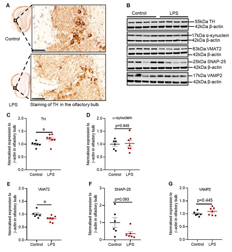
Int. J. Mol. Sci. 2021, 22, 7380                                                                                                      3 of 20

                                   as the olfactory bulb and colon have not been well examined in intrastriatal LPS models,
                                   and these are crucial aspects of PD. Therefore, this study aimed to further characterize the
                                   intrastriatal LPS model of PD including motor and non-motor symptoms and associated
                                   pathology in the striatum, olfactory bulb, and colon.

                                   2. Results
                                   2.1. Effects of Intrastriatal Injection of LPS on Motor Behaviour, Olfactory Function and Mood in
                                   C57BL/6 Mice
                                             The rotarod test, which measures motor behaviour, showed that there was no dif-
                                        ference in latency time in the LPS group compared to control at 4 weeks post-treatment;
                                        however, the latency time was significantly decreased by 27.4% in the LPS group com-
                                        pared to control at 8 weeks post-treatment, indicative of impaired motor function (base-
                                        line: 190.31 versus 170.42 s, p = 0.702; week 4: 143.23 versus 172.06 s, p = 0.410; week 8:
                                        140.36 versus 193.47 s, p = 0.033) (Figure 1A).
                                             Intrastriatal LPS did not induce a significant change in latency time in the buried
                                        food-seeking
Int. J. Mol. Sci. 2021, 22, x FOR PEER REVIEW          test at 4 and 8 weeks post-treatment compared to the control group,     indi-
                                                                                                                             4 of 21
                                        cating no differences in olfactory function (week 4: 333.57 versus 262 s, p = 0.633; week 8:
                                        300.36 versus 167.64 s, p = 0.058) (Figure 1B).

       Figure 1. Administration of LPS into the striatum induced motor impairment, but not olfactory dysfunction or anxiety-like
      Figure 1. Administration of LPS into the striatum induced motor impairment, but not olfactory dysfunction or anxiety-
       behaviour in C57BL/6 mice. (A) Set up and latency time in the rotarod test. (B) Set up and latency time in the buried
      like behaviour in C57BL/6 mice. (A) Set up and latency time in the rotarod test. (B) Set up and latency time in the buried
       food-seeking test. (C) Setup and distance travelled in the open field test. (D) Computerized tracing of the mice in the open
      food-seeking test. (C) Setup and distance travelled in the open field test. (D) Computerized tracing of the mice in the open
       fieldand
      field  andtime
                 timespent
                      spentin
                            inthe
                               thecentral
                                   centralzone
                                           zone of
                                                of the
                                                    the open
                                                        open field.
                                                              field. (E)
                                                                      (E)Computerized
                                                                          Computerizedtracing
                                                                                       tracingof
                                                                                               ofthe
                                                                                                  themice
                                                                                                      micein
                                                                                                           inthe
                                                                                                              theelevated
                                                                                                                  elevatedplus-maze
                                                                                                                           plus-maze
       and  time spent in the open  arm.  The results are presented     as mean  ± SEM (control n = 12–13; LPS  n = 13–14). Statistical
      and time spent in the open arm. The results are presented as mean ± SEM (control n = 12–13; LPS n = 13–14). Statistical
       analysis was performed   with  a two-way
      analysis was performed with a two-way ANOVAANOVA     test,  p <  0.05 (*).
                                                                    < 0.05 (*).

                                   2.2. Administration of LPS Altered the Expression of TH, α-Synuclein and Synaptic Proteins in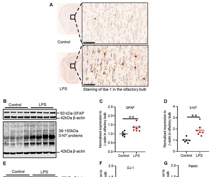
Int. J. Mol. Sci. 2021, 22, 7380 4 of 20
Furthermore, there was no difference in the time spent exploring the central zone of the
open field in the LPS group compared to the control group at 4 and 8 weeks post-treatment
(week 4: 29.58 versus 32.79 s, p = 0.875; week 8: 26.6 versus 19.53 s, p = 0.531) (Figure 1D),
and these findings were consistent with no change in time spent exploring the open arm
of elevated plus maze (week 4: 7.96 versus 11.35 s, p = 0.527; week 8: 3.94 versus 4.45 s,
p = 0.985) (Figure 1E). Collectively, the results for the open field and the elevated plus-maze
test showed that intrastriatal administration of LPS did not induce anxiety like-behaviour.
2.2. Administration of LPS Altered the Expression of TH, α-Synuclein and Synaptic Proteins in
the Striatum of C57BL/6 Mice
Intrastriatal administration of LPS induced degeneration of dopaminergic neuronal
fibres in the striatum depicted by reduced immunoreactivity for tyrosine hydroxylase (TH)
(Figure 2A), and this finding was congruent with the reduced expression of TH protein
(1.59-fold) in the striatum (p = 0.022) (Figure 2C). TH protein was also assessed in the SN
Int. J. Mol. Sci. 2021, 22, x FOR PEER REVIEW 5 of 21
(refer to Figure A1 in Appendix A), but there was no significant difference, suggesting that
intrastriatal LPS did not induce degeneration in the SN.
Figure 2. Administration of LPS altered the expression of TH, α-synuclein and synaptic proteins in the striatum of
Figure 2. Administration of LPS altered the expression of TH, α-synuclein and synaptic proteins in the striatum of C57BL/6
C57BL/6
mice.mice. (A) Representative
(A) Representative images images for immunohistochemical
for immunohistochemical detection ofdetection of TH in (B)
TH in the striatum. theRepresentative
striatum. (B)immunob-
Representative
immunoblots forα-synuclein,
lots for TH, TH, α-synuclein,
VMAT2, VMAT2,
SNAP-25 SNAP-25
and VAMP2and proteins
VAMP2in proteins in the(C)
the striatum. striatum. (C) Densitometric
Densitometric analysis of THanalysis
protein. of TH
(D)(D)
protein. Densitometric analysis
Densitometric of α-synuclein
analysis protein.protein.
of α-synuclein (E) Densitometric analysis of
(E) Densitometric VMAT2of
analysis protein.
VMAT2 (F)protein.
Densitometric analy-
(F) Densitometric
sis of SNAP-25 protein. (G) Densitometric analysis of VAMP2 protein. The results are presented as mean ± SEM (control
analysis of SNAP-25 protein. (G) Densitometric analysis of VAMP2 protein. The results are presented as mean ± SEM
n = 6; LPS n = 6–7). Statistical analysis was performed with a Mann–Whitney test, p< 0.05 (*). Immunohistochemical detec-
(control n=
tion of6;TH
LPS n =bar:
(scale 6–7).50Statistical analysis
µm; microscopic was performed
magnification: with
X400) a Mann–Whitney
(control test, p < 0.05 (*). Immunohistochemical
n = 5; LPS n = 5).
detection of TH (scale bar: 50 µm; microscopic magnification: X400) (control n = 5; LPS n = 5).
2.3. Effects of LPS on the Inflammatory and Oxidative Stress Markers, and Proteins Involved in
Defence Mechanisms Against Oxidative Stress in the Striatum of C57BL/6 Mice
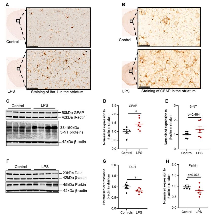
Intrastriatal administration of LPS induced inflammation in the striatum, which was
characterized by increased Iba-1 positive microglia cells (Figure 3A) and GFAP positive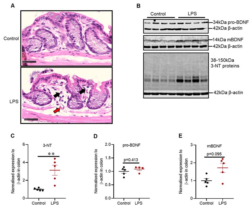
Int. J. Mol. Sci. 2021, 22, 7380                                                                                                                  5 of 20

                                      2.3. Effects of LPS on the Inflammatory and Oxidative Stress Markers, and Proteins Involved in
                                      Defence Mechanisms against Oxidative Stress in the Striatum of C57BL/6 Mice
                                            Intrastriatal administration of LPS induced inflammation in the striatum, which was
                                       characterized by increased Iba-1 positive microglia cells (Figure 3A) and GFAP positive
                                       astrocytes (Figure 3B), and these findings were consistent with the increased expression of
                                       GFAP protein in the striatum (1.44-fold; p = 0.022) (Figure 3D).
                                            LPS also significantly decreased the protein expression of striatal DJ-1 by 1.23-fold
Int. J. Mol. Sci. 2021, 22, x FOR PEER REVIEW                                                                                 6 of 21
                                       (p = 0.036) (Figure 3G), with no significant decrease in the expression of Parkin protein
                                       (p = 0.073) (Figure 3H); or in 3-NT, a marker of oxidative stress (p = 0.484) (Figure 3E).

      Figure
      Figure 3.
              3. LPS
                 LPSaltered
                       alteredthe
                                theinflammatory
                                     inflammatoryand  andoxidative
                                                          oxidative stress
                                                                      stress markers,
                                                                             markers, and
                                                                                        and proteins
                                                                                            proteins involved
                                                                                                      involved in
                                                                                                                in defence
                                                                                                                   defence mechanisms
                                                                                                                             mechanisms against
                                                                                                                                            against
      oxidative   stress in
      oxidative stress    inthe
                             thestriatum
                                  striatumofofC57BL/6
                                                C57BL/6mice.
                                                           mice.Representative
                                                                  Representative     images
                                                                                   images forfor immunohistochemical
                                                                                              immunohistochemical          detection
                                                                                                                        detection      of Iba-1(A)
                                                                                                                                   of Iba-1(A) and
      and  GFAP(B)
      GFAP(B)         instriatum.
                 in the    the striatum.   (C) Representative
                                      (C) Representative         immunoblots
                                                            immunoblots            for GFAP
                                                                            for GFAP         and 3-NT
                                                                                        and 3-NT        proteins
                                                                                                  proteins  in theinstriatum.
                                                                                                                      the striatum.  (D) Densito-
                                                                                                                               (D) Densitometric
      metric analysis of GFAP protein. (E) Densitometric analysis of 3-NT proteins. (F) representative immunoblots for DJ-1 and
      analysis of GFAP protein. (E) Densitometric analysis of 3-NT proteins. (F) representative immunoblots for DJ-1 and Parkin
      Parkin in the striatum (involved in defence against oxidative stress). (G) Densitometric analysis of DJ-1 protein. (H) Den-
      in the striatum (involved in defence against oxidative stress). (G) Densitometric analysis of DJ-1 protein. (H) Densitometric
      sitometric analysis of parkin protein. The results are presented as mean ± SEM (control n = 6; LPS n = 6–7). Statistical
      analysis was
      analysis  of parkin    protein.
                     performed      withThe  results are presented
                                          a Mann–Whitney       test, pas mean
                                                                       < 0.05 (*).±Immunohistochemical
                                                                                    SEM (control n = 6; LPS   n = 6–7).
                                                                                                           detection   ofStatistical
                                                                                                                          Iba-1 and analysis   was
                                                                                                                                      GFAP (scale
      performed     with   a Mann–Whitney       test, p < 0.05 (*). Immunohistochemical
      bar: 50 µm; microscopic magnification: X400) (control n = 5; LPS n = 5).                detection  of Iba-1  and  GFAP    (scale bar: 50 µm;
      microscopic magnification: X400) (control n = 5; LPS n = 5).
                                     2.4. Intrastriatal Administration of LPS Altered the Expression of TH and Synaptic Proteins in
                                     the Olfactory Bulb of C57BL/6 Mice
                                           There was no difference in TH positive cells in the glomerular layer of the olfactory
                                     bulb between control and LPS treated mice based on immunohistochemical staining (Fig-
                                     ure 4A); however, the expression of TH protein was significantly increased by 1.2-fold (p
                                     = 0.035) (Figure 4C), without alteration in α-synuclein protein (p = 0.945) (Figure 4D).
                                           Intrastriatal LPS significantly decreased the expression of VMAT2 protein by 1.2-fold
                                     (p = 0.0140), with no significant change in SNAP-25 (p = 0.093) (Figure 4F) or VAMP2 pro-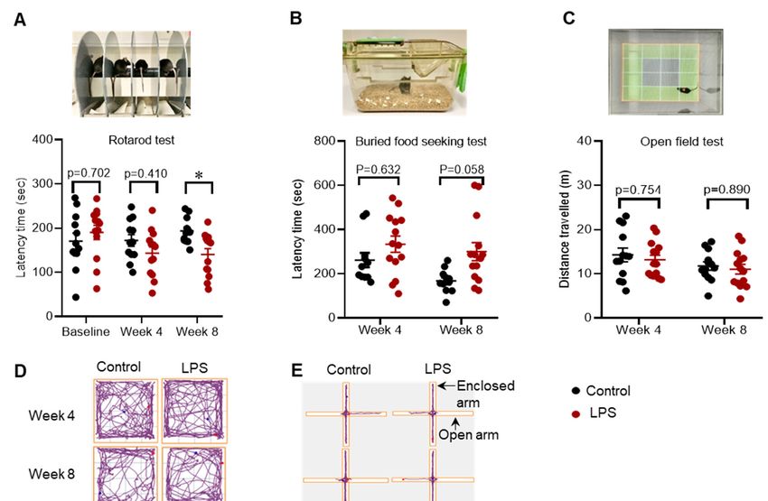
Int. J. Mol. Sci. 2021, 22, 7380                                                                                                                6 of 20

                                     2.4. Intrastriatal Administration of LPS Altered the Expression of TH and Synaptic Proteins in the
                                     Olfactory Bulb of C57BL/6 Mice
                                             There was no difference in TH positive cells in the glomerular layer of the olfac-
                                        tory bulb between control and LPS treated mice based on immunohistochemical staining
Int. J. Mol. Sci. 2021, 22, x FOR PEER REVIEW                                                                                 7 of 21
                                        (Figure 4A); however, the expression of TH protein was significantly increased by 1.2-fold
                                        (p = 0.035) (Figure 4C), without alteration in α-synuclein protein (p = 0.945) (Figure 4D).

      Figure 4. Intrastriatal administration of LPS altered the expression of TH and synaptic proteins in the olfactory bulb of
     Figure 4. Intrastriatal administration of LPS altered the expression of TH and synaptic proteins in the olfactory bulb of
      C57BL/6 mice. (A) Representative images for immunohistochemical detection of TH in the olfactory bulb. (B) Representative
     C57BL/6 mice. (A) Representative images for immunohistochemical detection of TH in the olfactory bulb. (B) Representa-
      immunoblots for TH, α-synuclein, VMAT2, SNAP-25 and VAMP2 proteins in the olfactory bulb. (C) Densitometric
     tive immunoblots for TH, α-synuclein, VMAT2, SNAP-25 and VAMP2 proteins in the olfactory bulb. (C) Densitometric
      analysisof
     analysis    ofTH
                    THprotein.
                         protein.(D)(D)Densitometric
                                         Densitometricanalysis
                                                         analysisofofα-synuclein  protein.(E)
                                                                      α-synucleinprotein.  (E)Densitometric
                                                                                               DensitometricanalysisanalysisofofVMAT2
                                                                                                                                 VMAT2protein.
                                                                                                                                         protein.
     (F) Densitometric analysis of SNAP-25. (G) Densitometric analysis of VAMP2. The results are presented as mean ±±SEM
      (F) Densitometric     analysis    of SNAP-25.   (G) Densitometric   analysis of VAMP2.   The   results    are  presented   as mean   SEM
      (control nn ==6;
     (control        6; LPS
                        LPS nn == 6–7).
                                  6–7). Statistical
                                        Statistical analysis was
                                                             was performed
                                                                 performedwith
                                                                             withaaMann–Whitney
                                                                                    Mann–Whitneytest, test,p p<
Int. J. Mol. Sci. 2021, 22, 7380                                                                                                                    7 of 20

                                      2.5. Effects of LPS on the Inflammatory and Oxidative Stress Markers, and Proteins Involved in
                                      Defence Mechanisms against Oxidative Stress in the Olfactory Bulb of C57BL/6 Mice
                                            Intrastriatal LPS has induced inflammation in the olfactory bulb illustrated by in-
                                       creased
  Int. J. Mol. Sci. 2021, 22, x FOR PEER REVIEW
                                                expression of Iba-1 positive microglia cells in the granular cell layer (Figure 85A),
                                                                                                                                  of 21
                                       and this finding coincided with the increased expression of GFAP protein (1.34-fold) in the
                                       olfactory bulb (p = 0.005) (Figure 5C).

      Figure 5. Intrastriatal administration of LPS altered the expression of inflammatory and oxidative stress markers and
         Figure 5. Intrastriatal administration of LPS altered the expression of inflammatory and oxidative stress markers and pro-
      proteins involved in defence mechanisms against oxidative stress in the olfactory bulb of C57BL/6 mice. (A) Representative
         teins involved in defence mechanisms against oxidative stress in the olfactory bulb of C57BL/6 mice. (A) Representative
      images
         imagesforfor
                    immunohistochemical
                      immunohistochemical     detection
                                                detectionof of
                                                             Iba-1
                                                               Iba-1in in
                                                                       thethe
                                                                            olfactory bulb.
                                                                               olfactory    (B)(B)
                                                                                         bulb.  Representative
                                                                                                   Representativeimmunoblots
                                                                                                                    immunoblots  forforGFAPGFAP  andand
      3-NT   proteins  in the  olfactory bulb. (C) Densitometric     analysis   of GFAP  protein. (D) Densitometric   analysis  of
         3-NT proteins in the olfactory bulb. (C) Densitometric analysis of GFAP protein. (D) Densitometric analysis of 3-NT pro-  3-NT     protein.
      (E)tein.
          Representative      immunoblots
               (E) Representative            for DJ-1for
                                      immunoblots      andDJ-1
                                                             parkin
                                                                 andinparkin
                                                                         the olfactory  bulb (involved
                                                                                in the olfactory         in defence
                                                                                                  bulb (involved   inagainst
                                                                                                                      defenceoxidative       stress).
                                                                                                                                against oxidative
      (F)stress).
          Densitometric    analysis ofanalysis
                  (F) Densitometric     DJ-1 protein.
                                                of DJ-1(G)  Densitometric
                                                          protein.            analysis ofanalysis
                                                                    (G) Densitometric     parkin protein.  The
                                                                                                   of parkin    resultsThe
                                                                                                             protein.   are results
                                                                                                                            presented areas   mean
                                                                                                                                           presented
      ± as
         SEMmean    ± SEM
               (control   n =(control
                               6; LPS n = 6;
                                          7).LPS  n = 7). analysis
                                              Statistical  Statisticalwas
                                                                        analysis   was performed
                                                                           performed               with a Mann–Whitney
                                                                                        with a Mann–Whitney                  test,
                                                                                                                 test, p < 0.05 (*),p p<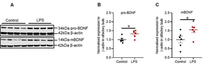
Int. J. Mol. Sci. 2021, 22, 7380 8 of 20
Int. J. Mol. Sci. 2021, 22, x FOR PEER REVIEW 9 of
Inflammation in the olfactory bulb was accompanied by no significant reduction in 21
DJ-
1 protein (p = 0.101) (Figure 5F), but a 1.22-fold decrease in the expression of Parkin protein
(p = 0.035) (Figure 5G) and a 1.89-fold increase in 3-NT proteins (p = 0.002) (Figure 5D).
2.6. Intrastriatal Administration of LPS Altered the Expression of Brain-Derived Neurotrophic
2.6. Intrastriatal Administration of LPS Altered the Expression of Brain-Derived Neurotrophic
FactorProtein
Factor Proteinininthe
theOlfactory
OlfactoryBulb
Bulbbut
butNot
Notininthe
theStriatum
StriatumofofC57BL/6
C57BL/6Mice Mice
Brain-derived
Brain-derived neurotrophic
neurotrophic factor
factor (BDNF)
(BDNF) is synthesized
is synthesized in its precursor
in its precursor form (pro-
form (pro-BDNF),
and it is enzymatically cleaved to generate the mature form (mBDNF). IntrastriatalIntrastria-
BDNF), and it is enzymatically cleaved to generate the mature form (mBDNF). adminis-
tal administration
tration of LPS increased of LPS increased
protein protein
expression expression
of pro-BDNF by of pro-BDNF
1.27-fold by 1.27-fold
(p = 0.015) (p =
(Figure 6B)
0.015) (Figure 6B) and mBDNF by 1.44-fold
and mBDNF by 1.44-fold (p = 0.041) (Figure 6C). (p = 0.041) (Figure 6C).
Additionally,the
Additionally, theprotein
proteinexpression
expressionofofpro-BDNF
pro-BDNFand andmBDNF
mBDNFwas wasassessed
assessedininthe
the
striatumvia
striatum viaWestern
Westernblot, blot,but
butthe
theproteins
proteinsmentioned
mentionedwere werenotnotaltered
altered(refer
(refertotoFigure
FigureA2
A2
in Appendix).
in Appendix A).
Figure 6. Intrastriatal administration of LPS altered the expression of pro-BDNF and mBDNF in the olfactory bulb of
Figure 6.mice.
C57BL/6 Intrastriatal administration
(A) Representative of LPS altered
immunoblots the expression
for pro-BDNF and of pro-BDNF
mBDNF and
in the mBDNFbulb.
olfactory in the
(B)olfactory bulb of
Densitometric
C57BL/6 mice. (A) Representative immunoblots for pro-BDNF and mBDNF in the olfactory bulb. (B) Densitometric anal-
analysis of pro-BDNF protein. (C) Densitometric analysis of mBDNF protein. The results are presented as mean ± SEM
ysis of pro-BDNF protein. (C) Densitometric analysis of mBDNF protein. The results are presented as mean ± SEM (control
(control n = 6; LPS n = 6–7). Statistical analysis was performed with a Mann–Whitney test, p < 0.05 (*).
n = 6; LPS n = 6–7). Statistical analysis was performed with a Mann–Whitney test, p< 0.05 (*).
2.7.Intrastriatal
2.7. IntrastriatalAdministration
AdministrationofofLPS
LPSInduced
InducedMild
MildInflammatory
InflammatoryChanges
Changesand
andAlteration
Alterationinin
OxidativeStress
Oxidative StressMarkers
Markersininthe
theDistal
DistalColon
ColonofofC57BL/6
C57BL/6Mice
Mice
Hematoxylinand
Hematoxylin andEosin
Eosinstaining
stainingfor
fordistal
distalcolon
colonindicated
indicatedthat
thatcontrol
controlmice
micehad
hadintact
intact
epitheliumlining
epithelium liningwith
withdistinct
distinctmuscularis
muscularismucosa.
mucosa.In Incontrast,
contrast,LPS
LPStreated
treatedmice
miceshowed
showed
intact
intactepithelium withaaslight
epithelium lining with slightincrease
increase in in mucosal
mucosal vasculature
vasculature (red(red arrow)
arrow) and and
lym-
lymphocytes
phocytes (black(black arrow)
arrow) (Figure
(Figure 7A).
7A).
There
Therewas
wasaa3-fold
3-foldincrease
increaseininthe
theexpression
expressionofof3-NT3-NTproteins
proteins(p(p==0.008)
0.008)(Figure
(Figure7C);
7C);
however, intrastriatal administration of LPS did not alter the expression of pro-BDNF
however, intrastriatal administration of LPS did not alter the expression of pro-BDNF (p (p = 0.413)
(Figure
= 0.413)7D) or mBDNF
(Figure 7D) or in the distal
mBDNF colon
in the (p =colon
distal 0.095)(p(Figure
= 0.095)7E).
(Figure 7E).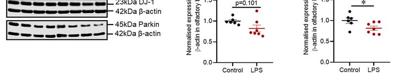
Int. J. Mol. Sci. 2021, 22, x FOR PEER REVIEW                                                                                                  10 of 21
Int. J. Mol. Sci. 2021, 22, 7380                                                                                                                 9 of 20

     Figure 7. Intrastriatal administration of LPS induced mild inflammatory changes and alteration in oxidative stress mark-
     Figure 7. Intrastriatal administration of LPS induced mild inflammatory changes and alteration in oxidative stress markers
     ersthe
     in  in distal
             the distal
                    coloncolon     of C57BL/6
                            of C57BL/6      mice.mice.    (A) Light
                                                   (A) Light          microscopy
                                                               microscopy    of mouse  of mouse   distal stained
                                                                                           distal colon   colon stained   with Hematoxylin
                                                                                                                   with Hematoxylin           and
                                                                                                                                       and Eosin,
     Eosin,   whereby    control    mice   showed    intact  epithelium   lining   with    distinct muscularis    mucosa,
     whereby control mice showed intact epithelium lining with distinct muscularis mucosa, and LPS treated mice showed     and  LPS  treated mice
     showed
     intact    intact epithelium
            epithelium    lining with lining  with increase
                                          a slight  a slight increase  in mucosal
                                                              in mucosal   vasculaturevasculature   (red arrow)
                                                                                            (red arrow)           and lymphocytes
                                                                                                          and lymphocytes           (black arrow)
                                                                                                                             (black arrow)  (Scale
     (Scale
     bar    bar = microscopic
         = 50µm,   50µm, microscopic        magnification:
                                    magnification:             X400).
                                                      X400). (B)      (B) Representative
                                                                 Representative     immunoblotsimmunoblots
                                                                                                     for 3-NT,for  3-NT, pro-BDNF
                                                                                                                pro-BDNF   and mBDNF andproteins.
                                                                                                                                          mBDNF
     (C) Densitometric
     proteins.              analysis analysis
                (C) Densitometric        of 3-NTofproteins.    (D) Densitometric
                                                     3-NT proteins.   (D) Densitometric analysis of pro-BDNF.
                                                                                              analysis             (E) Densitometric
                                                                                                        of pro-BDNF.                   analysis
                                                                                                                       (E) Densitometric        of
                                                                                                                                          analysis
     mBDNF.
     of mBDNF.  The
                  Theresults
                       results arearepresented
                                       presentedasasmean
                                                      mean± ± SEM
                                                                SEM(control
                                                                     (controln n= =5;5;LPS
                                                                                         LPSnn==4–5).
                                                                                                 4–5).Statistical
                                                                                                       Statisticalanalysis
                                                                                                                   analysiswas
                                                                                                                            wasperformed
                                                                                                                                performed with
                                                                                                                                            with aa
     Mann–Whitney
     Mann–Whitney test, test, pp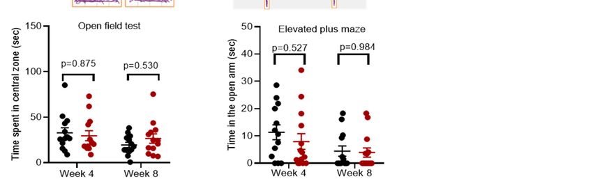
Int. J. Mol. Sci. 2021, 22, 7380                                                                                                 10 of 20

                                   Table 1. Summary of the proteins examined by Western blot. Downwards (↓) and upwards (↑)
                                   arrows indicate the direction of “significance”, left-right arrow (↔) refers to “no significance”, and
                                   dash (-) refers to “not assessed”.

                                                                                  Changes in Protein Expression
                                           Proteins                 Striatum              Olfactory Bulb                Colon
                                             TH                        ↓                          ↑                        -
                                            α-syn                      ↑                         ↔                         -
                                           VMAT2                       ↓                          ↓                        -
                                          SNAP-25                      ↓                         ↔                         -
                                           VAMP2                       ↔                         ↔                         -
                                            GFAP                       ↑                         ↑↑                        -
                                             DJ-1                      ↓                         ↔                         -
                                            Parkin                     ↔                          ↓                        -
                                        3-NT proteins                  ↔                         ↑↑                       ↑↑
                                          Pro-BDNF                     ↔                          ↑                       ↔
                                           mBDNF                       ↔                          ↑                       ↔

                                   3.2. Motor Function and Striatal Pathology
                                         LPS induced inflammation in the striatum, which was characterized by an increased
                                   number of Iba-1 positive microglia cells and GFAP positive astrocytes/GFAP protein,
                                   consistent with other neurotoxin models of PD [29,32,37,38]. This event was accompanied
                                   by a significant decrease in DJ-1 protein, but no significant alteration in Parkin. There were
                                   also no changes in 3-NT formation (a marker of oxidative stress). Prolonged inflammation
                                   mediated by microglia cells and astrocytes results in excessive production of inflammatory
                                   cytokines, chemokines and free radicals, which are detrimental to dopaminergic neurons
                                   and such pathological process is combated by the antioxidant system. Aside from their
                                   role in synaptic function, DJ-1 and Parkin have a key role in protection against oxidative
                                   stress via quenching of reactive oxygen species and clearance of damaged mitochondria,
                                   respectively. Thus, LPS- induced inflammation could have reduced the expression of DJ-1
                                   protein, and in return, reduced DJ-1 could have enhanced susceptibility to LPS induced
                                   oxidative stress. Additionally, LPS did not alter the expression of 3-NT proteins. 3-NT
                                   is a marker of nitric oxide-dependent post-translation modification. Thus, it is possible
                                   that other indicators of oxidative stress that were not examined in this study could be
                                   changed such as the level of specific reactive oxygen species and antioxidants. Furthermore,
                                   intrastriatal LPS induced degeneration of dopaminergic terminals in the striatum illustrated
                                   by reduced immunoreactivity of TH positive cell terminals and TH protein; thus, providing
                                   consistent results with the previous in vivo studies [32,37,39]. Dopaminergic neurons are
                                   susceptible to cytotoxic molecules release via pathological activation of microglia cells
                                   and astrocytes; thus, degeneration of dopaminergic terminals may be a consequence of
                                   LPS induced inflammation [17,18,29]. Degeneration of dopaminergic synaptic terminals
                                   interferes with synaptic processes in the striatum, and coordination of motor function
                                   at the cortical level, which is consistent with impaired motor function observed in LPS
                                   mice and other in vivo studies [31,32]. The rotarod test indicated that LPS induced motor
                                   impairment at 8 weeks post-treatment, which was consistent with the TH loss in the
                                   striatum. Intrastriatal LPS did not induce a significant change in TH protein in the SN, a
                                   marker of dopaminergic neuronal integrity, inconsistent with Hunter and colleagues [32].
                                   However, Hunter et al. in 2009 administered 20 µg of LPS bilaterally compared to our
                                   treatment regimen (10 µg unilaterally), and this could dictate the rate of progression of
                                   intrastriatal pathology to the SN, and resultant degeneration. Thus, our model is milder,
                                   and as a result, did not cause the SN degeneration. A longer time than 8 weeks may be
                                   required for the neurodegeneration to progress to the SN in our model. Administration of
                                   LPS reduced the levels of VMAT2 and SNAP-25 accompanied by increased α-synuclein
                                   protein in the striatum in our study. Defects in VMAT2 have been reported in patients and
                                   animal models of PD; however, the expression of SNAP-25 and VAMP2 proteins has not
Int. J. Mol. Sci. 2021, 22, 7380 11 of 20
been widely examined in animal models of PD [12,13,40]. In our study, the reduction of
VMAT2 and SNAP-25 proteins in the striatum could be due to the loss of dopaminergic
terminals in this region as shown by the loss of TH. In addition, α-synuclein protein
directly interacts with VAMP2 to promote the assembly of the SNARE complex; however,
an increase in α-synuclein protein is evident to disrupt the release of neurotransmitters
via defective clustering of synaptic vesicles [8,9]. Thus, an increase in α-synuclein protein
and reduction of VMAT2 and SNAP-25 in our study could reduce dopamine release
into the striatum and compromise neuronal transmission to the motor cortex, which can
affect the control of movement. Furthermore, in addition to the increased α-synuclein
protein expression, phosphorylated and aggregated forms have been identified in human
PD [41,42]; therefore, future studies are required to investigate if similar changes occur
in this model. Collectively, LPS induced inflammation in the striatum has led to the
degeneration of dopaminergic fibres, reduction in synaptic proteins and DJ-1 protein,
and these pathological processes could be responsible for motor impairment observed in
C57BL/6 mice.
3.3. Olfactory Function and Pathology
Hyposmia is evident in up to 90% of PD patients, and it is reported to precede the
motor symptoms of PD [43]. Thus, understanding olfactory function and pathology could
be invaluable for the diagnosis of clinical PD at the early stages. LPS induced inflammation
in the striatum led to an inflammatory response in the olfactory bulb characterized by
increased Iba-1 positive microglia cells in the granular cell layer and GFAP protein. Al-
though neuronal connections are yet to be thoroughly established between the nigrostriatal
pathway and olfactory bulb, the prominent increase of Iba-1 positive microglia cells in the
granular layer associated with intrastriatal LPS administration implies that inflammatory
cells could have migrated from the striatum via the rostral migratory stream to the olfactory
bulb, and this pathway needs to be investigated. Additionally, activation of inflammatory
cells leads to the secretion of free radicals, consistent with increased expression of 3-NT
proteins in the olfactory bulb of LPS treated mice. 3-NT is a marker of peroxynitrite in-
duced nitration of tyrosine residues, and peroxynitrite is a compound of nitric oxide and
superoxide, which are common free radicals associated with inflammation. Interestingly,
LPS induced a significant decrease in Parkin protein with no significant reduction in DJ-1
protein in the olfactory bulb, which is the opposite to the striatum where we found reduc-
tions in DJ-1, but not Parkin. Parkin protects against oxidative stress; thus, reduced Parkin
protein coincided with increased 3-NT proteins; however, further studies are critical to
elucidate the function of Parkin and DJ-1 proteins in the olfactory bulb in PD models.
The olfactory bulb is the first relay station of olfactory sensory information, and it
has a large group of dopaminergic interneurons in its glomerular layer. The literature
is unsettled regarding the function of dopaminergic interneurons in the olfactory bulb.
Some studies proposed that dopaminergic neurons have an inhibitory role in olfactory
neurotransmission and have reported an increase in the number of dopaminergic neurons
in vivo and PD patients [44–46]. In contrast, other studies have reported that dopamine
neurotransmission in the olfactory bulb promotes neurogenesis, a phenomenon unique
to this region and critical in olfaction [47]. Neural stem and progenitor cells from the
subventricular zone of the lateral ventricle are evident to migrate via rostral migratory
stream to the olfactory bulb, whereby these cells differentiate and integrate into GABAergic
and dopaminergic interneurons [47]. Intrastriatal LPS did not induce a noticeable change in
the number of TH positive dopaminergic cells in the glomerular layer; however, there was
a small but significant increase in TH protein expression when assessed by Western blots.
Our findings suggest that LPS did not affect the number of TH positive cells, but increased
the expression of TH protein in these cells, suggesting increased dopamine signalling.
Concomitant with these findings, LPS increased the expression of pro-BDNF and mBDNF
proteins in the olfactory bulb, which indicates the increased expression of the BDNF gene.Int. J. Mol. Sci. 2021, 22, 7380 12 of 20
The olfactory bulb is a junction whereby sensory olfactory neurons synapse with
mitral/tufted cells that extend to secondary olfactory structures and intrastriatal LPS could
disrupt synaptic transmission. Our results showed that intrastriatal administration of LPS
significantly decreased VMAT2 with no significant reduction in SNAP-25 and VAMP2
proteins. Decreased VMAT2 protein could be associated with LPS induced inflammation
and oxidative stress in the olfactory bulb as a pathological pathway to increase cytosolic
dopamine, dopamine-induced oxidative stress, and reduced neurotransmitter release
[48,49]. It has been reported that reduced VMAT2 protein can augment hyposmia [49];
however, synaptic communication in the olfactory bulb has not been widely examined
in models of PD and requires further clarification. Cumulatively, intrastriatal LPS has
induced inflammation within the olfactory bulb, leading to alterations in synaptic proteins,
but this did not induce a significant impairment in olfactory function as indicated by the
buried food-seeking test. These findings indicate that pathological alterations found in the
olfactory bulb were not at the threshold to produce a significant change in olfaction.
3.4. Colonic Pathology
PD is associated with a constellation of gastrointestinal complications such as drooling,
dysphagia, delayed gastric emptying and constipation, and some of these complications
precede motor symptoms in PD [2,50]. The most common gastrointestinal complication
of PD is constipation, and it can be associated with colonic and anorectal dysmotility [51].
Therefore, we aimed to explore the effects of the nigrostriatal lesion (via intrastriatal
LPS) on the distal colon. Hematoxylin and eosin staining of the distal colon indicated
that control mice had an intact epithelium lining with distinct muscularis mucosa while
LPS treated mice showed intact epithelium with a mild increase in mucosal vasculature
and lymphocytes, indicative of mild inflammation; these findings were congruent with
the increase in 3-NT proteins. Collectively, these findings indicate that intrastriatal LPS
induced mild colonic inflammation and oxidative stress, which can induce pathological
changes in the colon; thus, congruent with in vivo studies utilizing intranigral lesion to the
nigrostriatal pathway [52]. The mechanisms linking CNS pathology and gastrointestinal
complications are poorly understood in PD. In vivo studies have shown that there is a
bidirectional relationship between the nigrostriatal dopaminergic system and gut system
via the vagus nerve; however, there is limited experimental evidence [52]. Interestingly,
vagotomy alleviates the pathology of the nigrostriatal dopaminergic system induced via
gut dysfunction, and gut pathology induced via nigrostriatal lesions; however, additional
studies are critical to thoroughly understand the involved mechanisms [52,53].
Intrastriatal LPS did not alter the expression of mBDNF and pro-BDNF proteins in
the colon. Colonic mBDNF is expressed in epithelial cells and neurons of the myenteric
plexus [54], and it is proposed to be involved in the regulation of colonic motility and
visceral hyperalgesia [54]. Moreover, it has been shown that colonic mBDNF protein is
increased in patients with irritable bowel syndrome, and it correlates with disease severity.
Thus, future models of PD with potent gut abnormalities could examine the involvement
of colonic mBDNF in the inflammatory processes [54].
In conclusion, intrastriatal administration of LPS has induced inflammation not only
in the striatum, but also in the olfactory bulb and within the distal colon. LPS induced
inflammation was responsible for pathological changes observed in the striatum such as
degeneration of dopaminergic neuronal fibres, reduction of synaptic proteins, and proteins
involved in defence against oxidants, and these pathological changes produced a motor
phenotype similar to PD. Moreover, LPS has induced inflammation within the olfactory
bulb, which caused alteration in synaptic proteins, but did not induce significant impair-
ment in olfactory function. Our model recapitulated various aspects of human PD; thus, it
could be useful for understanding the role of inflammation in motor and non-motor symp-
toms and associated pathology in PD. Future studies could modify our treatment regimen
by utilizing bilateral intrastriatal injection of LPS (10 µg per hemisphere) to establish a
greater lesion to the nigrostriatal pathway, which could better resemble the symptomaticInt. J. Mol. Sci. 2021, 22, 7380 13 of 20
stage of PD. Intrastriatal administration of LPS has induced changes in the olfactory bulb,
striatum, and colon; however, we did not thoroughly explore the mechanisms of how LPS
induced degeneration in each of the regions mentioned and neuronal connections between
the regions, and this is a limitation. Moreover, reduced sense of smell and gastrointestinal
complications (e.g., constipation) are common early non-motor symptoms in PD patients.
Our study suggests that localized lesion to the nigrostriatal pathway induces pathologi-
cal changes in the olfactory bulb and colon, which could be associated with the onset of
olfactory and gastrointestinal complications. Therefore, future studies could extensively
investigate early non-motor symptoms of PD such as impaired olfaction, impaired sleep-
wake cycle, and gastrointestinal complications in the intrastriatal LPS model to establish
their relationship with nigrostriatal lesions. Future studies could utilize tests such as habit-
uation/dishabituation test for olfactory deficits, solid gastric emptying, and one-hour stool
collection as measures of gastroparesis and colon motility, respectively, and finally, sleep
latency to behavioural signs of sleep followed by polysomnography/electromyography to
characterize alteration in the sleep-wake cycle [49,55]. This approach could help to estab-
lish a relationship between early non-motor symptoms of PD and lesions to nigrostriatal
pathway and onset of motor symptoms; thus, it could aid in early diagnosis of PD and
implementation of timely treatment.
4. Materials and Methods
4.1. Animals
This research was approved by the Animal Ethics Committee of the University of
South Australia. Twenty-seven, 12 weeks old, C57BL/6 male mice were purchased from
Animal Resources Centre, Western Australia and were housed at Core Animal Facility
at the University of South Australia for the duration of the experiment. The mice were
housed in groups of 3–4 in a pathogen-free environment at a room temperature of 22 ◦ C,
with a 12 h alternating light/dark cycle, and had access to food and water ad libitum.
4.2. Experimental Design
Mice were randomly allocated into two groups, a control group to be given phosphate-
buffered saline (PBS) (n = 13) and an LPS group (n = 14). Each mouse was deeply anaes-
thetized through inhalation of isoflurane (3–4% isoflurane for induction, and 1–2% for
maintenance) and mounted onto the stereotaxic frame (Stoelting, Wood Dale, IL, USA).
The skin on the cranium was cleaned with 2% chlorhexidine/70% ethanol, and an incision
was made on the scalp with surgical scissors to expose the cranium. 3% hydrogen peroxide
was applied to the exposed cranium to easily define the bregma point. The following
coordinates were used, starting from the bregma point to locate two injection sites in the
right striatum: point A: +1.2 mm anterior-posterior, –1.5 mm medial-lateral, 3.5 mm deep,
and point B: –0.34 mm anterior-posterior, +2.5 mm medial-lateral, and 3.2 mm deep. A fine
needle was then used to drill a hole at each of the striatal injection sides. A 30 gauge 10 µL
Hamilton syringe containing 1 µL of PBS for control mice or LPS solution (5 µg/µL LPS,
Sigma-Aldrich, St. Louis, MI, USA) was slowly lowered ventrally to each of the injection
sites and left in place for 2 min. The solution was slowly injected, and the needle was
left in place for additional 2 min before it was gently withdrawn. After completion of
the injection, 100 µL of a local analgesic mixture of lignocaine (2.5 mg/mL) and bupiva-
caine (0.63 mg/mL) was applied to the surgical wound, and the two ends of the scalp
were glued together with surgical glue (Lyppard Australia Pty Ltd, Adelaide, South Aus-
tralia, Australia). Each mouse was given 500 µL of sterile saline (0.9% sodium chloride)
subcutaneously and kept warm on the heat pad to aid recovery post-surgery.
Behavioural testing was then conducted post-surgery at 4 and 8 weeks as outlined
in Figure 8. Subsequently, the mice were humanely killed, and tissues were collected for
biochemical and immunohistochemical analyses after the last behavioural test.Int. J. Mol. Sci. 2021, 22, x FOR PEER REVIEW 15 of 21
Int. J. Mol. Sci. 2021, 22, 7380 14 of 20
Figure 8. Timeline for intrastriatal administration of LPS in C57BL/6 mice. LPS was administered on day 1, followed
Figure
by 8. Timeline
behavioural for intrastriatal
testing administration
at 4 and 8 weeks of LPSThe
post-treatment. in C57BL/6 mice.collected
tissues were LPS wasfor
administered
subsequent on day 1, after
analyses followed by
the last
behavioural test.
behavioural testing at 4 and 8 weeks post-treatment. The tissues were collected for subsequent analyses after the last
behavioural test.
4.3. Behavioural
4.3. Behavioural Testing
Testing
The mice
The mice werewere housed
housedon onaa1212hhlight/dark
light/dark cycle,
cycle, lights
lights off off at 7:00
at 7:00 p.m.,
p.m., andand all
all be-
behavioural tests started at 9:00 a.m. on the day of the test. Behavioural testing
havioural tests started at 9:00 a.m. on the day of the test. Behavioural testing was con- was
conducted
ducted at 4 at
and 4 and 8 weeks
8 weeks afterafter
the the injection
injection of LPS
of LPS as depicted
as depicted in Figure
in Figure 8. 8.
4.3.1. Buried Food-Seeking Test
4.3.1. Buried Food-Seeking Test
The buried food-seeking test is used to measure olfactory function in mice, and it
The buried food-seeking test is used to measure olfactory function in mice, and it is
is based on the ability of mice to use olfactory cues for foraging [56,57]. Before the test,
based on the ability of mice to use olfactory cues for foraging [56,57]. Before the test, the
the mice were fasted overnight for 14 h and then were allowed to individually search for
mice were fasted overnight for 14 h and then were allowed to individually search for a
a standard food pellet buried beneath 4 cm of bedding in an individually cleaned home
standard food pellet buried beneath 4 cm of bedding in an individually cleaned home
cage. The mice were given a maximum time of 10 min to complete the task. An increase in
cage. The mice were given a maximum time of 10 min to complete the task. An increase
time taken to find the buried food, which is referred to as latency time, is associated with
in time taken to find the buried food, which is referred to as latency time, is associated
olfactory impairment [57].
with olfactory impairment [57].
Open Field Test
4.3.2. Open
Open field test assesses both voluntary movement and anxiety in mice mice [58].
[58]. After
acclimatization for
acclimatization for10
10min,
min,each
eachmouse
mousewas was placed
placed in in
thethe centre
centre of an
of an open open field
field arenaarena
(40
(40 length
cm cm length× 40× cm40width
cm width
× 40 cm× height),
40 cm height),
and its and its activity
activity was over
was tracked tracked
5 minover 5 min
using an
using an overhead
overhead camera connected
camera connected to ANY-maze,
to ANY-maze, a video tracking
a video tracking softwaresoftware
(ANY-maze (ANY-maze
version
version
7.01, 7.01, Stoelting,
Stoelting, Wood
Wood Dale, IL,Dale,
USA).IL,Mice
USA). Mice
have have a aversion
a natural natural aversion
to open to open but
spaces, spaces,
are
but are also explorative in their environment; therefore, a decrease in time
also explorative in their environment; therefore, a decrease in time spent in the central spent in the
central
zone is zone is a characteristic
a characteristic of anxiety-like
of anxiety-like behaviour
behaviour [59]. [59].
4.3.3. Elevated Plus Maze
4.3.3. Elevated Plus Maze
Elevated plus maze test is used to assess anxiety-like behaviour, and it is based
Elevated plus maze test is used to assess anxiety-like behaviour, and it is based on
on rodents’ aversion to open spaces [60]. This repugnance for open spaces results in
rodents’ aversion to open spaces [60]. This repugnance for open spaces results in thigmo-
thigmotaxis, which refers to the avoidance of open spaces by confining movements to
taxis, which refers to the avoidance of open spaces by confining movements to enclosed
enclosed arms or the edges of bounded spaces. The test setup consists of a plus-shaped
arms or the edges of bounded spaces. The test setup consists of a plus-shaped apparatus
apparatus with two open and enclosed arms elevated at 40–70 cm from the floor. Each of
with two open and enclosed arms elevated at 40–70 cm from the floor. Each of the mice
the mice was placed in the central open area of the apparatus and were allowed to explore
was placed in the central open area of the apparatus and were allowed to explore for 5
for 5 min. The movement of the mice was tracked using ANY-maze software. A significant
min. The movement of the mice was tracked using ANY-maze software . A significant
decrease in time spent in the open arm of the maze is indicative of anxiety-like behaviour.
decrease in time spent in the open arm of the maze is indicative of anxiety-like behaviour.
4.3.4. Rotarod Test
4.3.4. Rotarod Test
Motor impairment is the major characteristic of clinical PD, and this feature was
Motor
assessed in impairment
mice using is thethe major test,
rotarod characteristic of clinical
which utilizes PD, and
a rotating rodthis
to feature was as-
assess balance
sessed in mice using
and coordination the rotarod
[61,62]. test,were
The mice which utilizes
given a rotating
10 min before rod
the to assess
test balance and
to acclimatize to
coordination [61,62]. The mice were given 10 min before the test to acclimatize
the test room. The mice were then placed in the correct orientation on the rotating rod to the test
room. The mice were
(3 cm diameter), and thethen placedapparatus
rotarod in the correct
(Ugo orientation on theVarese,
Basile, Gemonio, rotating rodwas
Italy) (3 cm
setdi-
to
ameter), and the rotarod apparatus (Ugo Basile, Gemonio, Varese, Italy)
accelerate from 5–30 rpm for 5 min. The test was ended when the mice fell off the rotating was set to accel-
erate from 5–30
rod, swung 360◦rpm for rotating
on the 5 min. The
rodtest was ended
instead whenor
of walking, thereached
mice felltheoff the rotating
maximum timerod,
of
swung
the test,360°
and onthisthe rotating
time rod instead
was recorded of walking,
as latency or reached
time. The the maximum
latency time time ofwas
of a given mouse the
test, and this time was recorded as latency time. The latency time of a given mouse wasInt. J. Mol. Sci. 2021, 22, 7380 15 of 20
an average of three trials. The mice were given at least 10 min of rest between each trial. A
decreased latency time is associated with motor impairment [61,62].
4.4. Fresh Tissue Collection and Homogenisation
Mice were sacrificed 24 h after completion of the rotarod test via an intraperitoneal
injection of 60 mg/kg of sodium pentobarbitone, and the following fresh tissues were col-
lected for biochemical analyses: olfactory bulb, striatum, substantia nigra/ventral tegmen-
tal area and distal colon. The tissues were homogenized in RIPA buffer (50 mM tris, 150mM
sodium chloride, 1mM Ethylenediaminetetraacetic acid, 0.5% Triton X-100, 0.5% Sodium
deoxycholate, pH 7.4) plus cocktail protease inhibitor (Sigma-Aldrich) using Precellys
24 Homogeniser (Bertin Technologies, Montigny-le-Bretonneux, France). Homogenates
were centrifugated at 13,000 rpm for 30 min, and the supernatants were collected. The pro-
tein concentration of the supernatants was measured with a Micro-BCATM protein Assay
kit (Thermo-scientific, Rockford, IL, USA) according to the manufacturer’s guidelines.
4.5. Western Blot
Proteins were separated by gel electrophoresis on 10–14% SDS-polyacrylamide gels
using the CBS gel system (C.B.S Scientific, San Diego, CA, USA) for 90 min at 110 volts.
The proteins were then transferred onto a 0.2 or 0.45µm nitrocellulose membrane (GE
Healthcare Australia Pty Ltd, Sydney, New South Wales, Australia) at 0.6 amps for 90 min.
The blots were air-dried for 90 min to enhance attachment of proteins to the nitrocellulose
membrane before blocking with 5% bovine serum albumin (BSA)/tris buffered saline-
tween (TBST) +0.05% azide or 5% skim milk/TBST + 0.05% azide (Sigma-Aldrich). After
blocking, the membranes were incubated overnight at 4ºC with respective antibodies (refer
to Table A1 in Appendix A). Following primary antibody incubation, the blots were washed
with TBST and then incubated with corresponding secondary antibodies for near-infrared
Western blot detection (Li-Cor Biosciences, Lincoln, NE, USA) for 1 h at room temperature.
Immunoblots were visualized using Odyssey CLx imaging system (LI-COR Biosciences)
and quantified with Image Studio Lite 5.2 (LI-COR Biosciences). Protein normalization
was performed with mouse anti-β-actin [63].
4.6. Immunohistochemistry
Each mouse (control n = 5; LPS n = 5) was deeply anaesthetized via an intraperitoneal
injection of 60 mg/kg of sodium pentobarbitone, and an incision was made into the abdom-
inal cavity to expose the heart. Transcranial perfusion with 10% formalin was performed
before brain collection. Subsequently, collected brains were fixed in 10% formalin for 48 h
followed by tissue processing, paraffin embedding and sectioning of the paraffin blocks
(4 µm). For staining, sections were deparaffinized in 2 changes of xylene and 2 changes
of 100% ethanol for 5 min each before blocking endogenous peroxidase activity with 0.5%
hydrogen peroxidase in methanol for 30 min. Antigen retrieval was carried out in a mi-
crowave using 10 mM sodium citrate buffer, then sections were blocked for non-specific
binding using 3% normal horse serum (NHS) followed by overnight incubation with spe-
cific primary antibodies ((TH (Sigma-Aldrich) 1:3000 in 1% NHS + PBS/0.3 triton-X-100,
GFAP (Dako, Denmark) 1:1000 in 1% NHS + PBS/0.3 triton-X-100 and Iba-1 (Wako, USA)
1:2000 in 1% NHS + PBS/0.3 triton-X-100)). Subsequently, sections were incubated with
corresponding secondary antibodies (biotinylated anti-rabbit and mouse, 1:250 in 1% NHS
+ PBS/0.3 triton-X-100, Vector Laboratories Inc, CA, USA) for 30 min, incubated with strep-
tavidin horseradish peroxidase-conjugated (Vector Laboratories Inc, CA, USA, 1:500 in 1%
NHS + PBS/0.3 triton-X-100) for 1 h, and developed with 3,30 -diaminobenzidine (DAB)
solution (0.05% DAB Sigma-Aldrich) and 0.015% hydrogen peroxide in 1X PBS) for 7 min.
Sections were then counterstained with Mayer’s hematoxylin (Sigma, St. Louis, MI, USA)
for 1 min, rinsed with water, differentiated in acid alcohol, rinsed with water, blued in
0.04% ammonia water, and lastly dehydrated, cleared and coverslipped using DPX mount-Int. J. Mol. Sci. 2021, 22, 7380 16 of 20
ing solution (Sigma-Aldrich). Images of the sections were taken using NanoZoomer S60
(Hamamatsu Photonics, Hamamatsu, City Shizuoka, Japan).
4.7. Hematoxylin and Eosin Staining for Distal Colon
Each mouse (control n = 5; LPS n = 5) was deeply anaesthetized via an intraperitoneal
injection of sodium pentobarbitone (60 mg/kg), and an incision was made into the abdom-
inal cavity to expose the intestines. Subsequently, the colon was dissected, flushed with
cold PBS to remove intestinal content, and then divided into proximal and distal segments.
The distal segment was further divided into two halves, one half was freshly frozen, and
the other half was longitudinally cut open and assembled flat in a cassette for fixation (10%
formalin). The tissues were fixed in 10% formalin for 24 h, processed using Leica ASP300
automated processor for 6 h and embedded in paraffin for sectioning. Subsequently, the
tissues were sectioned at 4 µm with Thermo Scientific Microm HM 325 Rotary Microtome
(Thermo Scientific). Hematoxylin and Eosin staining was conducted using Leica ST5010
Autostainer XL (Leica Biosystems, Melbourne, Victoria, Australia) and the histological
slides were coverslipped using Leica CV5050 Fully automated Glass Coverslipper. Images
of the sections were taken using NanoZoomer S60 (Hamamatsu Photonics).
4.8. Statistical Analysis
Behavioural and Western blot data were analysed with GraphPad Prism Software 8
(San Diego, CA, USA), and the results are presented as mean ± standard error of the mean
(SEM). Statistical analyses for behavioural tests were performed with a two-way ANOVA
test. Biochemical analyses comparing the control and LPS group were performed with a
Mann–Whitney test. A statistical significance is reached when p-value is ≤0.05.
Author Contributions: I.D. performed all experiments, behavioural testing, tissue analyses, and
wrote the manuscript. L.B. and X.-F.Z. were involved in conceptualization, methodology, supervision,
and editing the manuscript. F.C. and S.G. were involved in supervision and contributed to the
critical review of the manuscript. All authors have read and agreed to the published version of
the manuscript.
Funding: This research received no external funding.
Institutional Review Board Statement: The study was conducted according to the guidelines of the
Declaration of Helsinki, and approved by the Institutional Review Board (or Ethics Committee) of
the University of South Australia (U28–19).
Informed Consent Statement: Not applicable.
Data Availability Statement: Not applicable.
Acknowledgments: Isaac Deng is a recipient of the University of South Australia Postgraduate
Award (USAPA).
Conflicts of Interest: The authors declare no conflict of interest.You can also read