Systemic Treatment With Nicotinamide Riboside Is Protective in a Mouse Model of Light-Induced Retinal Degeneration - IOVS
←
→
Page content transcription
If your browser does not render page correctly, please read the page content below
Biochemistry and Molecular Biology

              Systemic Treatment With Nicotinamide Riboside Is
              Protective in a Mouse Model of Light-Induced Retinal
              Degeneration
              Xian Zhang,1,2 Nathaniel F. Henneman,1,3,5 Preston E. Girardot,1 Jana T. Sellers,1
              Micah A. Chrenek,1 Ying Li,1 Jiaxing Wang,1 Charles Brenner,4 John M. Nickerson,1 and
              Jeffrey H. Boatright1,5
              1
                Department of Ophthalmology, School of Medicine, Emory University, Atlanta, Georgia, United States
              2
                Department of Ophthalmology, Second Xiangya Hospital of Central South University, Changsha, Hunan, China
              3
                Institut Necker-Enfants Malades (INEM), INSERM U1151/CNRS UMR 8253, 75015 Paris, France
              4
                Department of Diabetes & Cancer Metabolism, City of Hope National Medical Center, Duarte, California, United States
              5
                Center for Visual & Neurocognitive Rehabilitation, Atlanta VAHS, Decatur, Georgia, United States

              Correspondence: Jeffrey H.                PURPOSE. Maintaining levels of nicotinamide adenine dinucleotide (NAD+ ), a coenzyme
              Boatright, Department of                  critical for cellular energetics and biosynthetic pathways, may be therapeutic in retinal
              Ophthalmology, Emory University,          disease because retinal NAD+ levels decline during retinal damage and degeneration. The
              B5500, Clinic B Building, 1365B           purpose of this study was to investigate whether systemic treatment with nicotinamide
              Clifton Road, NE, Atlanta, GA, 30322,     riboside (NR), a NAD+ precursor that is orally deliverable and well-tolerated by humans,
              USA;
              jboatri@emory.edu.
                                                        is protective in a mouse model of light-induced retinal degeneration.
              Received: December 12, 2019               METHODS. Mice were injected intraperitoneally with vehicle or NR the day before and
              Accepted: July 21, 2020                   the morning of exposure to degeneration-inducing levels of light. Retinal function was
              Published: August 27, 2020                assessed by electroretinography and in vivo retinal morphology and inflammation was
                                                        assessed by optical coherence tomography. Post mortem retina sections were assessed
              Citation: Zhang X, Henneman NF,
              Girardot PE, et al. Systemic
                                                        for morphology, TUNEL, and inflammatory markers Iba1 and GFAP. Retinal NAD+ levels
              treatment with nicotinamide               were enzymatically assayed.
              riboside is protective in a mouse         RESULTS. Exposure to degeneration-inducing levels of light suppressed retinal NAD+
              model of light-induced retinal            levels. Mice undergoing light-induced retinal degeneration exhibited significantly
              degeneration. Invest Ophthalmol Vis       suppressed retinal function, severely disrupted photoreceptor cell layers, and increased
              Sci. 2020;61(10):47.
              https://doi.org/10.1167/iovs.61.10.47
                                                        apoptosis and inflammation in the outer retina. Treatment with NR increased levels of
                                                        NAD+ in retina and prevented these deleterious outcomes.
                                                        CONCLUSIONS. This study is the first to report the protective effects of NR treatment in
                                                        a mouse model of retinal degeneration. The positive outcomes, coupled with human
                                                        tolerance to NR dosing, suggest that maintaining retinal NAD+ via systemic NR treatment
                                                        should be further explored for clinical relevance.
                                                        Keywords: nicotinamide adenine dinucleotide, retinal degeneration, nicotinamide ribo-
                                                        side

                                                                                     ation in mice.7,9,13 Conversely, treatment with NAD+
              M     etabolic dysregulation in photoreceptor or RPE
                    cells is associated with blinding diseases such as
              Leber congenital amaurosis type 9,1–3 Leber hereditary
                                                                                     biosynthetic precursors nicotinamide or nicotinamide
                                                                                     mononucleotide, protects against retinal degeneration in
              optic neuropathy,4 and Age-Related Macular Degenera-                   mice and rats.9,10
              tion (AMD).5 Nicotinamide adenine dinucleotide (NAD+ )                    Brenner and colleagues discovered an alternative NAD+
              is the central hydride-transferring cofactor in metabolism             salvage biosynthesis pathway in which nicotinamide ribo-
              and a substrate for NAD+ -consuming enzymes.6–8 Main-                  side (NR), a form of vitamin B3 that was found in milk
              taining NAD+ levels is critical to retinal health. Reti-               and other foods,14,15 can be taken up from oral dosing
              nal NAD+ levels decrease with age in mice,9 are dimin-                 in humans.16 NR enters cells through nucleoside trans-
              ished in several models of retinal degeneration,9–12 and               porters and is then converted to NAD+ by NR kinases
              are low in post mortem RPE from AMD patients.5                         (NMRK1 and NMRK2) and nicotinamide mononucleotide
              Diminished activities of nicotinamide mononucleotide                   adenylyltransferase isozymes.17,18 The NMRK enzymes are
              adenylyltransferase-1 or nicotinamide phosphoribosyltrans-             upregulated after neuronal injury or during extreme ener-
              ferase, enzymes required for NAD+ biosynthesis, cause                  getic stress.6 Treatment with NR maintains NAD+ levels in
              retinal dystrophy in humans1–3 and retinal degener-                    target tissues and cells and is protective in several models

              Copyright 2020 The Authors
              iovs.arvojournals.org | ISSN: 1552-5783                                                                                          1

                   This work is licensed under a Creative Commons Attribution-NonCommercial-NoDerivatives 4.0 International License.

Downloaded from iovs.arvojournals.org on 09/28/2020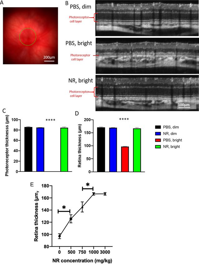
NR is Protective in Retinal Degeneration                                   IOVS | August 2020 | Vol. 61 | No. 10 | Article 47 | 2

              of neurodegeneration, including models of Alzheimer’s            LIRD model, ERGs were performed 1 week after toxic light
              disease, amyotrophic lateral sclerosis, and Parkinson’s          exposure.
              disease.19–27
                 We hypothesized that NR treatment will be protec-
              tive in retinal degeneration. In this study, we tested           In Vivo Ocular Imaging
              whether systemic delivery of NR is protective in a mouse
                                                                               Spectral domain optical coherence tomography (SD-OCT)
              model of light-induced retinal degeneration (LIRD).28–30 NR
                                                                               was conducted immediately after ERG measurement, when
              treatment increased retinal NAD+ , prevented LIRD-induced
                                                                               mice were still anesthetized and their pupils were still
              retinal NAD+ suppression, and protected retinal morphology
                                                                               dilated. A Micron IV SD-OCT system with fundus camera
              and function.
                                                                               (Phoenix Research Labs, Pleasanton, CA) and a Heidel-
                                                                               berg Spectralis HRA+OCT instrument with +25D lens
              METHODS                                                          (Heidelberg Engineering, Heidelberg, Germany) were used
                                                                               in tandem sequentially to assess ocular posterior segment
              Animal Models                                                    morphology in section and en face. Using the Micron IV
                                                                               system, image-guided OCT images were obtained for the left
              All mouse procedures were approved by the Emory Insti-           and right eyes after a sharp and clear image of the fundus
              tutional Animal Care and Use Committee and followed the          (with the optic nerve centered) was obtained. SD-OCT imag-
              ARVO Statement for the Use of Animals in Ophthalmic and          ing was a circular scan about 100 μm from the optic
              Vision Research. Adult (3 months old) male BALB/cAnNCrl          nerve head. Fifty scans were averaged. The retinal layers
              (BALB/c) mice were obtained from Charles River Labo-             were identified according to published nomenclature.32 Total
              ratory (Wilmington, MA, USA) and were housed under a             photoreceptor and retinal thickness were analyzed using
              12:12-hour light–dark cycle (7 AM on and 7 PM off). During       Adobe Photoshop CS6 (Adobe Systems Inc., San Jose, CA).
              the light cycle, light levels measured at the bottom of mouse    The number of pixels was converted into micrometers
              cages ranged from 5 to 45 lux. Induction of LIRD was previ-      by multiplying by the micrometers/pixel conversion factor
              ously described.28 Briefly, mice were exposed to 3000 lux        (1.3 microns = 1 pixel). Immediately after imaging on the
              light for 4 hours, then returned to home cages under normal      Micron IV system, a rigid contact lens was placed on the
              lighting conditions for the remainder of the experiment. At      eye (back optic zone radius: 1.7 mm, diameter: 3.2 mm,
              the time of light induction they weighed 22 to 28 g. NR          power: PLANO) and autofluorescence imaging at the layer
              treatment did not significantly alter weight (data not shown).   of the photoreceptor-RPE interface was conducted using the
              Mice were euthanized by asphyxiation with CO2 gas for all        Heidelberg Spectralis HRA+OCT instrument. During imag-
              experiments.                                                     ing and afterwards through anesthesia recovery, mice were
                                                                               kept on a water circulating heat pad set to 39 °C to maintain
                                                                               body temperature.
              Drug Administration
              NR chloride was from ChromaDex (Item #ASB-00014332-
              101, Lot# 40C910-18209-21). The drug vehicle was PBS             Histology, Immunofluorescence, and
              (VWRVK813, Cat#97063-660), which has a 1× solution               Morphometrics
              composition of 137 mM NaCl, 2.7 mM KCl, and 9.5 mM phos-
                                                                               BALB/c mice were euthanized 1 week after light expo-
              phate buffer. Solutions were made fresh each day. BALB/c
                                                                               sure. We followed the recommendations of Howland and
              mice received intraperitoneal injections of either vehicle or
                                                                               Howland33 for nomenclature of axes and planes in the
              NR (1000 mg/kg dose in PBS unless otherwise specified
                                                                               mouse eye, as indicated in Figure 1. Histologic and morpho-
              by experiment as reported in the Results) using an injec-
                                                                               metric procedures followed standard techniques as previ-
              tion volume of 10 μL of solution per gram of mouse body
                                                                               ously described.34,35 Briefly, eyes were dehydrated, embed-
              weight in accordance with in vivo rodent experiments of
                                                                               ded in paraffin, and sectioned through the sagittal plane on
              Brown et al.20 Unless otherwise specified, two injections
                                                                               a microtome at 5-μm increments through the optic nerve
              were administered before toxic light exposure. One injection
                                                                               head and the center of the cornea to ensure that consistent
              was performed the day before (at 4 PM), and the other injec-
                                                                               regions were examined between animals. The slides were
              tion was performed the morning of (at 9 AM) light insult.
                                                                               deparaffinized across five Coplin jars with 100 mL of xylene
              Toxic light exposure was from 10 AM to 2 PM.
                                                                               for 2 minutes each, consecutively. Then the slides were rehy-
                                                                               drated in a series of 100 mL ethanol solutions for 2 minutes
              Electroretinograms (ERG)                                         each: 100%, 90%, 80%, 70%, 60%, and 50% ethanol. After that,
                                                                               the slides were immersed in PBS for 5 minutes.
              The complete ERG protocol was previously detailed.31                 After rehydration, a TUNEL assay was performed on some
              Briefly, mice dark-adapted overnight were anesthetized with      sections according to the protocol for the DeadEnd Fluoro-
              intraperitoneal injections of ketamine (10 mg/mL; AmTech         metric TUNEL Kit (Promega, Fitchburg, WI). Stained sections
              Group Inc., Tempe, AZ) and xylazine (100 mg/mL; AKORN,           were imaged using fluorescent microscopy and TUNEL-
              Lake Forest, IL),31 placed on a heating pad inside a Faraday     positive cells in the outer nuclear layer (ONL) were manu-
              cage in front of a UBA-4200 Series desktop Ganzfeld stim-        ally counted for each whole retina using Adobe Photoshop
              ulator (MDIT-100, LKC Technologies, Gaithersburg, MD), a         CS6. Some sections were used for hematoxylin and eosin
              DTL fiber active electrode placed on top of each cornea,         (H&E) staining. ONL nuclei were counted within 100 μm-
              reference electrodes inserted into the cheeks, and a ground      wide segments spaced at 250, 750, 1250, and 1750 μm from
              electrode placed in the tail. ERGs were recorded for the         the optic nerve head in both the inferior and superior direc-
              scotopic condition (0.00039-25.3 cd s/m2 with increasing         tions. Mean counts of n = 3 to 6 mice per group were plotted
              flash stimulus intervals from 2.0 to 62.6 seconds). In the       as a spidergram.

Downloaded from iovs.arvojournals.org on 09/28/2020
NR is Protective in Retinal Degeneration                                            IOVS | August 2020 | Vol. 61 | No. 10 | Article 47 | 3

              FIGURE 1. Naming conventions recommended by Howland and
              Howland for planes and axes of the vertebrate eye regardless of
              species. A histologic section cut on the vertical plane through
              the great meridian is illustrated.79 The horizontal, vertical, and
              frontal planes are marked, as are the anterior–posterior (A–P) axis
              (also known as optic axis), the nasal–temporal (N–T) axis, and the
              superior–inferior (S–I; vertical) axis. Reprinted with permission from
              Wisard J, Faulkner A, Chrenek MA, et al. Exaggerated eye growth
              in IRBP-deficient mice in early development. Invest Ophthalmol Vis
              Sci 2011;52:5804–5811. Copyright 2011 CC BY-NC-ND 4.0 license.

                 For detecting Ionized calcium binding adaptor molecule
                                                                                       FIGURE 2. NR treatment increases retinal NAD+ and maintains reti-
              1 (Iba1), sections were blocked for 30 minutes in 0.1 M
                                                                                       nal NAD+ after toxic light exposure. Mice were treated with either
              Tris-buffered saline (TBS; Corning 46012CM pH 7.4, Fisher                NR (1000 mg/kg) or vehicle (PBS). The retinas were harvested and
              Scientific, Suwanee, GA) containing 2.5% donkey serum,                   assayed 48 hours after the end of light exposure. NAD+ concen-
              then incubated in the primary antibody (Rabbit anti-Iba1;                tration significantly decreased in retinas exposed to LIRD-inducing
              1:500; ab178847; Abcam; Cambridge, UK) diluted in the                    light (red bar) compared with those in control lighting (black and
              blocking serum overnight at 4 °C. Sections were rinsed                   blue bars). NR treatment prevented this decrease in NAD+ (green
                                                                                       bar). * P < 0.05, *** P < 0.001 by one-way ANOVA with Newman-Keuls
              twice with 0.1 M TBST (0.1% Tween-20) for 2 minutes
                                                                                       multiple comparisons post hoc test. N = 4–9 retinas per group. Error
              each following primary antibody incubation, then incubated               bars represent SEM.
              for 30 minutes at room temperature with the secondary
              antibody solution (donkey Anti-Rabbit IgG; 1:1000; Alexa
              Fluor 488; A21206; Life Technologies, Waltham, MA).                      gies). Sections were then washed three times with 0.1 M
              Sections were then washed twice with 0.1 M TBST for                      TBS for 15 minutes each, mounted with a 4’,6-diamidino-2-
              2 minutes each and then incubated in diluted PI solution                 phenylindole (DAPI) mounting medium, and coverslipped.
              (1:500 in 0.1 M TBS) for 2 minutes at room temperature.                     The slides immunostained for Iba1 or GFAP were stored
              The sections were washed in 0.1 M TBS for 5 minutes and                  in the dark at 4 °C until imaging using a Nikon Ti inverted
              mounted with mounting medium, and coverslipped. Bitmaps                  microscope with C1 confocal scanner (Nikon Instruments
              from the raw images were used for quantification. Using                  Inc., Melville, NY). Using an automated XY stage control
              ImageJ, the neural retina, including retinal ganglion cell               within the EZ-C1 software, the flatmount was imaged with a
              layer, inner plexiform layer, inner nuclear layer, outer plex-           20× objective lens. Then, confocal images from the entire
              iform layer, ONL, and outer segment were each selected                   section were photomerged using Adobe Photoshop CS6.
              with the lasso tool. The total pixels in this selection were             Bitmaps from the raw images were used for quantifica-
              recorded. The selection was isolated and split into RGB                  tion. Using ImageJ, all pixels outside the neural retina were
              channels. The green channel was thresholded into a black                 deleted. The remaining image was split into RGB channels.
              and white image, with the threshold set at 38 bits. Total black          The red channel was thresholded into a black and white
              pixels were recorded. Data are reported as the thresholded               image, with the threshold set at 38 bits. Total black pixels
              green pixels divided by the total pixels in the neural retina.           were recorded.
                 For detecting glial fibrillary acidic protein (GFAP) in
              ocular sections, sections were blocked for 30 minutes in                 NAD+ Measurements
              0.1 M TBS containing 5% donkey serum, then incubated
              in the primary antibody (rabbit anti-GFAP; 1:500; Z0334;                 Levels of NAD+ in homogenates were measured using
              Agilent Dako, Santa Clara, CA) diluted in the blocking serum             a commercially available kit by following manufacturer’s
              overnight at 4 °C. Sections were rinsed three times with                 instructions (Abcam; ab. 65348; #Lot: GR3226737-3; San
              0.1 M TBS for 15 minutes each following primary antibody                 Francisco, CA). In brief, retina samples were homoge-
              incubation, then incubated for 2 hours at room temper-                   nized in extraction buffer. Extracted samples were sepa-
              ature with the secondary antibody solution (donkey anti-                 rated into two aliquots. One was used to measure total
              rabbit IgG; 1:1000; Alexa Fluor 568; A10042; Life Technolo-              NAD (NADt). For NADH-specific measurements, samples

Downloaded from iovs.arvojournals.org on 09/28/2020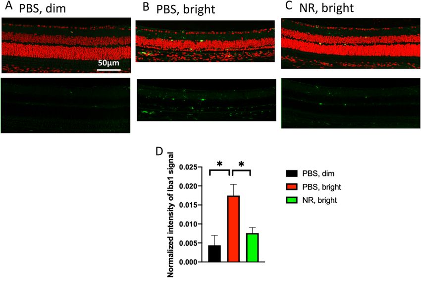
NR is Protective in Retinal Degeneration                                          IOVS | August 2020 | Vol. 61 | No. 10 | Article 47 | 4

              FIGURE 3. NR (1000 mg/kg) treatment preserves retinal function in LIRD mice. Representative ERG waveforms (A) of one eye of a single
              mouse from each treatment group. Scotopic ERG a-wave (B) and b-wave (C) mean amplitudes from LIRD mice at 1 week after LIRD induction.
              Mice treated with PBS and exposed to 3000-lux light for 4 hours to induce degeneration exhibited a- and b-wave mean amplitudes (red) that
              were statistically significantly diminished compared with those of dim groups (blue and black). However, the mean ERG amplitudes of mice
              undergoing retinal degeneration and treated with NR (green) were statistically indistinguishable from those treated with PBS and exposed
              to bright light. ** P < 0.01, *** P < 0.001, **** P < 0.0001 versus other groups by two-way ANOVA with Newman-Keuls multiple comparisons
              post hoc test. N = 4–6 mice per group. Error bars represent SEM.

              were heated to 60 °C for 30 minutes to decompose NAD+ .               RESULTS
              Extracted samples were placed in a 96-well plate and the
              NADH developer was added into each well. The plate was                NR Treatment Prevents Light-Induced Diminution
              placed into a hybrid reader and read every 30 minutes at              of Retinal NAD+ Levels
              OD 450 nm while the color was developing. Data from
              the 2-hour time point are presented. NADt and NADH                    NR is a NAD+ precursor that when given systemically
              concentrations were quantified against an NADH standard               increases NAD+ levels in many tissues, including central
              curve. In the end, NAD+ was calculated with the equation              nervous system structures.19,25,26 The protective effects of
              NAD+ = NADt – NADH.                                                   NR treatment are ascribed in part to local NAD+ increases
                                                                                    in target tissues.20–22,24,26 To test whether this is the case
                                                                                    in retina, BALB/c mice were intraperitoneal injected with
                                                                                    NR (1000 mg/kg) or vehicle (PBS). Each of these groups
              Statistical Analyses                                                  was split in half, one remaining in normal maintenance light
              Statistical analyses were conducted using Prism 8.4.2 Soft-           and the other exposed to a LIRD-inducing light level as
              ware (GraphPad Software Inc. La Jolla, CA). One-way or                described in the Methods. Forty-eight hours after the end of
              two-way ANOVA with Newman-Keuls’ post hoc test,36 were                light exposure, retinas were harvested and assayed for NAD+
              performed for ERG, biochemical, and morphometric data.                content. NAD+ concentration significantly decreased in reti-
              Unless otherwise noted, n is the number of animals per                nas exposed to LIRD-inducing light compared with those
              experimental group. For all analyses, results were consid-            in control (“dim”) lighting (Fig. 2). NR treatment prevented
              ered statistically significant if P < 0.05. All graphs display        this decrease in NAD+ , and indeed increased retinal NAD+
              data as mean ± SEM.                                                   in both lighting conditions (Fig. 2).

Downloaded from iovs.arvojournals.org on 09/28/2020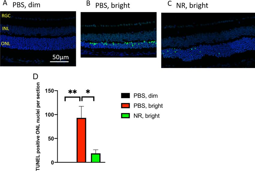
NR is Protective in Retinal Degeneration IOVS | August 2020 | Vol. 61 | No. 10 | Article 47 | 5
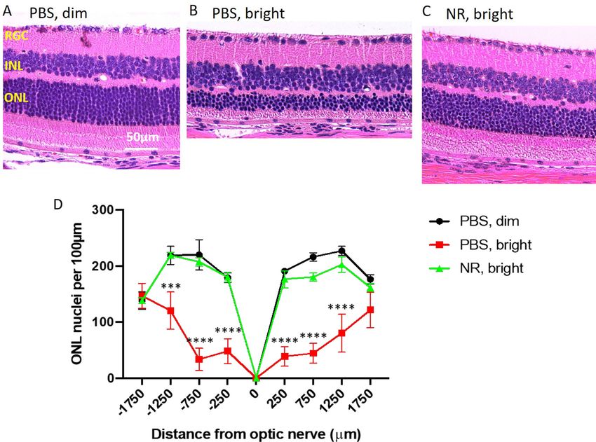
FIGURE 4. NR treatment preserves photoreceptor layer thickness and total retinal thickness as assessed in vivo following LIRD. (A) Repre-
sentative fundus image. (B) Representative OCT images from each group. The OCT image is a circular scan about 100 μm from the optic
nerve head. Photoreceptor thickness (C) and retinal thickness (D) from BALB/c mice 1 week after retinal degeneration induction. Mice
treated with PBS and exposed to 3000-lux light for 4 hours (red bar) exhibited great losses in thickness of the photoreceptor and retina
layers, whereas induced mice treated with NR (green bar) exhibited statistically significant preservation of layer thickness. (E) Treating with
increasing concentrations of NR up to 1000 mg/kg results in significantly increasing retina thicknesses in mice exposed to 3000-lux light for
4 hours. * P < 0.05 between two adjacent groups; **** P < 0.0001 versus all other group means; one-way ANOVA with Newman-Keuls multiple
comparisons post hoc test. N = 3–9 mice per group. Error bars represent SEM. Size marker = 200 μm or 100 μm.
Downloaded from iovs.arvojournals.org on 09/28/2020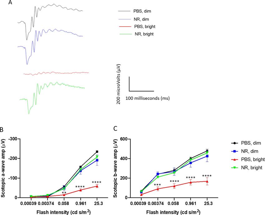
NR is Protective in Retinal Degeneration                                            IOVS | August 2020 | Vol. 61 | No. 10 | Article 47 | 6

              FIGURE 5. NR treatment preserves nuclei of the outer nuclear layer in the LIRD mice. (A–C) Representative H&E images of retina sections
              from each group at the region of 250 to 750 μm from the optic nerve head. Complete sections are shown in Supplementary Figure S1. (D).
              One week after LIRD induction, nuclei were counted in eight discrete regions of retinal sections starting at 250 μm from the optic nerve head
              and extending every 500 μm outward along both the dorsal/superior (positive values on abscissa) and ventral periphery/inferior (negative
              numbers on abscissa). PBS, bright–treated mice (red) showed significant loss of nuclei at six distances from the optic nerve head compared
              with the control group (black). However, NR treated mice (green), exhibited mean nuclei counts statistically indistinguishable from that of
              the control group (black) throughout the length of the retina. ***P < 0.001, ****P < 0.0001 versus other groups by two-way ANOVA with
              Newman-Keuls multiple comparisons post hoc test. N = 3–6 mice per group. Error bars represent SEM. Size marker = 50 μm.

              NR Treatment Preserves Retinal Function and                             and complete protection obtained at 1000 mg/kg (Fig. 4E)
              Morphology in LIRD Mice                                                 relative to non-LIRD control retinas (see Figs. 4C, 4D).
                                                                                         Photomicroscopy of H&E–stained sections of eyes
              ERG a- and b-wave mean amplitudes of PBS-treated BALB/c                 harvested 1 week after LIRD induction showed marked
              mice were significantly decreased 1 week after exposure to              degradation of morphology in the outer retina in LIRD
              3000 lux light for 4 hours. Representative ERG waveforms                mice treated with PBS compared with noninduced mice
              of one eye of a single mouse from each treatment group                  (Figs. 5A, 5B and Supplementary Fig. S1). In induced
              are shown in Figure 3A. This functional loss was entirely               mice treated with PBS, photoreceptor cell inner and outer
              prevented in mice treated with NR (Figs. 3B, 3C).                       segments and most of the nuclei of the ONL were elim-
                 As imaged in vivo by fundus photography and SD-                      inated, with loss predominantly centrally (Figs. 5A–D and
              OCT 1 week after degeneration induction, the retinas of                 Supplementary Fig. S1). Nearly all of this degeneration was
              LIRD mice treated with PBS exhibited significant damage                 prevented in mice treated with NR (Fig. 5C and Supplemen-
              (Fig. 4). The representative fundus image of Figure 4A                  tary Fig. S1). Quantification of ONL nuclei counts across reti-
              shows the region being measured. Corresponding SD-OCT                   nal sections confirmed significant losses owing to degen-
              images show considerable thinning of the photoreceptor                  eration in PBS-treated mice and confirmed nearly-complete
              layer that is prevented by NR treatment (Fig. 4B). Quantifi-            preservation in NR-treated mice (Fig. 5D and Supplementary
              cation of total retina and photoreceptor layer thicknesses              Fig. S1).
              shows that mice undergoing LIRD had significantly thinner
              retinas compared with noninduced mice, largely owing to
              nearly complete loss of the photoreceptor layer. This was
                                                                                      NR Treatment Prevents Accumulation of TUNEL
              entirely prevented with NR treatment (Figs. 4C, 4D). Preser-
              vation of retinal thickness in mice undergoing LIRD was                 Signal in Photoreceptor Cells of LIRD Mice
              dose dependent, with significant protection observed with               Paraffin-embedded ocular sections from mice euthanized
              a 500 mg/kg dose (Fig. 4E), the lowest concentration tested,            1 week after toxic light induction were stained for TUNEL

Downloaded from iovs.arvojournals.org on 09/28/2020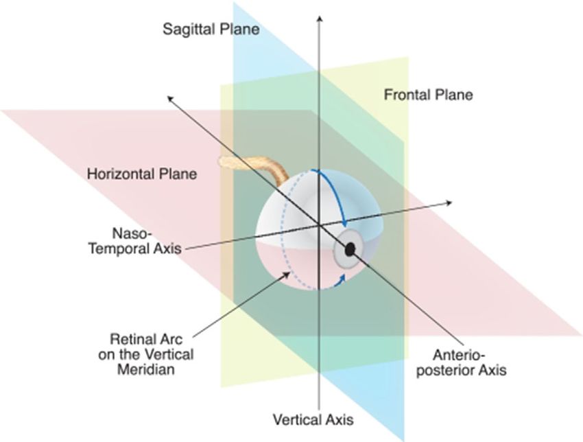
NR is Protective in Retinal Degeneration                                           IOVS | August 2020 | Vol. 61 | No. 10 | Article 47 | 7

              FIGURE 6. NR treatment prevents increases of TUNEL-positive cells following induction of LIRD. Both PBS- and NR-treated BALB/c mice
              received toxic light exposure and were euthanized at 1 week after exposure. Retinas from mice treated as described in text were fixed,
              sectioned, and used in a TUNEL assay. (A–C) Representative morphologic images of each group; the green signal is TUNEL and the blue
              signal is DAPI staining. Complete sections are shown in Supplementary Figure S2. (D) TUNEL-positive nuclei in ONL were counted from
              the entire retina. NR treated mice (green bar) exhibited significantly fewer TUNEL-positive cells compared with the PBS treated group (red
              bar). * P < 0.05, ** P < 0.01 by one-way ANOVA with Newman-Keuls multiple comparisons post hoc test. N = 4 mice per group. Error bars
              represent SEM. Size marker = 50 μm.

              and DAPI to label nuclei that contained double-stranded                in mice treated with NR, which exhibited fewer autofluores-
              DNA breaks (a marker of programmed cell death [apopto-                 cent spots (Fig. 7C), similar in number and pattern to the
              sis] or other forms of cell death).37 ONL TUNEL signal was             uninduced group, confirmed by statistical testing on counts
              high in retinas from LIRD mice treated with PBS compared               of these spots across several autofluorescent fundus images
              with retinas from uninduced mice (Figs. 6A, 6B and Supple-             (Fig. 7D). These in vivo data suggest that LIRD leads to
              mentary Fig. S2). Induced mice treated with NR exhibited               an inflammatory response that is largely prevented by NR
              significantly less TUNEL signal (Fig. 6C and Supplementary             treatment.
              Fig. S2). These data suggest that NR treatment diminished or              To further assess the effects of NR treatment on
              delayed apoptosis in photoreceptor cells.                              LIRD inflammatory responses, ocular sections from mice
                                                                                     euthanized 1 week after light exposure were stained
                                                                                     immunohistologically for Iba1, a marker for microglia and
              NR Treatment Prevents Inflammatory Responses                           monocytes.43,44 Eyes from mice not exposed to toxic light
              Following Induction of LIRD                                            and not undergoing LIRD showed few Iba1-positive cells
                                                                                     in the outer retina (Fig. 8A). In the eyes of mice that
              Subretinal autofluorescent spots observed in vivo by fundus            had undergone LIRD and treated with PBS, numerous and
              examination in patients and in animal models are consid-               widespread Iba1-positive cells were observed (Fig. 8B). This
              ered diagnostic markers for inflammatory responses in reti-            was prevented in mice treated with NR, which exhibited
              nal damage and disease.38–42 To test whether NR treatment              fewer Iba1-positive cells in outer retina (Fig. 8C), similar
              alters inflammatory responses to LIRD, in vivo fundus exam-            in number and pattern to the uninduced group, confirmed
              ination at the level of the subretinal space was conducted 1           by quantification of total Iba1 immunosignal in outer retina
              week after exposure of mice to toxic light. Eyes from mice             across several sections (Fig. 8D). These post mortem data
              not exposed to toxic light and not undergoing LIRD showed              suggest that LIRD leads to the appearance of Iba1-postive
              few autofluorescent spots at the level of the subretinal space         cells in outer retina, a response that is largely prevented by
              in vivo (Fig. 7A). In eyes of mice undergoing LIRD and                 NR treatment.
              treated with PBS, numerous and widespread autofluorescent                 Müller glia cell reactive gliosis was assessed by
              spots were observed (Fig. 7B). This outcome was prevented              immunofluorescence staining for GFAP, a marker for

Downloaded from iovs.arvojournals.org on 09/28/2020
NR is Protective in Retinal Degeneration IOVS | August 2020 | Vol. 61 | No. 10 | Article 47 | 8
FIGURE 7. NR treatment prevented subretinal autofluorescence observed in vivo in LIRD mice. (A–C) Representative morphology images
from each group at the level of the photoreceptor-RPE interface. In vivo Spectralis HRA+OCT images (with autofluorescence detection) were
taken 1 week after induction of degeneration. (D) Autofluorescent spots were counted across the fundus image field. Few were detected
in uninduced mice (black). Toxic light-exposed mice treated with NR exhibited significantly fewer autofluorescent spots compared with the
PBS treated group. **** P < 0.0001 one-way ANOVA with Newman-Keuls multiple comparisons post hoc test. N = 3–6 mice per group. Error
bars represent SEM. Size marker = 200 μm.
reactive Müller cells and astrocytes,45 on post mortem ocular protective effects of NR in a retinal degeneration mouse
sections prepared from mice euthanized 1 week after toxic model.
light exposure. In ocular sections from mice not exposed to Light-induced retinal damage is an acute model of reti-
toxic light and not undergoing LIRD, GFAP staining largely nal degeneration with a well-described oxidative stress
localized near the inner limiting membrane and less so in response.48 Similar to a report from Lin et al.,9 we found that
the outer plexiform layer (Fig. 9A). In eyes of mice under- retinal NAD+ levels were suppressed as early as 2 days after
going LIRD and treated with PBS, heavy GFAP staining was bright light exposure (Fig. 2). NR treatment increased retinal
observed in processes spanning the inner plexiform layer NAD+ levels in maintenance light conditions and prevented
(Fig. 9B). This additional signal was not in mice undergoing NAD+ diminution following toxic light exposure (Fig. 2).
LIRD but treated with NR (Fig. 9C). Quantification of GFAP To determine whether this maintenance of NAD+ could
immunosignal confirmed that GFAP expression was statis- protect the retina, we assessed (by ERG) the efficacy of
tically significantly increased in LIRD tissue, but that this NR treatment in preserving photoreceptor function in mice
increase was prevented with NR treatment (Fig. 9D). undergoing LIRD (Fig. 3). Remarkably, just two NR intraperi-
toneal injections completely preserved scotopic a- and b-
wave amplitudes. In vivo OCT imaging and fundus imag-
DISCUSSION ing showed that systemic NR treatment protected photore-
ceptors and whole retina from degeneration in a dose-
Treatment with NR has been shown to be protective in many dependent manner, with a maximal effect (no discernable
models of neurodegenerative diseases, such as Parkinson’s loss) at 1000 mg/kg dosage and a significant effect down to
disease25 and Alzheimer’s disease,19 and is well-tolerated 500 mg/kg (Fig. 4). Of consideration, a 500 mg/kg/d dose
via oral dosing in humans.46,47 In this study, we found that in mice allometrically scales to a 2.4 g/d human equiva-
systemic treatment with NR increased and maintained retinal lent dose (normalization of dose to body surface area).49,50
levels of NAD+ and protected photoreceptors in a model of Oral dosing of humans at 2000 mg/d is tolerated well,
retinal degeneration. Our study is the first to demonstrate and even lower doses increase circulating NAD+ levels and
Downloaded from iovs.arvojournals.org on 09/28/2020NR is Protective in Retinal Degeneration IOVS | August 2020 | Vol. 61 | No. 10 | Article 47 | 9
FIGURE 8. NR treatment prevents activation of Iba1-positive cells after induction of LIRD. Both PBS- and NR-treated BALB/c mice received
toxic light exposure and were euthanized at 1 week after exposure. (A–C) Representative morphologic images of each group; green is
Iba1 immunofluorescence signal and red is PI staining. (D) Iba1-positive signals were quantified from the entire retina. NR treated mice
(green bar) exhibited significantly less Iba1 signals compared with the PBS treated group (red bar). * P < 0.05 by one-way ANOVA with
Newman-Keuls multiple comparisons post hoc test. N = 4–5 mice per group. Error bars represent SEM. Size marker = 50 μm.
suppress circulating levels of inflammatory cytokines.46,47,51 ity that contributed to protection.55 Additionally, maintain-
Postmortem H&E staining provided more detailed infor- ing NAD+ levels may have permitted continued activity
mation on retinal morphology, indicating that NR treat- of enzymes for which NAD+ is a substrate and that have
ment protected retinal structure, both inferior and superior been shown to be critical to retinal health or function (e.g.,
(Fig. 5). Retinas of mice undergoing LIRD exhibited marked sirtuins,5,9,56–58 poly[ADP-ribose] polymerases,9,59 isocitrate
TUNEL staining, especially in the ONL, suggesting massive dehydrogenases60 ). For instance, maintaining NAD+ levels
apoptosis of photoreceptor cells (Fig. 6, compare Fig. 6A by treating with NR may allow continued enzyme-mediated
with Fig. 6B). NR treatment largely prevented this outcome DNA repair, which may underlie the observed prevention of
(Fig. 6C), suggesting that apoptosis was suppressed. This accumulation of DNA damage (Fig. 6), similar to effects of
effect, although not complete, apparently prevented enough NR treatment in a mouse Alzheimer’s disease model.26
subsequent cell death that bright light exposure had no Similar to what we and others reported previously,29,61–63
discernable impact in other measurements taken 1 week retinas undergoing LIRD exhibited inflammatory responses
after induction of LIRD, such as ERG amplitudes (Fig. 3) or (Figs. 7, 8, and 9). In vivo fundus imaging revealed autofluo-
morphometrics derived from OCT (Fig. 4) or post mortem rescent spots in retinas undergoing LIRD. These spots are
ONL nuclei counts (Fig. 5). Possibly these measurements are postulated to be components of inflammatory responses,
not sensitive enough to detect the impact of a small popu- including activated microglial cells, lipofuscin in RPE
lation of cells that were not protected from apoptosis and, cells, and bisretinoids in the photoreceptors,64–67 and RPE
presumbably, subsequent death. Possibly taking functional cells undergoing epithelial-mesenchymal transition that are
and morphologic measurements at times later than 1 week migrating into the neural retina.68,69 Confocal immunoflu-
after light exposure (e.g., 2 weeks, 1 month, etc.) would orescence microscopy on post mortem retina sections
show effects on these measurements. revealed that retinas undergoing LIRD had increased
Photoreceptors and RPE have high metabolic demands52 expression of Iba1 (Figs. 8B, 8C). This finding may indi-
and metabolic dysregulation, including suppressed NAD+ cate the presence of migrating, activated microglia.44 Retina
levels in either cell type, which are associated with retinal sections from LIRD mice also exhibited increased GFAP
degeneration or disease.10,53,54 It may be that the observed signal in processes spanning the inner plexiform layer and
increase in NAD+ after NR treatment, even after expo- into the inner nuclear layer, suggesting that Müller glia
sure to levels of light that would induce LIRD (Fig. 2), cells were activated.45 In retina sections from mice under-
allowed increased or maintained retinal metabolic capac- going LIRD but that had received intraperitoneal injections
Downloaded from iovs.arvojournals.org on 09/28/2020NR is Protective in Retinal Degeneration IOVS | August 2020 | Vol. 61 | No. 10 | Article 47 | 10
FIGURE 9. NR treatment prevents GFAP accumulating after induction of LIRD. Both PBS and NR treated BALB/c mice received toxic light
exposure and were euthanized at 1 week after exposure. (A–C) Representative morphologic images of each group; red is GFAP immunoflu-
orescence signal and blue is DAPI staining. (D) GFAP signals were quantified from the entire retina. NR-treated mice (green bar) exhibited
significantly less GFAP signals compared with the PBS-treated group (red bar). * P < 0.05, ** P < 0.01 by one-way ANOVA with Newman-Keuls
multiple comparisons post hoc test. N = 4–5 mice per group. Error bars represent SEM. Size marker = 50 μm.
of NR, the appearance of these inflammatory markers in vivo prevent retinal degeneration by preventing phagocytosis of
and post-mortem was significantly diminished (Figs. 7C, 7D, nonapoptotic photoreceptor cells, a mechanism known to
and 8C, 8D, and 9C, 9D). It is known that photoreceptors play a role in retinal degeneration.74
undergoing apoptosis induced by LIRD communicate with In summary, this study is the first to demonstrate that
microglia and Müller cells, causing inflammatory responses systemic treatment with NR is protective in an in vivo reti-
(e.g., Ccl2 upregulation and release from photoreceptor nal degeneration model. The protection is significant, and
cells).70 Suppression of photoreceptor cell apoptosis by NR may support the proposition of prospective human subject
treatment may preclude such signaling. Thus, it may be studies. Future studies ought to test the effects of NR treat-
that NR treatment prevented inflammatory stress responses ment in aged mice, since retinal health and NAD+ levels are
indirectly by preventing early rod photoreceptor injury that known to decline with age,9,75 NR effects in RPE-selective
would have led to apoptosis and subsequent stress signal- damage models,76,77 and putative protective effects of NR
ing. NR treatment may also more directly affect microglia and oral dosing in other mouse models of retinal degeneration.
Müller cells by some as-yet undefined actions. For instance,
NR treatment could increase intracellular NAD+ in microglia
or Müller cells, altering the transcriptional regulatory activ- Acknowledgments
ities of C-terminal–binding protein,71,72 or silent informa- Funded by the Abraham J. and Phyllis Katz Foundation
tion regulator 1,56,73 leading to decreased expression of ( JHB); The joint training program between Emory University
nuclear factor-κB and other proinflammatory genes. Such School of Medicine and Xiangya School of Medicine, Central
direct suppression of microglia activation could partially South University. China Scholarship Council (201806370277
Downloaded from iovs.arvojournals.org on 09/28/2020NR is Protective in Retinal Degeneration IOVS | August 2020 | Vol. 61 | No. 10 | Article 47 | 11
XZ); NIH R01EY028859 ( JHB); NIH R01EY021592 ( JMN); NIH 17. Bieganowski P, Brenner C. Discoveries of nicotinamide
R01EY028450 ( JMN); VA I01RX002806 ( JHB); VA I21RX001924 riboside as a nutrient and conserved NRK genes establish
( JHB); VARR&D C9246C (Atlanta VAMC); NIH P30EY06360 a Preiss-Handler independent route to NAD+ in fungi and
(Emory); an unrestricted departmental award from Research to humans. Cell. 2004;117:495–502.
Prevent Blindness. Inc. to the Ophthalmology Department at 18. Ratajczak J, Joffraud M, Trammell SA, et al. NRK1
Emory University, and the Roy J. Carver Trust (CB). Some data controls nicotinamide mononucleotide and nicotinamide
presented here were presented in abstract form at the annual riboside metabolism in mammalian cells. Nat Commun.
meeting of the Association for Research in Vision and Ophthal- 2016;7:13103.
mology.78 19. Gong B, Pan Y, Vempati P, et al. Nicotinamide riboside
restores cognition through an upregulation of proliferator-
Disclosure: X. Zhang, None; N.F. Henneman, None; P.E. Girar- activated receptor-gamma coactivator 1alpha regulated
dot, None; J.T. Sellers, None; M.A. Chrenek, None; Y. Li, None; beta-secretase 1 degradation and mitochondrial gene
J. Wang, None; C. Brenner, (I, C, P); J.M. Nickerson, None; expression in Alzheimer’s mouse models. Neurobiol Aging.
J.H. Boatright, None 2013;34:1581–1588.
20. Brown KD, Maqsood S, Huang JY, et al. Activation of SIRT3
References by the NAD(+) precursor nicotinamide riboside protects
from noise-induced hearing loss. Cell Metab. 2014;20:1059–
1. Chiang PW, Wang J, Chen Y, et al. Exome sequencing iden- 1068.
tifies NMNAT1 mutations as a cause of Leber congenital 21. Trammell SA, Weidemann BJ, Chadda A, et al. Nicotinamide
amaurosis. Nat Genet. 2012;44:972–974. riboside opposes type 2 diabetes and neuropathy in mice.
2. Falk MJ, Zhang Q, Nakamaru-Ogiso E, et al. NMNAT1 Sci Rep. 2016;6:26933.
mutations cause Leber congenital amaurosis. Nat Genet. 22. Hamity MV, White SR, Walder RY, Schmidt MS, Brenner C,
2012;44:1040–1045. Hammond DL. Nicotinamide riboside, a form of vitamin B3
3. Koenekoop RK, Wang H, Majewski J, et al. Mutations in and NAD+ precursor, relieves the nociceptive and aversive
NMNAT1 cause Leber congenital amaurosis and identify a dimensions of paclitaxel-induced peripheral neuropathy in
new disease pathway for retinal degeneration. Nat Genet. female rats. Pain. 2017;158:962–972.
2012;44:1035–1039. 23. Sasaki Y, Araki T, Milbrandt J. Stimulation of nicotinamide
4. Liao C, Ashley N, Diot A, et al. Dysregulated mitophagy and adenine dinucleotide biosynthetic pathways delays axonal
mitochondrial organization in optic atrophy due to OPA1 degeneration after axotomy. J Neurosci. 2006;26:8484–8491.
mutations. Neurology. 2017;88:131–142. 24. Vaur P, Brugg B, Mericskay M, et al. Nicotinamide riboside,
5. Zhang M, Jiang N, Chu Y, et al. Dysregulated metabolic a form of vitamin B3, protects against excitotoxicity-induced
pathways in age-related macular degeneration. Sci Rep. axonal degeneration. FASEB J. 2017;31:5440–5452.
2020;10:2464. 25. Schondorf DC, Ivanyuk D, Baden P, et al. The NAD+ precur-
6. Fletcher RS, Lavery GG. The emergence of the nicotinamide sor nicotinamide riboside rescues mitochondrial defects and
riboside kinases in the regulation of NAD+ metabolism. J neuronal loss in iPSC and fly models of Parkinson’s disease.
Mol Endocrinol. 2018;61:R107–R121. Cell Rep. 2018;23:2976–2988.
7. Lin JB, Apte RS. NAD(+) and sirtuins in retinal degenera- 26. Hou Y, Lautrup S, Cordonnier S, et al. NAD(+) supplementa-
tive diseases: a look at future therapies. Prog Retin Eye Res. tion normalizes key Alzheimer’s features and DNA damage
2018;67:118–129. responses in a new AD mouse model with introduced DNA
8. Belenky P, Bogan KL, Brenner C. NAD+ metabolism in repair deficiency. Proc Natl Acad Sci U S A. 2018;115:E1876–
health and disease. Trends Biochem Sci. 2007;32:12–19. E1885.
9. Lin JB, Kubota S, Ban N, et al. NAMPT-Mediated NAD(+) 27. Zhou Q, Zhu L, Qiu W, et al. Nicotinamide riboside
Biosynthesis Is Essential for Vision In Mice. Cell Rep. enhances mitochondrial proteostasis and adult neurogen-
2016;17:69–85. esis through activation of mitochondrial unfolded protein
10. Bai S, Sheline CT. NAD(+) maintenance attenuates response signaling in the brain of ALS SOD1(G93A) mice.
light induced photoreceptor degeneration. Exp Eye Res. Int J Biol Sci. 2020;16:284–297.
2013;108:76–83. 28. Lawson EC, Han MK, Sellers JT, et al. Aerobic exercise
11. Tam D, Tam M, Maynard KI. Nicotinamide modulates protects retinal function and structure from light-induced
energy utilization and improves functional recovery from retinal degeneration. J Neurosci. 2014;34:2406–2412.
ischemia in the in vitro rabbit retina. Ann N Y Acad Sci. 29. Mees LM, Coulter MM, Chrenek MA, et al. Low-intensity
2005;1053:258–268. exercise in mice is sufficient to protect retinal function
12. Sheline CT, Zhou Y, Bai S. Light-induced photoreceptor during light-induced retinal degeneration. Invest Ophthal-
and RPE degeneration involve zinc toxicity and are atten- mol Vis Sci. 2019;60:1328–1335.
uated by pyruvate, nicotinamide, or cyclic light. Mol Vis. 30. Henneman NF, Foster SL, Chrenek MA, et al. Xanthohumol
2010;16:2639–2652. protects morphology and function in a mouse model of reti-
13. Zabka TS, Singh J, Dhawan P, et al. Retinal toxicity, in nal degeneration. Invest Ophthalmol Vis Sci. 2018;59:45–53.
vivo and in vitro, associated with inhibition of nicotinamide 31. Boatright JH, Moring AG, McElroy C, et al. Tool from ancient
phosphoribosyltransferase. Toxicol Sci. 2015;144:163–172. pharmacopoeia prevents vision loss. Mol Vis. 2006;12:1706–
14. Bogan KL, Brenner C. Nicotinic acid, nicotinamide, and 1714.
nicotinamide riboside: a molecular evaluation of NAD+ 32. Huber G, Beck SC, Grimm C, et al. Spectral domain optical
precursor vitamins in human nutrition. Annu Rev Nutr. coherence tomography in mouse models of retinal degen-
2008;28:115–130. eration. Invest Ophthalmol Vis Sci. 2009;50:5888–5895.
15. Trammell SA, Yu L, Redpath P, Migaud ME, Brenner C. 33. Howland HC, Howland M. A standard nomenclature for
Nicotinamide riboside is a major NAD+ precursor vitamin the axes and planes of vertebrate eyes. Vision Res.
in cow milk. J Nutr. 2016;146:957–963. 2008;48:1926–1927.
16. Trammell SA, Schmidt MS, Weidemann BJ, et al. Nicoti- 34. Phillips MJ, Walker TA, Choi HY, et al. Tauroursodeoxy-
namide riboside is uniquely and orally bioavailable in mice cholic acid preservation of photoreceptor structure and
and humans. Nat Commun. 2016;7:12948. function in the rd10 mouse through postnatal day 30. Invest
Ophthalmol Vis Sci. 2008;49:2148–2155.
Downloaded from iovs.arvojournals.org on 09/28/2020NR is Protective in Retinal Degeneration IOVS | August 2020 | Vol. 61 | No. 10 | Article 47 | 12
35. Sun N, Shibata B, Hess JF, FitzGerald PG. An alterna- ity and promotes cellular senescence. Aging (Albany NY).
tive means of retaining ocular structure and improving 2018;10:1306–1323.
immunoreactivity for light microscopy studies. Mol Vis. 54. Jadeja RN, Thounaojam MC, Bartoli M, Martin PM. Implica-
2015;21:428–442. tions of NAD(+) metabolism in the aging retina and retinal
36. Zivin JA, Bartko JJ. Statistics for disinterested scientists. Life degeneration. Oxid Med Cell Longev. 2020;2020:2692794.
Sci. 1976;18:15–26. 55. Okawa H, Sampath AP, Laughlin SB, Fain GL. ATP consump-
37. Loo DT. In situ detection of apoptosis by the TUNEL assay: tion by mammalian rod photoreceptors in darkness and in
an overview of techniques. Methods Mol Biol. 2011;682:3– light. Curr Biol. 2008;18:1917–1921.
13. 56. Duarte DA, Rosales MA, Papadimitriou A, et al. Polyphenol-
38. Yung M, Klufas MA, Sarraf D. Clinical applications of fundus enriched cocoa protects the diabetic retina from glial
autofluorescence in retinal disease. Int J Retina Vitreous. reaction through the sirtuin pathway. J Nutr Biochem.
2016;2:12. 2015;26:64–74.
39. Zhang X, Girardot PE, Sellers JT, et al. Wheel running 57. Silberman DM, Ross K, Sande PH, et al. SIRT6 is required
exercise protects against retinal degeneration in the for normal retinal function. PLoS One. 2014;9:e98831.
I307N rhodopsin mouse model of inducible autoso- 58. Lin JB, Kubota S, Mostoslavsky R, Apte RS. Role of Sirtuins
mal dominant retinitis pigmentosa. Mol Vis. 2019;25:462– in retinal function under basal conditions. Adv Exp Med Biol.
476. 2018;1074:561–567.
40. Massengill MT, Ahmed CM, Lewin AS, Ildefonso CJ. Neuroin- 59. Nakagawa T, Guarente L. Sirtuins at a glance. J Cell Sci.
flammation in retinitis pigmentosa, diabetic retinopathy, and 2011;124:833–838.
age-related macular degeneration: a minireview. Adv Exp 60. Du J, Yanagida A, Knight K, et al. Reductive carboxylation
Med Biol. 2018;1074:185–191. is a major metabolic pathway in the retinal pigment epithe-
41. Silverman SM, Wong WT. Microglia in the retina: roles lium. Proc Natl Acad Sci U S A. 2016;113:14710–14715.
in development, maturity, and disease. Annu Rev Vis Sci. 61. Mandal MN, Patlolla JM, Zheng L, et al. Curcumin protects
2018;4:45–77. retinal cells from light-and oxidant stress-induced cell death.
42. Gargini C, Novelli E, Piano I, Biagioni M, Strettoi E. Pattern Free Radic Biol Med. 2009;46:672–679.
of retinal morphological and functional decay in a light- 62. Terao R, Honjo M, Ueta T, et al. Light stress-induced increase
inducible, rhodopsin mutant mouse. Sci Rep. 2017;7:5730. of sphingosine 1-phosphate in photoreceptors and its rele-
43. Couturier A, Bousquet E, Zhao M, et al. Anti- vance to retinal degeneration. Int J Mol Sci. 2019;20:3670.
vascular endothelial growth factor acts on retinal 63. Bian M, Zhang Y, Du X, et al. Apigenin-7-diglucuronide
microglia/macrophage activation in a rat model of ocular protects retinas against bright light-induced photorecep-
inflammation. Mol Vis. 2014;20:908–920. tor degeneration through the inhibition of retinal oxidative
44. Song D, Sulewski ME, Jr., Wang C, et al. Comple- stress and inflammation. Brain Res. 2017;1663:141–150.
ment C5a receptor knockout has diminished light- 64. Kim SY, Yang HJ, Chang YS, et al. Deletion of aryl hydrocar-
induced microglia/macrophage retinal migration. Mol Vis. bon receptor AHR in mice leads to subretinal accumulation
2017;23:210–218. of microglia and RPE atrophy. Invest Ophthalmol Vis Sci.
45. Heynen SR, Tanimoto N, Joly S, Seeliger MW, Samardz- 2014;55:6031–6040.
ija M, Grimm C. Retinal degeneration modulates intracel- 65. Bubis E, Sher I, Skaat A, et al. Blue Autofluorescence
lular localization of CDC42 in photoreceptors. Mol Vis. fundus imaging for monitoring retinal degeneration in Royal
2011;17:2934–2946. College of Surgeons Rats. Transl Vis Sci Technol. 2019;8:26.
46. Conze D, Brenner C, Kruger CL. Safety and Metabolism 66. Sparrow JR, Duncker T. Fundus autofluorescence and RPE
of long-term administration of NIAGEN (nicotinamide ribo- lipofuscin in age-related macular degeneration. J Clin Med.
side chloride) in a randomized, double-blind, placebo- 2014;3:1302–1321.
controlled clinical trial of healthy overweight adults. Sci Rep.
67. Sparrow JR, Wu Y, Nagasaki T, Yoon KD, Yamamoto K, Zhou
2019;9:9772.
J. Fundus autofluorescence and the bisretinoids of retina.
47. Elhassan YS, Kluckova K, Fletcher RS, et al. Nicoti- Photochem Photobiol Sci. 2010;9:1480–1489.
namide riboside augments the aged human skeletal muscle
68. Zanzottera EC, Ach T, Huisingh C, Messinger JD, Freund KB,
NAD(+) metabolome and induces transcriptomic and
Curcio CA. Visualizing retinal pigment epithelium pheno-
anti-inflammatory signatures. Cell Rep. 2019;28:1717–1728
types in the transition to atrophy in neovascular age-related
e1716.
macular degeneration. Retina. 2016;36(Suppl 1):S26–S39.
48. Demontis GC, Longoni B, Marchiafava PL. Molecular steps
69. Zanzottera EC, Ach T, Huisingh C, Messinger JD, Spaide RF,
involved in light-induced oxidative damage to retinal rods.
Curcio CA. Visualizing retinal pigment epithelium pheno-
Invest Ophthalmol Vis Sci. 2002;43:2421–2427.
types in the transition to geographic atrophy in age-related
49. Nair A, Morsy MA, Jacob S. Dose translation between labo- macular degeneration. Retina. 2016;36(Suppl 1):S12–
ratory animals and human in preclinical and clinical phases S25.
of drug development. Drug Dev Res. 2018;79:373–382.
70. Feng C, Wang X, Liu T, Zhang M, Xu G, Ni Y. Expression
50. Nair AB, Jacob S. A simple practice guide for dose conver- of CCL2 and its receptor in activation and migration of
sion between animals and human. J Basic Clin Pharm. microglia and monocytes induced by photoreceptor apop-
2016;7:27–31. tosis. Mol Vis. 2017;23:765–777.
51. Airhart SE, Shireman LM, Risler LJ, et al. An open-label, 71. Shen Y, Kapfhamer D, Minnella AM, et al. Bioenergetic state
non-randomized study of the pharmacokinetics of the nutri- regulates innate inflammatory responses through the tran-
tional supplement nicotinamide riboside (NR) and its effects scriptional co-repressor CtBP. Nat Commun. 2017;8:624.
on blood NAD+ levels in healthy volunteers. PLoS One.
72. Morris G, Puri BK, Maes M, Olive L, Berk M, Carvalho
2017;12:e0186459.
AF. The role of microglia in neuroprogressive disor-
52. Kanow MA, Giarmarco MM, Jankowski CS, et al. Biochemi- ders: mechanisms and possible neurotherapeutic effects of
cal adaptations of the retina and retinal pigment epithelium induced ketosis. Prog Neuropsychopharmacol Biol Psychia-
support a metabolic ecosystem in the vertebrate eye. Elife. try. 2020;99:109858.
2017;6:e28899.
73. McGettrick AF, O’Neill LA. How metabolism generates
53. Jadeja RN, Powell FL, Jones MA, et al. Loss of NAMPT in signals during innate immunity and inflammation. J Biol
aging retinal pigment epithelium reduces NAD(+) availabil- Chem. 2013;288:22893–22898.
Downloaded from iovs.arvojournals.org on 09/28/2020NR is Protective in Retinal Degeneration IOVS | August 2020 | Vol. 61 | No. 10 | Article 47 | 13
74. Zhao L, Zabel MK, Wang X, et al. Microglial phagocytosis of 77. Zhang N, Zhang X, Girardot P, et al. Optimization of a retinal
living photoreceptors contributes to inherited retinal degen- pigment epithelium damage model. Invest Ophthalmol Vis
eration. EMBO Mol Med. 2015;7:1179–1197. Sci. 2020;61:4441–4441.
75. Kolesnikov AV, Fan J, Crouch RK, Kefalov VJ. Age- 78. Zhang X, Henneman N, Girardot PE, et al. Systemic treat-
related deterioration of rod vision in mice. J Neurosci. ment with nicotinamide riboside is protective in four mouse
2010;30:11222–11231. models of retinal degeneration. Invest Ophthalmol Vis Sci.
76. Girardot P, Zhang X, Gefke I, et al. Systemic pentosan poly- 2020;61:2753–2753.
sulfate administration causes retinal function loss in the 79. Wisard J, Faulkner A, Chrenek MA, et al. Exaggerated eye
C57Bl/6J mouse. Invest Ophthalmol Vis Sci. 2019;60:2352– growth in IRBP-deficient mice in early development. Invest
2352. Ophthalmol Vis Sci. 2011;52:5804–5811.
Downloaded from iovs.arvojournals.org on 09/28/2020You can also read