Progerinin, an optimized progerin-lamin A binding inhibitor, ameliorates premature senescence phenotypes of Hutchinson-Gilford progeria syndrome
←
→
Page content transcription
If your browser does not render page correctly, please read the page content below
ARTICLE
                  https://doi.org/10.1038/s42003-020-01540-w                 OPEN

                  Progerinin, an optimized progerin-lamin A binding
                  inhibitor, ameliorates premature senescence
                  phenotypes of Hutchinson-Gilford progeria
1234567890():,;

                  syndrome
                  So-mi Kang1,7, Min-Ho Yoon1,7, Jinsook Ahn2, Ji-Eun Kim3, So Young Kim4, Seock Yong Kang4, Jeongmin Joo4,
                  Soyoung Park1, Jung-Hyun Cho1, Tae-Gyun Woo1, Ah-Young Oh1, Kyu Jin Chung3, So Yon An5,
                  Tae Sung Hwang5, Soo Yong Lee6, Jeong-Su Kim6, Nam-Chul Ha2, Gyu-Yong Song3 & Bum-Joon Park 1 ✉

                  Previous work has revealed that progerin-lamin A binding inhibitor (JH4) can ameliorate
                  pathological features of Hutchinson-Gilford progeria syndrome (HGPS) such as nuclear
                  deformation, growth suppression in patient’s cells, and very short life span in an in vivo
                  mouse model. Despite its favorable effects, JH4 is rapidly eliminated in in vivo pharmaco-
                  kinetic (PK) analysis. Thus, we improved its property through chemical modification and
                  obtained an optimized drug candidate, Progerinin (SLC-D011). This chemical can extend the
                  life span of LmnaG609G/G609G mouse for about 10 weeks and increase its body weight.
                  Progerinin can also extend the life span of LmnaG609G/+ mouse for about 14 weeks via oral
                  administration, whereas treatment with lonafarnib (farnesyl-transferase inhibitor) can only
                  extend the life span of LmnaG609G/+ mouse for about two weeks. In addition, progerinin can
                  induce histological and physiological improvement in LmnaG609G/+ mouse. These results
                  indicate that progerinin is a strong drug candidate for HGPS.

                  1 Department of Molecular Biology, College of Natural Science, Pusan National University, Busan, Korea. 2 Department of Food Science, College of Agricultural

                  Science, Seoul National University, Seoul, Korea. 3 Department of Pharmacy, College of Pharmacy, Chungnam National University, Daejeon, Korea. 4 New
                  Drug Development Center, Daegu-Gyeongbuk Medical Innovation Foundation, Daegu, Korea. 5 Institute of Animal Medicine, College of Veterinary Medicine,
                  Gyeongsang National University, Jinju, Korea. 6 Cardiovascular Center, Research Institute for Convergence of Biomedical Science and Technology, Pusan
                  National University Yangsan Hospital, Yangsan, Korea. 7These authors contributed equally: So-mi Kang, Min-Ho Yoon. ✉email: bjpark1219@pusan.ac.kr

                  COMMUNICATIONS BIOLOGY | (2021)4:5 | https://doi.org/10.1038/s42003-020-01540-w | www.nature.com/commsbio                                                  1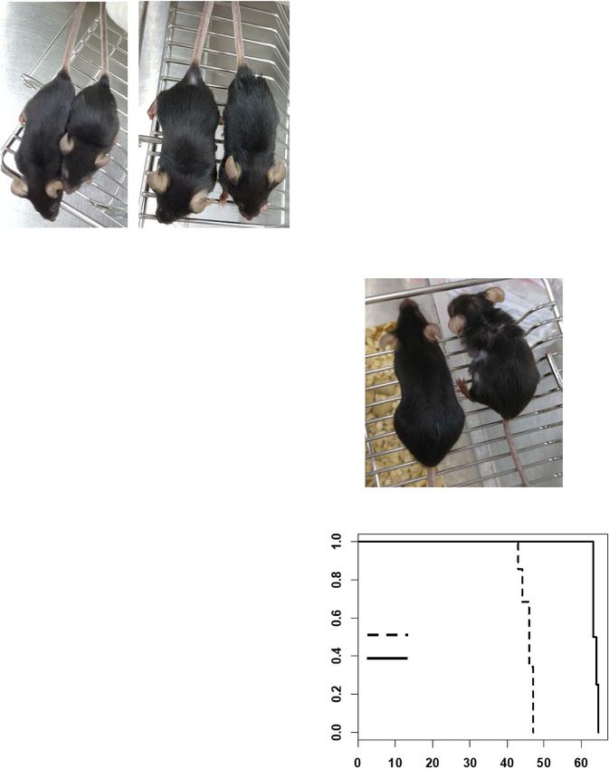
ARTICLE                                                              COMMUNICATIONS BIOLOGY | https://doi.org/10.1038/s42003-020-01540-w

H
         utchinson-Gilford progeria syndrome (HGPS) is a very               problem, we generated various JH4 modified chemicals (Fig. S3a)
         rare genetic disease1–3. It is also known as a premature           to improve its in vivo stability. We tested effects of JH4
         aging syndrome4,5. A single point mutation in LMNA                 derivatives on progerin-lamin A binding by GST-pull-down
(G608G) generates progerin (50 amino acid deleted lamin A) by               assay and progerin expression analysis (Figs. S3a and S3b).
abnormal splicing6–9. Lamin A is matured through a four-step                Among them, JH010 and SLC-D011 chemicals could block the
modifications. First, a 15-carbon farnesyl lipid is added to the             interaction of progerin and lamin A (Fig. 1c and Figs. S3c and
carboxyl terminal cysteine by farnesyltransferase. Second,                  S3d; chemical structures of them are depicted in Fig. S3e). In
Zmpste24 protease cleaves the last three amino acids. Third, the            addition, both JH010 and SLC-D011 could induce H3K9me3
newly exposed farnesylcysteine is carboxyl-methylated by a                  expression and reduce progerin expression in HGPS cells (Figs. 1d
prenylprotein-specific methyltransferase. Finally, Zmpste24                  and 1e and Figs. S3f and S3g). We also found that progerin levels
removes terminal 15 amino acids of the protein, resulting in the            were decreased after treatment with SLC-D011 in other kinds of
release of mature lamin A10–12. Since Zmpste24-cleavage site is             HGPS cells without alteration of mRNA expression of progerin
located in the deleted 50 AA region in progerin, progerin retains           (Fig. 1f and Figs. S3h and S3i). As expression of progerin were
farnesylated C-terminus12. Based on this fact, lonafarnib, a far-           reduced by SLC-D011 without a reduction of the mRNA levels,
nesyltransferase inhibitor (FTI), has been suggested as a putative          we speculated that progerin may lose its stability by dissociation
drug for HGPS13. Indeed, clinical trial of lonafarnib for HGPS has          from lamin A after treatment with SLC-D011. Indeed, we could
shown that it can extend the life span for ~2 years, although the           confirm that the expression of progerin was reduced by SLC-
trial has been performed without a placebo (single side experi-             D011 and restored by incubation with N-acetyl-leucinyl-norleuc-
ment). However, in in vitro experiment, lonafarnib has cytotoxic            inal, a proteasome inhibitor (Fig. S3j). We also tested that SLC-
effects, leading to the formation of donut-shaped nuclei14 and              D011 degraded progerin by autophagy activation. We observed
apoptosis15,16 rather than having a favorable effect. These results         that SLC-D011 did not change the expression of LC3 and beclin
suggested more effective therapeutic approaches were needed for             1, autophagy-related genes (Fig. S3k). We also confirmed that the
HGPS patients without severe side effect.                                   expression of beclin 1 could be reduced by 3-MA, an autophagy
   Here, we report that a binding inhibitor of lamin A (LA) and             inhibitor, but not by SLC-D011 (Fig. S3l) SA-β-gal staining, a
progerin called progerinin, improves aging-associated alterations           standard senescence assay20, also showed that SLC-D011 could
in both in vitro and in vivo HGPS models, and consequently                  block cellular senescence (Fig. S3m). We then observed the
suggest that progerinin can be an effective treatment strategy for          expression of CENP1, IL-6, IL-8, and BRCA1 at mRNA levels
patients with HGPS.                                                         were restored after treatment with SLC-D011 (Fig. S3n).
                                                                            Treatment with SLC-D011 could also rescue the expression of
                                                                            LAP2α in HGPS cells (Fig. S3o). In normal fibroblasts derived
Results                                                                     from healthy 9-year-old person, treatment with SLC-D011 did
SLC-D011 ameliorates the premature aging characteristics of                 not change significantly (Figs. S3n and S3o). Moreover, JH010
HGPS. To test whether abnormal processing and retaining of far-             and SLC-D011 could promote cell proliferation of HGPS cells
nesylation in progerin is critical for nuclear deformation in HGPS.         (Fig. 1g and Fig. S4a) and ameliorate nuclear abnormality (Fig. 1h
We generated a point mutation to the CAAX motif of LA and                   and Fig. S4b). Compared to normal fibroblasts, treatment with
progerin to mimic farnesylation inhibitory activity (Figs. S1a and          SLC-D011 increases cell proliferation in three different HGPS
S1b). We monitored nuclear morphologies of GFP-tagged LA, LA-               cells (Fig. S4c). We also observed a reduction of progerin level
C661A (LA-C661A), progerin (PG), and progerin-C611A (PG-                    and an increase of H3K9me3 in three kinds of HGPS cells after
C611A) positive HEK293 cells. However, farnesylation-deficient               treatment with JH4, JH010, or SLC-D011 (Figs. S4d and S4e).
mutants did not show obvious difference with wild-type LA and
progerin (Fig. 1a and Figs. S1c and S1d). The non-farnesylated
                                                                            Therapeutic effect of SLC-D011. In pharmacokinetic (PK)
progerin showed deformed nuclei to the similar extent as progerin.
                                                                            analysis, JH010 showed satisfactory bioavailability (B.A) result
Wild-type LA and LA-C661A also represented similar percentage of
                                                                            (~70%; Fig.2a and Fig. S5a). However, in in vitro absorption,
nuclear deformation (Fig. S1e). Indeed, progerin and progerin-
                                                                            distribution, metabolism, and excretion (ADME) analysis (Figs.
C611A reduced the expression of H3K9me3 (Fig. 1b and Fig. S1f and
                                                                            S5b–S5e), JH010 inhibited hERG (human Ether-à-go-go-Related
S1g), a marker of senescence17. We also observed increases in
                                                                            Gene), an ion channel (~75% inhibition at 1 μM; Fig. 2b). Thus,
binding affinity of progerin, progerin-C611A, and LA-C661A (Fig.
                                                                            we performed in vitro ADME test for SLC-D011. SLC-D011
S1h). The biological meaning of such increase of LA-C661A is cur-
                                                                            showed a B.A similar to JH010 (Fig. 2c and Fig. S6a) without
rently unclear. These results suggest that farnesylation in progerin is
                                                                            severe hERG inhibition (Fig. 2b). Based on other indicators such
not critical for nuclear abnormality.
                                                                            as CYP inhibition, plasma stability, liver microsomal stability, and
   Our recent study has suggested that nuclear abnormality, a
                                                                            plasma protein binding, it appeared that SLC-D011 did not cause
featured phenotype of HGPS cells, might be caused by strong
                                                                            serious adverse effects (Figs. S6b–S6e). Thus, we focused on the
binding between lamin A and progerin17. Indeed, an inhibitor
                                                                            therapeutic effect of SLD-D011. To check the efficacy of SLC-
(JH4) of progerin for lamin A binding can ameliorate nuclear
                                                                            D011, we measured progerin expression in HGPS cells and found
deformation in HGPS cells and restore senescence-related
                                                                            that 100 nM was enough for eliminating progerin (Fig. 2d, Figs.
markers such as p16/INK4A, DNA-PK, and H3K9me3 expres-
                                                                            S7a–S7c). Exogenous progerin expression could be reduced by
sion17. Moreover, treatment with JH4 via intraperitoneal injection
                                                                            treatment with SLC-D011 (from 100 nM; Fig. 2e and Fig. S7d).
(i.p) can extend the life span of LmnaG609G/G609G mouse for
                                                                            Indeed, 100 nM of SLC-D011 seemed to be enough for inducing
~4 weeks (~25% increase)17. To use this chemical as a therapeutic
                                                                            Ki67 (a cell proliferation marker21) expression in HGPS cells
drug, we tested its chemical property. Considering that patients
                                                                            (Fig. 2f, Figs. S7e and S7f). A direct cell counting assay also
with HGPS are children who need to take medication for their
                                                                            revealed that 100 nM SLC-D011 could promote cell proliferation
whole life18,19, intravenous injection (i.v) is not a suitable delivery
                                                                            and ameliorate nuclear abnormality (Figs. S7g and S7h).
method. Thus, we first checked the possibility of oral adminis-
tration. However, the half-life of JH4 was extremely short after
in vivo oral administration (undetectable level; Figs. S2a–S2c) that        SLC-D011 improves premature aging features of HGPS model
we could not calculate its bioavailability (B.A). To overcome this          mouse. Next, the effect of SLC-D011 was tested in a mouse model

2                         COMMUNICATIONS BIOLOGY | (2021)4:5 | https://doi.org/10.1038/s42003-020-01540-w | www.nature.com/commsbio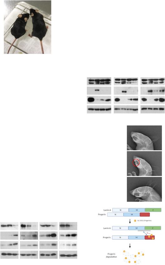
COMMUNICATIONS BIOLOGY | https://doi.org/10.1038/s42003-020-01540-w                                                                 ARTICLE

of progeria22. Treatment with SLC-D011 via i.p. injection (20 mg/             (Fig. 3f). We also observed improved morphologies such as the
kg, twice a week; Fig. 3a and Fig. S8) increased body weight                  status of hair and body size of mouse (Fig. 3d). These results
(Fig. 3b) and extended life span of LmnaG609G/G609G mouse up to               indicate that SLC-D011 is a strong drug candidate for HGPS.
21 weeks (Fig. 3c). Gross morphology was also obviously                       Thus, we chose SLC-D011 as a final drug candidate and renamed
improved after treatment with SLC-D011 (increase of body                      it as progerinin (progerin inhibitor).
size; Fig. 3a). A more interesting feature was obtained for
LmnaG609G/+ mice (Fig. 3d and Fig. S8). Treatment with SLC-
D011 increased their body weights (Fig. 3e) and extended their                Oral administration of SLC-D011 prevents progeroid pheno-
life span from 46.7 weeks to 63 weeks (>1 year; Fig. 3f). This is a           types in HGPS mouse. Progerinin (SLC-D011) has very low solu-
very exciting result because SLC-D011 can extend the life span of             bility in water, which is one barrier for oral administration. Thus, we
LmnaG609G/+ for ~16 weeks longer than the control (untreated)                 screened various solvents and found that a monoolein-based solution

COMMUNICATIONS BIOLOGY | (2021)4:5 | https://doi.org/10.1038/s42003-020-01540-w | www.nature.com/commsbio                                          3
ARTICLE                                                               COMMUNICATIONS BIOLOGY | https://doi.org/10.1038/s42003-020-01540-w

Fig. 1 SLC-D011 ameliorates premature aging features of HGPS. A. HEK293 cells were transiently transfected with expression vector encoding wild-type
lamin A (WT-LA), lamin A-C661A (LA-C661A), progerin, or progerin-C611A. LA-C661A or progerin-C611A with point mutation in CaaX motif was
predicted to be lack of farnesylation, indistinguishable from that of authentic WT-LA or progerin. GFP-conjugated empty expression vector was used as a
negative control. Cells were visualized at 24 h after transfection. B Transient transfection of HEK293 cells with expression vector encoding progerin or
progerin-G611A leads to reduced expression of H3K9me3 (n = 3 independent experiments; two-tailed Student’s t test). C. JH4 derivatives (JH010 and SLC-
D011(D011)) inhibit lamin A and progerin interaction. For binding inhibition assay, bead-conjugated lamin A (GST-LA) was incubated with HEK293 cells,
transiently transfected with GFP-progerin (PG), and treated with indicated chemicals. Actin was used as a loading control. PPT, co-precipitated materials
with recombinant proteins; Sup supernatant. 5 μM of each chemical was used for binding assay. D JH010 and SLC-D011 induce H3K9me3 expression.
H3K9me3 reduction is a well-known marker for premature aged cells. Thus, H3K9me3 expression was evaluated by immunostaining. Both JH010 and SLC-
D011 could induce H3K9me3 expression in HGPS cells (AG03198, n = 3 independent experiments; two-tailed Student’s t test). E SLC-D011 obviously
suppresses progerin expression. For progerin expression analysis, each chemical was used to treat HGPS cells (AG03198, Coriell Cell Repositories) for
7 days. Compared to JH4 or JH010, SLC-D011 obviously suppressed progerin expression (n = 3 independent experiments; two-tailed Student’s t test).
F SLC-D011 reduces the expression of progerin in other HGPS cells (AG11513 and AG11498, Coriell Cell Repositories). Treatment with SLC-D011 (2 μM) for
7 days reduced the expression of progerin and p16/INK4A but induced the expression of H3K9me3. However, normal fibroblast (N9; GM00038, foreskin
fibroblast from a 9-year-old healthy child) did not respond to SLC-D011 (n = 3 independent experiments; two-tailed Student’s t test). G JH4, JH010, and
SLC-D011 promote cell proliferation. Cell viability was determined by MTT assay after incubation with chemicals for 7 days (n = 3 independent
experiments; two-tailed Student’s t test). H JH4, JH010, and SLC-D011 show similar activity for ameliorating nuclear deformation. After incubation with
chemicals for 7 days, cells were stained with progerin and H3K9me3 antibodies (n = 3 independent experiment; two-tailed Student’s t test), **p < 0.001.
Data are reported as mean ± SD. “Con” means Dimethyl sulfoxide (DMSO)-treated control. The data are normalized to DMSO-treated cells.

was useful for dissolving progerinin (Fig. S9a). Monoolein-based              progerinin could restore trabecular and cortical bone thickening (Fig.
solution has been used to increase intestinal absorption without              S11f). Moreover, progerinin could improve grip strength (Fig. 4e)
causing toxicity23, suggesting that it would be suitable as a carrier for     and heart beat rate (Fig. 4f) known to be reduced in a mouse model
progerinin. Despite heating and sonicating to making a clear solution         of HGPS29. Acquisition of kyphosis and dental abnormality are
(Fig. S9a), progerinin was very stable in the solution (Fig. S9b). In PK      characteristics of LmnaG609G transgenic mice22,28,30. The mean
analysis, progerinin was well recovered in the blood (Fig. S9c) in a          kyphosis index (KI) of wild-type mice was >4. Incisors were normal
dose-dependent manner (Figs. S9c and S9d). Oral administration of             at 41 weeks of age. The mean KI of untreated LmnaG609G/+ mice was
progerinin (50 mg/kg, daily administration) into LmnaG609G/G609G              lower than that of Lmna+/+ mice. Such low level of KI was recovered
mice ameliorated gross morphology (Fig. 4a), increased body weight            after treatment with progerinin (Figs. S11g and S11h). LmnaG609G/+
(Fig. 4b), and extended the life span of LmnaG609G/G609G mice for             mice had abundant adipose tissues in the abdomen after treatment
10 weeks compared to untreated mice (Fig. 4c). Progerin levels in             with progerinin (Figs. S11g). Untreated LmnaG609G/+ mice showed
tissues of LmnaG609G/+ mice after treatment with progerinin (oral             abnormalities of incisors (Fig. 4g). Their lower incisors grew toward
administration at 50 mg/kg daily for 8 weeks) were also determined.           the palate because of malocclusion28. Progerinin ameliorated these
Gross morphology (hair of mice) was improved after treatment with             abnormalities of LmnaG609G/+ mice (Fig. 4g). All of LmnaG609G/+
progerinin for 4 weeks (Fig. S10a). Such treatment also increased             mice treated with progerinin showed histological improvements
body weight (Fig. S10b) and life span (from 49 weeks to 65 weeks;             compared with untreated LmnaG609G/+ mice. Our results indicate
Fig. S10c). Obvious reduction of progerin and the increase of                 that progerinin (monoolein-based SLC-D011 solution) could be used
H3K9me3 were observed in progerinin-treated LmnaG609G/+ mice                  as a medication for patients with HGPS.
compared with untreated LmnaG609G/+ mice (Fig. 4d and Figs. S10d                 Next, we performed a comparative study for lonafarnib and
and S10e). Histological analysis was then performed by hematoxylin            progerinin. Differentially from lonafarnib, SLC-D011 at a lower
and eosin (H&E) staining or Masson Trichrome staining for several             concentration reduced the expression of transfected progerin in
tissues of Lmna+/+ and LmnaG609G/+ mice (Figs. S11a–S11f). In                 HEK293 cells (Fig. S12a). To confirm this finding, we measured
general, fibrosis occurs in tissues such as the heart, liver, and lungs in     the expression level of progerin in HGPS cells after chemical
aged mice24–26. As the Masson Trichrome staining procedure stains             treatment. Although responses of different HGPS cells to
the collagen-rich fibrotic regions in blue, it is suitable for assessing       lonafarnib were different (such as a reduction of progerin by
and visualizing the extent of fibrosis in tissues27. Therefore, we per-        lonafarnib in AG03199 and an induction of H3K9me3 in
formed Masson Trichrome staining to check the fibrosis in the heart,           AG03198), SLC-D011 commonly reduced progerin level but
liver, and lungs of mice. It was confirmed that fibrosis was increased          induced H3K9me3 and cyclin B1, a cell cycle marker (Fig. 4h). In
especially in the heart and liver of untreated LmnaG609G/+ mice               addition, long-term treatment with lonafarnib induced cell death
compared with Lmna+/+ mice, which was confirmed to be reduced                  in both normal fibroblasts and HGPS cells (Fig. S12b). In life span
by progerinin (Figs. S11a–S11c). We also observed a relatively loose          analysis of LmnaG609G/+ mice, lonafarnib only extended 2 weeks
connection between blood vessel walls and tissues in the liver (Fig.          longer, whereas SLC-D011 could extend ~14 weeks longer (Fig.
S11b) and lungs (Fig. S11c) of untreated LmnaG609G/+ mice com-                S12c). Finally, we investigated the effect of combination treatment
pared with Lmna+/+ mice and progerin-treated LmnaG609G/+ mice.                of SLC-D011 with lonafarnib. In long-term treatment experiment
LmnaG609G transgenic mice are known to show defects in skin tis-              (2-week-treatment), we observed decreased level of progerin but
sues28. In the present study, epidermis of foot pad skin was thickened        increased expression levels of H3K9me3 and cyclin B1 in SLC-
and dermal connective tissue was enriched in progerinin-treated               D011-treated HGPS cells regardless of the presence of lonafarnib
LmnaG609G/+ mice (Fig. S11d). We observed bone marrow reduction               (Figs. S12d and S12e). However, co-treatment with lonafarnib
and loss of spinal cord cells in vertebrae of untreated LmnaG609G/+           seemed to interrupt the induction of cyclin B1 by SLC-D011 (Fig.
mice (Fig. S11e). Defects of bone marrow and spinal cord in vertebral         S12e). Indeed, lonafarnib induced donut-shaped nuclei (Fig.
column of LmnaG609G/+ mice were also reduced after treatment with             S12f), similar to a previous report14. Considering these results,
progerinin (Fig. S11e). Staining of femur (thigh bone) from untreated         SLC-D011 (progerinin) seems to be a more suitable candidate
LmnaG609G/+ mice with H&E revealed trabecular bone loss com-                  drug for HGPS than lonafarnib. We tested the toxicity of SLC-
pared with age-matched Lmna+/+ mice. We observed that                         D011 in dogs and rats and found that it had no severe toxicity at

4                          COMMUNICATIONS BIOLOGY | (2021)4:5 | https://doi.org/10.1038/s42003-020-01540-w | www.nature.com/commsbio
COMMUNICATIONS BIOLOGY | https://doi.org/10.1038/s42003-020-01540-w ARTICLE
A B
10000 Compound % Inhibition Conc n number
B.A : 69.9% JH10140203 74.98993.72 % 1uM 51
Conc of JH010 (ng/ml)
1000 Positive control
48.5246.87 % 1uM 14
(Quinidine)
100 14.6734.74 % 1uM 21
SLC-D011
66.3116.77 % 10uM 3
IV_1mg/kg
10
PO_10mg/kg Positive control
52.348.52 % 1uM 11
(Quinidine)
1
hERG inhibition
0.08 0.25 0.5 1 2 4 6 8
Time (hr)
100 nM
500 nM
C D
10 nM
50 nM
1 μM
2 μM
5 μM
Con
100 D011
BA : 65.9% (5 mg/kg)/58.1% (10 mg/kg)
Conc of SLC-D011 (log ng/ml)
75
Progerin
75
Lamin A/C
10
H3K9me3 15
IV_1mg/kg
54
PO_5mg/kg Cyclin B1
PO_10mg/kg 54
1 Actin
0.08 0.25 0.5 1 2 4 8
AG11498 (72 hr)
Time (hr)
E F
AG11498
100 nM
500 nM
0.8
10 nM
50 nM
*
1 μM
2 μM
5 μM
Con
D011 0.7
(Ki67 positive cell/total cell)
**
GFP (PG) 90 0.6
Cell proliferation
75
0.5 ** ** **
Lamin A/C **
0.4
**
54
Actin 0.3
0.2
HEK293 (TF: PG-G) 0.1
Treatment for 24 hr
0
Con 10nM 50nM 100nM500nM 1μM 2μM 5μM
Concentration of D011
(3day)
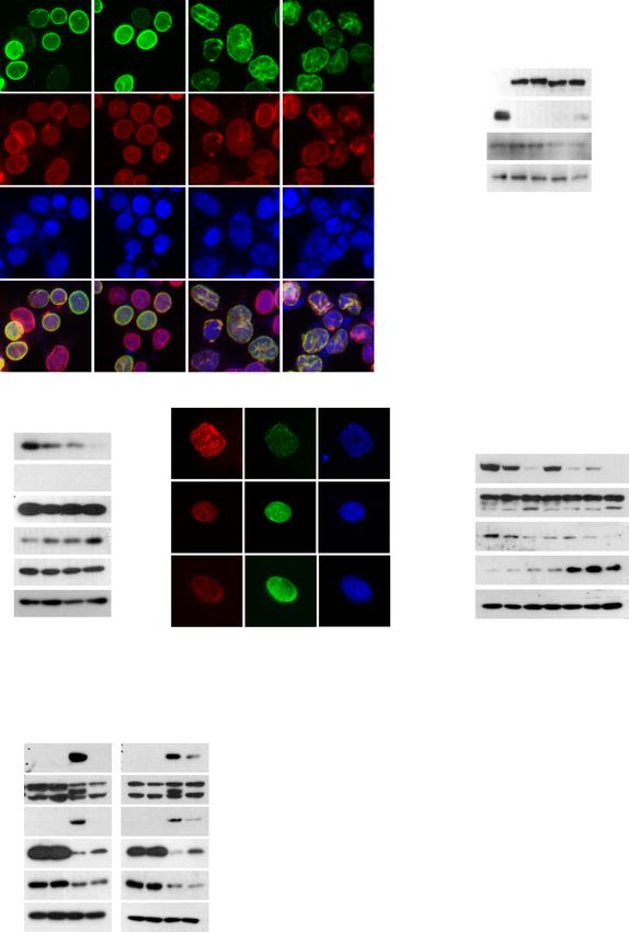
Fig. 2 Therapeutic effect of SLC-D011. A. In vivo pharmacokinetic (PK) analysis of JH010 (performed in rats). JH010 showed proper pharmacokinetic
profile and very high Bioavailability (B.A). B Effect of SLC-D011 on human ERG (hERG). SLC-D011 did not show severe hERG inhibition. C In vivo
pharmacokinetic analysis of SLD-D011. D Treatment with low concentration of SLC-D011 (100 nM) for 3 days reduces progerin expression but induces
H3K9me3 and cyclin B1 expression in HGPS patient-derived cells (AG11498, Coriell Cell Repositories, n = 3 independent experiment; two-tailed Student’s t
test). E SLC-D011 eliminates exogenous progerin expression. HEK293 cells transiently transfected with GFP-PG vectors were treated with SLC-D011 for
24 hr (n = 3 independent experiment; two-tailed Student’s t test). F SLC-D011 induces expression of Ki67 (a cell proliferating marker) in HGPS cells
(AG11498, Coriell Cell Repositories). Ki67-positive cells were visualized at 3 days after treatment, and counted from photomicrographs (n = 3 independent
experiment; two-tailed Student’s t test), **p < 0.001, *p < 0.05. “Con” means dimethyl sulfoxide (DMSO)-treated control.
very high concentrations (supplementary information1, page addition, treatment with FTIs can enhance the growth and life
10–11). Thus, we strongly suggest that progerinin (SLC-D011) is span of Zmpste24-deficient mice39. Based on these studies, a
a very plausible drug for HGPS without adverse effects. clinical trial of an inhibitor of farnesyltransferase called lona-
farnib has been performed for patients with HGPS16,40,41.
Discussion Although it has some efficacies for HGPS, lonafarnib has several
Understanding molecular mechanisms and pathological features side effects. In addition, it does not work in all patients42.
of progerin is essential for developing clinical treatment for Lonafarnib also showed cytotoxic effects including the formation
patients with HGPS. Over the past decade, active progress in of donut-shaped nuclei and cell death in in vitro experiments
HGPS research has led to an increasing number of treatment after a long-term treatment (Figs. S12b and S1f; ref. 16–18). Thus,
strategies31–36. Nevertheless, most of previous studies had insuf- we generated farnesylation-deficient (ASIM) progerin. It showed
ficient preclinical data for transposition to patients with HGPS. In no difference compared with progerin (Fig. 1a and Figs. S1c).
fact, farnesylation of progerin has been a main target for clinical Why the ASIM progerin showed inconsistent results with pre-
drug development to prevent aging process in HGPS. Blocking vious reports43,44 is unclear, but it is possible that eliminating
farnesylation by (FTIs in HGPS cells can restore nuclear struc- protein farnesylation would not be sufficient to abolish the
ture, cell proliferation, and chromatin organization37,38. In nuclear deformation, as some studies have suggested that
COMMUNICATIONS BIOLOGY | (2021)4:5 | https://doi.org/10.1038/s42003-020-01540-w | www.nature.com/commsbio 5ARTICLE COMMUNICATIONS BIOLOGY | https://doi.org/10.1038/s42003-020-01540-w
A B
1.8 LmnaG609G/G609G
10 weeks old 10 weeks old (male)
(female) 1.6
D011 Con D011 Con 1.4
Body weight (ratio)
1.2
1
0.8
0.6
0.4
Con
0.2 D011 * : p < 0.05
0
4 5 6 7 8 9 10 11 12 13 14 15 16 17 18 19 20
Age (weeks)
C D
Kaplan-Meier survival curve in LmnaG609G/G609G
1.0
45 weeks old
Ave=14.8
0.8
Max=15.5
Survival probability
Ave=19
0.6
Con (n=8) Max=21
*
D011 (n=8)
0.4
0.2
* P < 0.05
Con
0.0
0 5 10 15 20
D011 20mg/kg
Age (weeks)
Kaplan-Meier survival curve in LmnaG609G/+
E F **
LmnaG609G/+
1.2
Ave=46.7 Ave=63
Max=48 Max=64
Survival probability
1
Body weight (ratio)
0.8
Con (n=10)
0.6
D011 (n=11)
0.4
0.2 Con
D011 * P < 0.05 ** p < 0.001
0
32 34 36 38 40 42 44 46 48 50 52 54 56 58 60 62 64
Age (weeks)
Age (weeks)
Fig. 3 In vivo favorable effect of SLC-D011. A Gross morphology of LmnaG609G/G609G mice (10 weeks old) was ameliorated after injection with SLC-D011.
At the same age, the experimental group (D011) of mice were larger than vehicle-injected control (Con) subjects regardless of gender. B, C SLC-D011
increases body weight B and extends life span C of LmnaG609G/G609G progeria model mouse (Con: n = 8; SLC-D011: n = 8). Compared with 14.8 weeks of
average (ave) life span with maximum (max) life span of 15.5 weeks, 20 mg/kg of intraperitoneal (i.p) injection (twice a week) of SLC-D011 could extend
the life span upto 19 weeks (max = 21 weeks). LmnaG609G/G609G mice were injected with SLC-D011 from age of 5-week old, *p < 0.05. D Gross morphology
of SLC-D011 injected LmnaG609G/+ mouse. Sickly and weak features were observed in 45 weeks old LmnaG609G/+ mouse, but not observed in treated
mouse, although they had the same age. E SLC-D011 increases body weights of LmnaG609G/+ mice (Con: n = 10; SLC-D011: n = 11), *p < 0.05. F Favorable
effect of SLC-D011 on life span of LmnaG609G/+ mice. Compared with vehicle-injected control group (ave = 46.7 weeks and max = 48 weeks), SLC-D011
treatment obviously extended the average life span to 63 weeks (max = 64 weeks). The injection was started from 32 weeks old, **p < 0.001. “Con” means
vehicle (a solvent composed of DMSO and PBS)-injected mouse group.
6 COMMUNICATIONS BIOLOGY | (2021)4:5 | https://doi.org/10.1038/s42003-020-01540-w | www.nature.com/commsbioCOMMUNICATIONS BIOLOGY | https://doi.org/10.1038/s42003-020-01540-w ARTICLE
B 2
A LmnaG609G/G609
Body weight (ratio)
1.5
1
0.5
Con
D011 * : p=0.016
D011 0
Con
50 mg/kg 4 6 8 10 12 14 16 18 20 22 24 25 26
8 weeks old Age (weeks)
Lmna G609G/+
Lmna G609G/+
Lmna G609G/+
Lmna G609G/+
Lmna G609G/+
Lmna G609G/+
Lmna G609G/+
Lmna G609G/+
Lmna G609G/+
C D
Lmna +/+
Lmna +/+
Lmna +/+
Kaplan-Meier survival curve in LmnaG609G/G609G
1.0
**
D011 - - + + - - + + - - + +
0.8
Survival probability
Ave=16.8
Max=18 Lamin A/C 54
0.6
Con (n=8) Ave=25.2
Max=26 Progerin
D011 (n=8)
0.4
54
15
H3K9me3
0.2
54
** : pARTICLE COMMUNICATIONS BIOLOGY | https://doi.org/10.1038/s42003-020-01540-w
Fig. 4 Oral administration of SLC-D011 suppresses premature aging features. A Gross morphology of SLC-D011-treated (D011) mice at 8 weeks old.
Body size of treated mouse was apparently larger than that of the untreated control (Con) mouse. B Oral administration of SLC-D011 increases body
weights of LmnaG609G/G609G mice. Compared with untreated control group, body weight of treated mouse was increased by ~35%, *p < 0.05. C Oral
administration of SLC-D011 extends the life span of LmnaG609G/G609G mouse. SLC-D011 in monoolein-based solution was treated by oral gavage (50 mg/
kg, daily) from 5 weeks old. Compared with untreated control group (ave = 16.8 weeks and max = 18 weeks), treated group showed extended life span
(ave = 25.2 weeks and max = 26 weeks), **p < 0.001. D Reduction of progerin expression and increase of H3K9me3 expression in tissues of LmnaG609G/+
mice after treatment with SLC-D011 by oral gavage (50 mg/kg, daily) for 8 weeks (n = 3 independent experiments; two-tailed Student’s t test). E Grip test
of LmnaG609G/+ mice at 41-week-old was performed after administration of SLC-D011 for 6 weeks. The test was repeated 10 times for each mouse (n > 4),
**p < 0.001. Data are presented as mean ± SD. F Electrocardiographic analysis of LmnaG609G/+ mice (untreated: n = 4; SLC-D011: n = 4) and Lmna+/+
mice (n = 3) after treatment with SLC-D011 for 6 weeks (unpaired t test). Heart rate is shown as beats per minute (bpm). G Dental abnormalities in
LmnaG609G/+ mice (untreated: n = 4; SLC-D011: n = 5) and Lmna+/+ mice (n = 4). X-ray lateral projection of the skull of untreated LmnaG609G/+ mouse
(untreated: n = 4; SLC-D011: n = 5) at 41 weeks of age compared to Lmna+/+ mouse (n = 4) at same age shows abnormalities of incisors (unpaired t test).
Lower incisors of LmnaG609G transgenic mice grow toward the palate because of malocclusion. Oral administration of SLC-D011 for 6 weeks alleviates
dental abnormalities. H Anti-aging effects of single treatment of SLC-D011 or lonafarnib were measured in HGPS patients-derived cells (AG11513,
AG03199, AG11498, and AG03198, Coriell Cell Repositories). Single treatment of SLC-D011, compared to lonafarnib, reduced the expression of progerin
but induced the expression of H3K9me3 and cyclin B1 in HGPS cells more effectively. HGPS patients-derived cells were treated with SLC-D011 (500 nM) or
lonafarnib (500 nM) for 3 days. I Scheme of interaction between lamin A and progerin. SLC-D011 interrupts the interaction between lamin A and progerin
through direct binding to the C-terminal region of progerin. “Con” and “untreated” mean vehicle (monoolein-based solution)-treated mouse group or
vehicle (DMSO)-treated cells.
feeding. Progerinin was fed constantly and could be maintained (1:100; sc-594; Santa Cruz Biotechnology), and anti-p16-INK4A (1:500; 10883-1-
in the mouse’s body without stress. Oral administration of pro- AP; Proteintech).
gerinin relieved the stress of mice and resulted in better anti-aging
effects including life extension than i.p. injection (Figs. 4a–4c and Purification of recombinant proteins. To obtain selenomethionyl-labeled protein
Figs. S10a–S10c). Results of the present study suggest that pro- for crystallization, Escherichia coli strain B834 (DE3; Novagen, USA) harboring
plasmids encoding lamin A/C fragments (residue 250–400 or 556–664) were cul-
gerinin is more stable and effective than other potential drugs. tured in M9 medium supplemented with L-( + )-selenomethionine. Protein
Thus, progerinin might be a promising treatment for children expression was induced by 0.5 mM isopropyl β- d-1-thiogalactopyranoside at
with HGPS without causing serious adverse effects. 30 °C. Cells were harvested by centrifugation and resuspended in lysis buffer
containing 20 mM Tris-HCl (pH 8.0) and 150 mM NaCl. Cells were disrupted with
a sonicator. Cell debris was then removed by centrifugation. The supernatant of
Methods middle region fragment (residue 250–400) was loaded onto Ni-NTA affinity
Animal experiments. Animal experiments were performed in a facility certified by agarose resin (Qiagen, The Netherlands), pre-incubated with lysis buffer while the
the Association for Assessment and Accreditation of Laboratory Animal Care in supernatant of C-terminal region fragment (residue 556–664) was loaded onto
compliance with animal policies approved by Pusan National University. The glutathione affinity agarose resin (Qiagen, The Netherlands), pre-incubated with
mouse work was performed under the study protocol PNU-2019-2181, as approved lysis buffer. Target proteins were eluted with lysis buffer supplemented with 250
by the Institutional Animal Care and Use Committee. LmnaG609G/609G mice were mM imidazole. These eluted fractions were further purified using anion-exchange
generated by timed mating of heterozygous LmnaG609G/+ provided by Carlos chromatography (Hitrap Q HP, GE Healthcare, Chicago, IL, USA).
López-Otín (Universidad de Oviedo, Asturias, Oviedo, Spain). SLC-D011 was
mixed with dimethyl sulfoxide (DMSO) and phosphate-buffered saline (PBS). It Transfection of expression vectors. GFP tag conjugated EV (GFP-EV), lamin A
was then intraperitoneally injected into mice (20 mg/kg twice per week from 5- (GFP-LA), lamin A-C661A (GFP-LA-C661A), progerin (GFP-PG), and progerin-
week-old). SLC-D011 and lonafarnib were orally administrated to mice daily at a C611A (GFP-PG-C611A) vectors were used. GFP-PG and GFP-LA expression
concentration of 10 mg/ml in monoolein-based solution. Control mice were treated vectors were kindly provided by Misteli T. (National Cancer Institute [NCI],
with monoolein-based solution alone in the same way. LmnaG609G/609G mice were Frederick, MD, USA). GFP-LA-C661A and GFP-PG-C611A expression vectors
treated with clear chemical solution throughout the life span, starting from 5 weeks were created by a single point mutation in CaaX motif. The CSIM sequence of
of age. LmnaG609G/+ mice were treated via intraperitoneal and oral administra- lamin A, or progerin was mutated to ASIM. jetPEI (Polyplus Transfection, NY,
tions, starting from 32 weeks of age. USA) was used for transfection of expression vectors. Vectors were mixed with
1.5 μl of jetPEI in 150 mM NaCl solution. The mixture was added to HEK293 cells.
After 4 h of incubation, medium was replaced with a new medium supplemented
Cell culture and reagents. Human fibroblast cells from HGPS patients (AG03198,
with 10% FBS.
10-year-old female; AG03199, 10-year-old female; AG11513, 8-year-old female;
AG11498, 14-year-old male), and normal person (GM00038, 9-year-old female
N9) were obtained from Coriell Cell Repositories (Camden, NJ, USA) and main- Immunoblotting. Protein was extracted from cells using a radioimmunoprecipitation
tained in Eagle’s minimal essential medium supplemented with 15% fetal bovine assay (RIPA) buffer (50 mM Tris-Cl, pH 7.5, 150 mM NaCl, 1% NP-40, 0.1% SDS,
serum, and 2 mM glutamine without antibiotics. HEK293 cells were obtained from and 10% sodium deoxycholate). Samples were separated by sodium dodecyl sulfate-
the American Type Culture Collection (ATCC, Manassas, VA, USA) and main- polyacrylamide gel electrophoresis (SDS-PAGE) and transferred to polyvinylidene
tained in liquid medium containing 10% FBS, and 1% penicillin–streptomycin at difluoride (PVDF) membranes. Blotted membranes were blocked with 3% skimmed
37 °C with 5% CO2. milk in TBS-T buffer (20 mM Tris pH 7.6, 150 mM NaCl, and 0.05% Tween 20) for
1 h followed by incubation with specific primary antibodies. Horseradish peroxidase
(HRP)-conjugated goat anti-mouse, goat anti-rabbit, and mouse anti-goat IgG anti-
Chemical synthesis and characterization data. Synthetic scheme of SLC-D011 is bodies (Pierce, Thermo Fisher Scientific, Inc., Rockford, IL, USA) were used as sec-
provided in the Supplementary information (2, page 1) file. The molecular struc- ondary antibodies. Peroxidase activity was detected by chemiluminescence using an
ture of SLC-D011 was investigated using 1H-NMR Spectroscopy, 13C-NMR ECL kit (Intron, Seoul, Korea) following the manufacturer’s instructions. Bands were
spectroscopy, mass spectrometry, and X-ray diffractometry (Supplementary quantified using Image J software (National Institute of Health, NIH).
information 2, page 1–4).
Western blot for tissue samples. Lamin A/C and progerin amounts in tissue
Antibodies and reagents. Antibodies used for experiments included anti-GFP samples including lung, liver, and kidney analysis of two Lmna+/+, four untreated
(1:1000; sc-9996; Santa Cruz Biotechnology, Dallas, TX, USA); anti-GST (1:5000; LmnaG609G/+, and four D011-treated LmnaG609G/+ mice were evaluated by
sc-138; Santa Cruz Biotechnology), anti-His (1:1000; 66005-1-lg; Proteintech, western blotting. Proteins were extracted with RIPA lysis buffer supplemented
Rosemont, IL, USA), anti-Actin (1:10000; sc-47778; Santa Cruz Biotechnology), with protease inhibitors. Tissues were sonicated and centrifuged at 15,000 rpm for
anti-Lamin A/C (1:10000; sc-376248; Santa Cruz Biotechnology), anti-Progerin 15 min at 4 °C. Protein concentration was determined by Coomassie-blue staining.
(1:100; sc-81611; Santa Cruz Biotechnology), anti-Progerin (1:300; ab66587; Cellular lysates in SDS-containing sample buffer were inactivated by heating at
Abcam, Cambridge, UK), anti-Ki67 (1:200; Ab15580; Abcam), anti-cyclin B1 98 °C for 7 min. Proteins were loaded onto 8% SDS-PAGE gels and transferred to
8 COMMUNICATIONS BIOLOGY | (2021)4:5 | https://doi.org/10.1038/s42003-020-01540-w | www.nature.com/commsbioCOMMUNICATIONS BIOLOGY | https://doi.org/10.1038/s42003-020-01540-w ARTICLE
PVDF membranes. These membranes were blocked with 3% skimmed milk in KI28,30 and abnormalities of the incisors were evaluated. Data processing and analysis
TBS-T buffer for 1 h and incubated with primary antibodies including mouse were performed using a RadiAnt DICOM system in Institute of Animal Medicine,
monoclonal anti-progerin (1:100; sc-81611; Santa Cruz Biotechnology), mouse College of Veterinary Medicine, Gyeongsang National University (Jinju, Korea).
monoclonal anti-lamin A/C (1:500; MANLAC1; Developmental Studies Hybri-
doma Bank), and rabbit polyclonal anti-H3K9me3 (1;2000; Ab8898; Abcam) at 4°C
Histology analysis. Tissue specimens were fixed in 4% PFA and embedded in
overnight. Blots were then incubated with 1:10,000 goat anti-mouse (Pierce,
paraffin. Paraffin blocks were sectioned and transferred onto adhesive-coated
Thermo Fisher Scientific) or 1:10,000 goat anti-rabbit (Pierce, Thermo Fisher
slides. After deparaffinization and rehydration, tissue sections were stained with
Scientific) IgG (HRP conjugated) in 1% skimmed milk and washed with TBS-T
Masson Trichrome. Histology analysis was performed by BioLead Inc. (Seoul,
buffer. Bands were developed using and Intron ECL detection system and quan-
Korea).
tified using Image J (National Institute of Health, NIH).
Nuclear deformation counting. For nuclear deformation cell counting, immu-
RNA isolation and RT-PCR. For RT-PCR, total cellular RNA was extracted using nofluorescence staining was performed with lamin A or progerin antibodies. After
RNA extraction kit (Qiagen). Gene expression studies were performed using cDNA staining, abnormal nuclear membrane was counted in randomly selected fields and
synthesized from total RNA with MMLV RT (Invitrogen, Carlsbad, USA) and expressed as percentages or actual numbers of total cells counted. Abnormalities of
random hexamers. PCR from genomic DNA was perform using DiaStar Taq DNA nuclear membrane were determined based on the following: (1) lamin A/C or
polymerase (SolGent, Daejeon, Korea) and 50 ng of cDNA. Wild-type prelamin A progerin lining was extruded or engulfed, (2) having at least one bleb, and (3)
cDNA was synthesized by PCR amplification using a forward primer, 5′-AAG- irregular contour. Counting of cells with nuclear deformation was performed by
GAGATGACCTGCTCCATC-3′ and a reverse primer, 5′-TTTCTTTGGCTTCAA three independent observers who were blinded to chemical treatment group. To
GCCCCC-3′. The thermal cycling conditions were as follows: 94°C for 3 min analyze histone H3K9me3 intensity or expression, images were quantified using
(activation), 94°C for 1 min (denaturation), 61°C for 1 min (annealing) and 72°C “color histogram” function of Image J software (National Institute of Health, NIH).
for 42 sec (extension) up to 35 cycles. The PCR products were analyzed through 2% Fluorescence intensities were subtracted by background signals.
agarose electrophoresis and DNA was visualized by ethidium bromide staining and
UV photography. Levels of other transcripts were also performed with the fol-
lowing oligonucleotide primers: CENP-A, 5′-ACAAGGTTGGCTAAAGGA-3′ and Chemical PK analysis and in vitro ADME test. For PK analysis, 5 mg/kg of JH4
5′-ATGCTTCTGCTGCCTCTT-3′; BRCA1, 5′-AGAGTGTCCCATCTGTCTGG-3′ in 10% DMSO, 5% Tween 90, and 95% saline solution were intravenously injected
and 5′-CGCTGCTTTGTCCTCAGAG-3′; IL-6, 5′-AAATGCCAGCCTGCTGAC- and 10 mg/kg of JH4 in 10% NMP and 90% PEG400 solution were orally delivered.
GAAC-3′ and 5′-AACAACAATCTGAGGTGCCCATGCTA-3′; IL-8, 5′-TGGCA At pre-set time-points, blood concentration of JH4 was determined by LC-MS/MS
GCCTTCCTGATTTCTG-3′ and 5′-AACTTCTCCACAACCCTCTGC-3′ and analysis22,23. Other chemicals were also tested using similar protocol. In vitro
GAPDH 5′-ATCTTCCAGGAGCGAGATCCC-3′ and 5′-AGTGAGCTTCCCGT ADME studies (plasma protein binding, CYP inhibition, microsomal stability,
TCAGCTC-3′. plasma stability, and hERG inhibition) were performed by New Drug Development
Center (Daegu, Korea) using standard protocols24,25.
Protein–protein interaction analyses. For the analysis of protein–protein inter-
action, glutathione S-transferase (GST) pull-down assay and His pull-down assay Toxicity studies in rats. The protocol and procedures involving the care and use
were performed. To detect the interaction between wild-type lamin A and mutant of animals in this study was reviewed and approved by IACUC of QuBEST BIO
lamin A, GST or His-bead-fused lamin A recombinant protein was incubated with prior to conduct (Approval no.: QBSIACUC-A18122). During the study, the care
GFP-tagged lamin A-C661A (GFP-LA-C661A), progerin (GFP-progerin), or and use of animals will be conducted in accordance with all applicable guidelines of
progerin-C611A transfected HEK293 cells for 30 min at room temperature (RT). Animal Welfare Act. Eleven specific pathogen-gree Sprague-Dawley rats (~6 weeks
After washing once with PBS, precipitated materials were collected and subjected to old) were obtained from SAMTAKO Ltd. (Osan, Korea) and nine rats were used
SDS-PAGE and western blot analysis with anti-GFP and anti-GST. for the study. After overnight fasting (~16 h, food but not water should be withheld
overnight), test article formulations were dosed using plastic disposable feeding
needle attached to a plastic disposable syringe. Food was withheld for a further 3–4
Immunofluorescence staining. Cells were cultured on coverslips, washed with h after dosing. Each dose was based on the most recent body weight of each animal
PBS, fixed with 4% paraformaldehyde (PFA) for 30 min at RT, and then per- and the dose volume was 10 mg/kg. The dosing day was designated as Day 1. All
meabilized with 0.2% Triton X-100 at RT for 5 min. After treatment with blocking animals were observed twice daily for mortality and moribundity during the study.
solution (anti-Human antibody diluted 1:400 in PBS) for 1 h, cells were incubated A clinical observation was performed for all animals at the time of dosing and
with anti-lamin A/C (1:400), anti-progerin (1:100), Ki67 (1:200), and anti- approximately 1, 2, and 4 h post-dose on dosing day, and once daily during 7-day
H3K9me3 (1:200) in blocking buffer overnight at 4 °C. Finally, cells were incubated observation period. Observations were included, but are not limited to, changes in
with fluorescein isothiocyanate and rhodamine-conjugated secondary antibodies at the skin, fur, eyes and mucous membrane; respiratory, circulatory, autonomic and
4 °C for 7 h. Nuclei were stained with DAPI (4, 6-diamidino-2-phenylindole) at RT central nervous system function; somatomotor activity and behavior patterns.
for 10 min. After cells were washed three times with PBS, coverslips were mounted Individual body weights were measured for all animals on the day of animal
with mounting solution (H-5501; Vector Laboratories, Burlingame, CA, USA). receipt, randomization, prior to dosing start (Day 1) and study period (Days 4 and
Immunofluorescence signal was detected with a fluorescence microscopy (Zeiss 7). Those animals survived on completion of the 7-day observation period were
and Logos). weighed body weight prior to necropsy and CO2 inhalation anesthesia and
exsanguinated from the abdominal aorta. In order to avoid autolytic change, a
MTT assay. To determine the cellular viability, cells were treated with lonafarnib complete gross pathology examination of the carcass was performed as soon as
or SLC-D011 for 2 weeks. For MTT assay, cells were incubated with 0.5 mg/ml of possible after euthanasia of all animals. Necropsy was consisted of an external
3-(4,5-dimethythiazol-2-yl)-2,5-diphenyl tetrazolium bromide (MTT) solution for examination, including identification of all clinically recorded lesions, as well as a
6 hr at 37 °C. After removing MTT solution, the precipitated materials were dis- detailed internal examination. Toxicity studies were performed by QuBEST BIO
solved in 200 μM DMSO and quantified by measuring the absorbance at 540 nm. Co., Ltd (Yongin, Korea).
Toxicity studies in dogs. The protocol and procedures involving the care and use
Physiological analysis. For analysis of heart rate, mice were anesthetized with
of animals in this study was reviewed and approved by IACUC of QuBEST BIO
2.5% isoflurane and monitored using LOGIQ E9 (GE Healthcare). Data processing
prior to conduct (Approval no.: QBSIACUC-A18142). During the study, the care
and analysis were performed using a TOMTEC system in Cardiovascular Center,
and use of animals will be conducted in accordance with all applicable guidelines of
Pusan National University Yangsan Hospital (Yangsan, Korea). For grip strength
Animal Welfare Act. Non-naive one male and one female Beagle dogs
test, a tension meter measuring force was stationed horizontally on the platform.
(~16–24 months old, original supplier: ORIENTBIO Co., Ltd, Korea) were selected
Mouse was allowed to grip the tension bar with front paws before being pulled
from the stock colony and assigned for the study. The route of administration was
slowly away from the bar until its grip was broken. Four female LmnaG609G/+ mice,
the oral (by gavage) route, which is the anticipated clinical route of exposure. Test
four LmnaG609G/+ male mice, three female Lmna+/+ mice, and three male
article formulations were administered once via oral gavage. The day of 1st dosing
Lmna+/+ mice (all adults at 41 weeks old) were used for this test. Each mouse was
was designated as Day 1. Each dose was administered via a syringe attached with
subjected to the test ten times to determine its forelimb strength. Each estimate
12-french feeding tube. A dose volume of 5 ml/kg was used and individual dose
given corresponded to total reads measured ten times, excluding the minimum and
volumes were based on the most recent body weight. Each dose was followed by a
the maximum from each group of animals.
distilled water of 5 ml. Each animal was observed twice daily (a.m. and p.m.) for
mortality and moribundity; findings were recorded as they were observed. Cage
Radiological examinations. The radiographic study was performed in anaesthetized side observations were made for each animal once daily; abnormal findings were
living mice using a human radiographic system (College of Veterinary Medicine, recorded. Detailed observations were made for each animal once prior to treat-
Gyeongsang National University, Korea). A single radiograph of right lateral view of the ment; abnormal findings (ranked/graded, if appropriate) or an indication the
whole body was obtained. Age-matched (at 41 weeks old) Lmna+/+ mice, untreated animal appears normal was recorded. Body weights were measured prior to each
LamnG609G/+ mice, and progerinin-treated LmnaG609G/+ mice were radiographed. The dosing. Prior to each dosing and ~24 h post each dose, blood samples for hema-
images were recorded in DICOM format and then transferred to a personal computer. tology examination were collected from the via cephalic vein into tubes with
COMMUNICATIONS BIOLOGY | (2021)4:5 | https://doi.org/10.1038/s42003-020-01540-w | www.nature.com/commsbio 9ARTICLE COMMUNICATIONS BIOLOGY | https://doi.org/10.1038/s42003-020-01540-w
K2EDTA anticoagulant and following parameters were examined (ADVIA®2120, 20. Dimri, G. P. et al. A biomarker that identifies senescent human cells in culture
Germany). Prior to dosing and ~24 h post each dose, blood was collected as the and in aging skin in vivo. Proc. Natl Acad. Sci. USA 92, 9363–9367 (1995).
same method and frequency with hematology analysis using no anticoagulant and 21. Scholzen, T. & Gerdes, J. The Ki‐67 protein: from the known and the
then serum was separated by centrifugation and stored at freezer until analyze (AU unknown. J. Cell. Physiol. 182, 311–322 (2000).
400, Olympus, Japan; RAPIDCHEM 744 Na+/K+/Cl+ Analyzer, SIEMENS, 22. Osorio, F. G. et al. Splicing-directed therapy in a new mouse model of human
Germany). In terminal procedures, all surviving animals were returned to the stock accelerated aging. Sci. Transl. Med. 3, 106ra107 (2011).
colony without necropsy. Toxicity studies were performed by QuBEST BIO Co., 23. Ganem-Quintanar, A., Quintanar-Guerrero, D. & Buri, P. Monoolein: a review
Ltd (Yongin, Korea). of the pharmaceutical applications. Drug Dev. Ind. Pharm. 26, 809–820 (2000).
24. Biernacka, A. & Frangogiannis, N. G. Aging and cardiac fibrosis. Aging Dis. 2,
Statistics and reproducibility. Data were analyzed with an unpaired or paired 158–173 (2011).
two-sample Student’s t test. P value < 0.05 was considered statistically significant. 25. Delire, B. et al. Aging enhances liver fibrotic response in mice through
Error bars indicate standard deviation (SD). Data for all figures were represented as hampering extracellular matrix remodeling. Aging 9, 98–113 (2016).
mean values ± SD of at least three replicates. Details of statistical analyses and 26. Hecker, L. et al. Reversal of persistent fibrosis in aging by targeting Nox4-Nrf2
number of replicates (n) can be found in the figure legends. redox imbalance. Sci. Transl. Med. 6, 231ra47 (2014).
27. Dileto, C. L. & Travis, E. L. Fibroblast radiosensitivity in vitro and lung fibrosis
in vivo: comparison between a fibrosis-prone and fibrosis-resistant mouse
Reporting summary. Further information on research design is available in the Nature
strain. Radiat. Res. 146, 61–67 (1996).
Research Reporting Summary linked to this article.
28. Zaghini, A. et al. Long term breeding of the Lmna G609G progeric mouse:
characterization of homozygous and heterozygous models. Exp. Gerontol. 130,
Data availability 110784 (2020).
Toxicity analysis of chemicals is available in Supplementary Data 1. Full blots are shown 29. Beyret, E. et al. Single-dose CRISPR–Cas9 therapy extends lifespan of mice
in Supplementary Information. All other data that support the findings of this study are with Hutchinson–Gilford progeria syndrome. Nat. Med. 25, 419–422 (2019).
available from the corresponding author upon reasonable request. 30. Laws, N. & Hoey, A. Progression of kyphosis in mdx mice. J. Appl. Physiol. 97,
1970–1977 (2004).
31. Fong, L. G. et al. A protein farnesyltransferase inhibitor ameliorates disease in
Received: 12 February 2020; Accepted: 1 December 2020; a mouse model of progeria. Science 311, 1621–1623 (2006).
32. Yang, S. H. et al. Blocking protein farnesyltransferase improves nuclear
blebbing in mouse fibroblasts with a targeted Hutchinson-Gilford progeria
syndrome mutation. Proc. Natl Acad. Sci. USA 102, 10291–10296 (2005).
33. You, L. et al. Advancements and obstacles of CRISPR-Cas9 technology in
References translational research. Mol. Ther. Methods Clin. Dev. 13, 359–370 (2019).
1. Gordon, L. B., Rothman, F. G., López-Otín, C. & Misteli, T. Progeria: a 34. Santiago-Fernández, O. et al. Development of a CRISPR/Cas9-based therapy
paradigm for translational medicine. Cell 156, 400–407 (2014). for Hutchinson–Gilford progeria syndrome. Nat. Med. 25, 423–426 (2019).
2. Burke, B. & Stewart, C. L. Life at the edge: the nuclear envelope and human 35. Guilbert, S. M., Cardoso, D., Lévy, N., Muchir, A. & Nissan, X. Hutchinson-
disease. Nat. Rev. Mol. Cell Biol. 3, 575–585 (2002). Gilford progeria syndrome: rejuvenating old drugs to fight accelerated ageing.
3. Kipling, D., Davis, T., Ostler, E. L. & Faragher, R. G. What can progeroid Methods S1046-2023, 30302-0 (2020).
syndromes tell us about human aging? Science 305, 1426–1431 (2004). 36. Rahman, M. A. et al. The Huchinson-Gilford progeria syndrome and
4. Ahmed, M. S., Ikram, S., Bibi, N. & Mir, A. Hutchinson–Gilford progeria treatment: updated review of the literature. Sch. Acad. J. Pharm. (2019).
syndrome: a premature aging disease. Mol. Neurobiol. 55, 4417–4427 (2018). 37. Gabriel, D., Shafry, D. D., Gordon, L. B. & Djabali, K. Intermittent treatment with
5. Kashyap, S., Shanker, V. & Sharma, N. Hutchinson - Gilford progeria farnesyltransferase inhibitor and sulforaphane improves cellular homeostasis in
syndrome: a rare case report. Indian. Dermatol. Online J. 5, 478–481 (2014). Hutchinson-Gilford progeria fibroblasts. Oncotarget 8, 64809–64826 (2017).
6. Eriksson, M. et al. Recurrent de novo point mutations in lamin A cause 38. Mehta, I. S., Eskiw, C. H., Arican, H. D., Kill, I. R. & Bridger, J. M.
Hutchinson–Gilford progeria syndrome. Nature 423, 293–298 (2003). Farnesyltransferase inhibitor treatment restores chromosome territory
7. McClintock, D., Gordon, L. B. & Djabali, K. Hutchinson-Gilford progeria positions and active chromosome dynamics in Hutchinson-Gilford progeria
mutant lamin A primarily targets human vascular cells as detected by an anti- syndrome cells. Genome Biol. 12, 1–14 (2011).
Lamin A G608G antibody. Proc. Natl Acad. Sci. USA 103, 2154–2159 39. Harhouri, K. et al. An overview of treatment strategies for Hutchinson-Gilford
(2006). Progeria syndrome. Nucleus 9, 265–276 (2018).
8. Goldman, R. D. et al. Accumulation of mutant lamin A causes progressive 40. Gordon, L. B. et al. Association of lonafarnib treatment vs no treatment with
changes in nuclear architecture in Hutchinson-Gilford progeria syndrome. mortality rate in patients with Hutchinson-Gilford progeria syndrome. JAMA
Proc. Natl Acad. Sci. USA 101, 8963–8968 (2004). 319, 1687–1695 (2018).
9. McClintock, D. et al. The mutant form of lamin A that causes Hutchinson- 41. Gordon, L. B. et al. Survey of plasma proteins in children with progeria pre-
Gilford progeria is a biomarker of cellular aging in human skin. PloS ONE 2, therapy and on-therapy with lonafarnib. Pediatr. Res. 83, 982–992 (2018).
e1269 (2007). 42. Gordon, L. B. et al. Clinical trial of the protein farnesylation inhibitors
10. Coutinho, H. D. M., Falcão-Silva, V. S., Gonçalves, G. F. & da Nóbrega, R. B. lonafarnib, pravastatin, and zoledronic acid in children with Hutchinson-
Molecular ageing in progeroid syndromes: Hutchinson-Gilford progeria Gilford progeria syndrome. Circulation 134, 114–125 (2016).
syndrome as a model. Immun. Ageing 6, 1–7 (2009). 43. Capell, B. C. et al. Inhibiting farnesylation of progerin prevents the
11. Gordon, L. B., Cao, K. & Collins, F. S. Progeria: translational insights from cell characteristic nuclear blebbing of Hutchinson-Gilford progeria syndrome.
biology. J. Cell Biol. 199, 9–13 (2012). Proc. Natl Acad. Sci. USA 102, 12879–12884 (2005).
12. Young, S. G., Meta, M., Yang, S. H. & Fong, L. G. Prelamin A farnesylation 44. Mallampalli, M. P., Huyer, G., Bendale, P., Gelb, M. H. & Michaelis, S.
and progeroid syndromes. J. Biol. Chem. 281, 39741–39745 (2006). Inhibiting farnesylation reverses the nuclear morphology defect in a HeLa cell
13. Rusinol, A. E. & Sinensky, M. S. Farnesylated lamins, progeroid syndromes model for Hutchinson-Gilford progeria syndrome. Proc. Natl Acad. Sci. USA
and farnesyl transferase inhibitors. J. Cell Sci. 119, 3265–3272 (2006). 102, 14416–14421 (2005).
14. Verstraeten, V. L. et al. Protein farnesylation inhibitors cause donut-shaped 45. Varela, I. et al. Combined treatment with statins and aminobisphosphonates
cell nuclei attributable to a centrosome separation defect. Proc. Natl Acad. Sci. extends longevity in a mouse model of human premature aging. Nat. Med. 14,
USA 108, 4997–5002 (2011). 767–772 (2008).
15. Blondel, S. et al. Drug screening on Hutchinson Gilford progeria pluripotent
stem cells reveals aminopyrimidines as new modulators of farnesylation. Cell
Death Dis. 7, e2105–e2105 (2016). Acknowledgements
16. Basso, A. D., Kirschmeier, P. & Bishop, W. R. Lipid posttranslational This work was supported by National Research Foundation of Korea (NRF) grant funded
modifications. Farnesyl transferase inhibitors. J. Lipid Res. 47, 15–31 (2006). by the Korea government (MSIT) (NRF-2020R1A4A1019322 to B.J.P.; NRF-
17. Lee, S. et al. Interruption of progerin–lamin A/C binding ameliorates 2020R1F1A1075370 to B.J.P.; and NRF-2017R1A2B2005851 to G.Y.S.), and the Progeria
Hutchinson-Gilford progeria syndrome phenotype. J. Clin. Invest. 126, Research Foundation (Grant #PRF 2019-75 to B.J.P.).
3879–3893 (2016).
18. Stehbens, W. E., Delahunt, B., Shozawa, T. & Gilbert-Barness, E. Smooth
muscle cell depletion and collagen types in progeric arteries. Cardiovasc. Author contributions
Pathol. 10, 133–136 (2001). S.K., M.H.Y., J.A., S.P., J.H.C., T.G.W., A.Y.O., S.Y.A., and S.Y.L. performed the
19. Olive, M. et al. Cardiovascular pathology in Hutchinson-Gilford progeria: experiments. J.E.K., K.J.C., G.Y.S., S.Y.K., S.Y.K., and J.J. synthesized and offered the
correlation with the vascular pathology of aging. Arterioscler. Thromb. Vasc. chemicals. S.K., M.H.Y., and B.J.P. conceived the experimental designs. S.K. and B.J.P.
Biol. 30, 2301–2309 (2010). wrote the manuscript. S.K., T.S.H., J.S.K., N.C.H., and B.J.P. analyzed the data.
10 COMMUNICATIONS BIOLOGY | (2021)4:5 | https://doi.org/10.1038/s42003-020-01540-w | www.nature.com/commsbioYou can also read