Xenopus polo-like kinase Plx1 regulates XErp1, a novel inhibitor of APC/C activity
←
→
Page content transcription
If your browser does not render page correctly, please read the page content below

Downloaded from genesdev.cshlp.org on January 12, 2012 - Published by Cold Spring Harbor Laboratory Press

         First publ. in: Genes & development ; 19 (2005), 4. - S. 502-513

Xenopus polo-like kinase
Plx1 regulates XErp1, a novel
inhibitor of APC/C activity
Andreas Schmidt,1 Peter I. Duncan,2,4 Nadine R. Rauh,1 Guido Sauer,2 Andrew M. Fry,3
Erich A. Nigg,2 and Thomas U. Mayer1,5
1
 Chemical Biology, Independent Research Group, and 2Department of Cell Biology, Max-Planck-Institute of Biochemistry,
82152 Martinsried, Germany; 3Department of Biochemistry, University of Leicester, Leicester LE1 7RH, UK

Metaphase-to-anaphase transition is a fundamental step in cell cycle progression where duplicated
sister-chromatids segregate to the future daughter cells. The anaphase-promoting complex/cyclosome (APC/C)
is a highly regulated ubiquitin-ligase that triggers anaphase onset and mitotic exit by targeting securin and
mitotic cyclins for destruction. It was previously shown that the Xenopus polo-like kinase Plx1 is essential to
activate APC/C upon release from cytostatic factor (CSF) arrest in Xenopus egg extract. Although the
mechanism by which Plx1 regulates APC/C activation remained unclear, the existence of a putative APC/C
inhibitor was postulated whose activity would be neutralized by Plx1 upon CSF release. Here we identify
XErp1, a novel Plx1-regulated inhibitor of APC/C activity, and we demonstrate that XErp1 is required to
prevent anaphase onset in CSF-arrested Xenopus egg extract. Inactivation of XErp1 leads to premature APC/C
activation. Conversely, addition of excess XErp1 to Xenopus egg extract prevents APC/C activation. Plx1
phosphorylates XErp1 in vitro at a site that targets XErp1 for degradation upon CSF release. Thus, our data
lead to a model of APC/C activation in Xenopus egg extract in which Plx1 targets the APC/C inhibitor XErp1
for degradation.
[Keywords: Cell cycle; anaphase-promoting complex/cyclosome; Xenopus; polo-like kinase; cytostatic factor;
mitotic exit]
Received August 9, 2004; revised version accepted December 16, 2004.

Before fertilization, vertebrate eggs are arrested in meta-                  Data from various research groups have firmly estab-
phase of meiosis II by a biochemical activity that was                    lished a role for the germ-cell specific c-Mos kinase in
named cytostatic factor (CSF) in a seminal publication                    establishing CSF activity (Sagata et al. 1989; Tunquist
over 30 years ago (Masui and Markert 1971). CSF was                       and Maller 2003). c-Mos mediates its effect by activating
defined as an activity present in the cytoplasm of frog                   a mitogen-activated protein kinase (MAPK) cascade (Ko-
eggs arrested in metaphase of meiosis II that when in-                    sako et al. 1994) leading to the phosphorylation and
jected into one cell of a two-cell embryo produced a                      thereby activation of the protein kinase p90Rsk (Sturgill
cleavage arrest in the injected cell. Since then, numerous                et al. 1988). Components of the spindle assembly check-
attempts have been made to identify the molecular na-                     point seem to be the downstream targets of the c-Mos/
ture of CSF in vertebrate eggs and to dissect the mecha-                  MAPK/p90Rsk pathway, because the checkpoint pro-
nism underlying the meiotic metaphase arrest. Fertiliza-                  teins Mad1, Mad2, and Bub1 are required to mediate the
tion of the egg causes a transient rise in free intracellular             c-Mos-initiated CSF arrest in Xenopus egg extract
calcium mediating CSF release and thus anaphase-pro-                      (Schwab et al. 2001; Tunquist et al. 2002, 2003). How-
moting complex/cyclosome (APC/C) activation. Active                       ever, the c-Mos/MAPK/p90Rsk pathway is not the only
APC/C mediates the degradation of securin and cyclin B                    activity contributing to CSF arrest. For instance, it has
and thereby promotes anaphase onset (Lorca et al. 1993;                   been shown that cyclin-dependent kinase 2/cyclin E
Murray 2004). Extract from CSF-arrested Xenopus eggs                      (Cdk2/cyclin E) could mediate CSF arrest in the absence
faithfully recapitulates many morphological and bio-                      of c-Mos kinase (Tunquist et al. 2002), suggesting that
chemical events associated with release from CSF arrest                   Cdk2/cyclin E and c-Mos/MAPK/p90Rsk represent inde-
upon addition of calcium ions.                                            pendent pathways both needed for effective APC/C in-
                                                                          hibition. Furthermore, recent work identified a putative
                                                                          vertebrate homolog of the Drosophila regulator of cyclin
4
 Present address: Nestlé Research Center, Vers-chez-les-Blanc, PO Box
                                                                          A1 (Dong et al. 1997), Emi1 (early mitotic inhibitor 1), as
44, CH-1000 Lausanne 26, Switzerland.                                     an additional independent element contributing to CSF
5
 Corresponding author.                                                    activity in Xenopus eggs (Reimann et al. 2001a; Reimann
E-MAIL mayer@biochem.mpg.de; FAX 49-89-8578-3138.
Article and publication are at http://www.genesdev.org/cgi/doi/10.1101/   and Jackson 2002). Emi1, a conserved F-box protein with
gad.320705.Konstanzer Online-Publikations-System (KOPS)                   a putative zinc-binding region (ZBR) has been shown to
      URN: hhttp://nbn-resolving.de/urn:nbn:de:bsz:352-140483

502       GENES & DEVELOPMENT 19:502–513 © 2005 by Cold Spring Harbor Laboratory Press ISSN 0890-9369/05; www.genesdev.org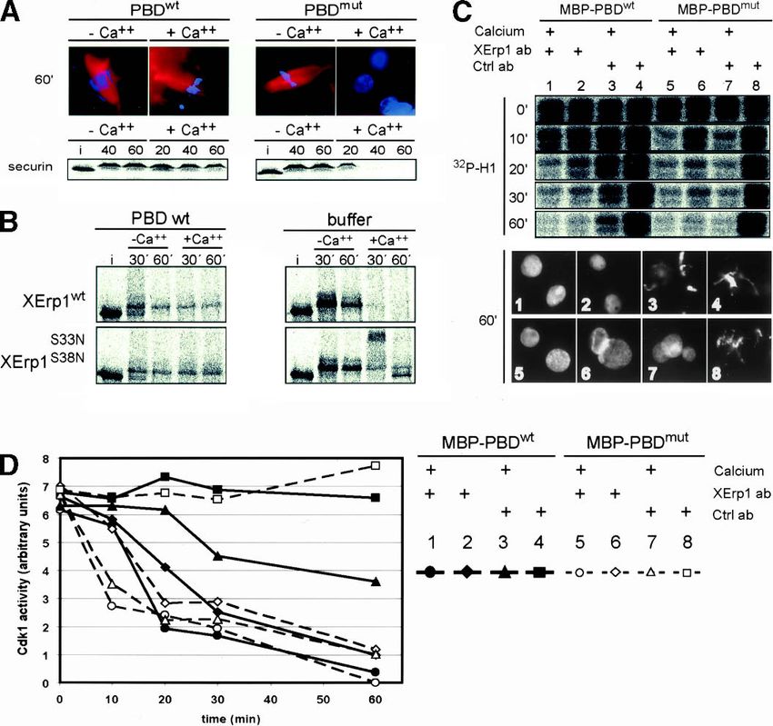
Downloaded from genesdev.cshlp.org on January 12, 2012 - Published by Cold Spring Harbor Laboratory Press

                                                                                             XErp1, a novel inhibitor of APC/C activity

bind to the APC/C activator CDC20 (Reimann et al.                    screen a Xenopus oocyte cDNA library. DNA sequence
2001a) thereby preventing APC/C activation.                          analysis of the 25 interactors identified revealed that two
   Polo-like kinases (Plks) play a pivotal role in regulating        independent clones were derived from the same gene.
progression through M phase in all eukaryotic cells (Barr            The full-length cDNA of these two clones encoded an
et al. 2004). Evidence from different model organisms                F-box protein of 651 amino acids which we named
suggests that Plk1 activity is required for the timely ac-           XErp1, for Xenopus Emi1-related protein 1 (formerly re-
tivation of the APC/C at anaphase onset (Descombes and               ferred to as Pxp17, acc. no. AAN76807). Protein se-
Nigg 1998; Shirayama et al. 1998; Donaldson et al. 2001).            quence analyses revealed that XErp1 contains a putative
These data are supported by studies showing that Plk1,               ZBR at its C terminus (Fig. 1A,B).
like Cdk1, can phosphorylate specific APC/C subunits                    The cDNA sequence of XErp1 is 97% identical to the
in vitro (Kotani et al. 1998; Golan et al. 2002). However,           cDNA sequence of Fbx26, which was previously identi-
it is still an open question whether the phosphorylation             fied as a novel Xenopus F-box containing protein (Regan-
of APC/C by Plk1 is a prerequisite for APC/C’s ubiquitin             Reimann et al. 1999). However, unlike XErp1 the protein
ligase activity. For example, whereas Golan et al. (2002)            predicted by the Fbx26 cDNA sequence does not contain
reported that both Cdk1 and Plk1 can stimulate APC/C                 a putative ZBR. To resolve the discrepancy between the
ubiquitin ligase activity, recent data suggest that the              two predicted protein sequences, we performed microse-
phosphorylation of APC/C by Cdk1 but not by Plk1 in-                 quence analyses of endogenous XErp1 protein immuno-
creases the activity of APC/C in vitro (Kraft et al. 2003).          purified from Xenopus egg extract. Liquid chromatogra-
Although the biological relevance of APC/C phosphory-                phy-tandem mass spectrometry (LC-MS/MS) analyses
lation by Plk1 remains to be determined, experiments in              identified several peptide sequences that match the pro-
Xenopus egg extract have clearly established a role of               tein sequence predicted by the XErp1 ORF but not by the
Plx1 in activating the APC/C in response to calcium                  Fbx26 ORF (Fig. 1B). Most of the discrepancies between
(Descombes and Nigg 1998) and in maintaining the                     the identified peptide sequences and the Fbx26 ORF arise
APC/C in its active form (Brassac et al. 2000). Xenopus              from frame shifts within the Fbx26 cDNA sequence, sug-
egg extract depleted of endogenous Plx1 or supplemented              gesting that the reported Fbx26 sequence is incorrect. In
with catalytically inactive Plx1N172A could not activate             light of these data we conclude that the XErp1 and Fbx26
the APC/C upon calcium addition and therefore did not                cDNAs encode identical proteins and that the XErp1
enter interphase (Descombes and Nigg 1998). Although                 ORF reported here provides the correct sequence. The N
that study did not address the mechanism by which Plx1               terminus of XErp1 displays no significant sequence ho-
allows the destruction of APC/C targets upon CSF re-                 mology to known proteins, whereas the C terminus of
lease, the existence of a putative Plx1-regulated APC/C              XErp1 shows significant sequence similarity to Emi1
inhibitor was proposed (Descombes and Nigg 1998).                    (Fig. 1A), a known APC/C inhibitor.
Such an inhibitor would be expected to be active in CSF                 Affinity-purified antibodies raised against an N-termi-
extract but would be inactivated by Plx1 upon fertiliza-             nal moiety of XErp1 fused to a six-histidine tag (His6-
tion.                                                                XErp1105–374) reacted with a polypeptide of an apparent
   We have identified XErp1 as a novel Plx1-interacting              molecular mass of ∼90 kDa in immunoblots of extract
protein. XErp1 is a conserved F-box protein with a puta-             from CSF-arrested Xenopus eggs (Fig. 1C, left panel). To
tive ZBR at its C terminus. XErp1 is required to maintain            confirm that the detected polypeptide is XErp1, we used
CSF-mediated metaphase arrest in Xenopus egg extract.                the affinity-purified antibody to probe immunoblots of
Excess XErp1 can prevent calcium-induced CSF release                 in vitro translated (IVT), 35S-labeled XErp1, Emi1, and
in Xenopus egg extract. In vitro, XErp1 can directly in-             unprogrammed wheat-germ extract (WGE). As shown in
hibit the ubiquitin ligase activity of APC/CCDC20. Plx1              the left panel of Figure 1C, the antibodies recognized IVT
phosphorylates XErp1 at a site in vitro that targets XErp1           XErp1 but failed to detect IVT Emi1 or any protein to a
for degradation at anaphase onset in Xenopus egg extract.            substantial degree in the unprogrammed WGE. The rea-
The activity of XErp1 is required to mediate the domi-               son for the observed difference in the electrophoretic
nant-negative effect of the Plx1 polo-box domain (PBD)               mobility between the endogenous XErp1 present in CSF
on CSF release. Our data lead to a model in which Plx1-              extract and IVT XErp1 will be addressed below. Autora-
mediated degradation of XErp1 is a prerequisite for                  diographic analysis of the immunoblots confirmed that
APC/C activation upon CSF release in Xenopus egg ex-                 both 35S-labeled IVT products, XErp1 and Emi1, were
tract.                                                               expressed at similar levels in the WGE (Fig. 1C, middle
                                                                     panel). Antibodies preincubated with the antigen His6-
                                                                     XErp1105–374 completely failed to detect XErp1 in CSF
Results                                                              extract and IVT XErp1 (Fig. 1C, right panel). Taken to-
                                                                     gether, these data demonstrate that our antibody specifi-
Identification of XErp1, a novel interactor of Plx1
                                                                     cally recognizes XErp1.
To identify the hypothetical Plx1-regulated APC/C in-
hibitor proposed by Descombes and Nigg (1998) we per-
                                                                     XErp1 is degraded upon CSF release
formed a yeast-two hybrid screen. To this end we fused
full-length catalytically inactive Plx1 (Plx1N172A) to the           If XErp1 represents the postulated APC/C inhibitor, then
DNA-binding domain of Gal4 and used this bait to                     XErp1 should be active in CSF extract and should be

                                                                                                  GENES & DEVELOPMENT              503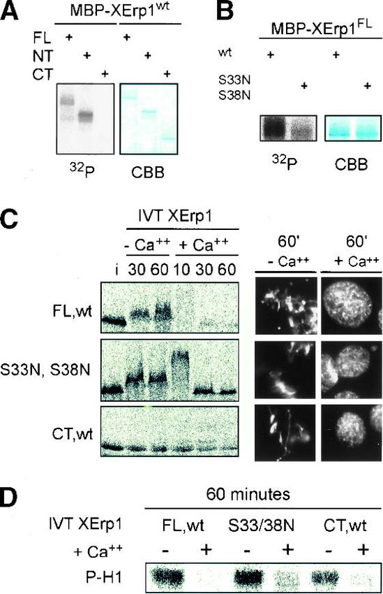
Downloaded from genesdev.cshlp.org on January 12, 2012 - Published by Cold Spring Harbor Laboratory Press
Schmidt et al.
inactivated upon CSF release. Immunoblot analysis tract was incubated with the proteasome inhibitor MG-
showed that XErp1 is present in CSF extract but rapidly 132 before calcium addition, 35S-labeled IVT XErp1 was
degraded upon calcium addition (Fig. 2A). Microscopic stabilized (data not shown), suggesting that the calcium-
examination of the extract revealed that the chromatin induced degradation of endogenous XErp1 is mediated by
decondensed upon calcium addition, confirming that the the ubiquitin–proteasome pathway. Once the released
extract had entered interphase (Fig. 2A). When CSF ex- extract had entered interphase (40–60 min), XErp1 reap-
peared in a higher-mobility form (Fig. 2A, left panel),
suggesting that XErp1 is phosphorylated in CSF but not
in interphase extract. To test this we immunopurified
35
S-labeled IVT XErp1 from CSF and interphase extract
and treated the CSF form with phosphatase. Autoradio-
graphic analysis revealed that phosphatase treatment
converted the slower-migrating form of XErp1 into the
higher-mobility form (Fig. 2B). These data indicate that
the mobility difference between the interphase and M
phase forms of XErp1 reflects meiosis-specific phos-
phorylation of XErp1. In extract treated with the protein
synthesis inhibitor cycloheximide, XErp1 was also de-
graded upon CSF release, but it did not reappear (Fig. 2A,
right panel). Taken together, these data indicate that
XErp1 is inactivated upon CSF release by proteasomal
degradation and resynthesized in interphase extract.
XErp1 accumulates during oocyte maturation
Next, we wanted to know whether XErp1 accumulates
during oocyte maturation. Oocyte maturation was in-
duced by progesterone treatment of stage VI oocytes. At
the indicated time points, equal numbers of oocytes
were collected from a synchronous population and pro-
cessed for immunoblot analysis (Fig. 2C). Immunoblot
analyses for phosphorylated MAPK (Erk1/2), which is
the active form of Erk1/2, and cyclin B levels (Fig. 2C)
confirmed that the progesterone treatment induced oo-
cyte maturation in our experiment. XErp1 was found to
be expressed in stage VI oocytes, and its protein level
increased significantly 4 h after progesterone treatment
(Fig. 2C). Interestingly, XErp1 showed a shift in electro-
phoretic mobility ∼2.5 h after progesterone treatment,
which remained until the oocytes entered meiosis II (Fig.
Figure 1. Domain structure of XErp1 and characterization of
the XErp1 antibody. (A) The N terminus of XErp1 contains the
putative SCF-TRCP recognition sequence DSGX3S. In addition,
XErp1 protein contains a putative C-terminal F-box and a zinc-
binding region (ZBR). The C terminus of XErp1 shares 39%
sequence identity with Xenopus Emi1. (B) ClustalW alignment
of XErp1 protein sequences from different vertebrate species
shows the evolutionary conservation of the XErp1 proteins. The
amino acid sequences of XErp1 peptides that were identified by
LC-MS/MS analysis of XErp1 are indicated (solid blue lines,
peptide sequences in agreement with both XErp1 and Fbx26
ORF; dashed blue lines, peptide sequences in agreement only
with XErp1 ORF). (C) Characterization of XErp1 antibodies. CSF
extract or in vitro translated (IVT) XErp1, IVT xEmi1, or unpro-
grammed wheat germ extract were immunoblotted with affin-
ity purified anti-XErp1 antibodies (left) or with affinity-purified
anti-XErp1 antibodies blocked with XErp1 antigen (right).
(Middle) The expression level of the 35S-labeled IVT proteins
was examined by autoradiographic analysis.
504 GENES & DEVELOPMENT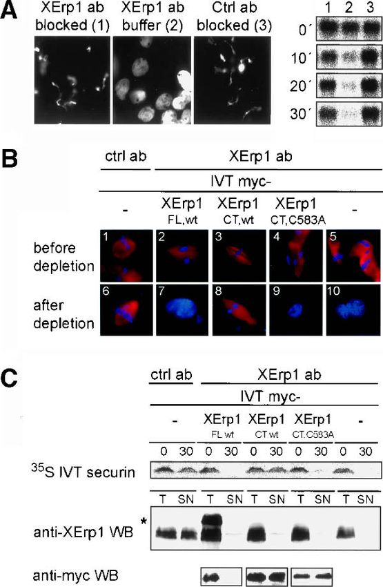
Downloaded from genesdev.cshlp.org on January 12, 2012 - Published by Cold Spring Harbor Laboratory Press

                                                                                             XErp1, a novel inhibitor of APC/C activity

                                                                    H1 as substrate (Fig. 3A, right panel). Autoradiographic
                                                                    analysis revealed that Cdk1 activity remained high in
                                                                    extract treated with immobilized anti-XErp1 antibodies
                                                                    preblocked with antigen or control antibodies (Fig. 3A,
                                                                    right panel, lanes 1,3). In contrast, Cdk1 activity dropped
                                                                    in extract incubated with immobilized anti-XErp1 anti-
                                                                    bodies in the absence of calcium (Fig. 3A, right panel,
                                                                    lane 2), confirming that the addition of anti-XErp1 anti-
                                                                    bodies to CSF extract induces premature entry into in-
                                                                    terphase.
                                                                       Next, we wanted to know whether the premature CSF
                                                                    release observed upon addition of immobilized anti-
                                                                    XErp1 is specific to the inactivation of XErp1. Thus, we
                                                                    tested whether exogenous XErp1 could rescue the phe-
                                                                    notype caused by XErp1 inactivation. Using an antibody
Figure 2. XErp1 accumulates during oocyte maturation and is
                                                                    raised against the N terminus of XErp1, we immunode-
destroyed upon CSF release. (A) CSF-arrested Xenopus egg ex-
tract was incubated at 20°C and treated with calcium to induce
CSF release in the presence or absence of the translation inhibi-
tor cycloheximide (CHX). At the indicated time points (min-
utes), samples were taken and immunoblotted for XErp1 pro-
tein. In addition, DNA morphology was observed to follow exit
from CSF arrest. (B) 35S-labeled IVT XErp1 was immunoprecipi-
tated from CSF-arrested or interphase extract and treated with
calf intestinal phosphatase (CIP). (C) Analysis of XErp1 protein
levels during oocyte maturation. Stage VI oocytes where iso-
lated and the maturation process induced by addition of proges-
terone. Oocytes were sampled at the indicated time points (min-
utes) after progesterone treatment and processed for immuno-
blot analysis of XErp1 and markers of the maturation process.

2C). The appearance of the upshifted XErp1 form coin-
cided with a transient decrease in the level of cyclin B
(Fig. 2C), which is characteristic for exit from meiosis I.
Since we have shown that the differences in electropho-
retic mobility between the M-phase and interphasic
forms of XErp1 are due to phosphorylation (Fig. 2B), it is
likely that the observed upshift of XErp1 upon exit from
meiosis I also reflects phosphorylation.

XErp1 is essential for CSF arrest                                   Figure 3. XErp1 is required for CSF activity. (A) On ice, CSF
Since XErp1 is present in CSF extract, we asked whether             extracts were treated with immobilized XErp1 antibody prein-
XErp1 is required to keep APC/C inactive in CSF extract.            cubated with a 10-fold molar excess of antigen (panel 1), buffer
                                                                    (panel 2), or control antibody preincubated with a 10-fold molar
To test this, we added anti-XErp1 antibody immobilized
                                                                    excess of antigen (panel 3). DNA morphology was examined 65
on protein-G beads to CSF extract. Microscopic exami-               min after warming the extracts to 20°C. Cdk1 activity was de-
nation of the extract revealed that the chromatin decon-            termined using an H1 kinase assay from samples withdrawn on
densed even in the absence of calcium (Fig. 3A, panel 2),           ice (0 min) and 10, 20, and 30 min after warming the extracts to
indicating that extract incubated with immobilized anti-            20°C. (B) CSF extracts were incubated for 20 min at 20°C with
XErp1 antibodies prematurely entered interphase. In                 myc-tagged IVT XErp1 proteins and subsequently incubated
contrast, extract treated with immobilized control anti-            with XErp1- or control-antibody beads on ice. Morphology of
bodies or anti-XErp1 antibodies preblocked with antigen             sperm nuclei and tubulin was examined before depletion and 55
maintained the CSF state, as indicated by condensed                 min after warming the XErp1- and mock-depleted samples to
chromatin (Fig. 3A, panels 1,3). To confirm these results           20°C. (C) Total Xenopus extract (T) and the supernatant of de-
                                                                    pleted extract (SN) were immunoblotted for endogenous XErp1
we next determined the cell cycle state of the differently
                                                                    (middle panel) and for myc-tagged IVT XErp1 proteins (lower
treated extracts by measuring the activity of Cdk1. Cdk1            panel). The asterisk (*) indicates myc-tagged IVT XErp1FL,wt on
activity is high in CSF extract and decreases upon the              the anti-XErp1 immunoblot. 35S-labeled IVT securin was de-
degradation of cyclin B at anaphase onset. At the indi-             tected by autoradiography. Samples were withdrawn before an-
cated time points, extract samples were taken and the               tibody addition (0) and 30 min after warming the extracts to
activity of Cdk1 was analyzed using exogenous histone               20°C (30).

                                                                                                  GENES & DEVELOPMENT              505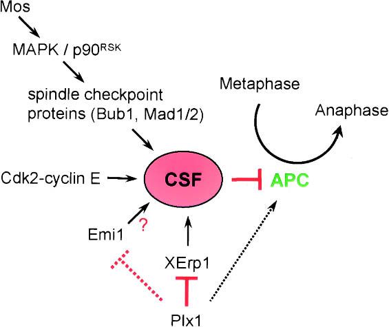
Downloaded from genesdev.cshlp.org on January 12, 2012 - Published by Cold Spring Harbor Laboratory Press

Schmidt et al.

pleted XErp1 from CSF extract supplemented with either               XErp1FL,wt. Examination of DNA morphology revealed
mock IVT or different myc-tagged IVT XErp1 proteins.                 that the incubation of extract with 500 nM MBP-
Since the inactivation of XErp1 triggers the irreversible            XErp1FL,wt prevented chromatin decondensation for >60
release of the extract from CSF arrest (Fig. 3A), the IVTs           min (Fig. 4A), suggesting that the calcium-induced re-
used for the rescue experiments had to be added to the               lease of CSF extract had not taken place. Consistently,
extract before the immunodepletion of XErp1 was initi-               immunoblot analysis revealed that CDC27, a core
ated. Immunoblot analysis confirmed that endogenous                  APC/C subunit that shows a prominent upshift upon
XErp1 was quantitatively removed from XErp1-depleted                 mitotic phosphorylation (Kotani et al. 1998), remained
extracts but not from the extract treated with control               upshifted after calcium addition (Fig. 4C, upper panel).
antibodies (Fig. 3C, middle panel). As expected, IVT full-           Moreover, the radiolabeled APC/C substrates IVT N-ter-
length XErp1 was recognized by the anti-XErp1 antibody               minal fragment of cyclin B1 (cycBNT) and securin were
and therefore depleted from the extract along with the               not degraded upon calcium addition in extract supple-
endogenous protein, but the two C-terminal fragments                 mented with MBP-XErp1FL,wt protein (Fig. 4C, middle
were not removed (Fig. 3C, lower panel). Microscopic                 and lower panels), indicating that the APC/C had not
examination revealed that control-depleted extract                   been activated. However, when CSF extract was supple-
maintained the CSF state as indicated by condensed                   mented with 500 nM full-length XErp1 protein carrying
chromatin and bipolar spindle structures (Fig. 3B, panels            a mutation in its ZBR (MBP-XErp1FL,C583A), the chroma-
1,6). Consistently, IVT securin, a substrate of the APC/             tin decondensed (Fig. 4A), CDC27 lost its M phase-spe-
C, remained stable in control-depleted extract (Fig. 3C,             cific upshift (Fig. 4C, upper panel), and the APC/C sub-
upper panel). As expected, XErp1-depleted extract                    strates IVT cycBNT and securin were degraded (Fig. 4C,
supplemented with mock IVT could not maintain the                    middle and lower panels). These data suggest that excess
CSF state but entered interphase prematurely, as indi-               XErp1 can prevent calcium-induced APC/C activation
cated by decondensed chromatin and degraded IVT se-                  and that XErp1 requires a functional ZBR for its inhibi-
curin (Fig. 3B [panels 5,10], C [upper panel]). XErp1-de-            tory effect on CSF release. This is in line with our pre-
pleted extract supplemented with full-length IVT XErp1               vious observation that IVT XErp1CT,wt but not IVT
(XErp1FL,wt) also could not maintain the CSF state (Fig.             XErp1CT,C583A could rescue the premature CSF release
3B, panels 2,7) because the exogenous IVT XErp1, like                induced by depletion of endogenous XErp1 (Fig. 3B,C).
the endogenous protein, was efficiently depleted from                Notably, when we added a C-terminal fragment of XErp1
the extract (Fig. 3C, lower panel). In contrast, in extract          (MBP-XErp1CT,wt) at concentrations as low as 100 nM,
supplemented with IVT XErp1 C terminus (XErp1CT,wt),                 the extract did not enter interphase for >60 min after
the chromatin stayed condensed, bipolar spindle struc-               calcium addition, as judged from DNA morphology (Fig.
tures remained, and IVT securin was not degraded (Fig.               4B), whereas equimolar concentrations of the N termi-
3B [panels 3,8, C [upper panel]), demonstrating that IVT             nus of XErp1 (MBP-XErp1NT,wt) or of MBP-XErp1FL,wt
XErp1CT,wt could rescue the premature CSF release in-                had no effect on CSF release (Fig. 4B). These data confirm
duced by depleting endogenous XErp1. However, a C-                   that the C terminus of XErp1 is sufficient to prevent CSF
terminal fragment of XErp1 carrying a mutation in the                release. In addition, the observation that the C terminus
ZBR (XErp1CT,C583A) could not rescue the premature                   of XErp1 is more potent in preventing CSF release than
CSF release induced by depleting endogenous XErp1 (Fig.              the full-length protein indicates that the N terminus of
3B [panels 4,9], C [upper panel]). Comparison of the sig-            XErp1 exerts a negative regulatory influence on the ac-
nal intensities derived from endogenous XErp1 and IVT                tivity of XErp1.
XErp1FL,wt (marked with an asterisk in Fig. 3C, anti-
XErp1 immunoblot) confirmed that the IVT product was
                                                                     MAPK pathway inhibition by UO126 does not prevent
present in the extract at a concentration similar to that
                                                                     XErp1 action
of endogenous XErp1. The same applied to the different
C-terminal XErp1 IVT products as judged from the anti-               Next, we asked whether XErp1 is dependent on the Mos/
myc immunoblot (Fig. 3C, lower panel). Taken together                MAPK pathway. If this were the case, then the activity of
these data demonstrate that XErp1 is essential to keep               the MAPK pathway should be essential for XErp1’s in-
the APC/C inactive in CSF extract, and that XErp1 re-                hibitory effect on CSF release. To test this we induced
quires a functional ZBR for its inhibitory effect on                 CSF release by adding calcium to Xenopus egg extract
APC/C activity.                                                      supplemented with MBP-XErp1FL,wt in the presence of
                                                                     the MAPK kinase inhibitor UO126 or DMSO as a solvent
                                                                     control. To confirm that the MAPK pathway was inac-
Excess XErp1 can prevent APC/C activation in
                                                                     tivated upon UO126 addition, we performed immunoblot
CSF extract
                                                                     analyses for the active form of MAPK (Erk1/2). As shown
Given that the inactivation of XErp1 results in the pre-             in Figure 4E, active MAPK was present in Xenopus ex-
mature activation of APC/C in Xenopus egg extract, we                tract treated with DMSO but barely detectably in
next asked whether excess XErp1 protein is sufficient to             UO126-treated extract. Autoradiographic analyses re-
prevent CSF release. To test this we induced CSF release             vealed that 35S-labeled IVT securin remained stable upon
by adding calcium to Xenopus egg extract incubated                   calcium addition in the presence of MBP-XErp1FL,wt and
with purified maltose-binding protein (MBP)-tagged                   UO126 (Fig. 4E), indicating that, under the conditions of

506      GENES & DEVELOPMENT
Downloaded from genesdev.cshlp.org on January 12, 2012 - Published by Cold Spring Harbor Laboratory Press
XErp1, a novel inhibitor of APC/C activity
the experiment, XErp1 does not depend on active MAPK
for its ability to block CSF release. In extract supple-
mented with control buffer, 35S-labeled IVT securin was
more rapidly degraded upon calcium addition in the pres-
ence of UO126 than in DMSO-treated extract, suggesting
that the inactivation of the MAPK pathway can acceler-
ate exit from meiosis II. Taken together, these data sug-
gest that the ability of XErp1 to block CSF release does
not strictly depend on the Mos/MAPK pathway.
XErp1 can inhibit APC/CCDC20 directly
Given the sequence similarity between XErp1’s C termi-
nus and Emi1 (Fig. 1A), we asked whether the C termi-
nus of XErp1, similar to Emi1, could directly inhibit
APC/C activity. To test this possibility we performed an
in vitro ubiquitylation reaction using purified compo-
nents, namely the ubiquitin-activating enzyme E1, the
ubiquitin-conjugating enzyme UbcX as an E2, the
APC/C activator CDC20, APC/C immunopurified from
mitotic Xenopus egg extract, and radiolabeled IVT cy-
cBNT as a substrate. In line with previous reports (Kra-
mer et al. 1998), autoradiographic analyses revealed that
the addition of CDC20 greatly enhanced APC/C’s ubiq-
uitin ligase activity towards its substrate cycBNT (Fig.
4D, groups 1,2). The addition of 300 nM MBP-XErp1CT,wt
strongly inhibited the APC/CCDC20-mediated ubiqui-
tylation of IVT cycBNT, whereas the addition of 1 µM
of MBP-XErp1CT,wt almost completely abolished the
CDC20-dependent ubiquitylation reaction (Fig. 4D,
groups 3,4). In contrast, addition of 1 µM or 3µM of MBP-
XErp1NT,wt had no significant effect on the Cdc20-de-
pendent ubiquitylation of IVT cyclin B1 N terminus (Fig.
Figure 4. Excess XErp1 protein blocks calcium-dependent CSF 4D, groups 5,6). These data strongly support our model
release independently of Mos/MAPK signaling. (A). Purified that XErp1 is an APC/C inhibitor and that the C termi-
MBP-tagged XErp1FL,wt or XErp1FL,C583A was added to Xenopus nus of XErp1 accounts for the inhibitory effect of XErp1
egg extract at 500 nM final concentration. Chromatin morphol- on CSF release by directly inhibiting APC/CCDC20. So
ogy was examined 20 and 60 min after calcium addition. (B) far, we have not been able to convincingly demonstrate a
Purified MBP-tagged XErp1FL,wt, XErp1NT,wt, or XErp1CT,wt
direct interaction between XErp1 and Cdc20, although
were separately added to Xenopus egg extract at 100 nM final
such an interaction has been reported for Emi1 and
concentration. Chromatin morphology was examined 30 min
and 60 min after calcium addition. (C) Xenopus extract was Cdc20 (Reimann et al. 2001b; Reimann and Jackson
treated as in A, and samples were taken at the indicated time 2002). Thus the precise mechanism of the inhibition of
points after calcium addition and immunoblotted for CDC27. APC/CCdc20 is presently unknown.
Samples of extract supplemented with 35S-labeled IVT securin
or N-terminal fragment of cyclin B1 (cycBNT) were taken at the
indicated time points before (CSF) and after calcium addition Plx1 phosphorylates XErp1 in vitro
and analyzed by autoradiography. (D) His-tagged Cdc20 was in-
cubated with buffer (group 2), MBP-XErp1CT,wt (group 3, 300 Because XErp1 was initially identified as a Plx1-interact-
nM; group 4, 1 µM), or MBP-XErp1NT,wt (group 5, 1 µM; group 6, ing protein, we next sought to explore the functional
3 µM). APC/C immunopurified from mitotic Xenopus extract relationship between XErp1 and Plx1 in the regulation of
was then mixed with buffer (group 1) or CDC20 incubated with CSF arrest and APC/C activity upon M-phase exit in
buffer (group 2) or the indicated MBP-XErp1 fragments (groups Xenopus egg extract. First, we tested whether XErp1 is a
3–6) and assayed for its ability to ubiquitylate 35S-labeled IVT substrate of Plx1 in vitro. Using MBP-fusion proteins we
cycBNT at 20°C. At the indicated time points, samples were could show that recombinant Plx1 efficiently phos-
taken and analyzed by autoradiography. (E) Purified MBP-tagged
phorylates full-length XErp1 and an N-terminal frag-
XErp1FL,wt or control buffer was added to CSF-arrested Xenopus
egg extract in the presence or absence of the MAPK kinase in-
ment of XErp1 in vitro (Fig. 5A). In contrast, XErp1CT,wt
hibitor UO126. Inactivation of the MAPK pathway was con- containing the C-terminal F-box and the ZBR was not
firmed by immunoblotting for active Erk1/2. The cell cycle detectably phosphorylated (Fig. 5A). The fact that Plx1
state of the extract was monitored by the stability of exog- efficiently phosphorylated XErp1FL,wt and XErp1NT,wt
enously added 35S-labeled IVT securin. but not equimolar concentrations of XErp1CT,wt (Fig. 5A)
GENES & DEVELOPMENT 507Downloaded from genesdev.cshlp.org on January 12, 2012 - Published by Cold Spring Harbor Laboratory Press
Schmidt et al.
the exogenous XErp1 did not block CSF release. IVT
XErp1FL,wt, like the endogenous protein, was stable in
CSF extract but was rapidly degraded upon calcium ad-
dition (Fig. 5C). In contrast, neither an IVT C-terminal
XErp1 fragment (amino acids 374–651) lacking the
DSGX3S motif nor IVT full-length XErp1 protein mu-
tated at Ser 33 and Ser 38 of the DSGX3S motif
(XErp1S33N,S38N) were degraded upon calcium addition
(Fig. 5C). Examination of DNA morphology (Fig. 5C,
right panel) and of Cdk1 kinase activity (Fig. 5D) con-
firmed that extracts incubated with the different IVT
XErp1 products entered interphase upon calcium addi-
tion. Intriguingly, IVT full-length XErp1 mutated in its
DSGX3S motif showed a transient hypershift upon cal-
cium addition (Fig. 5C, 10 min after calcium [or Ca++]
addition). This hypershift seems to be caused by phos-
phorylation, because the slower-migrating form ob-
served 10 min after calcium addition remained upshifted
when the extract was diluted into buffer containing the
phosphatase inhibitor okadaic acid but not when the ex-
tract was diluted into buffer only (data not shown). The
Figure 5. Plx1-dependent destabilization of XErp1 is required for
question of whether Plx1 contributes to this calcium-
CSF release. (A) MBP-tagged XErp1FL,wt, XErp1NT,wt, and
XErp1CT,wt were each incubated in an in vitro phosphorylation
dependent hypershift will be addressed below. These
reaction with His-tagged Plx1. Incorporation of 32P was analyzed data show that the DSGX3S sequence targets XErp1 for
by PAGE and autoradiography. (B) In vitro Plx1 kinase assay as in degradation in a cell cycle-dependent manner.
A using MBP-tagged wild-type XErp1FL,wt or XErp1FL,S33N,S38N. Next we wanted to know whether Plx1 could contrib-
(C) 35S-labeled IVT XErp1FL,wt, XErp1CT,wt, or a mutant form of ute to the degradation of XErp1 by directly phosphory-
XErp1 (XErp1FL,S33N,S38N) were incubated in CSF extract (i, input) lating the serine residues of the DSGX3S motif. We thus
in the presence or absence of calcium. At the indicated time performed additional in vitro Plx1 kinase assays using
points, samples were withdrawn and analyzed by autoradiogra- MBP-tagged XErp1FL,wt and XErp1S33N,S38N as sub-
phy. (D) Cdk1 activity of all samples shown in C withdrawn after strates. As shown in Figure 5B, the phosphorylation of
60 min was measured using an H1 kinase assay.
XErp1S33N,S38N was significantly reduced compared to
the wild-type protein, indicating that serine residues 33
and/or 38 of XErp1 are major Plx1 phosphorylation sites
indicates that the conditions of the in vitro assay did not
in vitro. Again, the fact that XErp1 carrying two point
promote unspecific protein phosphorylation by Plx1.
mutations was a very poor Plx1 substrate compared to
These data support our hypothesis that the activity of
equimolar concentrations of the wild-type protein sup-
XErp1 is regulated by Plx1-dependent phosphorylation of
ports the specificity of the observed phosphorylation.
XErp1’s N terminus. In line with these results, we ob-
Taken together, these data suggest that Plx1 phosphory-
served that Plx1 showed a yeast-two-hybrid interaction
lates XErp1 at its DSGX3S motif, which then serves as a
with the N terminus but not the C terminus of XErp1
“phospho-degron” (Fuchs et al. 2004) to target XErp1 for
(data not shown).
degradation upon CSF release. These data are consistent
with recent reports showing that Plk1 can phosphorylate
the DSGX2S motif of Emi1, thereby promoting its in
XErp1 is targeted for degradation via its
vitro ubiquitylation (Hansen et al. 2004; Moshe et al.
DSGX3S motif
2004). Thus, polo-like kinases are involved in the regu-
The N terminus of XErp1 contains a DSGX3S motif (Fig. lated degradation of both Emi1 and XErp1.
1A,B). It has been shown previously that the phosphory-
lation of the two serine residues of a DSGX2/3S motif
Dominant-negative polo-box domain of Plx1 (PBDwt)
targets substrates for degradation by the SCF (Skp1–Cul-
stabilizes XErp1
lin F-box) ubiquitin ligase complex that contains the
F-box protein -TRCP (SCF-TRCP) (Fuchs et al. 2004). Previously, it has been shown that the addition of ki-
We thus asked whether the degradation of XErp1 upon nase-dead Plx1 (Plx1N172A) to Xenopus egg extract pre-
CSF release is mediated by its DSGX3S motif. To test vents APC/C activation and CSF release (Descombes and
this possibility we added 35S-labeled IVT XErp1 to Xeno- Nigg 1998). A plausible model for the inhibitory effect of
pus egg extract and studied the stability of IVT XErp1 Plx1N172A on CSF release is that the APC/C inhibitor
upon calcium addition. For these experiments the differ- XErp1 is stabilized through sequestration by excess cata-
ent IVT XErp1 proteins were added to the CSF extract at lytically inactive Plx1N172A. If Plx1N172A sequesters po-
a concentration low enough (one-fourth of the amount tential substrates via its polo-box domain (PBD), then
used for the add-back experiment, Fig. 3C) to assure that the addition of PBD alone should be sufficient to exert a
508 GENES & DEVELOPMENTDownloaded from genesdev.cshlp.org on January 12, 2012 - Published by Cold Spring Harbor Laboratory Press
XErp1, a novel inhibitor of APC/C activity
dominant-negative effect on CSF release. Indeed, addi- 5C) could only be observed in the buffer control, but not
tion of recombinant MBP-tagged wild-type polo-box do- in extract supplemented with MBP-PBDwt (Fig. 6B). This
main of Plx1 (PBDwt) inhibited DNA decondensation, hypershift seems to be caused by phosphorylation (see
spindle disassembly, and the degradation of IVT securin above), suggesting that Plx1 activity is required—di-
upon calcium treatment (Fig. 6A). In contrast, the addi- rectly or indirectly—for the phosphorylation of endog-
tion of equimolar amounts of mutant PBD (Elia et al. enous XErp1 upon calcium addition at a site different
2003) (MBP-PBDmut) had no effect on CSF release, as from the DSGX3S motif. Perhaps this additional cal-
judged by the appearance of nuclei and the degradation of cium-induced phosphorylation event negatively regu-
IVT securin upon calcium addition (Fig. 6A). In line with lates the activity of XErp1 before XErp1 is targeted for
these results, it was recently reported that a point mu- degradation by the phosphorylation of its DSGX3S.
tation in the PBD of kinase-dead Plx1 (Plx1N172A,W408F) Taken together, these data suggest that that active Plx1
neutralizes the inhibitory effect of Plx1N172A on APC/C is required for the degradation of XErp1 via phosphory-
activation (Liu et al. 2004). lation of its DSGX3S motif at anaphase onset, and that
Next, we tested whether 35S-labeled IVT XErp1 is de- degradation of XErp1 is a prerequisite for CSF release.
graded upon calcium addition when CSF extract was pre-
incubated with MBP-PBDwt. As shown in Figure 6B, IVT
XErp1 is required for the PBDwt-induced
XErp1FL,wt remained stable upon calcium addition in the
block of CSF release
presence of MBP-PBDwt (compare 60-min time point be-
fore and after calcium addition). Since 35S-labeled IVT Thus far our data have shown that XErp1 is essential for
XErp1FL,wt became at least partially upshifted in CSF CSF arrest and that the activity of Plx1 is required to
extract supplemented with MBP-PBDwt (Fig. 6B, cf. input mediate the degradation of XErp1 upon CSF release. If
[i] and the 30-min time point in the absence of calcium), XErp1 is the critical downstream effector of Plx1 in regu-
it seems likely that the meiosis-specific phosphorylation lating the APC/C, one would predict that the activity of
of XErp1 does not completely depend on active Plx1. The XErp1 is crucial for the dominant-negative effect of the
identity of other kinases acting upon XErp1 is not pres- PBDwt on CSF release. To test this we examined whether
ently known, but Cdk1 is a likely candidate. In contrast PBDwt can still block CSF release under conditions
to the situation where the extract was supplemented where XErp1 is kept inactive by the addition of immo-
with dominant-negative MBP-PBD, IVT XErp1FL,wt was bilized anti-XErp1 antibodies. Since the inactivation of
rapidly degraded after calcium addition in the buffer con- XErp1 induces a premature calcium-independent CSF re-
trol (Fig. 6B). As expected, IVT XErp1S33N,S38N remained lease (Fig. 3), it was important to add the antibodies to
stable upon calcium addition in the presence of MBP- extract already incubated with PBDwt. When we added
PBDwt or in the buffer control (Fig. 6B). Interestingly, the immobilized anti-XErp1 antibodies to extract supple-
previously observed calcium-induced hypershift (see Fig. mented with MBP-PBDwt, the chromatin was decon-
Figure 6. XErp1 is required for effects of dominant-
negative PBD on CSF release (A) CSF extracts were in-
cubated with equimolar amounts of MBP-tagged either
wild-type (PBDwt) or mutant PBD (PBDmut) of Plx1.
Morphology of DNA and tubulin was examined after
extracts had been warmed to 20°C for 60 min in the
presence or absence of calcium. In addition, the stabil-
ity of exogenously added 35S-labeled IVT securin was
monitored by autoradiography. (B) 35S-labeled IVT
XErp1FL,wt or XErp1FL,S33N,S38N were incubated in CSF
extract containing PBDwt or control buffer. Extracts
were warmed to 20°C, and samples were taken and ex-
amined by autoradiography at the indicated time points
before and after addition of calcium. (C) Immobilized
anti-XErp1 antibodies (lanes 1,2,5,6) or immobilized
control antibodies (lanes 3,4,7,8) were added to CSF ex-
tract containing PBDWT (lanes 1–4) or PBDmut (lanes
5–8) in the presence (lanes 1,3,5,7) or absence (lanes
2,4,6,8) of calcium. Samples were taken at the indicated
time points after calcium and/or antibody addition and
assayed for Cdk1 activity. In addition, samples of the
reactions (panels 1–8) were withdrawn and chromatin
morphology was examined 65 min after warming the
extracts to 20°C. (D) Resulting bands of the H1 kinase
assay were quantified by densitometry. Band intensities
were corrected for background and then plotted against
time. Solid lines show reactions with PBDWT; dashed
lines show reactions with PBDmut.
GENES & DEVELOPMENT 509Downloaded from genesdev.cshlp.org on January 12, 2012 - Published by Cold Spring Harbor Laboratory Press
Schmidt et al.
densed 60 min after calcium addition (Fig. 6C, panel 1) here is actually due to Emi1 depletion or Emi1 codeple-
and Cdk1 activity rapidly dropped to interphase levels tion with XErp1. However, three independent lines of
(Fig. 6C [lane 1], D). In contrast, the chromatin remained evidence argue against this idea. First, the antibody used
condensed when calcium was added to extract supple- for immunodepletion did not detect IVT Emi1 in immu-
mented with MBP-PBDwt and immobilized control anti- noblot analyses (Fig. 1C). Second, we have observed no
bodies (Fig. 6C, panel 3). Under these conditions Cdk1 indication of an interaction between XErp1 and Emi1,
activity decreased by only ∼40%, consistent with a block since mass spectrometry analyses of proteins associated
in CSF release (Fig. 6C [lane 3], D). These data confirm with endogenous XErp1 did not identify Emi1 (G. Sauer
our model that PBDwt requires XErp1 to mediate its in- and T.U. Mayer, unpubl.). Third and most importantly,
hibitory effect on CSF release. decondensation of the sperm DNA, disassembly of the
Xenopus egg extract incubated with MBP-PBDwt and spindles, and degradation of securin upon XErp1-deple-
immobilized anti-XErp1 antibodies had low Cdk1 activ- tion could be rescued by the addition of IVT XErp1CT,wt
ity levels and decondensed chromatin even in the ab- at a concentration matching that of endogenous XErp1.
sence of calcium (Fig. 6C [lane 2], D [panel 2]), indicating These data clearly demonstrate that the observed prema-
that PBDwt cannot prevent the premature CSF release ture APC/C activation results from the loss of XErp1
induced by inactivation of XErp1. As expected, Xenopus protein.
extract supplemented with MBP-PBDmut entered inter- It is possible that XErp1 and Emi1 are part of parallel
phase only in the presence of calcium or upon addition of pathways that cooperate to regulate APC/C activity in
anti-XErp1 antibodies, as indicated by low Cdk1 activity Xenopus egg extract. If so, depleting one of these two
and decondensed chromatin (Fig. 6C [lanes 5–7], D [pan- proteins could lower the APC/C inhibitory activity in
els 5–7]). Taken together these observations show that the extract below a critical concentration, resulting in
XErp1 is required for the dominant-negative effect of premature APC/C activation. Similarly, the depletion of
PBDwt on CSF release and suggest that XErp1 acts down- Cdk2 protein by an antisense approach abolishes CSF
stream of Plx1. They are also consistent with our hy- arrest in Xenopus eggs despite the presence of an active
pothesis that XErp1 is a direct substrate of Plx1, impli- c-Mos/MAPK/p90Rsk pathway, Emi1, and XErp1 (Gabri-
cated in the regulation of the APC/C ubiquitin ligase. elli et al. 1993). These data suggest that the combined
activities of the c-Mos/MAPK/p90Rsk pathway, Cdk2/
cyclin E, Emi1, and XErp1 are required to assure com-
Discussion
plete APC/C inhibition during CSF arrest. Consistently,
Multiple independent pathways contribute to CSF activ- the APC/C substrate cyclin B is completely stabilized
ity in Xenopus eggs (Fig. 7). Here, we characterize XErp1, during CSF arrest but degraded to ∼50% of the meta-
a novel evolutionarily conserved protein essential for phase level during the meiosis I-to-II transition (Tun-
CSF arrest in Xenopus egg extract. Excess XErp1 can pre- quist and Maller 2003), a time when Cdk2/cyclin E is
vent calcium-induced CSF release, and the C terminus absent. Interestingly, XErp1 is present upon exit from
of XErp1 containing the ZBR is necessary and sufficient meiosis I, suggesting that the -TRCP-mediated degra-
to prevent CSF release. Consistently, the C terminus dation of XErp1 is not operating at this early stage of
of XErp1 inhibits the ubiquitin ligase activity of APC/ meiotic progression. However, XErp1 apparently does
CCDC20 directly. In addition to the ZBR, XErp1 contains
an F-box at its C terminus. Yeast two-hybrid analyses
revealed that the F-box of XErp1 is required for its inter-
action with the F-box-binding protein Skp1 (P.I. Duncan
and E.A. Nigg, unpubl.), making XErp1 a bona fide F-box
protein. However, the F-box does not seem to contribute
to XErp1’s inhibitory effect on APC/C activation, be-
cause IVT full-length XErp1 mutated in its F-box
XErp1L450A was equally potent as IVT XErp1WT in block-
ing calcium-induced CSF release (A. Schmidt, N.R.
Rauh, and T.U. Mayer, unpubl.). Similarly, the F-box of
Emi1 also seems to be dispensable for Emi1’s ability to
inhibit APC/C (Reimann et al. 2001a). Therefore, the
elucidation of the functional role of the F-Box of XErp1
and Emi1 will require further research. Figure 7. Model of different pathways regulating CSF activity.
The question arises of why Emi1 and XErp1, both of The c-Mos/MAPK/p90Rsk pathway seems to exert its inhibitory
which are able to inhibit APC/C activity directly, seem effect on APC/C by activating components of the spindle as-
sembly checkpoint. The underlying mechanism by which
to be essential to maintain the CSF arrest in Xenopus egg
Cdk2/cyclin E inhibits APC/C activity has not been identified.
extract. Our data and previous reports have shown that XErp1 and Emi1 mediate CSF activity by inhibiting APC/C di-
extract depleted of one of these proteins cannot maintain rectly. Plx1 targets XErp1 and possibly Emi1 for degradation,
the CSF arrest, although the remaining APC/C inhibit- thereby allowing APC/C activation. Additionally, Plx1 might
ing protein should still be present in the extract. One activate APC/C directly by phosphorylating specific APC/C
could imagine that the premature CSF release observed subunits.
510 GENES & DEVELOPMENTDownloaded from genesdev.cshlp.org on January 12, 2012 - Published by Cold Spring Harbor Laboratory Press
XErp1, a novel inhibitor of APC/C activity
not block APC/C during meiosis I. Whether this is due to in future studies. The regulation of XErp1 function and
insufficient XErp1 levels or its functional inactivation stability is undoubtedly complex. As indicated by the
(e.g., by the post-translational modification shown in several distinct electrophoretic mobilities associated
Fig. 2C) remains to be explored. with XErp1, this protein undergoes multiple post-trans-
In a recent publication, the role of Emi1 during CSF lational modifications, including phosphorylation not
arrest in Xenopus egg extracts was challenged (Ohsumi only within the DSGX3S degron, but also on other sites.
et al. 2004). In particular, Kishimoto and coworkers (Oh- The available evidence indicates that XErp1 is stable and
sumi et al. 2004) reported that wild-type Emi1 is rapidly active as an APC/C inhibitor in CSF extracts, although
degraded during oocyte maturation and highly unstable Plx1 can be recovered from such extracts in an active
in CSF extracts, raising the question of whether this pro- form. Thus, the Plx1-dependent inactivation of XErp1
tein could be present at sufficient levels in CSF-arrested clearly depends on a calcium signal. It is possible that
extracts to perform its alleged role as an APC/C inhibitor calcium sensitizes XErp1 to additional phosphorylation
(Reimann et al. 2001b; Reimann and Jackson 2002). by Plx1 (e.g., through the action of a calcium-activated
Moreover, contradictory data exist concerning the fate of kinase). Alternatively, it would seem premature to rig-
Emi1 upon calcium-induced CSF release. Whereas Emi1 orously exclude that the -TRCP system could be regu-
was originally reported to be stable upon CSF release lated by calcium. Regardless of these open questions, our
until the first mitotic division (Reimann et al. 2001b; present data clearly indicate that APC/C activation by
Reimann and Jackson 2002), a more recent study from polo-like kinase in Xenopus egg extract involves not
the same laboratory proposes that Plx1 triggers Emi1 de- only direct phosphorylation of APC/C subunits but also
struction (Hansen et al. 2004), similar to the results re- the derepression of APC/C by inactivation of inhibitory
ported here for XErp1. Clearly, further studies will be proteins.
required to clarify the precise role of Emi1 during CSF
arrest and release.
Irrespective of the controversy around the role of Emi1 Materials and methods
in regulating CSF release, our present data strengthen
the conclusion that the activity of Plx1 is required to Yeast two-hybrid screen
activate APC/C upon CSF release (Descombes and Nigg A yeast two-hybrid screen was performed as described previ-
1998) and to maintain APC/C in its active form (Brassac ously (Gietz and Woods 2002). Bait plasmid pGBDU/Plx1N172A
et al. 2000). Since polo-like kinases can phosphorylate was transformed into yeast strain PJ69-4A and selected by uracil
specific APC/C subunits in vitro, the model emerged auxotrophy. A Xenopus laevis oocyte matchmaker cDNA li-
that polo-like kinases can activate the APC/C directly. brary in pACT2 (Clontech) was subsequently transformed into
However, it is still controversial whether APC/C phos- yeast strain PJ69-4A carrying the bait plasmid pGBDU/
phorylation by polo-like kinases is sufficient (Kotani et Plx1N172A. Transformants were selected for growth on plates
al. 1998), required (Golan et al. 2002), or even dispens- lacking uracil, leucine, and adenine. pACT2 plasmids were res-
cued by transforming HB101 bacteria with yeast DNA prepara-
able for the activation of the APC/C (Kraft et al. 2003).
tions, followed by selection for leucine auxotrophy. Plasmid
Our present data lead to a model in which Plx1 controls
DNA from positive clones was retransformed, and the cDNA
APC/C activity at anaphase onset by targeting XErp1, an inserts of positive clones were sequenced to identify putative
inhibitor of APC/C, for degradation. According to this Plx1-interacting proteins.
model, the inhibition of CSF release observed upon ad-
dition of catalytically inactive Plx1N172A to Xenopus egg
Cloning and protein expression
extract is mediated by the sequestration of XErp1 by the
polo-box domain of Plx1N172A. Consistently, our data Full-length XErp1 (XErp1FL,wt) was initially subcloned from the
confirm that the polo-box domain of Plx1 requires XErp1 identified two-hybrid plasmid into a modified pCS2 vector in-
to exert its inhibitory effect on CSF release in Xenopus troducing restriction sites for the FseI and AscI restriction en-
zymes. Primers with the sequences 5⬘-ATTATGGCCGGC
egg extract. The phosphorylation of XErp1, like that of
CAGAGATGGCAAATCTCTTAGAG-3⬘ and 5⬘-ATTATGGC
other F-box proteins, seems to be a prerequisite for its GCGCCGGAAGACTAGCTTCAAAGTCTC-3⬘ were used to
degradation by the ubiquitin–proteasome pathway. Our amplify the coding sequence (start and stop codons are under-
data show that Plx1 can phosphorylate a DSGX3S motif lined). Site-directed mutagenesis to yield the C583A mutation
located within the N terminus of XErp1, and that the in the ZBR was carried out using the QuikChange kit (Strata-
DSGX3S motif is essential for the degradation of XErp1 gene) according to the manufacturer’s instructions. The N-ter-
upon CSF release. Previously, it has been shown for minal and C-terminal fragments of XErp1 comprise the amino
other proteins that the phosphorylation of both serine acids 1–424 and 374–651, respectively. Wild-type or mutant
residues of a DSGX2/3S motif creates a “phospho-de- fragments were subcloned from plasmids containing the desired
gron” recognized by the SCF-TRCP ubiquitin ligase com- full-length XErp1 as a template following the strategy described
above. The polo-box domain construct (PBDwt) comprising
plex (Fuchs et al. 2004). Therefore, it appears plausible
amino acids 358–598 of Plx1 was subcloned from full-length
that SCF-TRCP also mediates the degradation of phos- Plx1 in a pCS2-Myc plasmid using primers 5⬘-ATTATGGCC
phorylated XErp1 upon CSF release. GGCCGGAGTTCACG GAGCCTGC-3⬘ and 5⬘-ATTATGGC
The role of fertilization and the resulting calcium sig- GCGCCCTATGCCGAGGCCTTTAC-3⬘. A W408F, H532A,
nal in controlling the functional interaction between K534A mutation in polo box 1 (PBDmut) was introduced as de-
Plx1, XErp1, Emi1, and APC/C remain to be determined scribed previously (Liu et al. 2004). Wild-type, mutant, and frag-
GENES & DEVELOPMENT 511Downloaded from genesdev.cshlp.org on January 12, 2012 - Published by Cold Spring Harbor Laboratory Press
Schmidt et al.
ments of XErp1 protein as well as the polo-box domain of Plx1 pus, CDC27 and the Myc epitope-tag samples were blotted
were expressed from a modified pMal-vector (New England Bio- semi-dry on nitrocellulose membrane. The Myc epitope was
labs) as an N-terminal MBP-fusion. The purifications were per- detected with 9E10 hybridoma supernatant. As secondary anti-
formed according to the manufacturer’s instructions. Purifica- bodies, horseradish-peroxidase (HRP)-coupled anti-rabbit or
tion of his-tagged Plx1 from Sf9 cells was performed as described anti-mouse antibodies were typically used at a concentration of
(Descombes and Nigg 1998). 1 µg/mL. Detection was carried out with Amersham ECL re-
agents or Pierce SuperSignal femto substrate.
Antibody production
Kinase assays
6xHis-tagged N-terminal fragment of XErp1 (amino acids 105–
374) was expressed from a pQE31 plasmid in Escherichia coli For analyses of Cdk1/cyclin B activity, H1 kinase assays were
strain BL21 (DE3)pLysS, purified under denaturing conditions performed. The assay was started by mixing 1.62 µL Xenopus
with Ni2+NTA resins as recommended by the manufacturer extract with 18.6 µL of kinase assay mix containing 8 µg histone
(QIAGEN), and subsequently gel-purified after SDS-PAGE. Pu- H1, 14 µM ATP, and 6.5 µCi ␥-32P-ATP in H1 buffer (20 mM
rified protein was used to immunize New Zealand white rabbits -glycerophosphate, 3 mM MgCl2, 4 mM EGTA, 0.025% NP40).
(Elevage Scientifique des Dombes). Antibodies were affinity-pu- Reactions were carried out at room temperature and stopped by
rified following standard procedures. boiling samples in SDS-sample buffer. In vitro kinase assays
using recombinant his-tagged Plx1 were performed as described
(Descombes and Nigg 1998).
CSF extracts
Xenopus CSF egg extracts were prepared essentially as described In vitro ubiquitylation assays
previously (Murray 1991). CSF release was induced by adding
600 µM CaCl2 to the extract. DNA and spindle morphology Yeast E1 enzyme was purchased from Boston Biochem Inc. E2
were examined as described previously (Desai et al. 1999). All enzyme UbcX was expressed and purified from E. coli. Active
experiments were carried out with freshly prepared extracts. APC/C was immunoprecipitated from “⌬90 extract” (Stem-
mann et al. 2001) using monoclonal anti-CDC27 antibodies
(Sigma-Aldrich) immobilized on Protein G beads (Dynal). To
Mass spectrometry
obtain ⌬90 extract, CSF extract was released from CSF arrest as
Coomassie-stained protein bands were in-gel digested by tryp- described above and subsequently incubated with nonde-
sin, desalted, and concentrated using C18 extraction tips as re- structible cyclin B ⌬90 to a final concentration of 5 µg/mL.
ported (Shevchenko et al. 1996; Rappsilber et al. 2003). The Assays were done as described (Kramer et al. 1998) except that
peptide mixture was eluted from a pulled fused silica capillary 12 µg/µL UbcX was used and the buffer was CSF-XB. Briefly,
with an internal diameter of 75 µm and a tip opening of 8 µm his-tagged Cdc20 and MBP-tagged XErp1 proteins were preincu-
(New Objective) filled with ReproSil-Pur C18-AQ (Dr. Maisch, bated with the immunoprecipitated APC/C in buffer QA (10
GmbH, Ammerbuch, Germany) 3-mm reverse-phase material mM Tris-HCl at pH 7.5, 100 mM KCl, 1 mM MgCl2, 0.1 mM
directly into a quadrupole time-of-flight mass spectrometer CaCl2, 1 mM DTT) for 30 min. The beads were washed once in
(Q-ToF Ultima, Micromass) with a 60-min linear gradient of QA and twice in CSF-XB, then the reaction was started by mix-
0%–100% buffer B (80% acetonitrile, 0.5% acetic acid) from a ing the washed beads with a mix containing E1, E2, Ubiquitin,
CAPLC HPLC system (Waters) at a flow rate of ∼170 nL/min. an energy regeneration system, and radiolabeled, in vitro trans-
Combined peak lists were searched in the MSDB database using lated cycBNT. Samples were withdrawn after 0, 6, and 12 min
Mascot (http://www.matrixscience.com) allowing ±0.15 Da tol- and boiled in sample buffer.
erance for both peptide and MS/MS fragment ion mass values.
Acknowledgments
Immunodepletion and antibody addition
We thank Francis Barr, Stefan Jentsch, Edgar Kramer, Ingmar
Immunodepletion experiments were performed as described
Schön, and Olaf Stemmann for generously sharing reagents and
(Funabiki and Murray 2000). Briefly, anti-XErp1 or rabbit IgG
for critical reading of the manuscript. We are grateful to Thierry
antibodies were immobilized on Protein G beads (Dynal) in PBS
Lorca, Yong Wan, and Stephan Geley for reagents, and Jenny
buffer containing 0.1% Triton X-100 at 4°C. Washed beads were
Bormann and Samantha Wattam for excellent technical assis-
mixed with the extract and incubated on ice with occasional
tance. Research in the lab of T.U.M. is supported by Emmy
mixing. Beads were retrieved on a magnet twice before the ex-
Noether grant MA 1559/4-2 from the Deutschen Forschungsge-
tract was warmed to 20°C for analysis of DNA and spindle
meinschaft.
morphology, and H1 kinase activity.
For antibody addition experiments, antibody was coupled to
Protein G beads (Dynal) and incubated with a 10-fold molar
References
excess of antigenic protein or buffer. Beads were washed exten-
sively before mixing with extracts on ice. Extracts were then Barr, F.A., Sillje, H.H., and Nigg, E.A. 2004. Polo-like kinases
incubated at 20°C. and the orchestration of cell division. Nat. Rev. Mol. Cell
Biol. 5: 429–440.
Brassac, T., Castro, A., Lorca, T., Le Peuch, C., Doree, M.,
Immunoblotting
Labbe, J.C., and Galas, S. 2000. The polo-like kinase Plx1
Western blots were performed according to standard procedures. prevents premature inactivation of the APC(Fizzy)-depen-
Briefly, for immunodetection of XErp1, samples were wet-blot- dent pathway in the early Xenopus cell cycle. Oncogene 19:
ted on PVDF membrane after PAGE. The membrane was 3782–3790.
blocked extensively and then incubated with 200 ng affinity- Desai, A., Murray, A., Mitchison, T.J., and Walczak, C.E. 1999.
purified XErp1 rabbit antibody. For Western blots against Xeno- The use of Xenopus egg extracts to study mitotic spindle
512 GENES & DEVELOPMENTYou can also read