Pathological mutations in PNKP trigger defects in DNA single-strand break repair but not DNA double-strand break repair
←
→
Page content transcription
If your browser does not render page correctly, please read the page content below
6672–6684 Nucleic Acids Research, 2020, Vol. 48, No. 12                                                                Published online 6 June 2020
doi: 10.1093/nar/gkaa489

Pathological mutations in PNKP trigger defects in
DNA single-strand break repair but not DNA
double-strand break repair
Ilona Kalasova1 , Richard Hailstone2 , Janin Bublitz3 , Jovel Bogantes4 , Winfried Hofmann3 ,
Alejandro Leal5 , Hana Hanzlikova 1,2,* and Keith W. Caldecott 1,2,*
1
 Department of Genome Dynamics, Institute of Molecular Genetics of the Czech Academy of Sciences, Prague 4,
142 20, Czech Republic, 2 Genome Damage and Stability Centre, University of Sussex, Falmer, Brighton BN1 9RQ,

                                                                                                                                                              Downloaded from https://academic.oup.com/nar/article/48/12/6672/5854143 by guest on 01 December 2020
UK, 3 Department of Human Genetics, Hannover Medical School, Hannover, Germany, 4 Servicio de Cirugı́a
Reconstructiva, Hospital Rafael Ángel Calderón Guardia, Caja Costarricense de Seguro Social, San José, Costa
Rica and 5 Section of Genetics and Biotechnology, School of Biology, University of Costa Rica, San José, Costa Rica

Received March 11, 2020; Revised April 30, 2020; Editorial Decision May 27, 2020; Accepted June 04, 2020

ABSTRACT                                                                        disease, and the extent and nature of this reduction
                                                                                as the primary determinant of disease severity.
Hereditary mutations in polynucleotide kinase-
phosphatase (PNKP) result in a spectrum of neu-
                                                                                INTRODUCTION
rological pathologies ranging from neurodevelop-
mental dysfunction in microcephaly with early on-                               DNA strand breaks can arise endogenously or can result
set seizures (MCSZ) to neurodegeneration in ataxia                              from exogenous sources of DNA damage such as ionizing
oculomotor apraxia-4 (AOA4) and Charcot-Marie-                                  radiation and chemical genotoxins. To combat the geno-
Tooth disease (CMT2B2). Consistent with this, PNKP                              toxic impact of DNA damage, cells have evolved multiple
                                                                                biochemical pathways to detect and repair DNA strand
is implicated in the repair of both DNA single-
                                                                                breaks (1,2). Importantly, defects in DNA strand break re-
strand breaks (SSBs) and DNA double-strand breaks                               pair can result in a variety of different disease patholo-
(DSBs); lesions that can trigger neurodegeneration                              gies, highlighting the threat posed by DNA breaks to hu-
and neurodevelopmental dysfunction, respectively.                               man health (3–5). For example, microcephaly and devel-
Surprisingly, however, we did not detect a signifi-                             opmental delay are often present in individuals with de-
cant defect in DSB repair (DSBR) in primary fibrob-                             fects in non-homologous end-joining (NHEJ); one of the
lasts from PNKP patients spanning the spectrum                                  two major pathways by which DNA double-strand breaks
of PNKP-mutated pathologies. In contrast, the rate                              (DSBs) are repaired (4). In contrast, the primary pathol-
of SSB repair (SSBR) is markedly reduced. More-                                 ogy resulting from defects in the repair of DNA single-
over, we show that the restoration of SSBR in pa-                               strand breaks (SSBs) is neurodegeneration, and in partic-
tient fibroblasts collectively requires both the DNA                            ular progressive cerebellar ataxia (5,6). Examples of the
                                                                                latter are individuals with spinocerebellar ataxia with ax-
kinase and DNA phosphatase activities of PNKP, and
                                                                                onal neuropathy 1 (SCAN1), ataxia with oculomotor apraxia
the fork-head associated (FHA) domain that interacts                            type 1 (AOA1), and ataxia with oculomotor apraxia-XRCC1
with the SSBR protein, XRCC1. Notably, however, the                             (AOA-XRCC1), in which the DNA strand break repair pro-
two enzymatic activities of PNKP appear to affect dif-                          teins TDP1, aprataxin, and XRCC1 are mutated, respec-
ferent aspects of disease pathology, with reduced                               tively (7–10).
DNA phosphatase activity correlating with neurode-                                 Arguably one of the most common sources of neurolog-
velopmental dysfunction and reduced DNA kinase                                  ical disease associated with defects in DNA strand break
activity correlating with neurodegeneration. In sum-                            repair are mutations in the enzyme polynucleotide kinase-
mary, these data implicate reduced rates of SSBR,                               phosphatase (PNKP) (11). PNKP possesses both DNA 5 -
not DSBR, as the source of both neurodevelopmental                              kinase and DNA 3 -phosphatase activity and thereby can
and neurodegenerative pathology in PNKP-mutated                                 convert 5 -hydroxyl and 3 -phosphate termini to the canon-
                                                                                ical 5 -phosphate and 3 -hydroxyl moieties necessary for

* To
   whom correspondence should be addressed. Tel: +44 1273 877519; Email: k.w.caldecott@sussex.ac.uk
Correspondence may also be addressed to Hana Hanzlikova. Email: hana.hanzlikova@img.cas.cz


C The Author(s) 2020. Published by Oxford University Press on behalf of Nucleic Acids Research.
This is an Open Access article distributed under the terms of the Creative Commons Attribution License (http://creativecommons.org/licenses/by/4.0/), which
permits unrestricted reuse, distribution, and reproduction in any medium, provided the original work is properly cited.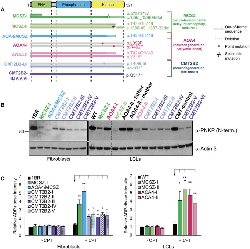
Nucleic Acids Research, 2020, Vol. 48, No. 12 6673
completion of DNA strand break repair (12,13). Whereas siRNA transfection
3 -phosphate termini are present at ∼70% of DNA breaks
Where indicated, cells were transfected with mix of ei-
induced by reactive oxygen species and ionising radiation,
ther PNKP siRNA #1: 5 -CCGGAUAUGUCCACGUG
both 3 -phosphate and 5 -hydroxyl termini are generated
AA-3 and PNKP siRNA #2: 5 -GGAAACGGGUCGC
at DNA strand breaks induced by the abortive activity
CAUCGA-3 or non-target siRNA #1: 5 -UGGUUUA
of topoisomerase 1 (TOP1) (14,15). Notably, PNKP inter-
CAUGUCGACUAA-3 and non-target siRNA #2: 5 -U
acts with protein complexes involved in the repair of SSBs
GGUUUACAUGUUGUGUGA-3 ) using Lipofectamine
and DSBs in mammalian cells via the interaction of its
RNAiMAX (Life Technologies) 48–72 h before experiment.
amino-terminal FHA domain with the SSBR and DSBR
scaffold proteins XRCC1 (16–18) and XRCC4 (19–22),
respectively. Constructs, protein purification and transfection
Mutations in PNKP result in three clinically distinct neu-
rological diseases. The most severe of these is microcephaly Wild-type recombinant human PNKP (PNKPWT ) and
Downloaded from https://academic.oup.com/nar/article/48/12/6672/5854143 by guest on 01 December 2020
with early onset seizures (MCSZ), a neurodevelopmental PNKP with a mutated FHA domain (R35A; PNKPFHA )
disease associated with microcephaly, early-onset seizures or catalytically inactive phosphatase domain (D171N;
and developmental delay (23). In addition, PNKP muta- PNKPPD ) harboured an N-terminal His tag if expressed
tions are the cause of the neurodegenerative disease ataxia from the bacterial expression construct pET16b or N-
with oculomotor apraxia 4 (AOA4), which exhibits progres- terminal EYFP if expressed from the mammalian expres-
sive cerebellar atrophy and ataxia oculomotor apraxia (24– sion construct pEYFP-C1. PNKP proteins were expressed
26). Strikingly, some affected individuals possess elements in E.coli from pET16b and purified by metal-chelate affin-
of both of these diseases; presenting with both microcephaly ity chromatography and gel filtration. For transfection of
and progressive cerebellar atrophy (26–30). Finally, PNKP human cells with purified recombinant PNKP, 2 g of con-
mutations were also identified recently in Charcot-Marie- trol bovine serum albumin (BSA) or the indicated PNKP
Tooth disease 2B2 (CMT2B2), which is associated with mild protein was electroporated into 2×10⁵ primary human fi-
axonal peripheral polyneuropathy and relatively late-onset broblasts using a NEON Transfection System (Invitrogen)
cerebellar ataxia (31–33). according to the manufacturer’s instructions. Cells were em-
Here, we have addressed the relationship between defects ployed for experiments 18 h post-transfection. For transfec-
in SSBR, DSBR, and neurological pathology in patient- tion with mammalian expression constructs, 1 g of con-
derived cells from individuals with mutations in PNKP trol pEYFP-C1 or pEYFP-C1 encoding wild type or the
spanning the spectrum of PNKP-associated pathologies. indicated mutant PNKP were electroporated into 2×10⁵
Surprisingly, our data implicate unrepaired SSBs as the primary human fibroblasts as described above. Transfected
source of both the neurodevelopmental and neurodegener- cells were employed in experiments 48 h post-transfection.
ative pathology in PNKP-associated disease, and the level
of residual SSBR as the primary determinant of disease Antibodies and western blotting
severity. Our data also implicate the two activities of PNKP
in different aspects of the disease pathology; with reduced Primary antibodies were anti-pan-ADP-ribose binding
DNA phosphatase and DNA kinase activity correlating reagent (MABE1016, Millipore), anti-PNKP N-terminal
best with neurodevelopmental dysfunction and neurode- (ab170954, Abcam), anti-PNKP C-terminal (ab18107,
generation, respectively. Abcam), anti- actin (66009, Proteintech), anti-␥ H2AX
(9718, Cell Signaling), and anti-PCNA (sc-56, Santa Cruz
Biotechnology). Secondary antibodies for western blotting
MATERIALS AND METHODS were horseradish peroxidase (HRP)-conjugated goat anti-
rabbit (170-6515, Bio-Rad) and goat anti-mouse (170-6516,
Cell lines Bio-Rad) and for indirect immunofluorescence were don-
The control human primary fibroblasts 1BR3 (denoted here key anti-rabbit Alexa 488 (A21206, Invitrogen) and donkey
as 1BR) and the PNKP patient-derived primary fibrob- anti-mouse Alexa 647 (A-31571, Invitrogen). Samples for
lasts AOA4/MCSZ (7,26), MCSZ-I (34), CMT2B2-(I-V) western blotting were lysed in SDS sample buffer and sub-
(32) and XRCC1 patient-derived primary fibroblasts (7) jected to SDS-PAGE, transferred onto nitrocellulose mem-
were grown in Minimum Essential Media (MEM, Gibco) brane and detected by the appropriate specific primary and
supplemented with 15% fetal bovine serum, 2 mM glu- HRP-conjugated secondary antibodies.
tamine and the antibiotics penicillin (100 units/ml) and
streptomycin (100 g/ml). Lymphoblastoid cells (LCLs)
Alkaline comet assays
derived from CMT2B2 patients with a pathogenic heterozy-
gous mutation (p.Y145S) in myelin protein zero (denoted The level of DNA strand breaks was evaluated by alkaline
here CMT-control), PNKP patients CMT2B2-(I-VI) (32), comet assays following DMSO treatment or treatment with
AOA4-I (25), AOA4-II and father and mother parental con- 10 M CPT for 60 min at 37◦ C or following irradiation with
trols (35), and ‘WT’ control LCLs were grown in RPMI- 20 Gy of X-rays (X-RAD 225XL, Accella) with indicated
1640 Media (Sigma) supplemented with 10% fetal bovine time of recovery. The average comet tail moment in 100 cells
serum and the antibiotics as above in a humidified atmo- per sample was evaluated by Comet Assay IV software (Per-
sphere of 5% CO2 at low oxygen (5%) at 37◦ C. ceptive Instruments).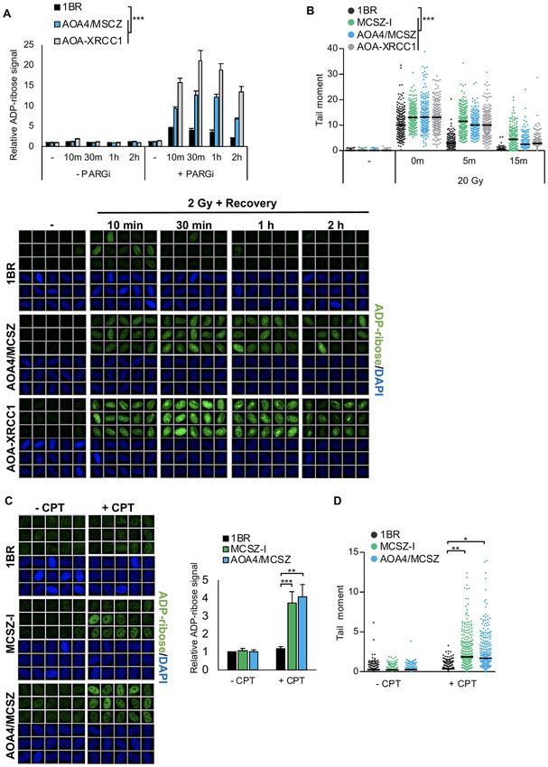
6674 Nucleic Acids Research, 2020, Vol. 48, No. 12
Indirect immunofluorescence microscopy lamide gel and analyzed on a PharosFX Molecular Imager
System (Bio-Rad).
For indirect immunofluorescence microscopy, cells were
cultured and treated where indicated with 10 M CPT
(C9911, Sigma) for 45 min, irradiated (X-RAD 225XL, RESULTS
Accella) with 20 Gy of X-rays on ice or 2 Gy of X-rays Reduced PNKP protein and activity in PNKP patient-derived
at RT in the presence of DMSO (D2650, Sigma), 10 M fibroblasts
PARG inhibitor (PDD 0017273; 5952, Tocris Bioscience)
or 5 M DNA-PK inhibitor (NU 7441; 3712, Tocris Bio- To identify the DNA strand break repair defects in PNKP-
science). Cells were fixed with 4% formaldehyde and im- associated neurological disease we initially employed pri-
munostained as described previously (7). Images were taken mary human fibroblasts derived from two PNKP-mutated
using a DMi6000 microscope (Leica) with 40× dry objec- individuals spanning the spectrum of PNKP-related disease
tive. Automated wide-field microscopy was performed on pathologies and, as a control, primary human fibroblasts
derived from an unaffected individual (1BR). One of the af-
Downloaded from https://academic.oup.com/nar/article/48/12/6672/5854143 by guest on 01 December 2020
scanR system (Olympus) with scanR Image Acquisition
and Analysis Software, 40 x/0.95NA (UPLSAPO 2 40×) fected individuals (MCSZ-I) is a patient with MCSZ and is
dry objective. a compound heterozygote harbouring a 1-bp duplication in
the FHA domain in one allele (c.63dupC) and a 10-bp dele-
tion spanning the exon14/intron14 splice site in the DNA
DSBR assays kinase domain in the second allele (c.1295 1298) (Figure
1A) (34). Both of these mutations are predicted to result in
To measure DSBR, cells were pre-extracted with 0.2%
translational frameshifts in, or upstream of, the DNA 5 -
Triton-X 100 for 2 min on ice prior fixation. Cells were
kinase domain. The second affected individual is a patient
stained for PCNA, ␥ H2AX, and counterstained for DNA
with combined AOA4/MCSZ and is homozygous for a 17-
using 1 g/ml DAPI (202710100, Acros). The cell cycle
bp duplication in exon 14 (c.1250 1266dup), which again
phase was determined based on the presence of PCNA sig-
results in a translational frameshift within the DNA kinase
nal and the DNA content of individual cell nuclei. PCNA-
domain (Figure 1A) (26).
positive cells were gated as S phase cells, and the DNA con-
Both MCSZ-I and AOA4/MCSZ cells possess a very
tent of PCNA-negative cells was compared to gate cells in
small amount of residual PNKP protein, as measured
G1 phase or G2 phase . Cells that could not be gated based
by western blotting (Figure 1B). The residual PNKP in
on these criteria were excluded from the analysis.
AOA4/MCSZ cells migrated faster than wild type PNKP
and was detected only by an N-terminal-specific antibody,
PNKP biochemical activity consistent with both PNKP alleles in this cell line en-
coding a severely truncated 5 -kinase domain. In contrast,
PNKP substrate was prepared by annealing equimolar the residual PNKP in MCSZ-I cells migrated at two po-
amounts of fluorophore-labeled deoxyriboligonucleotides sitions; one that was close to full-length PNKP and de-
(Midland Certified Reagent Company). To measure both 3 - tected by both N- and C-terminal antibodies and one that
phosphatase and 5 -kinase activities, oligonucleotides with was severely truncated and detected by N-terminal anti-
3 -phosphate ‘S1’ [5 -(TAMRA)-TAGCATCGATCAGTC body. AOA4/MCSZ cells lacked detectable 5 -kinase activ-
CTC-3 -P] and 5 -hydroxyl ‘S2’ [5 -OH-GAGGTCTAGCA ity, consistent with the truncated DNA kinase domain, but
TCGTTAGTCA-(6-FAM)-3 ] were annealed to a comple- did possess a small amount of residual 3 -phosphatase ac-
mentary strand oligonucleotide [5 -TGACTAACGATGC tivity, as measured by the appearance of a small amount
TAGACCTCTGAGGACTGATCGATGCTA-3 ] in an- of fully repaired (ligated) 3 -phosphate oligonucleotide sub-
nealing buffer (10 mM Tris pH 7.5, 200 mM NaCl, strate (Figure 1C). In contrast, consistent with our previous
1 mM EDTA) (36). To test only 3 -phosphatase ac- report (34), MCSZ-I cells exhibited significant levels of both
tivity, ‘S1’ was used in a combination with ‘C2’ [5 -P DNA 3 -phosphatase and 5 -kinase activity (Figure 1C).
-GAGGTCTAGCATCGTTAGTCA-(6-FAM)-3 ] and to The residual 5 -kinase activity in this cell line is most likely
test only 5 -kinase activity, ‘S2’ was used in a combina- encoded by the small amount of near-full length PNKP
tion with ‘C1’ [5 -(TAMRA)-TAGCATCGATCAGTCCT polypeptide detected in this cell line, resulting from alter-
C-3 -OH]. Cell-free protein extracts were prepared in lysis native splicing and/or translation-initiation downstream of
buffer [25 mM Tris, pH 7.5, 10 mM EDTA, 10 mM EGTA, the mutated FHA domain (34).
100 mM NaCl, 1 % Triton X-100, cOmplete protease in-
hibitors (Roche)], incubated on ice for 15 min and cen-
Normal rates of DSBR in PNKP patient-derived fibroblasts
trifuged at 16 000 × g, 20 min at 4◦ C. The indicated amounts
of purified PNKP proteins or cell-free protein extracts Defects in the repair of DSBs by NHEJ resulting
were incubated with 50 nM substrate and 1 M single- from hereditary mutations in XRCC4, DNA lig-
stranded nuclease competitor oligonucleotide [5 -AAAGA ase IV, or Cernunnos/XLF are strongly implicated in
TCACAAGCATAAAGAGACAGG-3 ] in reaction buffer neurodevelopmental/microcephalic pathologies similar to
(25 mM Tris, pH 7.5, 130 mM KCl, 10 mM MgCl2 , 1 mM those observed in individuals with mutations in PNKP
DTT, 1 mM ATP) at 37◦ C for 10 or 60 min. 50 l reactions (37–41). Consequently, because PNKP has been shown
were terminated by addition of 50 l quenching buffer (90% to be involved in NHEJ (19,20,42), it seemed plausible
formamide, 50 mM EDTA, 0.006% Orange G). 10 l of that unrepaired DSBs might contribute to PNKP-mutated
each reaction was separated on a 20% denaturing polyacry- disease. To address this possibility, we employed scanR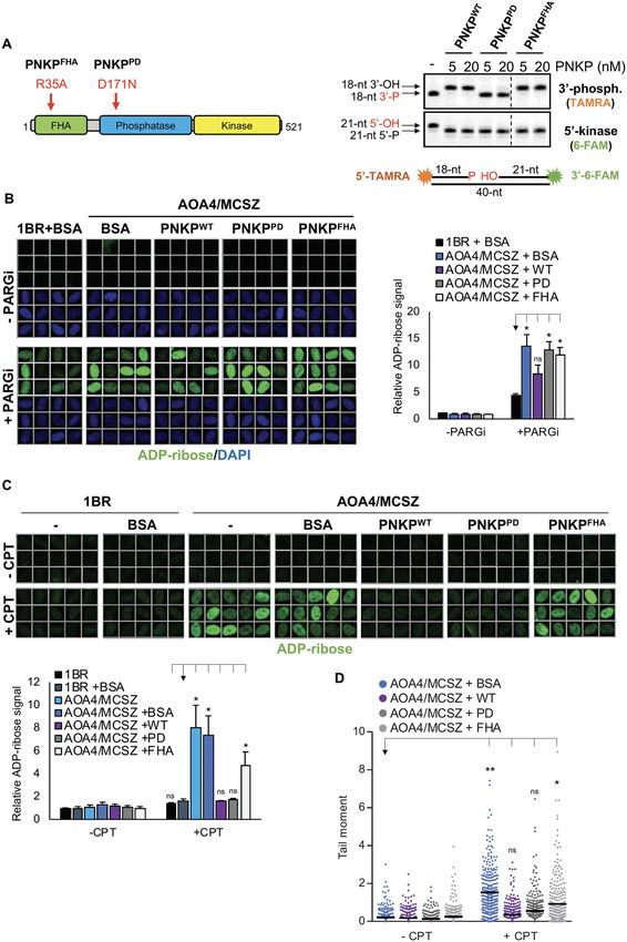
Nucleic Acids Research, 2020, Vol. 48, No. 12 6675
Downloaded from https://academic.oup.com/nar/article/48/12/6672/5854143 by guest on 01 December 2020
Figure 1. Reduced PNKP protein and activity in PNKP patient-derived fibroblasts. (A) Cartoon showing PNKP functional domains and the mutations
present in the patient-derived fibroblast cell lines MCSZ-I and AOA4/MCSZ. The former is compound heterozygous for two frame-shift mutations and
the latter is homozygous for a single frame-shift mutation. The location of the epitopes detected by the N- and C-terminal antibodies employed in this
study are indicated (black horizontal lines). (B) PNKP protein levels in control patient-derived fibroblasts from an unaffected individual (1BR) and the
PNKP patient-derived fibroblasts MCSZ-I and AOA4/MCSZ, measured by western blotting using N- and C-terminal specific anti-PNKP antibodies.
Black asterisk denotes an unspecific band.  actin was employed as a loading control. (C) PNKP activity in control (1BR) and PNKP patient-derived
(MCSZ-I, AOA4/MCSZ) fibroblast cell extracts. TAMRA- or 6-FAM-labelled oligonucleotide duplex harbouring a SSB with a 3 -phosphate or 5 -hydroxyl
terminus, respectively, was incubated with the indicated cell extracts (30 g total protein) for 10 min (to measure kinase and phosphatase activity) or 60 min
(to measure ligated product) at 37◦ C prior to fractionation by denaturing PAGE. Arrows indicate the positions of the TAMRA- labelled 3 -phosphatase
substrate (‘18-nt 3 -P’), 6-FAM-labelled 5 -kinase substrate (‘21-nt 5 -OH’), and intermediates of their repair resulting from 3 -phosphatase activity (‘18-nt
3 -OH’), 5 -kinase activity (‘21-nt 5 -P’), DNA gap filling (‘19-mer’), and DNA ligation (‘40-nt’).
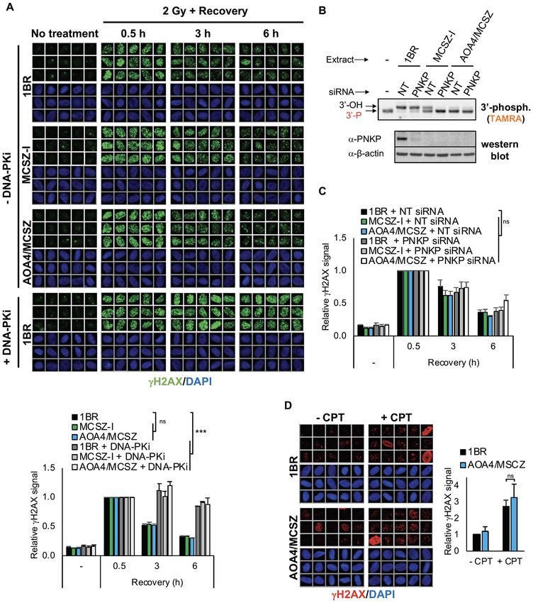
high-content imaging to quantify the level of ␥ H2AX, an ual PNKP activity we transfected the cells with PNKP
established and sensitive marker of DSBs (43,44), in PNKP siRNA (Figure 2B). However, while we detected a small
patient fibroblasts following treatment with ionizing radia- increase in the persistence of ␥ H2AX in one of the two
tion (IR) or camptothecin (CPT); physiologically relevant PNKP patient-derived cell lines (AOA4/MCSZ) following
sources of DSBs that are substrates for PNKP. ␥ H2AX IR, this increase was not statistically significant (Figure
levels rapidly increased in 1BR control fibroblasts within 2C and Supplementary Figure S1C). The absence of a de-
30 min following IR, and declined to nearly background tectable defect in DSBR was not restricted to DSBs in-
levels over a subsequent 6 h repair period (Figure 2A and duced by IR, because we also failed to detect any signif-
Supplementary Figure S1A). Surprisingly, however, the icant difference in the accumulation of ␥ H2AX between
rate at which levels of IR-induced ␥ H2AX declined in 1BR control and AOA4/MCSZ patient fibroblasts follow-
MCSZ and AOA4/MCSZ fibroblasts was similar to 1BR, ing treatment with camptothecin (CPT) (Figure 2D), which
suggesting that the rate of DSBR in PNKP patient-derived promotes abortive TOP1 activity and induces DNA breaks
fibroblasts was largely normal. That these experiments possessing 3 -phosphate and 5 -hydroxyl termini that are
measured NHEJ was ensured by quantifying ␥ H2AX only substrates for both activities of PNKP (15,46). Collectively,
in cells in G1-phase of the cell cycle at the time of analysis, these results suggest that a defect in DSBR is not a cause of
which based on their cell cycle profile (Supplementary the neuropathology that is associated with PNKP-mutated
Figure S1B) were primarily in G1 or G2 (>80%) at the time diseases.
of irradiation; cell cycle phases during which NHEJ is the
primary determinant of DSBR proficiency (45). Indeed,
Reduced Rates of SSBR in PNKP Patient-Derived Fibrob-
treatment of wild type and PNKP patient fibroblasts with
lasts
an inhibitor of the NHEJ enzyme DNA-dependent protein
kinase (DNA-PKi) resulted, as expected, in a profound Next, we examined the rate of SSBR in the PNKP patient-
defect in DSBR (Figure 2A and Supplementary Figure derived fibroblasts by quantifying the level of nuclear
S1A). poly(ADP-ribose) in cells following DNA damage, in the
To examine whether NHEJ proficiency in the PNKP presence of an inhibitor of poly(ADP-ribose) glycohydro-
patient-derived fibroblasts reflected the presence of resid- lase (PARG) to preserve the nascent polymer (7,47,48). This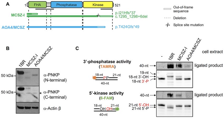
6676 Nucleic Acids Research, 2020, Vol. 48, No. 12
Downloaded from https://academic.oup.com/nar/article/48/12/6672/5854143 by guest on 01 December 2020
Figure 2. A normal rate of DSBR in PNKP patient-derived fibroblasts. (A) Representative scanR images (top) and quantification (bottom) of ␥ H2AX in
control (1BR) and PNKP patient-derived (MCSZ-I and AOA4/MCSZ) fibroblasts before and at the indicated times after ionizing radiation (2 Gy) in the
absence or presence of 5 M DNA-PK inhibitor (DNA-PKi was added 1 h before irradiation). Cell cycle populations were gated according to PCNA
positivity (S phase) and DNA content (G1 and G2) by DAPI staining. Only the data for G1 cells are shown. For quantification, data are normalized to
the 0.5 h time point and are the mean (±SEM) of three independent experiments. Statistical significance was determined by two-way ANOVA (ns, not
significant; ***P < 0.001). (B) DNA 3 -phosphatase activity and western blotting of cellular extracts from control 1BR and PNKP patient fibroblasts
(MCSZ-I and AOA4/MCSZ) transfected with non-target (NT) or PNKP siRNA. Oligonucleotide duplex substrate containing a SSB with a 3’-phosphate
terminus was incubated with cellular extracts for 10 min. (C) Quantification of ␥ H2AX in the indicated cell lines before and at the different times after
ionizing radiation (2 Gy). Cells were transfected with non-targeting siRNA (NT) or PNKP siRNA 48–72 h prior to irradiation. Quantification and statis-
tics were conducted as described in (A). Representative scanR images are shown in Supplementary Figure S1C. (D) Representative scanR images (left)
and quantification (right) of ␥ H2AX in the indicated cell lines after a 45 min incubation with DMSO vehicle or 10 M camptothecin (CPT). Data are
normalized to DMSO-treated control (1BR) cells and are the mean (±SEM) of three independent experiments. Statistical significance was determined by
one-tailed t-test (ns, not significant).Nucleic Acids Research, 2020, Vol. 48, No. 12 6677
sensitive immunofluorescence-based assay provides an indi- Whereas transfection with His-PNKPWT partially com-
rect measure of the level of chromosomal SSBs, analogous plemented the SSBR defect in AOA4/MCSZ patient fi-
to the measurement of ␥ H2AX as a marker of DSBs. In- broblasts following IR, as measured by a decrease in the
deed, incubation for short periods with PARG inhibitor un- level of nuclear ADP-ribose 30 min following irradiation,
covered a significant increase in the level of nuclear ADP- transfection with His-PNKPPD failed to do so (Figure 4B).
ribose in control 1BR fibroblasts following treatment with This is consistent with the presence of 3 -phosphate at
2 Gy of IR, and as expected this level was much higher in ∼70% of oxidative DNA breaks (14). We also examined
AOA-XRCC1 patient-derived fibroblasts in which SSBR is which PNKP activities were required to correct the de-
known to be reduced (7) (Figure 3A). More importantly, fect in repair of TOP1-induced SSBs, since this type of
ADP-ribose was also greatly elevated in the AOA4/MCSZ SSB possess both 3 -phosphate and 5 -hydroxyl termini
patient fibroblasts, indicative of a similar defect in SSBR (15). Surprisingly, both His-PNKPWT and His-PNKPPD
in these cells (Figure 3A). We also measured the level of completely suppressed the elevated accumulation of TOP1-
DSBs in these experiments in parallel, but did not detect induced SSBs in AOA4/MCSZ patient fibroblasts follow-
Downloaded from https://academic.oup.com/nar/article/48/12/6672/5854143 by guest on 01 December 2020
a difference between control and PNKP patient fibroblasts ing CPT treatment, as measured both by levels of nuclear
(Supplementary Figure S2A). Notably, we also detected a ADP-ribose and by alkaline comet assays (Figure 4C and
reduced rate of SSBR in both AOA4/MCSZ and MCSZ- D). Similar results were obtained if we employed pEYFP-
I patient fibroblasts in experiments in which we employed tagged PNKP cDNA expression constructs, instead of re-
a 10-fold higher dose of IR (20 Gy), to measure levels of combinant protein (Supplementary Figure S3A). This re-
poly(ADP-ribose) in the absence of PARG inhibitor (Sup- sult indicates that only the DNA 5 -kinase activity of PNKP
plementary Figure S2B). is rate limiting for rapid repair of TOP1-induced SSBs
To confirm the defect in SSBR in PNKP patient-derived in AOA4/MCSZ fibroblasts. Notably, recombinant His-
fibroblasts, we also quantified DNA strand breaks follow- PNKPFHA harbouring a mutated FHA domain failed to
ing IR directly, using alkaline comet assays. Whilst this restore full repair capacity in AOA4/MCSZ fibroblasts fol-
assay detects both SSBs and DSBs, >95% of the DNA lowing either IR or CPT, confirming that the recruitment of
strand breaks induced by IR are SSBs (49). Importantly, PNKP by XRCC1 is important for SSBR following either
in agreement with the nuclear ADP-ribose assay, the rate oxidative stress or abortive TOP1 activity. Note that the in-
at which SSBs declined following 20 Gy of IR was signif- ability of transfected His-PNKPFHA to restore SSBR was
icantly slower both in XRCC1 and PNKP patient-derived not due to differences in the level of transfected protein, be-
fibroblasts, when compared to the 1BR control (Figure 3B). cause protein extracts prepared from cells transfected with
Finally, we also measured the induction and repair of SSBs either His-PNKPWT or His-PNKPFHA possessed similar
induced by the abortive activity of TOP1, following treat- levels of PNKP biochemical activity, as determined by their
ment with CPT. Importantly, SSBs accumulated to a much ability to repair of an oligonucleotide substrate containing
higher level in both of the PNKP patient-derived fibroblasts both 5 -hydroxyl and 3 -phosphate termini (Supplementary
during incubation with CPT, when compared to 1BR con- Figure S3B). Together, these data indicate a collective re-
trol cells, as measured by either nuclear ADP-ribose levels quirement for all three domains of PNKP for efficient repair
or by alkaline comet assays (Figure 3C and D). We con- of the range of SSBs that arise in cells.
clude from these experiments that whilst AOA4/MCSZ and
MCSZ-I cells exhibit normal rates of DSBR, they possess
SSBR functionality and the severity of PNKP-associated dis-
reduced rates of SSBR.
ease
Next, we examined whether the severity of PNKP-
Requirement for the DNA 5 -kinase, DNA 3 -phosphatase,
associated disease is related to the level of residual PNKP
and FHA Domains of PNKP for SSBR
protein and/or activity during SSBR. To address this, we
To examine which PNKP activities are responsible for first compared the level of PNKP protein in cells derived
the SSBR defect in PNKP-mutated disease we conducted from individuals with MCSZ with those from individu-
complementation experiments, by transfecting purified als with AOA4 or CMT2B2; the milder forms of PNKP-
wild type and mutant recombinant PNKP proteins into mutated disease (Figure 5A). Whilst, PNKP protein lev-
AOA4/MCSZ fibroblasts. AOA4/MCSZ fibroblasts were els were greatly reduced in most of the patient-derived
employed because they possess the least amount of resid- LCLs and fibroblasts, there was no correlation between
ual DNA 3 -phosphatase activity and completely lack resid- the level of residual PNKP protein and pathological sever-
ual DNA 5 -kinase activity. As expected, recombinant ity (Figure 5B). For example, the level of PNKP protein
wild type histidine-tagged PNKP (His-PNKPWT ) and His- in the CMT2B2 cell lines was generally no higher than in
PNKPFHA harbouring a mutated FHA domain possessed the AOA4-I and MCSZ-I cell lines, despite the far milder
both DNA 5 -kinase activity and DNA 3 -phosphatase pathology associated with the former. Similarly, we did not
activity, whereas His-PNKPPD harbouring a mutated 3 - observe any strong correlation between disease severity and
phosphatase domain lacked the latter (Figure 4A). We also the level of residual DNA 5 -kinase or DNA 3 -phosphatase
generated recombinant PNKP protein containing a point activity (Supplementary Figure S4A). For example, the level
mutation (K378A) that greatly reduces or ablates DNA ki- of residual 3 -phosphatase activity in the CMT2B2 cell lines
nase activity, but this protein was relatively unstable when was generally no higher than in cell lines from the more se-
transfected into cells and so was not utilized further (un- vere diseases AOA4 and MCSZ, and the level of 5 -kinase
published observations). activity in MCSZ-I was higher than in cell lines from the6678 Nucleic Acids Research, 2020, Vol. 48, No. 12
Downloaded from https://academic.oup.com/nar/article/48/12/6672/5854143 by guest on 01 December 2020
Figure 3. A reduced rate of SSBR in PNKP patient-derived fibroblasts. (A) DNA single-strand breaks quantified by measuring nuclear ADP-ribose levels
in control (1BR) and PNKP patient-derived (AOA4/MCSZ) or XRCC1 patient-derived (AOA-XRCC1) fibroblasts before and at the indicated times
after treatment with ionizing radiation (2 Gy). Where indicated, cells were incubated in the presence of 10 M PARG inhibitor (PARGi) 30 min prior
to and after IR to suppress poly(ADP-ribose) degradation. Data were quantified by scanR high-content imaging and are the mean (±SEM) of three
independent experiments, with statistical significance determined by two-way ANOVA (***P < 0.001). Representative scanR images of single cell galleries
(from samples employing PARG inhibitor) are also shown (bottom). (B) DNA strand breaks quantified by alkaline comet assays in the indicated primary
fibroblasts before and at the indicated times after ionizing radiation (20 Gy). Data are the individual comet tail moments of 300 cells per sample combined
from three independent experiments. The horizontal bars show the mean tail moment of the 300 cells. Statistically significant differences were determined
using the individual means from the three independent experiments (100 cells/sample) by two-way ANOVA (***P < 0.001). (C) DNA strand breaks
quantified by measuring nuclear ADP-ribose levels in control (1BR) and PNKP patient-derived (MCSZ-I & AOA4/MCSZ) fibroblasts after incubation
for 45 min with DMSO vehicle or with 10 M camptothecin (CPT). Representative scanR images (left) and quantification (right) are shown. Data are
normalized to DMSO-treated 1BR cells and are the mean (±SEM) of seven independent experiments. Statistical analysis (one-tailed t-test) is indicated
(***P < 0.001; **P < 0.01). (D) DNA strand breaks quantified by alkaline comet assays in the indicated cell lines treated with DMSO vehicle or with 10
M CPT for 60 min. Data are as described in (B). Statistical significance was determined by one-tailed t-test (**P < 0.01; *P < 0.05).Nucleic Acids Research, 2020, Vol. 48, No. 12 6679
Downloaded from https://academic.oup.com/nar/article/48/12/6672/5854143 by guest on 01 December 2020
Figure 4. Importance of the PNKP DNA 5 -kinase, 3 -phosphatase, and FHA Domains for SSBR. (A) Cartoon (left) and enzymatic activity (right)
of the recombinant PNKP proteins employed here for complementation experiments. For activity assays, a 5 -TAMRA- and 3 -6-FAM- dual-labelled
oligonucleotide duplex (shown bottom) harbouring a SSB with both 3 -phosphate and 5 -hydroxyl termini was incubated for 10 min with the indicated
purified recombinant PNKP proteins prior to fractionation by denaturing PAGE. The positions of the TAMRA-labelled 3 -phosphatase substrate (‘18-nt 3 -
P’), 6-FAM-labelled 5 -kinase substrate (‘21-nt 5 -OH’), and the products of their processing by PNKP 3 -phosphatase activity (‘18-nt 3 -OH’) and 5 -kinase
activity (‘21-nt 5 -P’), respectively, are shown. (B) Representative scanR images (left) and quantification (right) of nuclear ADP-ribose in control fibroblasts
(1BR) and AOA4/MCSZ patient-derived fibroblasts 30 min following ionizing radiation (2 Gy) in the absence or presence of 10 M PARG inhibitor
(PARGi) as indicated. PARGi was added 30 min prior to irradiation. Cells were transfected with BSA or the indicated purified wild-type (PNKPWT ) or
mutant (PNKPPD , PNKPFHA ) PNKP proteins 18 h prior to irradiation. Data are normalized to DMSO-treated 1BR fibroblasts transfected with BSA
and are the mean (±SEM) of three independent experiments. Statistically significant differences were determined by one-tailed t-test (*P < 0.05; ns, not
significant). (C) Representative scanR images (top) and quantification (bottom) of nuclear ADP-ribose in the indicated cell lines after 45 min incubation
with DMSO vehicle or 10 M camptothecin (CPT). Cells were transfected in the presence of the indicated proteins 18 h prior to CPT treatment. Data are
as described in (B). (D) DNA strand breaks quantified by alkaline comet assays in the indicated cell lines following transfection with BSA or the indicated
recombinant PNKP proteins and treated 18 h later with DMSO vehicle or 10 M CPT for 60 min. Data are the individual comet tail moments of 300
cells per sample combined from three independent experiments. The horizontal bars show the mean tail moment. Statistically significant differences were
determined using the means from the three independent experiments (100 cells/sample by one-tailed t-test (*P < 0.05; **P < 0.01; ns, not significant).6680 Nucleic Acids Research, 2020, Vol. 48, No. 12
Downloaded from https://academic.oup.com/nar/article/48/12/6672/5854143 by guest on 01 December 2020
Figure 5. PNKP functionality and disease severity. (A) Cartoon depicting the location and type of PNKP mutations present in the MCSZ, AOA4 and
CMT2B2 patient-derived cell lines employed in this study. The details of the mutant alleles and the relevant citations are described in the materials and
methods. For homozygous mutations only one allele is depicted. (B) Western blot showing PNKP protein levels in the indicated control and PNKP patient-
derived fibroblasts (left) and LCLs (right).  actin was employed as a loading control. (C) Quantification of nuclear ADP-ribose in the indicated control
and PNKP patient-derived cells after 45 min incubation with DMSO vehicle or 10 M camptothecin (CPT). Data are normalized to DMSO-treated WT
cells and are the mean (±SEM) of four independent experiments. Statistically significant differences were determined by one-tailed t-test (**P < 0.01; *P
< 0.05). Representative scanR images are shown in Supplementary Figure S4B.
less severe diseases AOA4 and CMT2B2. Consequently, we lectively, these data implicate reduced rates of SSBR as a
employed the sensitive nuclear ADP-ribose assay to exam- cause of PNKP-associated disease, and suggest that the ex-
ine whether disease severity correlated with the overall ef- tent of this reduction is a determinant of disease severity.
ficiency of SSBR in the different patient-derived cell lines.
Indeed, we detected elevated levels of ADP-ribose in all DISCUSSION
of the patient-derived cell lines when compared to control
cells following treatment with CPT, and the level of CPT- Reduced rates of SSBR, but not DSBR, in PNKP-mutated
induced nuclear ADP-ribose in MCSZ and AOA4 cell lines disease
was higher than in cell lines from the far less severe disease, Mutations in PNKP result in several diseases spanning
CMT2B2 (Figure 5C and Supplementary Figure S4B). Col- a spectrum of neurological pathology, from neurodevel-Nucleic Acids Research, 2020, Vol. 48, No. 12 6681
opmental dysfunction in microcephaly with early-onset disease, and suggest that the severity of this disease is related
seizures (MCSZ) through to moderate or mild/late-onset to the level of SSBR capacity.
neurodegeneration in AOA4 and CMT2B2, respectively. A
possible explanation why mutations in PNKP might cause
MCSZ and reduced DNA 3 -phosphatase functionality
both neurodegeneration and neurodevelopmental dysfunc-
tion was the putative role for this protein in the repair It is currently unclear which of the two enzymatic activi-
of both SSBs and DSBs. Surprisingly, however, we found ties of PNKP contribute most to the neuroprotective role
here that primary fibroblasts derived from individuals with of this protein during SSBR. To address this, we compared
MCSZ or with combined AOA4/MCSZ exhibit normal patient-derived cell lines from the different PNKP-mutated
rates of DSBR following treatment with either ionising ra- diseases for levels of PNKP protein and its two biochemi-
diation (IR) or camptothecin (CPT); physiologically rele- cal activities. However, we failed to detect a correlation be-
vant sources of DSBs harbouring the 5 -hydroxyl and/or 3 - tween disease severity and any of these parameters individ-
phosphate termini that are substrates for PNKP (15). The ually. It is possible that other factors contribute to disease
Downloaded from https://academic.oup.com/nar/article/48/12/6672/5854143 by guest on 01 December 2020
lack of a significant DSBR defect in PNKP-mutated cells severity, such as an additive impact of the reduction in the
was surprising, because PNKP interacts with the critical two PNKP catalytic activities or as yet unidentified differ-
NHEJ protein XRCC4 (19,21,50) and has been reported ences in other DNA repair pathways. Alternatively, perhaps
to be required for efficient NHEJ both in vitro and in cells the level of residual PNKP activity detected in vitro fails to
(19,20,42,51). It is unlikely that this result reflects a limi- fully reflect the functionality of the mutant protein in cells.
tation of our ␥ H2AX assay, because we readily detected Indeed, consistent with this idea, only cell lines from pa-
the expected defect in DSBR if the cells were incubated tients with MCSZ exhibited a reduced ability to repair chro-
with an inhibitor of the critical NHEJ protein, DNA-PKcs. mosomal SSBs induced by IR [this work and (54)]. Since
Whilst we cannot rule out that the pathogenic mutations in IR induces SSBs with 3 -phosphate termini this observa-
PNKP result in measurable defects in DSBR specifically in tion suggests that the DNA phosphatase functionality of
neurones this seems unlikely, because the defect in NHEJ PNKP is lower in MCSZ cells than in AOA4 and CMT2B2
in similar microcephalic diseases resulting from mutations cells, despite the lack of a detectable difference in their DNA
in XRCC4, XLF/Cernnunos, or DNA ligase IV is read- phosphatase activity in cell extracts in vitro. One possible
ily detected using patient-derived fibroblasts and assays of explanation for this discrepancy is that our biochemical as-
the type employed here (38,40,52). One possible explana- says are not sufficiently sensitive to detect pathologically
tion for our results is that the residual PNKP activity that relevant differences between the DNA phosphatase activity
is present in the patient-derived fibroblasts is sufficient to of the different patient cell lines. Alternatively, perhaps the
maintain DSBR proficiency. Perhaps consistent with this large truncation mutations that are common in MCSZ af-
idea, we noted that PNKP siRNA slightly increased the fect not only the catalytic activity of PNKP but also its abil-
level of residual ␥ H2AX in AOA4/MCSZ cells following ity to be recruited and and/or stimulated by XRCC1. This
IR, albeit not statistically significantly. Alternatively, per- latter idea may explain why MCSZ-I cells, which retain rel-
haps other DNA 3 -end processing proteins can substitute atively high levels of residual PNKP activity but which har-
for PNKP during NHEJ (53). Irrespective of why DSBR bour a mutated FHA domain, possess a defect in SSBR fol-
was largely normal in the PNKP patient-derived fibrob- lowing IR. However, irrespective of why there is a discrep-
lasts employed here, our experiments suggest that a defect in ancy between our biochemical and cellular assays, the ob-
DSBR is unlikely to be the cause of PNKP-mutated disease. servation that only MCSZ cell lines exhibit a defect in repair
In contrast to DSBR, the proficiency of SSBR in MCSZ of IR-induced SSBs implicates reduced PNKP-dependent
patient-derived fibroblasts was greatly reduced following ei- DNA phosphatase activity in the development of MCSZ.
ther IR or CPT. This is consistent with our previous analy- In contrast to reduced DNA phosphatase activity, an
ses, in which we detected similar defects in SSBR in lym- involvement of reduced DNA kinase activity in causing
phoblastoid cell lines derived from different MCSZ pa- MCSZ is less likely. For example, the AOA4 cell lines
tients (54). The defect in SSBR detected here was appar- (AOA4-I and AOA4-II) exhibited levels of SSBR following
ent whether we quantified SSBs directly by alkaline comet CPT as low as those in MCSZ cells. Since we found here
assays or indirectly by measuring levels of nuclear ADP- that reduced SSBR following CPT is a measure of reduced
ribose. The additional use of the nuclear ADP-ribose assay 5 -DNA kinase function this suggests that reduced DNA
was important, because the greater sensitivity of this assay kinase activity does not, by itself at least, cause MCSZ.
allowed us to measure SSBs at the same low dose of IR This conclusion is also consistent with our previous obser-
as that employed for measuring DSBs (2 Gy). In contrast vation that the PNKP point mutation E326K, which results
to cell lines derived from patients with MCSZ however, we in MCSZ, reduces the proficiency of SSBR following IR but
failed to detect any defect in SSBR following IR in the less not CPT (54).
severe diseases AOA4 or CMT2B2 (unpublished observa-
tions). However, we did detect a defect in SSBR in AOA4
Neurodegeneration and reduced DNA 5 -kinase functionality
and CMT2B2 cell lines following treatment with CPT, using
this assay. Moreover, the defect in CMT2B2 cells following In contrast to the neurodevelopmental pathology that
CPT was less than that detected in either MCSZ or AOA4 typifies MCSZ, it seems unlikely that reduced DNA 3 -
cell lines, consistent with CMT2B2 being pathologically the phosphatase functionality can account for the neurodegen-
mildest of these diseases. Collectively, these experiments im- erative pathology that typifies AOA4 and CMT2B2. For ex-
plicate slower rates of SSBR as a cause of PNKP-mutated ample, as discussed above, whilst we readily detected a de-6682 Nucleic Acids Research, 2020, Vol. 48, No. 12
fect in the repair of IR-induced SSBs with 3 -phosphate ter- Author contributions: I.K. designed and performed most of
mini in MCSZ cells, we failed to do so in cell lines from ei- the experiments and wrote the first draft of the manuscript.
ther AOA4 or CMT2B2; the PNKP-mutated diseases that R.H. expressed and purified recombinant PNKP proteins.
are associated with neurodegeneration (data not shown). W.H. and J.B. generated and provided AOA4-II LCLs. A.L.
Indeed, our data suggest that it is reduced DNA 5 -kinase and J.B. identified CMT2B2 patients and obtained blood
activity that is the major contributor and/or cause of the samples and skin biopsies. H.H. conducted preliminary im-
neurodegeneration in PNKP-mutated disease, because we munofluorescence experiments and supervised the work.
detected reduced SSBR in all of our AOA4 and CMT2B2 H.H. and K.W.C. conceived the study, managed the project
cell lines following treatment with CPT, which as discussed and wrote the manuscript.
above is a measure of DNA 5 -kinase functionality. More-
over, the extent of this SSBR defect was greater in AOA4 FUNDING
than in CMT2B2 cells, consistent with the relative sever-
ity and/or age of onset of neurodegeneration in these dis- ERC Advanced Investigator Award (SIDSCA) [694996]
Downloaded from https://academic.oup.com/nar/article/48/12/6672/5854143 by guest on 01 December 2020
eases. If reduced DNA 5 -kinase is a cause of neurodegener- to KWC. MRC Programme Grant [MR/P010121/1] to
ation in PNKP-mutated disease then this pathology should K.W.C. K.W.C. is the recipient of a Royal Society
also be present in most patients with MCSZ, most of which Wolfson Research Merit Award. Access to the Olym-
based on our analyses harbor residual DNA kinase activity pus scanR and Leica microscopes at the Light Mi-
and rates of CPT-induced SSBR as low or lower than AOA4 croscopy Core Facility, IMG CAS, Prague; C.Z. was
and CMT2B2. Indeed, while cerebellar atrophy and ataxia supported by MEYS [LM2015062 Czech-BioImaging];
was not initially reported as a feature of MCSZ, more recent ERDF [CZ.02.1.01/0.0/0.0/16 013/0001775]; OPPK
case reports have reported this pathology (26–30). [CZ.2.16/3.1.00/21547]; NPU I [LO1419]. A.L. was sup-
Finally, our data have implications concerning the iden- ported by the Vice-Presidency for Research, University of
tity of the endogenous SSBs that trigger neurological dys- Costa Rica [Project 111-B8-372]. Funding for open access
function in PNKP-mutated disease. For example, if reduced charge: ERC; MRC.
3 -phosphatase is indeed the cause of neurodevelopmen- Conflict of interest statement. None declared.
tal dysfunction these data implicate SSBs arising from ox-
idative stress in MCSZ, because this is a major source of REFERENCES
DNA breaks with 3 -phosphate termini. Similarly, if re- 1. Caldecott,K.W. (2014) DNA single-strand break repair. Exp. Cell
duced DNA 5 -kinase activity is a cause of cerebellar ataxia Res., 329, 2–8.
and neurodegeneration in PNKP-mutated disease our re- 2. Scully,R., Panday,A., Elango,R. and Willis,N.A. (2019) DNA
sults implicate SSBs arising from abortive TOP1 activity double-strand break repair-pathway choice in somatic mammalian
cells. Nat. Rev. Mol. Cell. Biol., 20, 698–714.
in AOA4 and CMT2B2, because this is a major source 3. Tubbs,A. and Nussenzweig,A. (2017) Endogenous DNA damage as a
of DNA breaks with 5 -hydroxyl termini. The latter ob- source of genomic instability in cancer. Cell, 168, 644–656.
servation is consistent with the molecular defect in the re- 4. McKinnon,P.J. and Caldecott,K.W. (2007) DNA strand break repair
lated SSBR-defective neurodegenerative disease, SCAN1, and human genetic disease. Annu. Rev. Genom. Hum. G, 8, 37–55.
5. Yoon,G. and Caldecott,K.W. (2018) Nonsyndromic cerebellar ataxias
which is similarly required for the repair of SSBs induced associated with disorders of DNA single-strand break repair. Handb.
by abortive TOP1 activity (8,55). It is also consistent with Clin. Neurol., 155, 105–115.
recent reports that the repair of TOP1-induced SSBs is re- 6. Caldecott,K.W. (2008) Single-strand break repair and genetic disease.
duced in ataxia telangiectasia; the archetypal neurodegen- Nat. Rev. Genet., 9, 619–631.
erative disease associated with defects in DNA strand break 7. Hoch,N.C., Hanzlikova,H., Rulten,S.L., Tetreault,M.,
Komulainen,E., Ju,L., Hornyak,P., Zeng,Z., Gittens,W., Rey,S.A.
repair (56,57). et al. (2016) XRCC1 mutation is associated with PARP1
In summary, our data implicate reduced SSBR, but not hyperactivation and cerebellar ataxia. Nature, 541, 87–91.
DSBR, as a cause of PNKP-mutated disease, and suggest 8. Takashima,H., Boerkoel,C.F., John,J., Saifi,G.M., Salih,M.A.M.,
that the pathological severity of this disease is determined Armstrong,D., Mao,Y., Quiocho,F.A., Roa,B.B., Nakagawa,M. et al.
(2002) Mutation of TDP1, encoding a topoisomerase I-dependent
by the nature and extent of this reduction. Moreover, our DNA damage repair enzyme, in spinocerebellar ataxia with axonal
data also highlight the two enzymatic activities of PNKP neuropathy. Nat. Genet., 32, 267–272.
in different aspects of the disease pathology, with reduced 9. Moreira,M.C., Barbot,C., Tachi,N., Kozuka,N., Uchida,E.,
DNA phosphatase and DNA kinase activity correlating Gibson,T., Mendonça,P., Costa,M., Barros,J., Yanagisawa,T. et al.
with neurodevelopmental dysfunction and neurodegenera- (2001) The gene mutated in ataxia-ocular apraxia 1 encodes the new
HIT/Zn-finger protein aprataxin. Nat. Genet., 29, 189–193.
tion, respectively. 10. Date,H., Onodera,O., Tanaka,H., Iwabuchi,K., Uekawa,K.,
Igarashi,S., Koike,R., Hiroi,T., Yuasa,T., Awaya,Y. et al. (2001)
Early-onset ataxia with ocular motor apraxia and hypoalbuminemia
SUPPLEMENTARY DATA is caused by mutations in a new HIT superfamily gene. Nat. Genet.,
29, 184–188.
Supplementary Data are available at NAR Online. 11. Dumitrache,L.C. and McKinnon,P.J. (2016) Polynucleotide
kinase-phosphatase (PNKP) mutations and neurologic disease.
Mech. Ageing Dev., 161, 121–129.
ACKNOWLEDGEMENTS 12. Karimi-Busheri,F., Daly,G., Robins,P., Canas,B., Pappin,D.J.C.,
This work used instruments provided by C4Sys infrastruc- Sgouros,J., Miller,G.G., Fakhrai,H., Davis,E.M., Beau,M.M.L. et al.
(1999) Molecular Characterization of a Human DNA Kinase. J. Biol.
ture. We thank Martin Paucar, John Reynolds and Luis Chem., 274, 24187–24194.
Bermudez for thoughtful and helpful discussions, and ad- 13. Jilani,A., Ramotar,D., Slack,C., Ong,C., Yang,X.M., Scherer,S.W.
ditionally for the provision of AOA4-I cells. and Lasko,D.D. (1999) Molecular cloning of the human gene, PNKP,Nucleic Acids Research, 2020, Vol. 48, No. 12 6683
encoding a polynucleotide kinase 3 -phosphatase and evidence for its 31. Pedroso,J.L., Rocha,C.R.R., Macedo-Souza,L.I., Mario,V.D.,
role in repair of DNA strand breaks caused by oxidative damage. J. Marques,W., Barsottini,O.G.P., Oliveira,A.S.B., Menck,C.F.M. and
Biol. Chem., 274, 24176–24186. Kok,F. (2015) Mutation in PNKP presenting initially as axonal
14. Henner,W.D., Grunberg,S.M. and Haseltine,W.A. (1982) Sites and Charcot-Marie-Tooth disease. Neurol. Genet., 1, e30.
structure of gamma radiation-induced DNA strand breaks. J. Biol. 32. Leal,A., Bogantes-Ledezma,S., Ekici,A.B., Uebe,S., Thiel,C.T.,
Chem., 257, 11750–11754. Sticht,H., Berghoff,M., Berghoff,C., Morera,B., Meisterernst,M.
15. Pouliot,J.J., Robertson,C.A. and Nash,H.A. (2001) Pathways for et al. (2018) The polynucleotide kinase 3 -phosphatase gene (PNKP)
repair of topoisomerase I covalent complexes in Saccharomyces is involved in Charcot-Marie-Tooth disease (CMT2B2) previously
cerevisiae. Genes Cells, 6, 677–687. related to MED25. Neurogenetics, 19, 215–225.
16. Loizou,J.I., El-Khamisy,S.F., Zlatanou,A., Moore,D.J., Chan,D.W., 33. Previtali,S.C., Zhao,E., Lazarevic,D., Pipitone,G.B., Fabrizi,G.M.,
Qin,J., Sarno,S., Meggio,F., Pinna,L.A. and Caldecott,K.W. (2004) Manganelli,F., Mazzeo,A., Pareyson,D., Schenone,A., Taroni,F. et al.
The Protein Kinase CK2 Facilitates Repair of Chromosomal DNA (2019) Expanding the spectrum of genes responsible for hereditary
Single-Strand Breaks. Cell, 117, 17–28. motor neuropathies. J. Neurol. Neurosurg. Psychiatry, 90, 1171–1179.
17. Whitehouse,C.J., Taylor,R.M., Thistlethwaite,A., Zhang,H., 34. Kalasova,I., Hanzlikova,H., Gupta,N., Li,Y., Altmüller,J.,
Karimi-Busheri,F., Lasko,D.D., Weinfeld,M. and Caldecott,K.W. Reynolds,J.J., Stewart,G.S., Wollnik,B., Yigit,G. and Caldecott,K.W.
Downloaded from https://academic.oup.com/nar/article/48/12/6672/5854143 by guest on 01 December 2020
(2001) XRCC1 stimulates human polynucleotide kinase activity at (2019) Novel PNKP mutations causing defective DNA strand break
damaged DNA termini and accelerates DNA single-strand break repair and PARP1 hyperactivity in MCSZ. Neurol. Genet., 5, e320.
repair. Cell, 104, 107–117. 35. Scholz,C., Golas,M.M., Weber,R.G., Hartmann,C., Lehmann,U.,
18. Ali,A.A.E., Jukes,R.M., Pearl,L.H. and Oliver,A.W. (2009) Specific Sahm,F., Schmidt,G., Auber,B., Sturm,M., Schlegelberger,B. et al.
recognition of a multiply phosphorylated motif in the DNA repair (2018) Rare compound heterozygous variants in PNKP identified by
scaffold XRCC1 by the FHA domain of human PNK. Nucleic Acids whole exome sequencing in a German patient with ataxia-oculomotor
Res., 37, 1701–1712. apraxia 4 and pilocytic astrocytoma. Clin. Genet., 94, 185–186.
19. Chappell,C., Hanakahi,L.A., Karimi-Busheri,F., Weinfeld,M. and 36. Dobson,C.J. and Allinson,S.L. (2006) The phosphatase activity of
West,S.C. (2002) Involvement of human polynucleotide kinase in mammalian polynucleotide kinase takes precedence over its kinase
double-strand break repair by non-homologous end joining. EMBO activity in repair of single strand breaks. Nucleic Acids Res., 34,
J., 21, 2827–2832. 2230–2237.
20. Koch,C.A., Agyei,R., Galicia,S., Metalnikov,P., O’Donnell,P., 37. Bee,L., Nasca,A., Zanolini,A., Cendron,F., d’Adamo,P., Costa,R.,
Starostine,A., Weinfeld,M. and Durocher,D. (2004) Xrcc4 physically Lamperti,C., Celotti,L., Ghezzi,D. and Zeviani,M. (2015) A nonsense
links DNA end processing by polynucleotide kinase to DNA ligation mutation of human XRCC4 is associated with adult-onset progressive
by DNA ligase IV. EMBO J., 23, 3874–3885. encephalocardiomyopathy. EMBO Mol. Med., 7, 918–929.
21. Aceytuno,R.D., Piett,C.G., Havali-Shahriari,Z., Edwards,R.A., 38. Guo,C., Nakazawa,Y., Woodbine,L., Björkman,A., Shimada,M.,
Rey,M., Ye,R., Javed,F., Fang,S., Mani,R., Weinfeld,M. et al. (2017) Fawcett,H., Jia,N., Ohyama,K., Li,T.-S., Nagayama,Y. et al. (2015)
Structural and functional characterization of the XRCC4 deficiency in human subjects causes a marked neurological
PNKP-XRCC4-LigIV DNA repair complex. Nucleic Acids Res., 45, phenotype but no overt immunodeficiency. J. Allergy Clin. Immun.,
6238–6251. 136, 1007–1017.
22. Bernstein,N.K., Williams,R.S., Rakovszky,M.L., Cui,D., Green,R., 39. Ben-Omran,T.I., Cerosaletti,K., Concannon,P., Weitzman,S. and
Karimi-Busheri,F., Mani,R.S., Galicia,S., Koch,C.A., Cass,C.E. et al. Nezarati,M.M. (2005) A patient with mutations in DNA Ligase IV:
(2005) The molecular architecture of the mammalian DNA repair Clinical features and overlap with Nijmegen breakage syndrome. Am.
enzyme, polynucleotide kinase. Mol. Cell, 17, 657–670. J. Med. Genet. A, 137A, 283–287.
23. Shen,J., Gilmore,E.C., Marshall,C.A., Haddadin,M., Reynolds,J.J., 40. O’Driscoll,M., Cerosaletti,K.M., Girard,P.-M., Dai,Y., Stumm,M.,
Eyaid,W., Bodell,A., Barry,B., Gleason,D., Allen,K. et al. (2010) Kysela,B., Hirsch,B., Gennery,A., Palmer,S.E., Seidel,J. et al. (2001)
Mutations in PNKP cause microcephaly, seizures and defects in DNA Ligase IV mutations identified in patients exhibiting
DNA repair. Nat. Genet., 42, 245–249. developmental delay and immunodeficiency. Mol. Cell, 8, 1175–1185.
24. Bras,J., Alonso,I., Barbot,C., Costa,M.M., Darwent,L., Orme,T., 41. Buck,D., Malivert,L., Chasseval,R. de, Barraud,A.,
Sequeiros,J., Hardy,J., Coutinho,P. and Guerreiro,R. (2015) Fondanèche,M.-C., Sanal,O., Plebani,A., Stéphan,J.-L.,
Mutations in PNKP cause recessive ataxia with oculomotor apraxia Hufnagel,M., Deist,F. le et al. (2006) Cernunnos, a novel
type 4. Am. J. Hum. Genet., 96, 474–479. nonhomologous end-joining factor, is mutated in human
25. Paucar,M., Malmgren,H., Taylor,M., Reynolds,J.J., Svenningsson,P., immunodeficiency with microcephaly. Cell, 124, 287–299.
Press,R. and Nordgren,A. (2016) Expanding the ataxia with 42. Karimi-Busheri,F., Rasouli-Nia,A., Allalunis-Turner,J. and
oculomotor apraxia type 4 phenotype. Neurol. Genet., 2, e49. Weinfeld,M. (2007) Human polynucleotide kinase participates in
26. Poulton,C., Oegema,R., Heijsman,D., Hoogeboom,J., Schot,R., repair of DNA double-strand breaks by nonhomologous end joining
Stroink,H., Willemsen,M.A., Verheijen,F.W., van de Spek,P., but not homologous recombination. Cancer Res., 67, 6619–6625.
Kremer,A. et al. (2012) Progressive cerebellar atrophy and 43. Rogakou,E.P., Pilch,D.R., Orr,A.H., Ivanova,V.S. and Bonner,W.M.
polyneuropathy: expanding the spectrum of PNKP mutations. (1998) DNA double-stranded breaks induce histone H2AX
Neurogenetics, 14, 43–51. phosphorylation on serine 139. J. Biol. Chem., 273, 5858–5868.
27. Entezam,M., Razipour,M., Talebi,S., Toosi,M.B. and 44. Rogakou,E.P., Boon,C., Redon,C. and Bonner,W.M. (1999)
Keramatipour,M. (2018) Multi affected pedigree with congenital Megabase chromatin domains involved in DNA Double-Strand
microcephaly: WES revealed PNKP gene mutation. Brain Dev., 41, breaks in vivo. J. Cell Biol., 146, 905–916.
182–186. 45. Beucher,A., Birraux,J., Tchouandong,L., Barton,O., Shibata,A.,
28. Taniguchi-Ikeda,M., Morisada,N., Inagaki,H., Ouchi,Y., Takami,Y., Conrad,S., Goodarzi,A.A., Krempler,A., Jeggo,P.A. and Löbrich,M.
Tachikawa,M., Satake,W., Kobayashi,K., Tsuneishi,S., Takada,S. (2009) ATM and Artemis promote homologous recombination of
et al. (2018) Two patients with PNKP mutations presenting with radiation-induced DNA double-strand breaks in G2. EMBO J., 28,
microcephaly, seizure, and oculomotor apraxia. Clin. Genet., 93, 3413–3427.
931–933. 46. Hsiang,Y.H., Hertzberg,R., Hecht,S. and Liu,L.F. (1985)
29. Gatti,M., Magri,S., Nanetti,L., Sarto,E., Bella,D.D., Salsano,E., Camptothecin induces protein-linked DNA breaks via mammalian
Pantaleoni,C., Mariotti,C. and Taroni,F. (2019) From congenital DNA topoisomerase I. J. Biol. Chem., 260, 14873–14878.
microcephaly to adult onset cerebellar ataxia: Distinct and 47. Hanzlikova,H., Kalasova,I., Demin,A.A., Pennicott,L.E.,
overlapping phenotypes in patients with PNKP gene mutations. Am. Cihlarova,Z. and Caldecott,K.W. (2018) The importance of
J. Med. Genet. Part, 179, 2277–2283. Poly(ADP-Ribose) polymerase as a sensor of unligated okazaki
30. Rudenskaya,G., Marakhonov,A., Shchagina,O., Lozier,E., fragments during DNA replication. Mol. Cell, 71, 319–331.
Dadali,E., Akimova,I., Petrova,N. and Konovalov,F. (2019) Ataxia 48. Hanzlikova,H., Gittens,W., Krejcikova,K., Zeng,Z. and
with oculomotor apraxia type 4 with PNKP common “Portuguese” Caldecott,K.W. (2016) Overlapping roles for PARP1 and PARP2 in
and novel mutations in two belarusian families. J. Pediatric Genet., the recruitment of endogenous XRCC1 and PNKP into oxidized
08, 058–062. chromatin. Nucleic Acids Res., 45, 2546–2557.You can also read