Transcription Recovery after DNA Damage Requires Chromatin Priming by the H3.3 Histone Chaperone HIRA
←
→
Page content transcription
If your browser does not render page correctly, please read the page content below
Transcription Recovery after DNA
Damage Requires Chromatin Priming
by the H3.3 Histone Chaperone HIRA
Salomé Adam,1,2 Sophie E. Polo,1,2,3,* and Geneviève Almouzni1,2,*
1Chromatin   Dynamics, Institut Curie Research Centre, 75248 Paris Cedex 5, France
2Centre  National de la Recherche Scientifique, Unité Mixte de Recherche 218, 75248 Paris Cedex 5, France
3Present address: Epigenetics and Cell Fate Centre, UMR7216 CNRS/Université Paris Diderot, 75205 Paris Cedex 13, France

*Correspondence: sophie.polo@univ-paris-diderot.fr (S.E.P.), genevieve.almouzni@curie.fr (G.A.)
 http://dx.doi.org/10.1016/j.cell.2013.08.029

SUMMARY                                                               (Filipescu et al., 2013). However, whether H3.3 deposition drives
                                                                      or simply reflects transcriptional activity is still under debate.
Understanding how to recover fully functional and                     Recent data support the idea that chromatin marking by H3.3
transcriptionally active chromatin when its integrity                 incorporation is required for either the activation or the long-
has been challenged by genotoxic stress is a critical                 term maintenance of gene expression patterns (reviewed in
issue. Here, by investigating how chromatin dy-                       Skene and Henikoff, 2013; Szenker et al., 2011), illustrating the
namics regulate transcriptional activity in response                  importance of chromatin integrity in cell fate determination.
                                                                         DNA damage challenges chromatin integrity by eliciting the
to DNA damage in human cells, we identify a pathway
                                                                      destabilization of chromatin structure, followed by restoration
involving the histone chaperone histone regulator A                   of its organization (reviewed in Adam and Polo, 2012; Smerdon,
(HIRA) to promote transcription restart after UVC                     1991; Soria et al., 2012). In addition, in response to various types
damage. Our mechanistic studies reveal that HIRA                      of DNA lesions, a local inhibition of transcription ensues, as best
accumulates at sites of UVC irradiation upon detec-                   characterized for RNA polymerase II (RNAPII)-dependent tran-
tion of DNA damage prior to repair and deposits                       scription (Chou et al., 2010; Iacovoni et al., 2010; Moné et al.,
newly synthesized H3.3 histones. This local action                    2001; Pankotai et al., 2012; Shanbhag et al., 2010; Tornaletti,
of HIRA depends on ubiquitylation events associated                   2009). Such DNA damage-induced transcription arrest is critical
with damage recognition. Furthermore, we demon-                       to prevent the production of aberrant transcripts and to avoid
strate that the early and transient function of HIRA                  interference between transcription and repair machineries
in response to DNA damage primes chromatin for                        (Svejstrup, 2010). Much effort has been devoted to understand-
                                                                      ing how transcription inhibition is achieved, particularly in
later reactivation of transcription. We propose that
                                                                      response to bulky DNA lesions, which result in RNAPII stalling,
HIRA-dependent histone deposition serves as a                         followed by DNA damage bypass, RNAPII backtracking, or
chromatin bookmarking system to facilitate tran-                      RNAPII removal by proteasomal degradation (Hanawalt and
scription recovery after genotoxic stress.                            Spivak, 2008; Tornaletti, 2009). Displacement of stalled RNAPII
                                                                      allows completion of DNA repair and subsequent restart of tran-
INTRODUCTION                                                          scriptional activity in regions that have been silenced in response
                                                                      to DNA damage (Gaillard and Aguilera, 2013). However, what
To prevent the deleterious effects of DNA lesions that constantly     triggers and controls transcription restart once repair is com-
challenge genome integrity (Hoeijmakers, 2009), cells have            plete is still an open issue. Given the involvement of chromatin
developed mechanisms—collectively referred to as the DNA              marks in regulating gene expression and integrating damage sig-
damage response (DDR)—to signal and repair DNA damage                 nals (Li et al., 2007; Luijsterburg and van Attikum, 2011), it is
(Ciccia and Elledge, 2010; Giglia-Mari et al., 2011; Jackson          tempting to hypothesize that mechanisms involving chromatin
and Bartek, 2009). Beyond genome stability, the maintenance           marking could contribute to transcription recovery upon DNA
of a defined organization of the genome into chromatin, with spe-     damage.
cific patterns of histone variants and their marks (Talbert and He-      Significant progress has been made in characterizing key
nikoff, 2010), is critical for preserving genome functions and cell   players in chromatin dynamics that respond to DNA damage (re-
identity. Concerning histone H3 variants, as described in mam-        viewed in Soria et al., 2012). Among them, histone chaperones,
mals, the replicative variant H3.1 is incorporated into chromatin     which escort histones upon their synthesis up to their deposition
genome-wide in a manner coupled to DNA synthesis during               on to DNA (Burgess and Zhang, 2013; De Koning et al., 2007), are
replication and repair. In contrast, the replacement variant H3,      of particular interest. Several of these chaperones have been
while present at telomeric and pericentric heterochromatin,           involved in the DDR (reviewed in Avvakumov et al., 2011; Soria
has been strongly associated with actively transcribed genes          et al., 2012), including the H3 variant-specific chaperones

94 Cell 155, 94–106, September 26, 2013 ª2013 Elsevier Inc.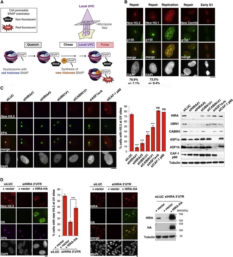
chromatin assembly factor 1 (CAF-1) and histone regulator A            upon HIRA knockdown cannot be due to a general loss of func-
(HIRA). Although recent data support a possible role for HIRA          tion of the transcription machinery, as attested by the levels and
in maintaining genome stability in yeast and human cells (Ander-       phosphorylation status of RNAPII large subunit, which return
son et al., 2009; Kapitzky et al., 2010; Yang et al., 2013), the       to normal 24 hr post-UVC irradiation in HIRA-depleted cells
actual function of HIRA in the DDR is still elusive. Indeed, HIRA      (Figure S1E).
is primarily characterized as a chaperone mediating the deposi-           Considering that these results may reflect a general role of
tion of H3.3 histone variants in transcribed chromatin regions (re-    HIRA associated to any disruption of transcription independent
viewed in Skene and Henikoff, 2013; Szenker et al., 2011). By          of DNA damage, we examined the effect of HIRA depletion on
contrast, CAF-1-mediated histone deposition is known to be             transcription in undamaged cells treated with 5,6-dichlorobenzi-
coupled to repair synthesis in response to DNA damage (Gaillard        midazole 1-b-D-ribofuranoside (DRB), a reversible transcription
et al., 1996). By means of its local recruitment to damaged chro-      inhibitor (Bensaude, 2011). Remarkably, in this setting, HIRA
matin regions where it promotes the incorporation of newly             downregulation did not affect transcription recovery (Figure S1F).
synthesized H3.1 histones, CAF-1 contributes to preserving             Thus, the capacity of HIRA to promote transcription restart after
chromatin organization during late repair steps in human cells         DNA damage is not a general response to transcription inhibi-
(Green and Almouzni, 2003; Polo et al., 2006).                         tion, and it may reflect a direct role of HIRA in UVC damage
   However, besides restoration of chromatin organization,             repair. To address this possibility, we first tested the ability of
whether and how chromatin dynamics could link with the recov-          HIRA-depleted cells to survive UVC damage by performing
ery of transcriptional activity in damaged regions is a critical but   clonogenic assays. Thus, we demonstrated that HIRA knock-
underappreciated question. Here, we explore the regulatory             down did not sensitize cells to UVC damage (Figure S1G). We
mechanisms of transcription recovery after DNA damage by               then examined more specifically the repair capacity of HIRA-
investigating how histone chaperones control transcription             depleted cells by measuring the removal kinetics of cyclobutane
restart in response to genotoxic stress. Our findings identify a       pyrimidine dimers (CPDs), the major UVC-induced DNA photo-
role of the H3.3 histone chaperone HIRA in priming damaged             products (Figure 2A). These photolesions are known to be
chromatin early after damage recognition, which is key to facili-      excised with slow kinetics (Ehmann et al., 1978); therefore, we
tating later reactivation of transcription once repair is complete.    focused our analysis on late time points after UVC (24–34 hr).
                                                                       Consistent with the fact that HIRA-depleted cells are not hyper-
RESULTS                                                                sensitive to UVC damage, we found that these cells did not
                                                                       display any significant delay in UVC damage removal when
Transcription Recovery after DNA Repair Involves the                   exposed to global irradiation (Figure 2A). In line with these find-
Histone Chaperone HIRA                                                 ings, the recruitment of the nucleotide excision repair factor
In order to address whether transcription regulation may be con-       Xeroderma Pigmentosum, complementation group B (XPB) to
nected to chromatin dynamics in response to DNA damage (Fig-           sites of local UVC irradiation was not affected by HIRA depletion,
ure 1A), we quantified nascent transcripts by 5-ethynyl uridine        and DNA repair synthesis revealed by 5-ethynyl 20 -deoxyuridine
(EU) labeling after global UV type C (UVC) irradiation in cells        (EdU) incorporation was similar to control (Figure 2B). Thus,
depleted of candidate histone chaperones (Figure 1B). Our              these results show that HIRA-depleted cells do not display over-
detailed kinetic analysis of nascent transcript production after       all defects in UV damage repair. Similar results were obtained
UVC irradiation revealed that transcription inhibition and recov-      upon CAF-1 and HIRA codepletion (data not shown), thus dis-
ery were best achieved at 2 hr and 24 hr postirradiation, respec-      carding the hypothesis of a functional redundancy between
tively (data not shown), which is consistent with previous reports     CAF-1 and HIRA in UVC damage repair. Collectively, our data
(Jensen and Mullenders, 2010; Nakazawa et al., 2010; Zhang             demonstrate that the histone chaperone HIRA plays a critical
et al., 2012). We thus selected these time points for subsequent       role in transcription recovery upon UVC damage and does so
analyses. The histone chaperone CAF-1, previously shown to             without significantly affecting the repair process per se.
be involved in the cellular response to UVC irradiation (reviewed
in Adam and Polo, 2012), did not affect transcription arrest and       HIRA Accumulation in Damaged Chromatin Regions
subsequent recovery after DNA damage as shown by depleting             To determine whether HIRA contribution to transcription recov-
its p60 subunit (Figure 1C; Table S1 [for small interfering RNAs,      ery after UVC irradiation involves a local action of this chaperone
siRNAs] and Table S2 [for antibodies]). Strikingly, HIRA downre-       at damage sites, we investigated the recruitment of HIRA to
gulation, while leaving transcription arrest unaffected, signifi-      damaged chromatin. As previously demonstrated (Green and
cantly impaired transcription recovery to an extent comparable         Almouzni, 2003; Polo et al., 2006), CAF-1 p60 and p150 subunits
to that observed in repair-deficient cells (excision-repair cross-     accumulated in regions of local UVC irradiation (Figure 2C).
complementing 6; siERCC6) (Figure 1C; Figure S1A available             Remarkably, we could detect all known subunits of the HIRA
online; note that knockdown efficiencies are comparable                complex—HIRA, ubinuclein 1 (UBN1) and calcineurin-binding
throughout the experiment). We verified that the inhibitory effect     protein 1 (CABIN1) (Amin et al., 2012)—enriched in damaged
of depleting HIRA on transcription recovery could not be ex-           chromatin regions (Figure 2C). By contrast, in our cell line model,
plained by significant alterations in cell cycle progression (Fig-     we did not observe any detectable recruitment of other known
ure S1B). Similar results obtained in U2OS cells (Figure S1D)          H3 histone chaperones (Burgess and Zhang, 2013), including
further indicate that our findings are not limited to a particular     death-domain associated protein (DAXX), nuclear autoantigenic
cell line. In addition, the defect in transcription restart observed   sperm protein (NASP), and antisilencing function 1 (ASF1)

                                                                           Cell 155, 94–106, September 26, 2013 ª2013 Elsevier Inc. 95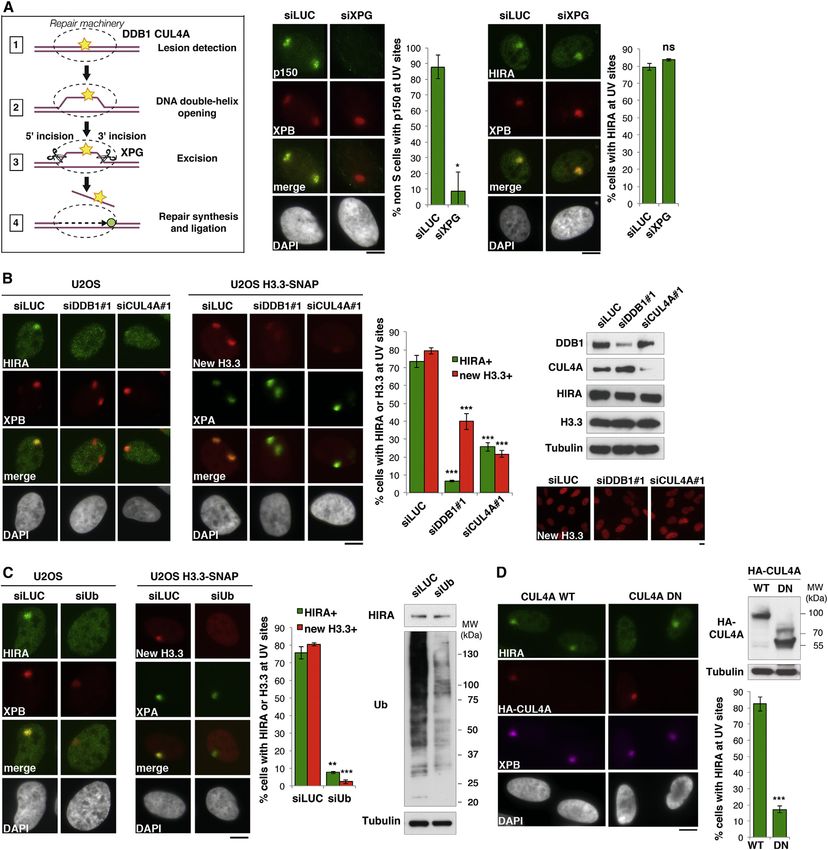
Figure 1. The Histone Chaperone HIRA Promotes Transcription Recovery after DNA Repair
(A) Both transcription activity (red) and chromatin organization are dynamically regulated at sites of DNA damage, raising the possibility that DNA damage-
induced chromatin rearrangements could be instrumental for transcription recovery after repair of DNA damage.
(B) Scheme of the assay for monitoring transcriptional regulation in response to global UVC irradiation. EU incorporation is measured by fluorescence microscopy
before (-), 2 hr after, and 24 hr after DNA damage.
(C) Transcriptional activity in UVC-irradiated HeLa cells treated with the indicated siRNAs (siLUC, control). Error bars on the graphs represent SEM from at least
two independent experiments. siRNA efficiencies are shown on the western blot panel. Black arrowhead, full-length ERCC6; white arrowhead, splice variant.
Scale bar, 10 mm.
See also Figure S1.

(Figure 2C). HIRA recruitment to DNA damage was observed in                          Taken together, these data put forward the histone chaperone
the vast majority of damaged cells, both in tumoral cells and                     complex HIRA as a UVC damage response factor that accumu-
primary fibroblasts (Figure 2C; Figure S2A). It occurred regard-                  lates locally in DNA damaged chromatin regions.
less of cell cycle stage and in a UVC dose-dependent manner
(Figures S2B and S2C). Individual depletion of HIRA complex                       HIRA-Mediated Accumulation of New H3.3 Histones in
subunits enabled us to establish that accumulation of the HIRA                    Damaged Chromatin Regions
subunit in damaged chromatin regions occurred independently                       Because the HIRA complex promotes de novo deposition of the
of UBN1 but was favored in the presence of CABIN1 (Fig-                           H3.3 variant into chromatin (reviewed in Szenker et al., 2011), we
ure S2D). Thus, besides the active role of UBN1 in stimulating                    examined new H3.3 histone deposition in damaged chromatin
the deposition activity of the HIRA subunit (Ray-Gallet et al.,                   regions. For this, we combined local UVC irradiation with specific
2011), we describe a function for CABIN1 in stabilizing HIRA at                   tracking of newly synthesized histones. We took advantage of
sites of DNA lesions.                                                             the SNAP-tag technology to fluorescently label new histones

96 Cell 155, 94–106, September 26, 2013 ª2013 Elsevier Inc.
(Bodor et al., 2012; see Experimental Procedures for details),          damaged chromatin regions to mediate H3.3 deposition and
using U2OS cells engineered to stably express SNAP-tagged               which parameters could control this key event. Given that
histone H3 variants (Dunleavy et al., 2011) (see Figure 3A and          H3.3 deposition by the HIRA complex has been linked to
Figure S3 for a characterization of the cell lines). Thus, we re-       RNAPII (Jullien et al., 2012; Placek et al., 2009; Ray-Gallet
vealed a local enrichment of newly synthesized H3.3 in UVC-             et al., 2011; Szenker et al., 2011; 2012; Yang et al., 2011), we
damaged chromatin, as observed in U2OS cells (Figure 3B),               first tested the effect of inhibiting transcription. However, cell
but also in HeLa cells stably expressing SNAP-tagged H3.3               treatment with RNAPII inhibitors such as a-amanitin or DRB
(data not shown). Similar to HIRA recruitment, new H3.3 histones        did not impair HIRA accumulation at sites of DNA damage (Fig-
accumulated in UVC-damaged regions in the vast majority of              ure S2G). Thus, HIRA recruitment to damaged chromatin re-
damaged cells and throughout interphase (Figures 3B and                 gions is not transcription dependent, arguing that HIRA is tar-
S2B). Furthermore, we reproduced these findings by monitoring           geted to DNA damage regardless of the transcription status
real-time accumulation of new H3.3 at sites of UVC laser micro-         of the region before damage infliction. To explore whether a
irradiation (Figure S2F). Although we also detected new H3.1            more specific connection with the DDR could be involved, we
histone accumulation both at repair sites and replication foci,         depleted candidate factors of the nucleotide excision repair
consistent with previous results (Polo et al., 2006; Ray-Gallet         (NER) pathway, which removes UVC damage (reviewed in
et al., 2011), we did not observe any detectable accumulation           Nouspikel, 2009) (Figure 4A). Although depletion of the endonu-
of newly synthesized CenH3, a centromeric histone variant (Fig-         clease xeroderma pigmentosum group G (XPG) abrogated
ure 3B). These data highlight that not all new histone H3 variants      CAF-1 recruitment to UVC damage because it impaired repair
are equally mobilized in response to DNA damage.                        synthesis (Green and Almouzni, 2003), HIRA accumulation still
   In order to characterize the molecular determinants of new           occurred in damaged chromatin regions (Figure 4A). These
H3.3 accumulation in UVC-damaged regions, we analyzed the               data indicate that HIRA recruitment is not coupled to late repair
effect of depleting candidate histone H3 chaperones. We                 steps. However, depletion of early NER factors involved in the
observed a significant reduction of H3.3 de novo accumulation           recognition of UVC damage, such as Cullin4A (CUL4A) and
in damaged chromatin regions in cells depleted of HIRA complex          DNA damage binding proteins 1 and 2 (DDB1 and DDB2,
subunits, with the most severe effect occurring on depletion of         respectively) (Nouspikel, 2009), markedly impaired HIRA
the HIRA subunit itself (Figure 3C), as confirmed with an indepen-      recruitment to damaged chromatin and de novo H3.3 accumu-
dent set of siRNAs (Figure S4A). This is consistent with the cen-       lation in damaged regions without affecting HIRA total levels
tral role of this protein in the overall stability of the complex, as   and H3.3 production (Figure 4B; Figure S5A and S5B for a sec-
shown by the reduced levels of the other subunits in HIRA-              ond set of siRNAs). Although HIRA targeting to DNA damage
depleted cells (Figure 3C; Ray-Gallet et al., 2011). In addition,       could involve an association—direct or indirect—between
expression of an siRNA-resistant HIRA construct restored the            HIRA complex subunits and these early NER proteins, as
accumulation of new H3.3 histones in UVC-damaged chromatin              recently reported for CABIN1 (Choi et al., 2013), it may also
regions (Figure 3D). These results establish that the defective         require the E3 ubiquitin ligase activity of DDB1-CUL4A-contain-
accumulation of H3.3 in damaged chromatin is a direct and spe-          ing complexes (reviewed in Nouspikel, 2011; Figure S6A).
cific consequence of HIRA loss of function. We also found some          Consistent with this possibility, by preventing all ubiquitylation
defect in H3.1 deposition in damaged chromatin regions on               reactions using RNA interference against ubiquitin, we inhibited
depletion of HIRA, indicating a more general impact of this             both HIRA and new H3.3 accumulation in UVC-damaged chro-
chaperone on chromatin integrity at damage sites (Figure S4B).          matin without any significant effect on HIRA total levels
In contrast to HIRA subunits, depletion of any of the other chap-       (Figure 4C). To strengthen these data, we perturbed ubiquitin-
erones tested, including ASF1, CAF-1, and the H3.3-specific             dependent signaling by proteasome inhibition (Figure S6B).
chaperone DAXX, did not affect new H3.3 accumulation in                 This treatment depletes the cellular pool of free ubiquitin (Dan-
damaged regions (Figure 3C; data not shown). We verified that           tuma et al., 2006), thus preventing de novo ubiquitylation reac-
the inhibitory effect of depleting HIRA complex subunits on             tions taking place at damage sites (Figure S6C; Mailand et al.,
new H3.3 accumulation could not be explained by alterations             2007). Consistent with a major role for such ubiquitylation
in the cell cycle profiles or marked changes in the total               reactions in controlling HIRA-mediated H3.3 deposition in
levels of new H3.3 (Figure S4C and S4D). It is interesting              damaged chromatin regions, treatment of cells with protea-
that new H3.3 accumulation in damaged chromatin remained                some inhibitors—MG132 or epoxomicin—severely impaired
undetectable 8 hr after local UVC irradiation in HIRA-depleted          the accumulation of HIRA and new H3.3 at sites of UVC dam-
cells, indicative of an impaired rather than a delayed process          age (Figure S6B; and data not shown). Of note, this inhibition
(Figure S4E).                                                           occurred even though CUL4A was still recruited to damaged
   From these results, we conclude that newly synthesized H3.3          chromatin (Figure S6D; Ishii et al., 2010). Furthermore, the
histones accumulate in damaged chromatin regions in a HIRA-             expression of a dominant-negative form of CUL4A, proficient
dependent manner. Thus, we identify a pathway for restoring             for UVC damage binding and deficient for ubiquitylation, in-
chromatin structure after genotoxic stress.                             hibited HIRA recruitment to damaged chromatin (Figure 4D).
                                                                        These results establish that the ubiquitylation activity of the
New H3.3 Accumulation on Detection of DNA Damage                        DDB1-CUL4A complex, rather than its presence at damage
Because the mechanism of HIRA recruitment to chromatin was              sites, is critical for stimulating HIRA and new H3.3 accumula-
still elusive, we wondered how HIRA could be targeted to                tion. Remarkably, once recruited, the HIRA complex remains

                                                                            Cell 155, 94–106, September 26, 2013 ª2013 Elsevier Inc. 97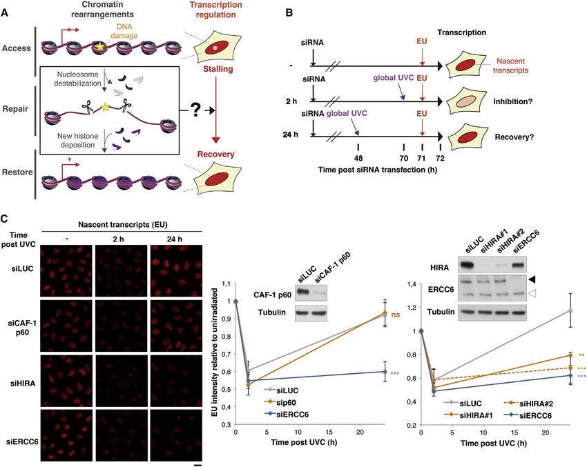
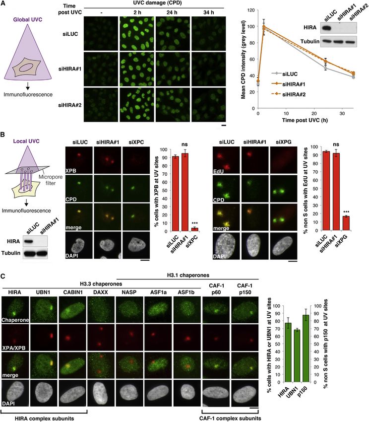
Figure 2. HIRA Function in the DNA Damage Response
(A) Repair activity analyzed by immunofluorescence against CPDs at different time points postglobal UVC irradiation (as depicted on the left) in HeLa cells treated
with the indicated siRNAs (siLUC, control). siRNA efficiencies are shown on the western blot panel.
(B) Recruitment of the repair protein XPB and repair synthesis (EdU incorporation) at DNA damage (CPD) analyzed by immunofluorescence 30 min and 4 hr after
local UVC irradiation, respectively, in U2OS cells treated with the indicated siRNAs. Local UVC damage is induced by irradiating cells with a UVC lamp through a
                                                                                                                               (legend continued on next page)

98 Cell 155, 94–106, September 26, 2013 ª2013 Elsevier Inc.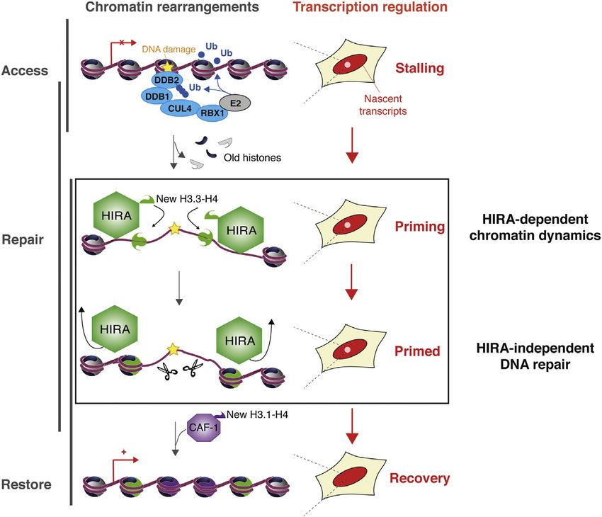
only transiently enriched in damaged chromatin. By exploring                    cient, to allow reactivation of transcription. Indeed, transcription
the molecular determinants of HIRA dissociation from damaged                    does not restart after UVC irradiation in repair-deficient XPG-
chromatin, we also shed light on a possible mechanism for                       depleted cells (Figure S1C), even if HIRA is efficiently recruited
HIRA recruitment. Indeed, we observed that HIRA release                         to damaged regions in these cells (Figure 4A). Thus, HIRA-inde-
from UVC-damaged chromatin regions is impaired in repair-                       pendent DNA damage repair events occur in the meantime to
deficient cells (siXPG) and significantly delayed on H3.3 deple-                allow reactivation of transcription.
tion (Figure S5C). These results show that completion of DNA                       Together, these data establish that HIRA acts early and tran-
repair and new histone deposition are both required to displace                 siently at sites of DNA damage, prior to repair completion and
HIRA from damaged chromatin, suggesting that this chaperone                     transcription recovery. Based on these findings, we propose
is recruited to nucleosome-free damaged DNA.                                    that HIRA recruitment to damaged regions acts as a priming
   Collectively, these data demonstrate that HIRA-mediated                      event making damaged chromatin prone for transcription restart
accumulation of new H3.3 is an early response to DNA damage,                    once repair is complete.
dependent on ubiquitylation events mediated by the DDB2-
DDB1-CUL4A complex, an early NER factor involved in DNA                         DISCUSSION
damage detection. Furthermore, these findings highlight that
completion of DNA repair is not a pre-requisite for new histone                 Our study defines a histone deposition pathway that primes
deposition into damaged chromatin but is critical for HIRA disso-               chromatin in response to DNA damage allowing transcription
ciation from UVC-damaged regions.                                               recovery after repair (Figure 6). This pathway involves early tar-
                                                                                geting of the histone chaperone HIRA to DNA lesions, where it
HIRA Priming Role for Transcription Recovery                                    promotes de novo deposition of H3.3 histones and renders chro-
To further explore the functional relationships between the                     matin prone for later reactivation of transcription. In light of these
histone chaperone HIRA and transcription regulation in response                 data, we propose a concept of chromatin bookmarking by his-
to DNA damage, we dissected the relative kinetics of HIRA                       tone deposition early on during the DDR that licenses the chro-
accumulation and transcription recovery in damaged chromatin                    matin substrate for transcription.
regions.
   Consistent with its dependency on early repair steps, we de-                 HIRA Targeting to Damaged Chromatin and New H3.3
tected HIRA recruitment to UVC-damaged chromatin within mi-                     Deposition
nutes after damage, reaching a maximum 40 min postirradiation.                  Although the yeast orthologs of the HIRA complex protect cells
Furthermore, HIRA recruitment preceded the accumulation of                      against genotoxic agents (Anderson et al., 2009; Kapitzky
the histone chaperone CAF-1 to DNA lesions (Figure 5A). Inter-                  et al., 2010), the actual role of this chaperone in response to
estingly and consistent with a histone chaperone function, we                   DNA damage is unknown, and in other eukaryotes, the contribu-
found that HIRA targeting to damaged chromatin was transient,                   tion of HIRA to the DDR remains elusive. Here, we characterize a
becoming undetectable 5 hr postdamage induction (Figures 5A                     transcription-associated function for HIRA in the DDR. Our ob-
and 5B). In contrast, the accumulation of new H3.3 at sites of                  servations indicate that, contrary to its yeast counterparts, hu-
DNA damage remained detectable up to 24 hr postirradiation                      man HIRA is not required for genome stability per se but instead
(Figures 5B and 5C). Thus, we conclude that newly synthesized                   contributes to transcription restart after repair. Furthermore,
H3.3 histones stably associate with DNA upon deposition by the                  HIRA exerts a local action in damaged chromatin regions by
HIRA chaperone in damaged chromatin and remain in place at                      depositing newly synthesized H3.3 histones. In this setting,
least until repair is complete.                                                 HIRA recruitment is not driven by the transcriptional activity but
   Next, we compared the relative kinetics of HIRA-dependent                    by the presence of DNA lesions. These findings have broad impli-
H3.3 deposition and transcription regulation at sites of local                  cations for our understanding of H3.3 deposition that, at least in
UVC irradiation. When HIRA was enriched at DNA damage                           this instance, does not simply occur as a consequence of tran-
(1 hr postdamage induction), we observed a local inhibition of                  scription. Considering that another set of new histones is depos-
transcription at these locations (Figure 5C), consistent with pre-              ited by CAF-1 during the late repair steps (Polo et al., 2006), the
vious observations (Moné et al., 2001). Remarkably, transcrip-                 impact of newly deposited histones on the chromatin landscape
tion was still inhibited 5 hr after damage, when HIRA was already               at damage sites is particularly intriguing. This is a critical issue
released from damaged chromatin regions. It was only long after                 given that new soluble histones show a distinct pattern of modi-
HIRA release from damaged chromatin that transcriptional activ-                 fications when compared to nucleosomal histones (Loyola et al.,
ity recovered (20–24 hr; Figure 5C). Thus, although HIRA deter-                 2006). How much and for how long newly incorporated histones
mines transcription restart after damage, its enrichment in                     stay in damaged chromatin regions will thus determine the extent
damaged chromatin is not concomitant with transcription recov-                  to which the original information conveyed by chromatin is ulti-
ery. These results imply that HIRA is necessary, but not suffi-                 mately modified, which will be the matter of future investigations.

micropore filter as shown on the scheme. siRNA efficiencies are controlled on the western blot panel. Depletion of the repair proteins XPC and XPG are used as
positive controls.
(C) Recruitment of H3 histone chaperones to DNA damage analyzed by immunofluorescence 30 min after local UVC irradiation in U2OS cells. Immunodetection of
the repair protein XPA or XPB labels damaged regions. Error bars on the graphs represent SD from at least two independent experiments. Scale bars, 10 mm.
See also Figure S2.

                                                                                      Cell 155, 94–106, September 26, 2013 ª2013 Elsevier Inc. 99
Figure 3. The HIRA Complex Promotes New H3.3 Accumulation in Damaged Chromatin Regions
(A) Scheme of the assay for monitoring accumulation of newly synthesized histones at UVC damage by fluorescence microscopy on cultured human cells stably
expressing SNAP-tagged histones. Pre-existing SNAP-tagged histones are quenched with a nonfluorescent substrate (block) so that only the histones neo-
synthetized during the chase period are labeled with the red fluorescent substrate tetramethylrhodamine (TMR)-star during the pulse step. Local UVC damage is
induced as in Figure 2B and cells are fixed 1 hr later.
(B) Accumulation of new H3.1 and H3.3, but not CenH3 variant, in UV-damaged regions analyzed by fluorescence microscopy in U2OS cells. CAF-1 p150 subunit
immunodetection labels replication and repair foci. As positive controls, we detect new H3.1 at replication foci and new CenH3 at centromeres in early G1 cells.
(legend continued on next page)
100 Cell 155, 94–106, September 26, 2013 ª2013 Elsevier Inc.In our study, we revealed clearly distinct functions for two regions actually makes damaged chromatin prone for transcrip-
chromatin assemblers, HIRA and CAF-1, in response to UVC tion restart is intriguing. An important function of HIRA at DNA
damage, as only HIRA is critical to facilitate transcription recov- damage that we identified in our study is its ability to deposit
ery after DNA repair. Another important feature that distinguishes new H3.3, which has been associated with transcriptional activa-
HIRA from CAF-1 is the timing of targeting to damaged chro- tion in various contexts and organisms—cross-tolerance in
matin. Our mechanistic studies reveal that HIRA recruitment is fission yeast, early embryonic development and nuclear reprog-
a very early and transient response that depends on DNA dam- ramming in Xenopus, muscle differentiation in mouse cells, and
age detection by DDB1-CUL4A-containing complexes. These response to viral infection in human cells (Chujo et al., 2012; Jul-
complexes promote ubiquitylation of various substrates in lien et al., 2012; Placek et al., 2009; Szenker et al., 2012; Yang
response to DNA damage, including NER factors and histones et al., 2011). Maintaining a certain level of H3.3 histones, which
(Nouspikel, 2011). Ubiquitylation of the HIRA complex itself is are prone to exclude repressive proteins and to carry specific
an attractive hypothesis to explain how HIRA is recruited to sets of posttranslational modifications promoting transcription
damaged chromatin. However, a recent systems-wide analysis (reviewed in Szenker et al., 2011), could ensure that repaired
did not identify ubiquitylation changes on HIRA or its associated chromatin remains a preferred substrate for RNA polymerase.
proteins UBN1 and CABIN1 after UVC damage in human cells Consistent with this model, we showed that the defect in tran-
(Povlsen et al., 2012). Another hypothesis that should be scription restart observed upon HIRA knockdown is not due to
explored in future studies is that the HIRA complex could a loss of function of the transcription machinery. In this context,
possibly recognize one or several of the numerous ubiquitylation it will be interesting to explore whether mutations in H3.3 recently
targets of DDB1-CUL4A at damage sites. However, in light of identified in pediatric brain tumors (Schwartzentruber et al.,
recent work in human cells and flies showing that HIRA can 2012; Sturm et al., 2012; Wu et al., 2012) could misregulate the
directly bind DNA (Ray-Gallet et al., 2011) and is recruited to response of HIRA to DNA damage and thereby affect the main-
nucleosome-free regions (Ray-Gallet et al., 2011; Schneiderman tenance of genome function.
et al., 2012), it is tempting to speculate that the ubiquitylation In addition, beyond a direct effect on H3.3 deposition, a more
activity of DDB1-CUL4A-containing complexes, which is general role of HIRA in the maintenance of chromatin integrity,
thought to increase DNA accessibility at damage sites (Nouspi- even indirectly by affecting H3.1 deposition, should also be inte-
kel, 2011), could expose naked DNA stretches, thereby trig- grated. Furthermore, while HIRA is the predominant histone
gering HIRA recruitment. Consistent with such a model where chaperone mediating de novo H3.3 deposition in damaged chro-
HIRA would be recruited to histone-free DNA, we showed that matin regions in our cell line models, we cannot exclude a
histone deposition promotes HIRA displacement from UV- possible contribution of other chaperones in distinct cellular
damaged chromatin. contexts such as in neurons, where DAXX-mediated H3.3 depo-
In addition, HIRA function in the DDR may not be restricted to sition controls the expression of genes responsive to neuronal
UVC photoproducts but could also be involved in response to activation (Michod et al., 2012).
other types of DNA lesions, including DNA breaks, as recently Most important, we have identified at the level of chromatin
described (Yang et al., 2013). Indeed, in support of this broader organization a key priming event for transcription restart that
function and consistent with a previous study (Adamson et al., occurs early in the DDR, independently of global DNA repair.
2012), we observed a transient accumulation of HIRA at sites While our study characterized HIRA-mediated reactivation of
of UVA laser microirradiation (Figure S2E). It is striking that, transcription after DNA damage on a genome-wide scale, it
once recruited, the HIRA complex remains only transiently in remains to be determined whether this mechanism applies simi-
damaged chromatin. Whether this release is itself critical for larly to gene-rich and noncoding regions as found in heterochro-
HIRA contribution to transcriptional reactivation is still to be matin. More subtle effects on transcription profiles at specific
determined. Most important, this transient presence delineates genomic regions could also be encountered upon milder alter-
a specific time window for HIRA action that will impact transcrip- ations in histone dynamics, which should be possible to detect
tion only after repair is complete. using emerging high-throughput technologies for nascent RNA
sequencing (Churchman and Weissman, 2011; Core et al.,
HIRA Function in Transcriptional Reactivation 2008).
Recovery of transcriptional activity after DNA damage repair, In conclusion, we provide an understanding of transcriptional
although critical for cell function, has remained essentially an un- control after DNA damage involving chromatin bookmarking by
characterized process to date. Here, we provide proof that spe- histone chaperones to license repaired DNA to be transcribed.
cific chromatin dynamics directly contribute to this process, by These findings open up avenues to unravel how genotoxic stress
showing that the H3.3-specific chaperone HIRA primes chro- could influence transcriptional programs via seemingly minor
matin for later recovery of transcription following DNA damage changes in chromatin resetting and, thus, have a profound
repair. How the early and transient action of HIRA at damaged impact on cell fate.
(C and D) Accumulation of new H3.3 in UV-damaged regions requires the HIRA complex. New H3.3 accumulation at DNA damage is revealed in U2OS cells
treated with the indicated siRNAs (siLUC, control) (C) or with the indicated combinations of siRNAs and plasmids (D). XPA labels repair sites, and hemagglutinin
(HA) immunodetection is used to visualize exogenous HIRA. Efficiencies of siRNA and plasmid transfections are shown on the western blots and on the right
immunofluorescence panel. Error bars on the graphs represent SD from three independent experiments. Scale bars, 10 mm.
See also Figures S3 and S4.
Cell 155, 94–106, September 26, 2013 ª2013 Elsevier Inc. 101Figure 4. HIRA-Mediated H3.3 Accumulation Is an Early Response Dependent on Ubiquitylation Events upon DNA Damage Detection (A) Scheme indicating the early and late NER proteins targeted by RNA interference in subsequent experiments. Recruitment of HIRA and CAF-1 p150 to DNA damage regions marked by XPB is analyzed by immunofluorescence 30 min and 1 hr after local UVC irradiation, respectively, in U2OS cells treated with the indicated siRNAs (siLUC, control). (B) HIRA and new H3.3 accumulation in UVC-damaged regions analyzed 30 min and 1 hr postlocal UVC irradiation respectively, in U2OS cells treated with the indicated siRNAs. siRNA efficiencies, HIRA, and H3.3 total levels are shown on the western blot panel. The levels of new H3.3 incorporated into chromatin are analyzed by SNAP labeling (bottom panel). (C) HIRA and new H3.3 accumulation at DNA damage analyzed as in (B). (D) HIRA accumulation at DNA damage analyzed 30 min postlocal UVC irradiation in U2OS cells transiently transfected with the indicated plasmids. The expression of CUL4A full-length (WT) and truncated mutant (DN) is shown on the western blot panel. Error bars on the graphs represent SD from at least two independent experiments. Scale bars, 10 mm. See also Figures S5 and S6. 102 Cell 155, 94–106, September 26, 2013 ª2013 Elsevier Inc.
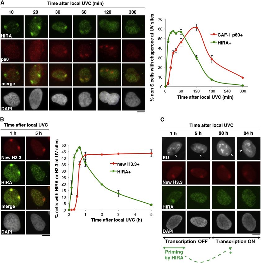
Figure 5. HIRA-Mediated H3.3 Deposition Does Not Coincide with Transcription Recovery
(A) Recruitment kinetics of HIRA and CAF-1 p60 to DNA damage analyzed by immunofluorescence at the indicated time points after local UVC irradiation in U2OS
cells.
(B) Accumulation kinetics of new H3.3 and its chaperone HIRA at DNA damage monitored at the indicated time points after local UVC irradiation in U2OS cells.
(C) Nascent RNA synthesis (EU labeling) analyzed at the indicated time points after local UVC irradiation (100 J/m2) in U2OS cells in parallel with the accumulation
of new H3.3 and its chaperone HIRA at DNA damage. Arrowheads point to sites of irradiation. Error bars on the graphs represent SD from three independent
experiments. Scale bars, 10 mm.

EXPERIMENTAL PROCEDURES                                                             Aldrich). For proteasome inhibition, cells were treated with 10 mM MG132
                                                                                    (Merck) in dimethyl sulfoxide (DMSO) for 2 hr. DNA breaks were induced by
Cell Culture and Drug Treatment                                                     treating cells with 50 mg/ml phleomycin (Sigma-Aldrich) for 30 min.
HeLa and U2OS cells were grown at 37 C and 5% CO2 in Dulbecco’s modified
Eagle’s medium (DMEM, Invitrogen) supplemented with 10% fetal calf serum            UV Irradiation
(EUROBIO), 100 U/ml penicillin, and 100 mg/ml streptomycin (Invitrogen). BJ         Cells were subjected to global or local UVC irradiation (254 nm), to laser-
primary fibroblasts were grown in the same medium supplemented with                 induced UVC damage (266 nm) and to UVA laser microirradiation (405 nm)
15% fetal calf serum and antibiotics. U2OS cells stably expressing H3.1-,           as described in the Extended Experimental Procedures.
H3.3-, or CenH3-SNAP (Dunleavy et al., 2011) were maintained in the presence
of 100 mg/ml G418 (Invitrogen).                                                    Flow Cytometry
  RNAPII-dependent transcription was inhibited by incubating cells in the          Cells were fixed in ice-cold 70% ethanol before DNA staining with 50 mg/ml pro-
presence of 100 mM DRB for 2 hr or 10 mg/ml a-amanitin for 8 hr (Sigma-            pidium iodide (Sigma-Aldrich) in PBS containing 0.05% Tween and 0.5 mg/ml

                                                                                       Cell 155, 94–106, September 26, 2013 ª2013 Elsevier Inc. 103
Figure 6. Model for Chromatin Bookmark-
ing by HIRA-Dependent Histone Deposition
Allowing Transcription Restart after Repair
of DNA Damage
Scheme representing how H3 histone chaperones
contribute to restoring nucleosomal organization
in response to DNA damage. Early ubiquitylation
associated with damage detection targets HIRA to
damaged chromatin regions where it deposits new
H3.3 histones and promotes subsequent events in
chromatin dynamics, including CAF-1-mediated
H3.1 deposition. Although not essential for global
repair, these HIRA-dependent chromatin changes
are key in restoring a proper chromatin organiza-
tion and in bookmarking damaged chromatin
for subsequent transcription restart (‘‘priming’’
function).
Nascent RNA Labeling
Nascent RNA labeling by EU incorporation was
performed with Click-iT RNA Imaging kits (Invitro-
gen). Refer to the Extended Experimental Proce-
dures for details.
Visualization of DNA Repair Synthesis
EdU was incorporated into cells during 4 hr
immediately after local UVC irradiation and re-
vealed using the Click-iT EdU Alexa Fluor 594
RNase A (USB/Affymetrix). DNA content was analyzed by flow cytometry using a Imaging kit (Invitrogen) according to manufacturer’s instructions before
C6 flow cytometer (Accuri). Proportion of cells in each step of the cell cycle was CPD labeling by immunofluorescence.
estimated using the Dean/Jett/Fox algorithm on FlowJo software (TreeStar).
SNAP Labeling of Histones
siRNA and Plasmid Transfection The SNAP labeling protocol is as described in Bodor et al. (2012). Refer to the
siRNA purchased from Dharmacon or Eurofins MWG Operon (Table S1) were Extended Experimental Procedures for details.
transfected into cells using Lipofectamine RNAiMAX (Invitrogen) following
manufacturer’s instructions. The final concentration of siRNA in the culture SUPPLEMENTAL INFORMATION
medium was 30–50 nM. Cells were harvested 24–72 hr posttransfection.
Cells were transiently transfected with plasmid DNA (1 mg/ml final) using Lip- Supplemental Information includes Extended Experimental Procedures, six
ofectamine 2000 (Invitrogen) according to manufacturer’s instructions 48 hr figures, and two tables and can be found with this article online at http://dx.
before subsequent cell treatment. For rescue experiments, cells were doi.org/10.1016/j.cell.2013.08.029.
concomitantly transfected with siRNA (30 nM final) and plasmid DNA
(0.375 mg/ml final) using Lipofectamine 2000 (Invitrogen) according to manu- ACKNOWLEDGMENTS
facturer’s instructions 72 hr before subsequent cell treatment. Plasmids are
described in the Extended Experimental Procedures. We are grateful to J. Haber, M. Papamichos-Chronakis, and D. Ray-Gallet for
critical reading of the manuscript and to members of UMR218 for helpful dis-
Colony-Forming Assays cussions. We thank E. Dunleavy and C. de Noronha for cell lines and reagents
Cells were replated 48 hr after siRNA transfection and exposed to global UVC and O. Renaud, O. Leroy, and P. LeBaccon from the PICT-IBiSA imaging facil-
irradiation the following day. Colonies were stained 12 days later with 0.5% ity. This work was supported by la Ligue Nationale contre le Cancer (Equipe
crystal violet/20% ethanol and counted. Results were normalized to plating labellisée Ligue 2010), the European Commission Network of Excellence ‘‘Epi-
efficiencies and analyzed by a two-way analysis of variance followed by Bon- GeneSys’’ (HEALTH-F4-2010-257082), the European Research Council
ferroni posttests with Prism 5 software (ns, nonsignificant, *p < 0.05, **p < 0.01, Advanced Grant ‘‘Eccentric’’ (2009-AdG_20090506), and the French National
and ***p < 0.001). Research Agency (Labex ‘‘DEEP’’) in G.A.’s laboratory. Research in S.E.P.’s
group is supported by the French National Research Agency (ANR-12-JSV6-
Cell Extracts and Western Blot 0002-01), the ‘‘Who am I’’? Laboratory of Excellence (ANR-11-LABX-0071)
Cell extracts were obtained as described in the Extended Experimental funded by the French Government through its ‘‘Investments for the Future’’
Procedures and run on 4%–20% Mini-PROTEAN TGX gels (Bio-Rad). Proteins program (ANR-11-IDEX-0005-01), and the Fondation ARC. S.A. is a recipient
of interest were probed using the appropriate primary (Table S2) and of a PhD fellowship from University Pierre & Marie Curie, and S.E.P. is funded
horseradish peroxidase (HRP)-conjugated secondary antibodies (Jackson by INSERM (Institut National de la Santé et de la Recherche Médicale) and a
Immunoresearch). Marie Curie Intra-European fellowship.
Immunofluorescence Received: April 26, 2013
Cells were subjected to immunostaining with primary (Table S2) and second- Revised: July 17, 2013
ary antibodies conjugated to Alexa-Fluor 488, 594, or 680 (Invitrogen) as Accepted: August 16, 2013
described in the Extended Experimental Procedures. Published: September 26, 2013
104 Cell 155, 94–106, September 26, 2013 ª2013 Elsevier Inc.REFERENCES Gaillard, P.H., Martini, E.M., Kaufman, P.D., Stillman, B., Moustacchi, E., and
Almouzni, G. (1996). Chromatin assembly coupled to DNA repair: a new role for
Adam, S., and Polo, S.E. (2012). Chromatin dynamics during nucleotide exci- chromatin assembly factor I. Cell 86, 887–896.
sion repair: histones on the move. Int. J. Mol. Sci. 13, 11895–11911. Giglia-Mari, G., Zotter, A., and Vermeulen, W. (2011). DNA damage response.
Adamson, B., Smogorzewska, A., Sigoillot, F.D., King, R.W., and Elledge, S.J. Cold Spring Harb. Perspect. Biol. 3, a000745.
(2012). A genome-wide homologous recombination screen identifies the RNA-
Green, C.M., and Almouzni, G. (2003). Local action of the chromatin assembly
binding protein RBMX as a component of the DNA-damage response. Nat.
factor CAF-1 at sites of nucleotide excision repair in vivo. EMBO J. 22, 5163–
Cell Biol. 14, 318–328.
5174.
Amin, A.D., Vishnoi, N., and Prochasson, P. (2012). A global requirement for
Hanawalt, P.C., and Spivak, G. (2008). Transcription-coupled DNA repair: two
the HIR complex in the assembly of chromatin. Biochim. Biophys. Acta
decades of progress and surprises. Nat. Rev. Mol. Cell Biol. 9, 958–970.
1819, 264–276.
Hoeijmakers, J.H.J. (2009). DNA damage, aging, and cancer. N. Engl. J. Med.
Anderson, H.E., Wardle, J., Korkut, S.V., Murton, H.E., López-Maury, L., Bäh-
361, 1475–1485.
ler, J., and Whitehall, S.K. (2009). The fission yeast HIRA histone chaperone is
required for promoter silencing and the suppression of cryptic antisense tran- Iacovoni, J.S., Caron, P., Lassadi, I., Nicolas, E., Massip, L., Trouche, D., and
scripts. Mol. Cell. Biol. 29, 5158–5167. Legube, G. (2010). High-resolution profiling of gammaH2AX around DNA dou-
ble strand breaks in the mammalian genome. EMBO J. 29, 1446–1457.
Avvakumov, N., Nourani, A., and Côté, J. (2011). Histone chaperones: modu-
lators of chromatin marks. Mol. Cell 41, 502–514. Ishii, T., Shiomi, Y., Takami, T., Murakami, Y., Ohnishi, N., and Nishitani, H.
(2010). Proliferating cell nuclear antigen-dependent rapid recruitment of
Bensaude, O. (2011). Inhibiting eukaryotic transcription: Which compound to
Cdt1 and CRL4Cdt2 at DNA-damaged sites after UV irradiation in HeLa cells.
choose? How to evaluate its activity? Transcription 2, 103–108.
J. Biol. Chem. 285, 41993–42000.
Bodor, D., Rodrı́guez, M., and Moreno, N. (2012). Analysis of protein turnover
Jackson, S.P., and Bartek, J. (2009). The DNA-damage response in human
by quantitative SNAP-based pulse-chase imaging. Curr. Protoc. Cell Biol. 55,
biology and disease. Nature 461, 1071–1078.
8.8.1–8.8.34.
Jensen, A., and Mullenders, L.H.F. (2010). Transcription factor IIS impacts UV-
Burgess, R.J., and Zhang, Z. (2013). Histone chaperones in nucleosome
inhibited transcription. DNA Repair (Amst.) 9, 1142–1150.
assembly and human disease. Nat. Struct. Mol. Biol. 20, 14–22.
Jullien, J., Astrand, C., Szenker, E., Garrett, N., Almouzni, G., and Gurdon, J.B.
Choi, S.-Y., Jang, H., Roe, J.-S., Kim, S.-T., Cho, E.-J., and Youn, H.-D. (2013).
(2012). HIRA dependent H3.3 deposition is required for transcriptional reprog-
Phosphorylation and ubiquitination-dependent degradation of CABIN1 re-
ramming following nuclear transfer to Xenopus oocytes. Epigenetics Chro-
leases p53 for transactivation upon genotoxic stress. Nucleic Acids Res. 41,
matin 5, 17.
2180–2190.
Chou, D.M., Adamson, B., Dephoure, N.E., Tan, X., Nottke, A.C., Hurov, K.E., Kapitzky, L., Beltrao, P., Berens, T.J., Gassner, N., Zhou, C., Wüster, A., Wu,
Gygi, S.P., Colaiácovo, M.P., and Elledge, S.J. (2010). A chromatin localization J., Babu, M.M., Elledge, S.J., Toczyski, D., et al. (2010). Cross-species chemo-
screen reveals poly (ADP ribose)-regulated recruitment of the repressive poly- genomic profiling reveals evolutionarily conserved drug mode of action. Mol.
comb and NuRD complexes to sites of DNA damage. Proc. Natl. Acad. Sci. Syst. Biol. 6, 451.
USA 107, 18475–18480. Li, B., Carey, M., and Workman, J.L. (2007). The role of chromatin during tran-
Chujo, M., Tarumoto, Y., Miyatake, K., Nishida, E., and Ishikawa, F. (2012). scription. Cell 128, 707–719.
HIRA, a conserved histone chaperone, plays an essential role in low-dose Loyola, A., Bonaldi, T., Roche, D., Imhof, A., and Almouzni, G. (2006). PTMs on
stress response via transcriptional stimulation in fission yeast. J. Biol. Chem. H3 variants before chromatin assembly potentiate their final epigenetic state.
287, 23440–23450. Mol. Cell 24, 309–316.
Churchman, L.S., and Weissman, J.S. (2011). Nascent transcript sequencing Luijsterburg, M.S., and van Attikum, H. (2011). Chromatin and the DNA dam-
visualizes transcription at nucleotide resolution. Nature 469, 368–373. age response: the cancer connection. Mol. Oncol. 5, 349–367.
Ciccia, A., and Elledge, S.J. (2010). The DNA damage response: making it safe Mailand, N., Bekker-Jensen, S., Faustrup, H., Melander, F., Bartek, J., Lukas,
to play with knives. Mol. Cell 40, 179–204. C., and Lukas, J. (2007). RNF8 ubiquitylates histones at DNA double-strand
Core, L.J., Waterfall, J.J., and Lis, J.T. (2008). Nascent RNA sequencing breaks and promotes assembly of repair proteins. Cell 131, 887–900.
reveals widespread pausing and divergent initiation at human promoters. Michod, D., Bartesaghi, S., Khelifi, A., Bellodi, C., Berliocchi, L., Nicotera, P.,
Science 322, 1845–1848. and Salomoni, P. (2012). Calcium-dependent dephosphorylation of the histone
Dantuma, N.P., Groothuis, T.A.M., Salomons, F.A., and Neefjes, J. (2006). A chaperone DAXX regulates H3.3 loading and transcription upon neuronal acti-
dynamic ubiquitin equilibrium couples proteasomal activity to chromatin re- vation. Neuron 74, 122–135.
modeling. J. Cell Biol. 173, 19–26. Moné, M.J., Volker, M., Nikaido, O., Mullenders, L.H., van Zeeland, A.A., Ver-
De Koning, L., Corpet, A., Haber, J.E., and Almouzni, G. (2007). Histone chap- schure, P.J., Manders, E.M., and van Driel, R. (2001). Local UV-induced DNA
erones: an escort network regulating histone traffic. Nat. Struct. Mol. Biol. 14, damage in cell nuclei results in local transcription inhibition. EMBO Rep. 2,
997–1007. 1013–1017.
Dunleavy, E.M., Almouzni, G., and Karpen, G.H. (2011). H3.3 is deposited at Nakazawa, Y., Yamashita, S., Lehmann, A.R., and Ogi, T. (2010). A semi-auto-
centromeres in S phase as a placeholder for newly assembled CENP-A in mated non-radioactive system for measuring recovery of RNA synthesis and
G1 phase. Nucleus 2, 146–157. unscheduled DNA synthesis using ethynyluracil derivatives. DNA Repair
(Amst.) 9, 506–516.
Ehmann, U.K., Cook, K.H., and Friedberg, E.C. (1978). The kinetics of thymine
dimer excision in ultraviolet-irradiated human cells. Biophys. J. 22, 249–264. Nouspikel, T. (2009). DNA repair in mammalian cells : Nucleotide excision
repair: variations on versatility. Cell. Mol. Life Sci. 66, 994–1009.
Filipescu, D., Szenker, E., and Almouzni, G. (2013). Developmental roles of his-
tone H3 variants and their chaperones. Trends Genet. Published online July 2, Nouspikel, T. (2011). Multiple roles of ubiquitination in the control of nucleotide
2013. http://dx.doi.org/10.1016/j.tig/2013.06.002. excision repair. Mech. Ageing Dev. 132, 355–365.
Gaillard, H., and Aguilera, A. (2013). Transcription coupled repair at the inter- Pankotai, T., Bonhomme, C., Chen, D., and Soutoglou, E. (2012). DNAPKcs-
face between transcription elongation and mRNP biogenesis. Biochim. dependent arrest of RNA polymerase II transcription in the presence of DNA
Biophys. Acta 1829, 141–150. breaks. Nat. Struct. Mol. Biol. 19, 276–282.
Cell 155, 94–106, September 26, 2013 ª2013 Elsevier Inc. 105Placek, B.J., Huang, J., Kent, J.R., Dorsey, J., Rice, L., Fraser, N.W., and mutations in H3F3A and IDH1 define distinct epigenetic and biological sub-
Berger, S.L. (2009). The histone variant H3.3 regulates gene expression during groups of glioblastoma. Cancer Cell 22, 425–437.
lytic infection with herpes simplex virus type 1. J. Virol. 83, 1416–1421.
Svejstrup, J.Q. (2010). The interface between transcription and mechanisms
Polo, S.E., Roche, D., and Almouzni, G. (2006). New histone incorporation maintaining genome integrity. Trends Biochem. Sci. 35, 333–338.
marks sites of UV repair in human cells. Cell 127, 481–493.
Szenker, E., Lacoste, N., and Almouzni, G. (2012). A developmental require-
Povlsen, L.K., Beli, P., Wagner, S.A., Poulsen, S.L., Sylvestersen, K.B., Poul-
ment for HIRA-dependent H3.3 deposition revealed at gastrulation in Xeno-
sen, J.W., Nielsen, M.L., Bekker-Jensen, S., Mailand, N., and Choudhary, C.
pus. Cell Rep. 1, 730–740.
(2012). Systems-wide analysis of ubiquitylation dynamics reveals a key role
for PAF15 ubiquitylation in DNA-damage bypass. Nat. Cell Biol. 14, 1089– Szenker, E., Ray-Gallet, D., and Almouzni, G. (2011). The double face of the
1098. histone variant H3.3. Cell Res. 21, 421–434.
Ray-Gallet, D., Woolfe, A., Vassias, I., Pellentz, C., Lacoste, N., Puri, A., Talbert, P.B., and Henikoff, S. (2010). Histone variants—ancient wrap artists of
Schultz, D.C., Pchelintsev, N.A., Adams, P.D., Jansen, L.E.T., and Almouzni, the epigenome. Nat. Rev. Mol. Cell Biol. 11, 264–275.
G. (2011). Dynamics of histone H3 deposition in vivo reveal a nucleosome
gap-filling mechanism for H3.3 to maintain chromatin integrity. Mol. Cell 44, Tornaletti, S. (2009). DNA repair in mammalian cells: Transcription-coupled
928–941. DNA repair: directing your effort where it’s most needed. Cell. Mol. Life Sci.
66, 1010–1020.
Schneiderman, J.I., Orsi, G.A., Hughes, K.T., Loppin, B., and Ahmad, K. (2012).
Nucleosome-depleted chromatin gaps recruit assembly factors for the H3.3 Wu, G., Broniscer, A., McEachron, T.A., Lu, C., Paugh, B.S., Becksfort, J., Qu,
histone variant. Proc. Natl. Acad. Sci. USA 109, 19721–19726. C., Ding, L., Huether, R., Parker, M., et al.; St. Jude Children’s Research Hos-
Schwartzentruber, J., Korshunov, A., Liu, X.-Y., Jones, D.T.W., Pfaff, E., pital–Washington University Pediatric Cancer Genome Project. (2012).
Jacob, K., Sturm, D., Fontebasso, A.M., Quang, D.-A.K., Tönjes, M., et al. Somatic histone H3 alterations in pediatric diffuse intrinsic pontine gliomas
(2012). Driver mutations in histone H3.3 and chromatin remodelling genes in and non-brainstem glioblastomas. Nat. Genet. 44, 251–253.
paediatric glioblastoma. Nature 482, 226–231. Yang, J.-H., Song, Y., Seol, J.-H., Park, J.Y., Yang, Y.-J., Han, J.-W., Youn, H.-
Shanbhag, N.M., Rafalska-Metcalf, I.U., Balane-Bolivar, C., Janicki, S.M., and D., and Cho, E.-J. (2011). Myogenic transcriptional activation of MyoD medi-
Greenberg, R.A. (2010). ATM-dependent chromatin changes silence transcrip- ated by replication-independent histone deposition. Proc. Natl. Acad. Sci.
tion in cis to DNA double-strand breaks. Cell 141, 970–981. USA 108, 85–90.
Skene, P.J., and Henikoff, S. (2013). Histone variants in pluripotency and dis- Yang, X., Li, L., Liang, J., Shi, L., Yang, J., Yi, X., Zhang, D., Han, X., Yu, N., and
ease. Development 140, 2513–2524. Shang, Y. (2013). Histone acetyltransferase 1 promotes homologous recombi-
Smerdon, M.J. (1991). DNA repair and the role of chromatin structure. Curr. nation in DNA repair by facilitating histone turnover. J. Biol. Chem. 288, 18271–
Opin. Cell Biol. 3, 422–428. 18282.
Soria, G., Polo, S.E., and Almouzni, G. (2012). Prime, repair, restore: the active Zhang, X., Horibata, K., Saijo, M., Ishigami, C., Ukai, A., Kanno, S.-I., Tahara,
role of chromatin in the DNA damage response. Mol. Cell 46, 722–734. H., Neilan, E.G., Honma, M., Nohmi, T., et al. (2012). Mutations in UVSSA
Sturm, D., Witt, H., Hovestadt, V., Khuong-Quang, D.-A., Jones, D.T.W., cause UV-sensitive syndrome and destabilize ERCC6 in transcription-coupled
Konermann, C., Pfaff, E., Tönjes, M., Sill, M., Bender, S., et al. (2012). Hotspot DNA repair. Nat. Genet. 44, 593–597.
106 Cell 155, 94–106, September 26, 2013 ª2013 Elsevier Inc.You can also read