Adipokines and Autoimmunity in Inflammatory Arthritis - MDPI
←
→
Page content transcription
If your browser does not render page correctly, please read the page content below
cells
Review
Adipokines and Autoimmunity in Inflammatory Arthritis
Elena Neumann * , Rebecca Hasseli, Selina Ohl, Uwe Lange, Klaus W. Frommer                                    and Ulf Müller-Ladner

                                           Department of Rheumatology and Clinical Immunology, Campus Kerckhoff, Justus Liebig University Giessen,
                                           61231 Bad Nauheim, Germany; r.hasseli@kerckhoff-klinik.de (R.H.); s.ohl@kerckhoff-klinik.de (S.O.);
                                           U.lange@kerckhoff-klinik.de (U.L.); K.frommer@kerckhoff-klinik.de (K.W.F.);
                                           u.mueller-ladner@kerckhoff-klinik.de (U.M.-L.)
                                           * Correspondence: e.neumann@kerckhoff-klinik.de

                                           Abstract: Adipokines are adipose tissue-derived factors not only playing an important role in
                                           metabolism but also influencing other central processes of the body, such as inflammation. In au-
                                           toimmune diseases, adipokines are involved in inflammatory pathways affecting different cell types.
                                           Many rheumatic diseases belong to the group of autoimmune diseases, for example rheumatoid
                                           arthritis (RA) and psoriatic arthritis. Due to the autoimmune responses, a chronic inflammatory
                                           milieu develops, which affects the whole body, including adipose tissue. Metabolic alterations such
                                           as obesity influence inflammatory responses in autoimmune diseases. Adipokines are bioactive
                                           mediators mainly produced by adipose tissue. Due to alterations of systemic adipokine levels, their
                                           role as biomarkers with diagnostic potential has been suggested in the context of rheumatic diseases.
                                           In the affected joints of RA patients, different synoviocytes but also osteoclasts, osteoblasts, and chon-
                                           drocytes produce several adipokines, contributing to the unique inflammatory microenvironment.
                                           Adipokines have been shown to be potent modulatory effectors on different cell types of the immune
                                           system but also local cells in synovial tissue, cartilage, and bone. This review highlights the most
                                           recent findings on the role of adipokines in the pathophysiology of inflammatory arthritis with a
                                           distinct focus on RA in the quickly developing research field.


Citation: Neumann, E.; Hasseli, R.;
                                           Keywords: adipokines; adipocytokines; rheumatic diseases; rheumatoid arthritis; autoimmunity
Ohl, S.; Lange, U.; Frommer, K.W.;
Müller-Ladner, U. Adipokines and
Autoimmunity in Inflammatory
Arthritis. Cells 2021, 10, 216. https://   1. Introduction
doi.org/10.3390/cells10020216                    Disorders affecting the joint can be divided in two central groups. Primary inflamma-
                                           tory autoimmune arthritides consist of rheumatoid arthritis (RA) and spondyloarthritides,
Received: 30 November 2020                 including psoriatic arthritis (PsA) and ankylosing spondylitis. Findings in inflammatory
Accepted: 19 January 2021
                                           arthritis are often compared to non-autoimmune osteoarthritis (OA). OA is usually induced
Published: 22 January 2021
                                           by previous joint injury, biomechanical stress on joints due to malposition, or overweight
                                           and obesity. Metabolic diseases, such as diabetes or hyperlipidemia, are considered key
Publisher’s Note: MDPI stays neutral
                                           risk factors for OA development [1]. Though cartilage degradation is a central feature of
with regard to jurisdictional claims in
                                           OA, resulting in an inflammatory response due to mechanical joint damage, the whole
published maps and institutional affil-
                                           joint is affected, including adjacent bone, which involves the formation of osteophytes [1].
iations.
                                           In contrast, RA is an autoimmune disorder characterized by severe chronic inflammation
                                           early within the disease, leading to irreversible joint damage if left untreated [2]. RA
                                           affects mainly peripheral joints symmetrically, leading to progressive joint damage. The
                                           clinical presentation of RA varies, and environmental factors and epigenetic mechanisms
Copyright: © 2021 by the authors.          have been associated with this disorder [3,4]. A persistent inflammatory infiltration of
Licensee MDPI, Basel, Switzerland.
                                           the synovial tissue contributes to local synovial cell activation, which causes the release
This article is an open access article
                                           of pro-inflammatory and matrix-degrading factors to the tissue and joint space. RA syn-
distributed under the terms and
                                           ovial fibroblasts (RASFs) exhibit an autonomous activated phenotype contributing to the
conditions of the Creative Commons
                                           inflammatory milieu as well as synovial hyperplasia [4,5].
Attribution (CC BY) license (https://
                                                 PsA and RA are both rheumatic chronic inflammatory diseases sharing similarities
creativecommons.org/licenses/by/
4.0/).
                                           such as synovitis but also differences in the pathophysiology. This includes a different

Cells 2021, 10, 216. https://doi.org/10.3390/cells10020216                                                     https://www.mdpi.com/journal/cells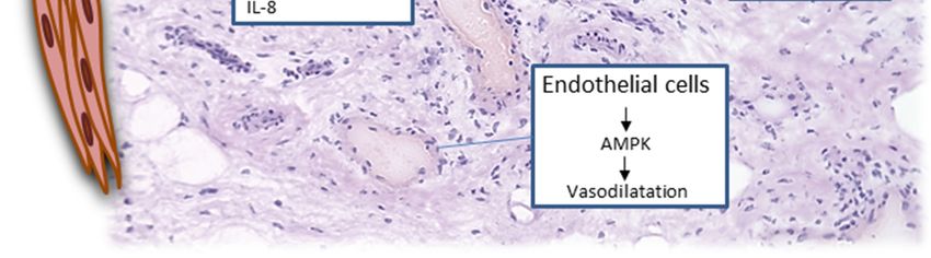
Cells 2021, 10, 216                                                                                            2 of 15

                      vascular pattern in the affected joints [6], a less pronounced synovial hyperplasia in PsA
                      compared to RA patients [7], and the cell infiltrates at the inflamed synovial–entheseal
                      complex being more prominent in PsA patients [8]. Therefore, enthesitis is one of the
                      frequent features of PsA according to the Classification of Psoriatic Arthritis (CASPAR)
                      criteria [9,10] but rare in the case of RA as well as OA. PsA patients also respond differently
                      to treatment. For example, the anti-IL17A biologic secukinumab is more effective in PsA
                      than in RA patients.
                            Dysregulation of immune-endocrine circuits is involved in the development of chronic
                      metabolic disorders, such as obesity, metabolic syndrome, and diabetes, but also plays a
                      role in chronic inflammatory diseases, such as RA [11]. Understanding the mechanisms of
                      both immune regulation and resolution of inflammation is crucial for the development of
                      successful treatment approaches to achieve and maintain remission or low disease activity
                      in RA and PsA. Regulatory mechanisms include mediators such as cytokines, chemokines,
                      hormones, neurotransmitters, and their receptors from inflamed tissue but also the pe-
                      riphery, including the adipose tissue. Many diseases are based on chronic inflammation
                      lasting for years or for the patients’ whole life if left untreated. Due to alterations of
                      tissues or tissue components in the course of the disease as well as specifically due to
                      autoimmune responses, the organism is no longer able to terminate the pathophysiological
                      inflammatory circle, resulting in continuous tissue damage and, finally, loss of function
                      and quality-of-life impairment.

                      2. Adipose Tissue and Adipokines
                           Adipose tissue is the key tissue regulating energetic homeostasis. It also serves as an
                      endocrine organ due to secretion of a large number of bioactive substances. The factors
                      that are released by adipose tissue are called adipocytokines. This group of mediators
                      includes adipokines, cytokines, chemokines, complement factors, and hormones [12].
                      Cytokines produced by an excess of adipose tissue can affect the whole body, leading
                      to the development of a so-called low-level inflammation, which can be observed in
                      obese individuals.
                           Adipokines are factors mainly produced by adipocytes in the white adipose tissue.
                      Adipose tissue secretes a large number of highly biologically active factors. However,
                      many adipokines such as leptin and adiponectin are also known modulators of immune
                      responses. Systemic alterations of adipokines have been identified for a large number of
                      chronic inflammatory diseases, and the potential of adipokines such as adiponectin and
                      leptin has been discussed. Therefore, adipokines have been investigated for many years
                      in the context of chronic inflammatory and degenerative rheumatic diseases systemically
                      as well as on the local level in cells and tissues. Furthermore, pro- and anti-inflammatory
                      properties of adipokines have been identified. Therefore, it has been accepted in the past
                      years that adipokines play an important role in immune-mediated rheumatic disease and
                      degenerative OA.

                      2.1. Adiponectin
                            Adiponectin, encoded by the ADIPOQ gene, has been described as a mainly anti-
                      inflammatory adipokine. Adiponectin is produced in large amounts by adipocytes of the
                      white adipose tissue [13]. Adiponectin concentrations are inversely correlated with the
                      body mass index (BMI). Adiponectin is a complex molecule. Adiponectin monomers form
                      different isoforms, depending on the degree of oligomerization: the trimer (low molecular
                      weight (LMW)), the hexamer (middle molecular weight (MMW)), and the multimeric
                      (high molecular weight (HMW)) adiponectin consisting of 12–18 monomers. Globular
                      adiponectin, consisting of the monomeric head-domain, is formed by proteolytic cleavage.
                      The monomer occurs as an intermediate in adipocytes, while in the circulation the main
                      forms are the multimeric adiponectin isoforms. Besides the two main adiponectin recep-
                      tors, AdipoR1 and AdipoR2, which are able to bind globular and full-length adiponectin
                      isoforms with different affinities, other receptors such as T-cadherin and PAQR3 (pro-
Cells 2021, 10, 216 3 of 15
gestin and AdipoQ receptor family member 3) have been described [12–17]. Recently,
Tanaka and colleagues showed that adiponectin overexpression increased the regenera-
tion of myofibers promoting muscle regeneration in a T-cadherin-dependent manner [17].
Adiponectin has several central biological functions, such as fatty acid biosynthesis and
inhibition of gluconeogenesis within the liver [12,13,15]. However, adiponectin not only
shows potential as a biomarker due to the systemic alterations under inflammatory con-
dition, which were found to decrease [18–20] or increase [21–23] in physiological and
pathophysiological conditions, but is also actively involved in inflammatory responses and
affects different cell types. In type 2 diabetes, atherosclerosis, and metabolic syndrome,
predominantly anti-inflammatory effects have been described [12,18,24]. However, in the
context of rheumatoid arthritis, the role of adiponectin is not fully understood. On cellular
and tissue level, opposite effects have been described for rheumatic diseases, such as RA,
where it seems to promote inflammation and tissue damage in the affected joints. Recent
findings will be outlined in the sections below.
2.2. Leptin
The main adipokine produced by adipocytes is leptin. Leptin concentrations are posi-
tively correlated with white adipose tissue mass. In addition, leptin has central functions in
metabolism and also plays a role in inflammation and inflammatory disorders [25]. Leptin
is encoded by the LEP (ob) gene and is a 16 kDa non-glycosylated protein. The effects
of leptin are mediated by binding to the long form of the leptin receptor LEPR [26]. By
inducing anorexigenic factors, leptin is known to be a central protein in appetite regu-
lation and obesity. However, leptin is also involved in many processes besides insulin
secretion and basal metabolism, such as reproduction, bone mass regulation, and (chronic)
inflammatory diseases [14,25]. Leptin, in turn, is induced in adipose tissue, depending
on the energy status, by sex hormones and by inflammatory mediators [14,25]. In con-
trast to adiponectin, leptin is considered a pro-inflammatory adipokine. It is involved in
low-grade inflammation due to overweight in obesity [26]. Both the innate and adaptive
immune responses are affected by leptin, and the LEPR is expressed at the surface of most
immune cells. Leptin increases the phagocytosis of macrophages, induces proliferation
of monocytes and macrophages, alters the cytotoxicity of natural killer cells as well as
the proliferation of CD4 T cells, suppresses type 2 T helper cells (Th2), and increases Th1
responses as well as regulatory T cell (Treg) responses [25,27]. Leptin itself is induced by
pro-inflammatory cytokines during acute infection and sepsis, but it is also induced during
chronic inflammatory autoimmune diseases [25,28].
2.3. Visfatin
Visfatin, or pre-B-cell colony-enhancing factor (PBEF), is a multifunctional protein that
has the ability to promote B cell differentiation and possesses nicotinamide phosphoribosyl-
transferase (Nampt) enzymatic activity. These different mechanisms of action make visfatin
a very interesting protein. Whether altered local visfatin levels are associated with its
extracellular interaction with cells or due to its intracellular enzymatic Nampt activity,
leading to changes in the nicotinamide adenine dinucleotide content, remains to be clarified.
Visfatin is produced by adipose tissue as well as other tissues, such as liver, bone marrow,
and muscle. It can be induced by pro-inflammatory factors, chemokines, hypoxia, and
visfatin itself, and in turn visfatin induces a pro-inflammatory response in many cell types
and tissues by itself [12,15,24]. The cell surface receptor for visfatin is unknown, and
several studies have shown that the visfatin effects are in part due to its Nampt activity [14].
However, an interaction with insulin-like growth factor (IGF)-1 signaling has been reported.
Here, it can been shown that visfatin inhibits IGF-1-mediated function independently
of the IGF-1 receptor activation [29], suggesting another mechanism of action besides
its Nampt activity. Interestingly, it has been recently observed that several micro RNAs
(miRNAs) are involved in the visfatin-mediated effects specifically in the context of OA
chondrocytes [30]. Visfatin significantly reduced viability and induced apoptosis in OACells 2021, 10, 216 4 of 15
chondrocytes, involving the NFκB pathway as well as decreasing miR-140 and miR-146a
and increasing iR-let7e expression in this study [30].
2.4. Resistin
Resistin is a homodimeric cysteine-rich protein that is mainly produced by macrophages
in humans, whereas in animals such as mice, the source of resistin is the adipose tis-
sue resident adipocytes [14,31]. Therefore, results from animal studies can often not
directly be translated to the human situation. Human resistin was described to be a mainly
pro-inflammatory protein promoting immune cell recruitment and immune cell activa-
tion [14,32] because it is produced by macrophages. Furthermore, a role in the context of
the development of, for example, coronary artery disease, atherosclerosis, type 2 diabetes,
as well as OA and psoriasis has been reported [14,33], showing a mainly inflammation-
promoting effect. However, anti-inflammatory effects of resistin have also been described
on cellular and tissue level [31]. Therefore, the effect of resistin may depend on the tissue
and pathophysiological condition studied. In addition, an immunomodulatory role in the
context of rheumatic diseases, including RA, PsA, and OA, has been described [14].
2.5. Chemerin, Vaspin, and Omentin
Chemerin precursors consist of a hydrophobic signal peptide sequence, a cysteine
fold-containing domain, and a labile C domain. Chemerin is activated after hydroliza-
tion by cysteine or serine proteases. Removal of the signal sequence leads to a secreted
preform with low biological activity. Different cleaved isoforms and underlying mech-
anisms of cleavage have been described in the past years [34], which are in the focus of
current investigations. Chemerin is involved in immune responses, and its role in coronary
atherosclerosis and metabolic syndrome development, among other diseases, has been
described [14]. Mainly anti-inflammatory chemerin properties have been described for
macrophages as well as in an LPS-induced acute lung injury mouse model [35]. On the
cellular level, chemerin acts as a chemoattractant for natural killer cells, macrophages, and
dendritic cells [14,36].
Vaspin belongs to a family of serine protease inhibitors. Vaspin has been associated
with insulin resistance and metabolic syndrome as well as atherosclerosis and cardiovas-
cular disease [37]. Besides subcutaneous adipose tissue, vaspin is also expressed and
produced in other tissues such as skin and skeletal muscle. Interestingly, vaspin has been
described to be involved in skeletal muscle inflammation [38]. Vaspin overexpression in
mice showed altered metabolism and inflammation with improved glucose tolerance and
resistance to high-fat diet-induced obesity with lower systemic IL-6 levels [14]. On the cel-
lular level, vaspin alters adipocyte differentiation and glucose homeostasis. In the context
of coronary atheromatous plaques, the pro-inflammatory phenotype of macrophages was
suppressed by vaspin [14]. However, limited knowledge regarding the evaluation of the
specific role of vaspin in the context of autoinflammation in RA is available.
Omentin is a glycoprotein that binds to galactofuranosyl residues of microorganisms
and to the lactoferrin-binding protein. It is expressed mainly in omental adipose tissue.
However, omentin is abundant in the plasma of healthy donors [39]. Omentin has been
described to mainly have anti-inflammatory effects. Anti-atherogenic effects in obese
individuals have been described, as well as a negative association with inflammatory
bowel disease and metabolic syndrome [14,39].
2.6. Progranulin, Lipocalin-2, and Nesfatin
Progranulin (PGRN, granulin/epithelin precursor) consists of seven granulin/epithelin
repeats that can be cleaved into small homologous subunits. Full-length protein as well
as the resulting peptides after cleavage are biologically active [40]. PGRN is an autocrine
factor promoting different physiological processes, such as chondrocyte differentiation and
proliferation as well as enchondral ossification [14]. Pro- and anti-inflammatory effects
of PGRN have been described. The anti-inflammatory properties are mainly mediatedCells 2021, 10, 216 5 of 15
by competitive binding to tumor necrosis factor (TNF) receptors disturbing the TNFα-
mediated responses [25,41]. PGRN is produced by many different cells such as adipocytes,
macrophages, and chondrocytes and has been suggested as a potential biomarker in in-
flammatory disease [42].
Nesfatin (nesfatin-1) is an adipokine involved in satiety induction and in energy
homeostasis. It is secreted by the hypothalamus and acts as an anorexigenic factor. In
addition, it is produced by subcutaneous adipose tissue and other tissues within the gut,
pancreas, and testes [43]. Lipocalin-2 (LCN2) is an adipokine induced by pro-inflammatory
factors, such as IL-1beta, LPS, and other cytokines, as well as dexamethasone and other
adipokines, such as leptin and adiponectin [14,44,45].
3. Adipokines in Autoimmune Rheumatoid Arthritis
3.1. Rheumatoid Arthritis
RA affects 1% of the population worldwide, leading to loss of physical joint func-
tion. It is a systemic chronic autoimmune disease developing in genetically susceptible
individuals due to environmental factors and involving epigenetic mechanisms. Being a
very heterogeneous disorder with different clinical forms and dominant pathomechanisms,
treatment of RA still remains challenging [2–4,13]. Regarding rheumatic diseases, RA
was the first to be evaluated in the context of adipokines, and their systemic levels are
mostly compared with those in primarily non-inflammatory degenerative osteoarthritis
and healthy donors. Due to the limited availability of control cells and tissues from healthy
donors, findings on the cellular level are usually compared to cells from OA patients.
Many different cells are involved in the pathogenesis of chronic inflammatory joint
diseases such as RA. Friction-free joint mobility is ensured by the synovial membrane and
synovial fluid. The synovial membrane consists of a thin layer at the border to the synovial
fluid with two main cell types, the macrophage-like synovial cells (called type A) and
fibroblast-like synoviocytes (called type B). Both cell types are also present in the sublining,
which consists mainly of connective tissue and adipose tissue in the deeper layers of the
sublining. Synovial macrophages consist of resident cells but also originate from blood
monocytes. Synovial fibroblasts produce hyaluronic acids and proteoglycans, important
factors of the synovial fluid. In addition, variable leukocyte populations and blood vessels
are present in the synovial tissue. In chronic inflammatory arthritis such as RA and PsA,
the synovial tissue is the central site of inflammation, leading to synovial hyperplasia and
local activation of synovial cells over time and, finally, resulting in irreversible cartilage
and bone damage [2–4]. Synovial inflammation is characterized by increased permeability
of the vessels, leading to leukocyte infiltration. Local proliferation and activation of RASFs
is a typical and early feature of RA and, together with an increased influx of monocytes,
differentiating into RA synovial macrophages, leads to the formation of the hyperplastic
synovial lining layer and an increased cellular density within the sublining [2–4,13]. Due
to the interaction of the different cell types and the high amounts of adipokines present
within the synovial fluid and tissue, many efforts to evaluate the effects of adipokines on
the different effector cells in the pathophysiology of RA have been made.
3.1.1. Adiponectin in Rheumatoid Arthritis
Increased levels of adiponectin have been shown in the synovial fluid of the affected
joints in RA [12,14,15]. Systemic adiponectin levels have been described to be increased in
RA as well as associated with disease activity and disease progression [12,24,46]. However,
not all studies have confirmed the correlation with disease activity [24,47]. This is most
likely due to the different methods of normalization but also due to the heterogeneity of the
disease. However, most studies have shown a correlation of adiponectin with inflammation
markers such as C-reactive protein (CRP), including a recent study showing that total and
HMW adiponectin are positively correlated with CRP levels [47]. A positive correlation be-
tween serum adiponectin and the disease activity score disease activity score (DAS)28 - ESR
in RA patients was shown in another cross-sectional study [48]. Interestingly, subcutaneousCells 2021, 10, 216 6 of 15
adipose tissue from RA patients produced more adiponectin compared to OA patients
and correlated with disease activity and disease duration in these patients [49]. Along this
line, an increased risk for obese subjects with high serum adiponectin levels at baseline to
develop RA, specifically with high adiponectin and CRP levels, was recently described in a
study during a follow-up for up to 29 years [50]. The response to anti-TNF-alpha treatment
seems to be associated with better improvement in patients with higher adiponectin levels
at baseline [51]. Another study showed increased adiponectin and reduced chemerin levels
in RA patients after anti-IL-6 treatment in both monotherapy or combined therapy with
methotrexate independent of the treatment response [51]. Another recent study showed
that tocilizumab treatment of RA patients with active RA despite previous conventional
synthetic disease-modifying antirheumatoic drug (DMARD) and/or biological DMARD
treatment was associated with an increase in total and HMW adiponectin, especially early
after treatment onset, then declining until month 6 to 12 [52]. In this study, anti-IL-6 treat-
ment also induced a gain in lean mass, while fat mass remained unchanged. Baricitinib, a
JAK inhibitor blocking central inflammatory signaling pathways, was recently shown to
decrease systemic inflammation biomarkers, such as IL-6, CRP, as well as adiponectin, in
rheumatoid arthritis patients [53].
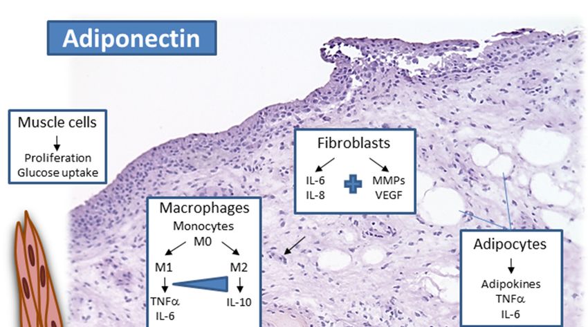
On the cellular level, numerous studies showed that cultured RASF respond to
adiponectin by induction of pro-inflammatory factors, including prostaglandin E2, IL-6,
IL-8, as well as matrix-metallo proteinases such as MMP-1 and MMP-13 [12,24]. Specifically,
the HMW adiponectin isoforms seem to display stronger pro-inflammatory effects in RASF.
Adiponectin stimulation of RASF indirectly affected T follicular helper cells, which was
mainly mediated by the induction of IL-6 [54]. HMW adiponectin induced the production
of IL-6 in unstimulated but not LPS-activated human monocytes, while LMW reduced IL-6
and increased IL-10 in LPS-activated monocytes [55]. Adiponectin seems to promote the
differentiation of naïve T cells into Th17 cells, contributing to synovial inflammation and
bone erosions, which may be mostly dependent on AdipoR1, demonstrated in conditional
AdipoR1 knockout models [56]. Other cell types such as chondrocytes,
Cells 2021, 10, x FOR PEER REVIEW 7 of 15
endothelial cells,
and lymphocytes also showed a pro-inflammatory response to adiponectin [12,24], sug-
gesting a mainly pro-inflammatory effect of adiponectin locally within the affected joints
adiponectin concentrations within the affected joints of RA patients. Besides central cells
(Figure 1). It ofwas shown
cartilage and bonethat adiponectin
erosion, inflammatory cellsis
areable
altered,to increase
showing differentthe interaction of RASF with
reactivity
to the different adiponectin isoforms, representing an interesting approach to target ad-
endothelial cells—for example,
iponectin-mediated effects. a cell-to-cell binding assay [57].
Figure 1. Adiponectin activates local synovial cells, such as synovial macrophages and synovial fibroblasts in the lining
layer and sublining, as well as endothelial cells and adipose tissue and muscle in the deeper and adjacent areas of the
Figure 1. Adiponectin activates local synovial cells, such as synovial macrophages and synovial
joint.
fibroblasts in the lining layer and sublining, as well as endothelial cells and adipose tissue and muscle
3.1.2. Leptin in Rheumatoid Arthritis
in the deeper andIncreased
adjacent areas
systemic oflevels
leptin the in
joint.
RA have been discussed to be related with disease
progression and disease activity [12,14,58,59]. Systemic leptin concentrations were shown
to correlate with body fat percentage in RA patients [46,60], and an association between
systemic leptin and IL-6 levels with cardiovascular risk of RA patients was reported [61].
In RASF and OASF, leptin induced the expression of IL-6 and IL-8 involving the
JAK2/STAT3 and other signaling pathways. In leptin-deficient mice, the severity of ar-
thritis was shown to be reduced together with IL-1beta and TNFalpha levels [14]. Inter-
estingly, high levels of systemic leptin and vaspin were identified in early RA compared
to healthy controls [62]. However, another study showed increased leptin levels in met-Cells 2021, 10, 216 7 of 15
Fibroblasts are central cells in wound healing promoting angiogenesis, and increased
angiogenesis is a prominent feature of RA. In addition, different bone cells also respond to
adiponectin in RA, but this has also been shown for OA [15]. In osteoblasts, adiponectin in-
duced pro-inflammatory cytokine production, which was reduced by targeting MMW/HMW
adiponectin in osteoblasts but also ameliorated collagen-induced arthritis (CIA) in mice,
showing a central contribution of those adiponectin isoforms to inflammatory processes in
RA [54]. Therefore, different cell types respond to altered local adiponectin concentrations
within the affected joints of RA patients. Besides central cells of cartilage and bone erosion,
inflammatory cells are altered, showing different reactivity to the different adiponectin
isoforms, representing an interesting approach to target adiponectin-mediated effects.
3.1.2. Leptin in Rheumatoid Arthritis
Increased systemic leptin levels in RA have been discussed to be related with disease
progression and disease activity [12,14,58,59]. Systemic leptin concentrations were shown
to correlate with body fat percentage in RA patients [46,60], and an association between
systemic leptin and IL-6 levels with cardiovascular risk of RA patients was reported [61]. In
RASF and OASF, leptin induced the expression of IL-6 and IL-8 involving the JAK2/STAT3
and other signaling pathways. In leptin-deficient mice, the severity of arthritis was shown
to be reduced together with IL-1beta and TNFalpha levels [14]. Interestingly, high levels of
systemic leptin and vaspin were identified in early RA compared to healthy controls [62].
However, another study showed increased leptin levels in metabolic syndrome as well as
patients with spondyloarthritis but not in patients with RA after one year of treatment with
DMARD [63], and a decrease in disease activity correlated with decreased leptin levels
could be observed in this study. In contrast, another study addressed RA patients with long
disease duration (≥5 years), showing that pro-inflammatory markers, such as TNFalpha as
well as resistin and leptin, were highest in long-duration RA, although they also increased
in short-duration RA (Cells 2021, 10, 216 8 of 15
relation to visfatin [69]. The potential of visfatin as a therapeutic target has been discussed
for RA as small-molecule visfatin inhibitors are available and have already been attained
in oncology [70,71].
Interestingly, collagen-induced arthritis (CIA) in visfatin-deficient mice showed re-
duced bone destruction, disease progression, and inflammatory activity in these ani-
mals [72]. In addition, this recent study showed that visfatin is required for osteoclastogen-
esis and that this requirement is most likely due to its Nampt activity. This is in line with
another study using the APO866, an inhibitor blocking the enzymatic Nampt activity, lead-
ing to reduced CIA severity and production of inflammatory factors [29]. Regarding RASF,
visfatin was recently shown to increase RASF adhesion to endothelial cells under static as
well as flow conditions [57], potentially promoting angiogenesis and vessel guiding in RA
synovial tissue. Visfatin is one of the adipokines with potential as a biomarker but also
affecting different cell types in the affected joints in arthritis. Although the cell surface
receptor for visfatin remains unclear, the enzymatic Nampt activity, which can be targeted
using specific inhibitors, makes visfatin one of the most interesting adipokine targets in
chronic inflammatory rheumatic diseases.
3.1.4. Resistin in Rheumatoid Arthritis
Altered systemic resistin levels in the context of RA remain controversial [14]. How-
ever, a recent study showed highest levels of resistin in long-duration RA, but resistin
levels were also elevated in short-duration RA compared to healthy controls [64]. Therefore,
resistin levels may depend on RA disease duration and severity. Resistin is expressed in
synovial tissue as well as in synovial fluid of RA patients compared to OA [14,73]. Increased
synovial resistin seems to be correlated with disease activity and inflammatory param-
eters such as IL-6 levels and leukocyte count [14,32]. Several studies reported systemic
resistin levels to be correlated with inflammatory biomarkers such as CRP, erythrocyte
sedimentation rate (ESR), or TNFalpha [58,73,74]. However, other studies could not show
a correlation of synovial fluid resistin with systemic CRP levels or systemic resistin levels
with inflammation parameters [32,73]. On the other hand, intra-articular injection of resistin
in a mouse model induced inflammation and hyperplasia in the synovial tissue, similar to
arthritis [14,32]. Locally, stromal cells, such as RASF and osteoblasts, and immune cells,
such as macrophages and B cells, express resistin in the affected RA joints [12,15,73]. Inflam-
matory cells such as human macrophages, synovial fluid leukocytes, or peripheral blood
mononuclear cells (PBMC) respond in a pro-inflammatory manner to resistin [14,32] and
so do RASF, specifically with the release of pro-inflammatory factors and chemokines [75].
Interestingly, anti-TNFalpha therapy rapidly reduced resistin serum levels in RA patients
in close association with CRP [76]. Along this line, resistin expression was reduced in
CD4 T cells and CD14 monocytes in RA patients responding to anti-TNFalpha therapy
in contrast to patients who failed therapy response [77]. It was reported recently that
after tocilizumab treatment for 24 weeks, resistin levels were significantly increased [66,78].
However, regarding anti-IL-6 treatment with tocilizumab of patients with active RA despite
previous csDMARDS and/or bDMARDs, no significant changes in systemic resistin along
with leptin and ghrelin during follow-up after 12 months were observed in contrast to
adiponectin [52]. In addition, resistin was found not to be associated with the metabolic
syndrome (by National Cholesterol Education Program’s ATP III and clinical parameters)
at baseline and after one year of treatment of RA patients with DMRDs [63]. However, the
decrease in resistin correlated with a decrease in disease activity in these patients [63]. It
remains unclear whether the impact of disease activity may be more prominent compared
to the metabolic syndrome in this treatment approach using DMARDs. In summary, the
role of resistin as a biomarker and its pathophysiological role in the treatment response
of RA patients are not fully understood and may depend on treatment approach, start of
treatment within the course of the disease, as well as dosage.Cells 2021, 10, 216 9 of 15
3.1.5. Chemerin, Vaspin, and Omentin in Rheumatoid Arthritis
Cleaved chemerin isoforms are increased in RA synovial fluid [34], and elevated
chemerin plasma levels are correlated with BMI and disease activity in RA [79,80]. Re-
cently, it has been postulated that visfatin and chemerin are increased in RA patients and
chemerin can be used as an inflammation marker for RA patients [81]. Interestingly, total
chemerin was found to be increased in RA serum, although it was recently revealed using a
proteomics approach that mainly 155A, 156F, and 157S chemerin isoforms are present in RA
serum, which is different from the distribution in polycystic ovary syndrome, also showing
increased total chemerin levels [79]. A reduction in chemerin levels after treatment of RA
patients with the anti-IL-6 inhibitor tocilizumab was observed [51], showing its potential
as an inflammatory biomarker. Locally, chondrocytes and RASF express chemerin as well
as its receptor [34,36,44]. Stimulation of RASF and OASF with chemerin induced IL-6,
chemokines, and MMP-3 in these cells, and IL-1β in chondrocytes. In addition, chemerin
induced RASF and leukocyte migration [44,45].
Increased systemic vaspin levels have been described to be associated with inflam-
mation in RA [14,76]. In synovial fluid, vaspin levels were higher in RA compared to OA,
but no correlation with inflammatory markers was observed [82]. Although the role of
vaspin in the cells of RA pathophysiology is limited, there is some evidence that vaspin
may affect different cells in RA development. However, it has been described that human
osteoblasts are protected from apoptosis by vaspin, and vaspin was shown to inhibit osteo-
clastogenesis and osteoclast activity using a murine RAW264.7 macrophage cell line [14].
Increased vaspin levels have also been recently described for PsA patients compared to
healthy controls [83]. It was shown in this study that the levels of vaspin and neutrophil
gelatinase-associated lipocalin as well as the apolipoprotein B1/A1 ratios were significantly
higher in PsA compared to controls, but none of the factors were correlated with disease
activity [83]. In ankylosing spondylitis, low vaspin levels were found to be related to
endothelial dysfunction [84], showing similar regulations and functions in inflammatory
arthritis. However, studies evaluating the role of vaspin in the context of rheumatoid
arthritis are limited.
Omentin was found to be lower in RA in synovial fluid compared to OA patients [82].
However, an association of systemic omentin with CRP has been described for RA pa-
tients [85]. In contrast, no elevated omentin concentrations were observed in RA tissues
compared to OA [86]. This is in line with a study showing a weak response of both RASF
and OASF to omentin stimulation, suggesting only weak effects of omentin in RA and on
other effector cells in response to omentin [86]. However, serum omentin was also found
to be increased in PsA compared to healthy controls [87].
Chemerin and vaspin showed a positive correlation with inflammation and, in part,
disease progression. However, additional studies are required to fully assess the potential
of these adipokines as biomarkers for disease progression and treatment response in
RA. Furthermore, the effects of these adipokines on the cellular level require additional
evaluation to fully estimate their role in chronic inflammation and contribution to cartilage
and bone destruction in RA.
3.1.6. Progranulin, Nesfatin, and Lipocalin-2 in Rheumatoid Arthritis
Progranulin (PGRN) levels were found to be increased in the synovial fluid and serum
of RA patients compared to OA patients and healthy donors [14]. A correlation between
systemic PGRN and disease activity and RA progression has been described [14,88], and
thus PGRN may represent a promising marker for inflammatory responses in RA. Inter-
estingly, the presence of anti-PGRN antibodies in RA patients was recently found to be
associated with a higher disease activity compared to anti-PGRN-negative patients [89].
In addition, PGRN is an important mediator in the maintenance of cartilage integrity [14].
A protective effect on osteoblast differentiation under inflammatory conditions was also
shown [90]. The observed increased PGRN concentrations in RA may, however, not suffice
to inhibit the pro-inflammatory effects under chronic autoimmune conditions [40]. ThisCells 2021, 10, 216 10 of 15
is in line with findings in animal models of inflammatory arthritis, showing that reduced
PGRN expression promotes more severe disease [41], and atsttrin, an engineered protein
composed of three progranulin fragments, was shown to have similar effects to progranulin
with beneficial effects in arthritis but apparently also anti-neuroinflammatory effects [91].
Interestingly, progranulin autoantibodies were detectable in approximately 25% of seropos-
itive RA patients and in 21% rheumatoid factor and ACPA-negative RA patients [89]. The
mean DAS28 values were significantly higher in progranulin autoantibody-positive RA
patients in this study.
Nesfatin was found to be positively associated with rheumatoid factor in RA patients,
correlated with MMP-2 concentrations and reduced atherosclerosis in these patients [69].
However, nesfatin-1 was not found to be associated with BMI or disease activity of RA
patients in a recent study [92].
Lipocalin-2 is elevated in RA synovial fluid compared to OA patients [14,93,94] as
well as in the serum of RA patients [95]. LCN2 is induced in joint tissues due to me-
chanical loading and inflammatory factors [45,93,94] but also by adipokines, such as
leptin and adiponectin in chondrocytes [14,45]. TNFalpha and IL-17 are able to induce
LCN2 in osteoblasts [14], and LCN2 induced in neutrophils by granulocyte macrophage
colony-stimulating factor (GM-CSF) was associated with synovial cell proliferation and
cell infiltration into RA synovial tissue [94]. In ankylosing spondylitis, elevated lipocalin-
2 values were found, and in the ank/ank mutant mice, a mouse model for ankylosing
spondylitis, increased lipocalin-2 levels were associated with the coexistence of ankylosis
and gut inflammation observed in these animals [96].
Additional studies evaluating adipokines such as lipocalin-2 and nesfatin are required
to elucidate the potential of these factors as biomarkers and immunomodulatory proteins.
In contrast, several studies addressing the potential of progranulin as an inflammatory
marker in the context of RA showed a clear correlation with disease activity in humans
as well as in animal models of inflammatory arthritis. Therefore, PGRN represents an
interesting adipokine with potential as a biomarker, as well as an immunomodulatory
protein affecting different cell types.
3.1.7. Adipokine Network in Rheumatoid Arthritis
Many studies evaluate several adipokines, contributing to the knowledge of cross-
talking adipokine networks and relationships between adipokines as molecular mediators
in RA. Systemic chemerin and visfatin were both increased in RA compared to healthy
controls with chemerin, showing the highest specificity for RA [81]. On the cellular level,
both visfatin and resistin alter the expression of miRNA in chondrocytes, leading to both
reduced viability and increased apoptosis [30], thus showing the role of these adipokines
in cartilage erosion. Regarding the relationship of adiponectin and leptin, serum leptin
and adiponectin normalized to body fat mass were both significantly higher compared to
healthy controls, and adiponectin concentrations correlated positively with the degree of
bone destruction and serum MMP-3 in contrast to leptin [48], suggesting a relationship of
adiponectin with bone erosion and both adipokines with inflammation. However, another
study found both systemic leptin and resistin levels to be correlated with inflammatory
markers and RA activity independent of the subjects’ BMI in contrast to adiponectin
when comparing patients with low, moderate, and high disease activity according to
DAS28 [74]. In RA patients with active disease, treatment with tocilizumab significantly
reduced chemerin, while increasing adiponectin, and leptin and resistin were not altered in
this study [51]. However, another study addressing tocilizumab treatment of RA patients
found significantly higher adiponectin levels in RA at baseline, while after tocilizumab
treatment, resistin levels and the leptin–adiponectin ratio were increased; in contrast, BMI-
adjusted adiponectin levels were decreased and leptin levels remained unchanged [78].
These studies show that adipokine adjustments have to be taken into account besides
disease activity to clearly elucidate the role of adipokines and adipokine relationships
systemically. However, these and many previous studies clearly show that adipokinesCells 2021, 10, 216 11 of 15
are altered in the context of chronic inflammatory diseases and represent biomarkers of
inflammation as well as disease activity, joint erosion, and treatment response.
3.1.8. Summary and Conclusions
Chronic inflammation leads to tissue damage, which, in case of chronic inflammatory
joint diseases, results in irreversible damage of cartilage and bone. Long-term pain and
loss of locomotor function have a negative impact on the health care and social system.
In the past years, it was shown that metabolic factors, specifically factors produced in
high amounts by adipose tissue and adipocytes, contribute to inflammatory processes not
only in RA but also in many other autoimmune diseases. Many autoimmune diseases are
very heterogeneous, leading to the need for different treatment options. Finding common
markers to allow a better and early identification as novel biomarkers and diagnostic tools,
including evaluation of treatment efficiency, would be of high value. In addition, it is well
established that not only protective effects are mediated by adipokines but that they also
contribute to inflammatory responses on immune cells. Besides, connective tissue cells and
in RA bone cells are altered by adipokines such as adiponectin and visfatin. Adipokines are
able to interact and in part induce each other, forming a complex adipokine network. Due
to partially anti- as well as pro-inflammatory functions of some adipokines, a specific role
is difficult to assess in a complex and multifactorial disease such as RA. The high interest
in evaluating the role of adipokines in synovial inflammation as well as cartilage and bone
erosion, leading to progressive destruction of affected joints, is visible in the number of
studies performed in the past years, which contributes to our knowledge regarding the role
of adipokines in autoimmunity as well as specifically in the pathophysiology of chronic
inflammatory arthritis.
Funding: There was no funding involved in this review.
Conflicts of Interest: There are no conflicts of interest regarding the review.
References
1. Mobasheri, A.; Batt, M. An update on the pathophysiology of osteoarthritis. Ann. Phys. Rehabil. Med. 2016, 59, 333–339. [CrossRef]
[PubMed]
2. Smolen, J.S.; Aletaha, D.; McInnes, I.B. Rheumatoid arthritis. Lancet 2016, 388, 2023–2038. [CrossRef]
3. Chen, Z.; Bozec, A.; Ramming, A.; Schett, G. Anti-inflammatory and immune-regulatory cytokines in rheumatoid arthritis. Nat.
Rev. Rheumatol. 2019, 15, 9–17. [CrossRef] [PubMed]
4. Firestein, G.S.; McInnes, I.B. Immunopathogenesis of Rheumatoid Arthritis. Immunity 2017, 46, 183–196. [CrossRef]
5. Neumann, E.; Lefevre, S.; Zimmermann, B.; Geyer, M.; Lehr, A.; Umscheid, T.; Schonburg, M.; Rehart, S.; Muller-Ladner, U.
Migratory potential of rheumatoid arthritis synovial fibroblasts: Additional perspectives. Cell Cycle 2010, 9, 2286–2291. [CrossRef]
6. Reece, R.J.; Canete, J.D.; Parsons, W.J.; Emery, P.; Veale, D.J. Distinct vascular patterns of early synovitis in psoriatic, reactive, and
rheumatoid arthritis. Arthritis Rheum. 1999, 42, 1481–1484. [CrossRef]
7. Kruithof, E.; Baeten, D.; De Rycke, L.; Vandooren, B.; Foell, D.; Roth, J.; Canete, J.D.; Boots, A.M.; Veys, E.M.; De Keyser, F.
Synovial histopathology of psoriatic arthritis, both oligo- and polyarticular, resembles spondyloarthropathy more than it does
rheumatoid arthritis. Arthritis Res. Ther. 2005, 7, R569–R580. [CrossRef]
8. McGonagle, D.; Lories, R.J.; Tan, A.L.; Benjamin, M. The concept of a “synovio-entheseal complex” and its implications for
understanding joint inflammation and damage in psoriatic arthritis and beyond. Arthritis Rheum. 2007, 56, 2482–2491. [CrossRef]
9. Sakkas, L.I.; Alexiou, I.; Simopoulou, T.; Vlychou, M. Enthesitis in psoriatic arthritis. Semin. Arthritis Rheum. 2013, 43, 325–334.
[CrossRef]
10. Taylor, W.; Gladman, D.; Helliwell, P.; Marchesoni, A.; Mease, P.; Mielants, H. Classification criteria for psoriatic arthritis:
Development of new criteria from a large international study. Arthritis Rheum. 2006, 54, 2665–2673. [CrossRef]
11. Pongratz, G.; Straub, R.H. The sympathetic nervous response in inflammation. Arthritis Res. Ther. 2014, 16, 504. [CrossRef]
[PubMed]
12. Neumann, E.; Junker, S.; Schett, G.; Frommer, K.; Muller-Ladner, U. Adipokines in bone disease. Nat. Rev. Rheumatol. 2016, 12,
296–302. [CrossRef]
13. Szumilas, K.; Szumilas, P.; Sluczanowska-Glabowska, S.; Zgutka, K.; Pawlik, A. Role of Adiponectin in the Pathogenesis of
Rheumatoid Arthritis. Int. J. Mol. Sci. 2020, 21, 8265. [CrossRef] [PubMed]
14. Carrion, M.; Frommer, K.W.; Perez-Garcia, S.; Muller-Ladner, U.; Gomariz, R.P.; Neumann, E. The Adipokine Network in
Rheumatic Joint Diseases. Int. J. Mol. Sci. 2019, 20, 4091. [CrossRef] [PubMed]Cells 2021, 10, 216 12 of 15
15. Neumann, E.; Frommer, K.W.; Vasile, M.; Muller-Ladner, U. Adipocytokines as driving forces in rheumatoid arthritis and related
inflammatory diseases? Arthritis Rheum. 2011, 63, 1159–1169. [CrossRef] [PubMed]
16. Obata, Y.; Kita, S.; Koyama, Y.; Fukuda, S.; Takeda, H.; Takahashi, M.; Fujishima, Y.; Nagao, H.; Masuda, S.; Tanaka, Y.; et al.
Adiponectin/T-cadherin system enhances exosome biogenesis and decreases cellular ceramides by exosomal release. JCI Insight
2018, 3, e99680. [CrossRef] [PubMed]
17. Tanaka, Y.; Kita, S.; Nishizawa, H.; Fukuda, S.; Fujishima, Y.; Obata, Y.; Nagao, H.; Masuda, S.; Nakamura, Y.; Shimizu, Y.; et al.
Adiponectin promotes muscle regeneration through binding to T-cadherin. Sci. Rep. 2019, 9, 16. [CrossRef]
18. Cesari, M.; Pessina, A.C.; Zanchetta, M.; De Toni, R.; Avogaro, A.; Pedon, L.; Dorigatti, F.; Maiolino, G.; Rossi, G.P. Low plasma
adiponectin is associated with coronary artery disease but not with hypertension in high-risk nondiabetic patients. J. Intern. Med.
2006, 260, 474–483. [CrossRef]
19. Dzielinska, Z.; Januszewicz, A.; Wiecek, A.; Demkow, M.; Makowiecka-Ciesla, M.; Prejbisz, A.; Kadziela, J.; Mielniczuk, R.;
Florczak, E.; Janas, J.; et al. Decreased plasma concentration of a novel anti-inflammatory protein–adiponectin–in hypertensive
men with coronary artery disease. Thromb. Res. 2003, 110, 365–369. [CrossRef]
20. Ouchi, N.; Shibata, R.; Walsh, K. Cardioprotection by adiponectin. Trends Cardiovasc. Med. 2006, 16, 141–146. [CrossRef]
21. Galler, A.; Gelbrich, G.; Kratzsch, J.; Noack, N.; Kapellen, T.; Kiess, W. Elevated serum levels of adiponectin in children,
adolescents and young adults with type 1 diabetes and the impact of age, gender, body mass index and metabolic control: A
longitudinal study. Eur. J. Endocrinol. 2007, 157, 481–489. [CrossRef] [PubMed]
22. Kato, K.; Osawa, H.; Ochi, M.; Kusunoki, Y.; Ebisui, O.; Ohno, K.; Ohashi, J.; Shimizu, I.; Fujii, Y.; Tanimoto, M.; et al. Serum total
and high molecular weight adiponectin levels are correlated with the severity of diabetic retinopathy and nephropathy. Clin.
Endocrinol. 2008, 68, 442–449. [CrossRef] [PubMed]
23. Leth, H.; Andersen, K.; Frystyk, J.; Tarnow, L.; Rossing, P.; Parving, H.; Flyvbjerg, A. Elevated levels of high-molecular-weight
adiponectin in type 1 diabetes. J. Clin. Endocrinol. Metab. 2008, 93, 3186–3191. [CrossRef] [PubMed]
24. Fatel, E.C.S.; Rosa, F.T.; Simao, A.N.C.; Dichi, I. Adipokines in rheumatoid arthritis. Adv. Rheumatol. 2018, 58, 25. [CrossRef]
[PubMed]
25. Abella, V.; Scotece, M.; Conde, J.; Pino, J.; Gonzalez-Gay, M.A.; Gomez-Reino, J.J.; Mera, A.; Lago, F.; Gomez, R.; Gualillo, O.
Leptin in the interplay of inflammation, metabolism and immune system disorders. Nat. Rev. Rheumatol. 2017, 13, 100–109.
[CrossRef]
26. Lago, F.; Gomez, R.; Gomez-Reino, J.J.; Dieguez, C.; Gualillo, O. Adipokines as novel modulators of lipid metabolism. Trends
Biochem. Sci. 2009, 34, 500–510. [CrossRef]
27. Hasenkrug, K.J. The leptin connection: Regulatory T cells and autoimmunity. Immunity 2007, 26, 143–145. [CrossRef]
28. Feijoo-Bandin, S.; Portoles, M.; Rosello-Lleti, E.; Rivera, M.; Gonzalez-Juanatey, J.R.; Lago, F. 20 years of leptin: Role of leptin in
cardiomyocyte physiology and physiopathology. Life Sci. 2015, 140, 10–18. [CrossRef]
29. Busso, N.; Karababa, M.; Nobile, M.; Rolaz, A.; Van Gool, F.; Galli, M.; Leo, O.; So, A.; De Smedt, T. Pharmacological inhibition of
nicotinamide phosphoribosyltransferase/visfatin enzymatic activity identifies a new inflammatory pathway linked to NAD.
PLoS ONE 2008, 3, e2267. [CrossRef]
30. Cheleschi, S.; Giordano, N.; Volpi, N.; Tenti, S.; Gallo, I.; Di Meglio, M.; Giannotti, S.; Fioravanti, A. A Complex Relationship
between Visfatin and Resistin and microRNA: An In Vitro Study on Human Chondrocyte Cultures. Int. J. Mol. Sci. 2018, 19, 3909.
[CrossRef]
31. Zhao, C.W.; Gao, Y.H.; Song, W.X.; Liu, B.; Ding, L.; Dong, N.; Qi, X. An Update on the Emerging Role of Resistin on the
Pathogenesis of Osteoarthritis. Mediat. Inflamm. 2019, 2019, 1532164. [CrossRef] [PubMed]
32. Bokarewa, M.; Nagaev, I.; Dahlberg, L.; Smith, U.; Tarkowski, A. Resistin, an adipokine with potent proinflammatory properties.
J. Immunol. 2005, 174, 5789–5795. [CrossRef] [PubMed]
33. Tu, C.; He, J.; Wu, B.; Wang, W.; Li, Z. An extensive review regarding the adipokines in the pathogenesis and progression of
osteoarthritis. Cytokine 2019, 113, 1–12. [CrossRef] [PubMed]
34. Zhao, L.; Yamaguchi, Y.; Ge, X.; Robinson, W.H.; Morser, J.; Leung, L.L.K. Chemerin 156F, generated by chymase cleavage of
prochemerin, is elevated in joint fluids of arthritis patients. Arthritis Res. Ther. 2018, 20, 132. [CrossRef] [PubMed]
35. Luangsay, S.; Wittamer, V.; Bondue, B.; De Henau, O.; Rouger, L.; Brait, M.; Franssen, J.D.; de Nadai, P.; Huaux, F.; Parmentier, M.
Mouse ChemR23 is expressed in dendritic cell subsets and macrophages, and mediates an anti-inflammatory activity of chemerin
in a lung disease model. J. Immunol. 2009, 183, 6489–6499. [CrossRef] [PubMed]
36. Berg, V.; Sveinbjornsson, B.; Bendiksen, S.; Brox, J.; Meknas, K.; Figenschau, Y. Human articular chondrocytes express ChemR23
and chemerin; ChemR23 promotes inflammatory signalling upon binding the ligand chemerin(21-157). Arthritis Res. Ther. 2010,
12, R228. [CrossRef] [PubMed]
37. Dimova, R.; Tankova, T. The role of vaspin in the development of metabolic and glucose tolerance disorders and atherosclerosis.
Biomed Res. Int. 2015, 2015, 823481. [CrossRef]
38. Nicholson, T.; Church, C.; Baker, D.J.; Jones, S.W. The role of adipokines in skeletal muscle inflammation and insulin sensitivity. J.
Inflamm. (Lond) 2018, 15, 9. [CrossRef]
39. Schaffler, A.; Neumeier, M.; Herfarth, H.; Furst, A.; Scholmerich, J.; Buchler, C. Genomic structure of human omentin, a new
adipocytokine expressed in omental adipose tissue. Biochim. Biophys. Acta 2005, 1732, 96–102. [CrossRef]
40. Wei, J.; Hettinghouse, A.; Liu, C. The role of progranulin in arthritis. Ann. N. Y. Acad. Sci. 2016, 1383, 5–20. [CrossRef]Cells 2021, 10, 216 13 of 15
41. Tang, W.; Lu, Y.; Tian, Q.Y.; Zhang, Y.; Guo, F.J.; Liu, G.Y.; Syed, N.M.; Lai, Y.; Lin, E.A.; Kong, L.; et al. The growth factor
progranulin binds to TNF receptors and is therapeutic against inflammatory arthritis in mice. Science 2011, 332, 478–484.
[CrossRef] [PubMed]
42. Abella, V.; Pino, J.; Scotece, M.; Conde, J.; Lago, F.; Gonzalez-Gay, M.A.; Mera, A.; Gomez, R.; Mobasheri, A.; Gualillo, O.
Progranulin as a biomarker and potential therapeutic agent. Drug Discov. Today 2017, 22, 1557–1564. [CrossRef] [PubMed]
43. Ayada, C.; Toru, U.; Korkut, Y. Nesfatin-1 and its effects on different systems. Hippokratia 2015, 19, 4–10. [PubMed]
44. Kaneko, K.; Miyabe, Y.; Takayasu, A.; Fukuda, S.; Miyabe, C.; Ebisawa, M.; Yokoyama, W.; Watanabe, K.; Imai, T.; Mu-
ramoto, K.; et al. Chemerin activates fibroblast-like synoviocytes in patients with rheumatoid arthritis. Arthritis Res. Ther. 2011,
13, R158. [CrossRef] [PubMed]
45. Conde, J.; Gomez, R.; Bianco, G.; Scotece, M.; Lear, P.; Dieguez, C.; Gomez-Reino, J.; Lago, F.; Gualillo, O. Expanding the adipokine
network in cartilage: Identification and regulation of novel factors in human and murine chondrocytes. Ann. Rheum. Dis. 2011,
70, 551–559. [CrossRef]
46. Chihara, K.; Hattori, N.; Ichikawa, N.; Matsuda, T.; Saito, T. Re-evaluation of serum leptin and adiponectin concentrations
normalized by body fat mass in patients with rheumatoid arthritis. Sci. Rep. 2020, 10, 15932. [CrossRef] [PubMed]
47. Lei, Y.; Li, X.; Gao, Z.; Liu, Y.; Zhang, B.; Xia, L.; Lu, J.; Shen, H. Association Between Adiponectin and Clinical Manifestations in
Rheumatoid Arthritis. J. Interferon Cytokine Res. 2020, 40, 501–508. [CrossRef]
48. Minamino, H.; Katsushima, M.; Yoshida, T.; Hashimoto, M.; Fujita, Y.; Shirakashi, M.; Yamamoto, W.; Murakami, K.; Murata,
K.; Nishitani, K.; et al. Increased circulating adiponectin is an independent disease activity marker in patients with rheumatoid
arthritis: A cross-sectional study using the KURAMA database. PLoS ONE 2020, 15, e0229998. [CrossRef]
49. Kontny, E.; Zielinska, A.; Ksiezopolska-Orlowska, K.; Gluszko, P. Secretory activity of subcutaneous abdominal adipose tissue in
male patients with rheumatoid arthritis and osteoarthritis—association with clinical and laboratory data. Reumatologia 2016, 54,
227–235. [CrossRef]
50. Zhang, Y.; Peltonen, M.; Andersson-Assarsson, J.C.; Svensson, P.A.; Herder, C.; Rudin, A.; Carlsson, L.; Maglio, C. Elevated
adiponectin predicts the development of rheumatoid arthritis in subjects with obesity. Scand. J. Rheumatol. 2020, 49, 452–460.
[CrossRef]
51. Fioravanti, A.; Tenti, S.; Bacarelli, M.R.; Damiani, A.; Li Gobbi, F.; Bandinelli, F.; Cheleschi, S.; Galeazzi, M.; Benucci, M.
Tocilizumab modulates serum levels of adiponectin and chemerin in patients with rheumatoid arthritis: Potential cardiovascular
protective role of IL-6 inhibition. Clin. Exp. Rheumatol. 2019, 37, 293–300. [PubMed]
52. Toussirot, E.; Marotte, H.; Mulleman, D.; Cormier, G.; Coury, F.; Gaudin, P.; Dernis, E.; Bonnet, C.; Damade, R.; Grauer, J.L.; et al.
Increased high molecular weight adiponectin and lean mass during tocilizumab treatment in patients with rheumatoid arthritis:
A 12-month multicentre study. Arthritis Res. Ther. 2020, 22, 224. [CrossRef] [PubMed]
53. d’Alessandro, M.; Perillo, F.; Metella Refini, R.; Bergantini, L.; Bellisai, F.; Selvi, E.; Cameli, P.; Manganelli, S.; Conticini, E.;
Cantarini, L.; et al. Efficacy of baricitinib in treating rheumatoid arthritis: Modulatory effects on fibrotic and inflammatory
biomarkers in a real-life setting. Int. Immunopharmacol. 2020, 86, 106748. [CrossRef] [PubMed]
54. Lee, Y.A.; Hahm, D.H.; Kim, J.Y.; Sur, B.; Lee, H.M.; Ryu, C.J.; Yang, H.I.; Kim, K.S. Potential therapeutic antibodies targeting
specific adiponectin isoforms in rheumatoid arthritis. Arthritis Res. Ther. 2018, 20, 245. [CrossRef]
55. Neumeier, M.; Weigert, J.; Schaffler, A.; Wehrwein, G.; Muller-Ladner, U.; Scholmerich, J.; Wrede, C.; Buechler, C. Different effects
of adiponectin isoforms in human monocytic cells. J. Leukoc. Biol. 2006, 79, 803–808. [CrossRef]
56. Zhang, Q.; Wang, L.; Jiang, J.; Lin, S.; Luo, A.; Zhao, P.; Tan, W.; Zhang, M. Critical Role of AdipoR1 in Regulating Th17 Cell
Differentiation Through Modulation of HIF-1alpha-Dependent Glycolysis. Front. Immunol. 2020, 11, 2040. [CrossRef]
57. Hasseli, R.; Frommer, K.W.; Schwarz, M.; Hulser, M.L.; Schreiyack, C.; Arnold, M.; Diller, M.; Tarner, I.H.; Lange, U.; Pons-
Kuhnemann, J.; et al. Adipokines and Inflammation Alter the Interaction Between Rheumatoid Arthritis Synovial Fibroblasts and
Endothelial Cells. Front. Immunol. 2020, 11, 925. [CrossRef]
58. Yoshino, T.; Kusunoki, N.; Tanaka, N.; Kaneko, K.; Kusunoki, Y.; Endo, H.; Hasunuma, T.; Kawai, S. Elevated serum levels of
resistin, leptin, and adiponectin are associated with C-reactive protein and also other clinical conditions in rheumatoid arthritis.
Intern. Med. 2011, 50, 269–275. [CrossRef]
59. Lee, Y.H.; Bae, S.C. Circulating leptin level in rheumatoid arthritis and its correlation with disease activity: A meta-analysis. Z.
Rheumatol. 2016, 75, 1021–1027. [CrossRef]
60. Guimaraes, M.; de Andrade, M.V.M.; Machado, C.J.; Vieira, E.L.M.; Pinto, M.; Junior, A.L.T.; Kakehasi, A.M. Leptin as an obesity
marker in rheumatoid arthritis. Rheumatol. Int. 2018, 38, 1671–1677. [CrossRef]
61. Batun-Garrido, J.A.J.; Salas-Magana, M.; Juarez-Rojop, I.E. Association between leptin and IL-6 concentrations with cardiovascular
risk in patients with rheumatoid arthritis. Clin. Rheumatol. 2018, 37, 631–637. [CrossRef] [PubMed]
62. Rodriguez, J.; Lafaurie, G.I.; Bautista-Molano, W.; Chila-Moreno, L.; Bello-Gualtero, J.M.; Romero-Sanchez, C. Adipokines and
periodontal markers as risk indicators of early rheumatoid arthritis: A cross-sectional study. Clin. Oral Investig. 2020. [Online
ahead of print]. [CrossRef] [PubMed]
63. Kononoff, A.; Vuolteenaho, K.; Hamalainen, M.; Kautiainen, H.; Elfving, P.; Savolainen, E.; Arstila, L.; Niinisalo, H.; Rutanen, J.;
Marjoniemi, O.; et al. Metabolic Syndrome, Disease Activity, and Adipokines in Patients With Newly Diagnosed Inflammatory
Joint Diseases. J. Clin. Rheumatol. 2020. [Online ahead of print]. [CrossRef] [PubMed]You can also read