Sea Anemones: Quiet Achievers in the Field of Peptide Toxins - Semantic Scholar
←
→
Page content transcription
If your browser does not render page correctly, please read the page content below
toxins
Review
Sea Anemones: Quiet Achievers in the Field of
Peptide Toxins
Peter J. Prentis 1,2 ID
, Ana Pavasovic 1,3 and Raymond S. Norton 4, * ID
1 School of Earth, Environmental and Biological Sciences, Queensland University of Technology (QUT),
Brisbane, QLD 4001, Australia; p.prentis@qut.edu.au (P.J.P.); a.pavasovic@qut.edu.au (A.P.)
2 Institute of Future Environments, Queensland University of Technology (QUT),
Brisbane, QLD 4001, Australia
3 Faculty of Health, Queensland University of Technology (QUT), Brisbane, QLD 4001, Australia
4 Medicinal Chemistry, Monash Institute of Pharmaceutical Sciences, Monash University,
Parkville, VIC 3052, Australia
* Correspondence: ray.norton@monash.edu; Tel.: +61-3-9903-9167
Received: 18 December 2017; Accepted: 4 January 2018; Published: 8 January 2018
Abstract: Sea anemones have been understudied as a source of peptide and protein toxins,
with relatively few examined as a source of new pharmacological tools or therapeutic leads. This is
surprising given the success of some anemone peptides that have been tested, such as the potassium
channel blocker from Stichodactyla helianthus known as ShK. An analogue of this peptide, ShK-186,
which is now known as dalazatide, has successfully completed Phase 1 clinical trials and is about
to enter Phase 2 trials for the treatment of autoimmune diseases. One of the impediments to
the exploitation of sea anemone toxins in the pharmaceutical industry has been the difficulty
associated with their high-throughput discovery and isolation. Recent developments in multiple
‘omic’ technologies, including genomics, transcriptomics and proteomics, coupled with advanced
bioinformatics, have opened the way for large-scale discovery of novel sea anemone toxins from a
range of species. Many of these toxins will be useful pharmacological tools and some will hopefully
prove to be valuable therapeutic leads.
Keywords: sea anemone; peptide; ShK; potassium channel; autoimmune disease; genomics;
transcriptomics; proteomics; evolution
Key Contribution: The sea anemone peptide ShK highlights the potential of these venomous animals
to produce valuable therapeutic leads, and the abundance in nature of peptides related to ShK
suggests that this scaffold can support a range of functions. Current genomic, transcriptomic and
proteomic studies of sea anemones promise to greatly expand the number of ShK analogues and to
identify a range of novel peptide families.
1. Introduction
Sea anemones are members of the phylum Cnidaria, class Anthozoa, subclass Hexacorallia and
order Actiniaria, one of the oldest extant orders of venomous animals. In common with many other
venomous animals, they produce venom that is a complex mixture of small molecules, peptides and
proteins [1–7]. Unlike some of the better known groups of venomous animals such as snakes
and spiders, however, or even their fellow cnidarians the Australian box jellyfish or Irukandji,
which (quite appropriately) attract considerable public attention [8,9], the potentially harmful
consequences of contact with sea anemones are relatively unknown. Indeed, many members of the
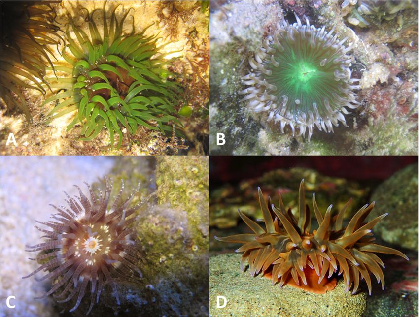
public are not even aware that they are animals rather than plants, let alone venomous ones (Figure 1).
Toxins 2018, 10, 36; doi:10.3390/toxins10010036 www.mdpi.com/journal/toxins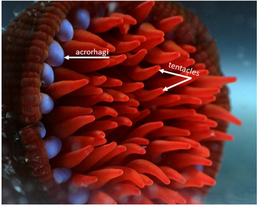
Toxins 2018, 10, 36 2 of 15
Toxins 2018, 10, 36 2 of 14
While most injuries
While most injuriescaused
causedbyby seasea anemones
anemones areare associated
associated withwith
skin skin rashes
rashes and oedema,
and oedema, more
more been reported for several species, including Actinodendron
extreme reactions have been reported for several species, including Actinodendron plumosum and
extreme reactions have plumosum and
other
other species
species from
from the
the family
family Actinodendronidae
Actinodendronidae (species(species from
from this
this family
family are
are collectively
collectively known
known as as
the hell’s fire anemones), Telmatactis species, Phyllodiscus semoni (night or wasp anemone),
the hell’s fire anemones), Telmatactis species, Phyllodiscus semoni (night or wasp anemone), Actinia Actinia equina
(beadlet anemone)
equina (beadlet and Anemonia
anemone) sulcata (the
and Anemonia snakelocks
sulcata anemone,
(the snakelocks synonomy
anemone, Anemonia
synonomy viridis) [10,11].
Anemonia viridis)
For
[10,11]. For example, a swimmer lost consciousness and underwent cardiopulmonary arreststung
example, a swimmer lost consciousness and underwent cardiopulmonary arrest after being after
by the stung
being sea anemone,
by the sea Actinia equina,
anemone, although
Actinia thisalthough
equina, may havethis beenmaya consequence
have been a of an anaphylactic
consequence of an
reaction
anaphylacticfollowing
reactionprior exposure
following to unknown
prior exposure to seaunknown
anemones sea[12]. Sea anemones
anemones [12]. Sea from the family
anemones from
Aliciideae are known to be particularly dangerous to humans, with severe
the family Aliciideae are known to be particularly dangerous to humans, with severe reactionsreactions observed following
contact
observed with both Triactis
following contactproducta
with [13]
bothand P. semoni
Triactis [10]. [13]
producta andP. P.
In fact, semoni is responsible
semoni [10]. In fact,forP.one of the
semoni is
few fatalitiesfor
responsible to one
resultof from seafatalities
the few anemonetoenvenomation
result from sea [14]. The venom
anemone from P. semoni
envenomation has caused
[14]. The venom
acute
from P. renal failure
semoni hasincaused
humans, with
acute a protein
renal failuretoxin (PsTX-115)
in humans, withfrom this venom
a protein toxincausing severe
(PsTX-115) kidney
from this
damage in rat models [15].
venom causing severe kidney damage in rat models [15].
Figure 1. Sea anemone species that are locally abundant in the waters off south-eastern Queensland,
Figure 1. Sea anemone species that are locally abundant in the waters off south-eastern Queensland,
Australia; these species have been largely unexplored for their toxin contents and are currently under
Australia; these species have been largely unexplored for their toxin contents and are currently under
investigation in
investigation in our
our labs
labs to
to identify
identify new
new peptide
peptide and
and protein
protein toxins
toxins with
with potentially
potentially novel
novel functions.
functions.
(A) Aulactinia veratra (B) undescribed species of Anthopleura (C) Calliactis polypus (D) Actinia
(A) Aulactinia veratra (B) undescribed species of Anthopleura (C) Calliactis polypus (D) Actinia australiensis.
australiensis.
Photo credit: Photo credit: Ana Pavasovic.
Ana Pavasovic.
2. Venom Apparatus
2. Venom Apparatus
Sea anemones, in common with other members of the phylum Cnidaria, possess numerous
Sea anemones, in common with other members of the phylum Cnidaria, possess numerous
specialized stinging cells (cnidocytes) that are widely distributed throughout the body [16]. These
specialized stinging cells (cnidocytes) that are widely distributed throughout the body [16].
stinging cells are equipped with organelles known as nematocysts (cnidae), which contain small
These stinging cells are equipped with organelles known as nematocysts (cnidae), which contain small
threads that are forcefully everted when stimulated mechanically or chemically [17]. These
threads that are forcefully everted when stimulated mechanically or chemically [17]. These nematocysts
nematocysts contain a complex cocktail of toxins that is used to envenomate predatory and prey
contain a complex cocktail of toxins that is used to envenomate predatory and prey species upon
species upon discharge [1,3,6]. Nematocysts show significant heterogeneity in their density and
discharge [1,3,6]. Nematocysts show significant heterogeneity in their density and morphology across
morphology across different structures within sea anemones [18]. For example, in Actinia tenebrosa
(or its northern hemisphere relatives Actinia equina and Actinia fragacea), acrorhagi (which are used in
aggressive intra-specific combat (Figure 2)) contain holotrichs and basitrichs [19] (Figure 3), while the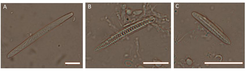
Toxins 2018, 10, 36                                                                                                3 of 15

different structures within sea anemones [18]. For example, in Actinia tenebrosa (or its northern
hemisphere       relatives Actinia equina and Actinia fragacea), acrorhagi (which are used in aggressive
Toxins 2018, 10, 36                                                                                         3 of 14
intra-specific combat (Figure 2)) contain holotrichs and basitrichs [19] (Figure 3), while the tentacles
(which
tentaclesare(which
              used inareprey  capture
                            used      andcapture
                                 in prey   defence)and
                                                    contain basitrichs
                                                       defence)        andbasitrichs
                                                                  contain  spirocysts; basitrichs
                                                                                     and          are also
                                                                                          spirocysts;      found
                                                                                                       basitrichs
in the  mesenteric      filaments,  column,  pedal  disc and  actinopharynx   [19] (Figure  3).  The
are also found in the mesenteric filaments, column, pedal disc and actinopharynx [19] (Figure 3). Thedifferences
in cnidae composition
differences                   are related toare
                in cnidae composition         differences in differences
                                                 related to  the functional  specialization
                                                                          in the  functional ofspecialization
                                                                                                 morphological   of
structures,   i.e., the capture  of prey (crustaceans, small fish), defence against predators   and intraspecific
morphological structures, i.e., the capture of prey (crustaceans, small fish), defence against predators
aggression     [19–22].
and intraspecific      aggression [19–22].

      Figure 2.
      Figure  2. This
                  Thisphotograph
                        photographshows
                                     showsthe
                                            thebright blue
                                                 bright    acrorhagi
                                                        blue          used
                                                              acrorhagi  usedin intraspecific combat
                                                                                in intraspecific combat[20][20]
                                                                                                            andand
                                                                                                                 red
      tentacles  used  in prey capture and  defence  against predators   of the  Australian  sea anemone,
      red tentacles used in prey capture and defence against predators of the Australian sea anemone,        Actinia
      tenebrosa.
      Actinia     Both structures
               tenebrosa.         have a high
                            Both structures     density
                                             have  a highof density
                                                            nematocysts,   but they have
                                                                     of nematocysts,     butdifferent nematocysts
                                                                                              they have    different
      complementscomplements
      nematocysts     (for example, (for
                                     holotrichs are abundant
                                         example,    holotrichsinare
                                                                   acrorhagi
                                                                     abundant  andinbasitrichs
                                                                                     acrorhagiinand
                                                                                                  tentacles). Photo
                                                                                                      basitrichs  in
      credit: Chloe
      tentacles).    vancredit:
                  Photo   der Burg.
                                Chloe van der Burg.

      Figure 3.3.Photomicrographs
                  Photomicrographs  of of cnidae
                                       cnidae fromfrom   Actinia
                                                    Actinia       tenebrosa.
                                                            tenebrosa.        (A) holotrich
                                                                       (A) holotrich         from acrorhagi;
                                                                                     from acrorhagi;            (B)
                                                                                                      (B) spirocyst
      spirocyst
      from       from tentacles;
            tentacles;           (C) basitrich
                        (C) basitrich           from tentacles.
                                       from tentacles.           Scale are
                                                          Scale bars    bars5 are
                                                                               µm5 in
                                                                                    µ in each
                                                                                       each    case. Photo
                                                                                            case.    Photo credit:
      Michela Mitchell.
                Mitchell.

3. Peptide
3. Peptide Toxins
           Toxins from
                  from Anemones
                       Anemones
     While many
     While   manyfamilies
                    familiesof of
                               seasea anemone
                                   anemone      toxins
                                            toxins havehave   been described
                                                        been described   alreadyalready  [7,23],still
                                                                                 [7,23], much     much    still
                                                                                                      remains
remains   to be discovered   about  toxins in this group. In fact, there are  only 236  peptide   or
to be discovered about toxins in this group. In fact, there are only 236 peptide or protein toxins in  protein
toxins in the manually curated ToxProt database [24], which have been isolated from just 45 sea
anemone species. This means that fewer than four percent (45 of more than 1100 species) of all sea
anemones have had their venom peptides and proteins examined. Currently, anemone toxins can be
classified into 15 known families (Table 1), but 19 toxin peptides and proteins remain largely
uncharacterized. The four most common toxin protein families isolated from anemones are the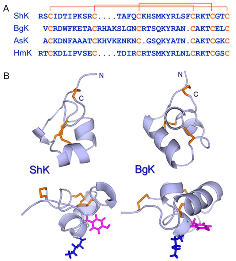
Toxins 2018, 10, 36                                                                                   4 of 15

the manually curated ToxProt database [24], which have been isolated from just 45 sea anemone
species. This means that fewer than four percent (45 of more than 1100 species) of all sea anemones
have had their venom peptides and proteins examined. Currently, anemone toxins can be classified
into 15 known families (Table 1), but 19 toxin peptides and proteins remain largely uncharacterized.
The four most common toxin protein families isolated from anemones are the actinoporin family,
sea anemone subfamily (30 proteins found in the ToxProt database); sea anemone sodium channel
inhibitory toxin family, type I subfamily (52 proteins found in the ToxProt database); sea anemone
type 3 (BDS) potassium channel toxin family (32 proteins found in the ToxProt database), and venom
Kunitz-type family, sea anemone type 2 potassium channel toxin subfamily (26 proteins found in the
ToxProt database). Of these four toxin protein families, only the actinoporin family, sea anemone
subfamily, has been found outside the Actinioidea superfamily of anemones based on sequences
currently available in the ToxProt database. In fact, 206 of the 236 toxin proteins in the ToxProt database
are from the Actinioidea superfamily. This indicates that the species that have been examined show a
strong taxonomic bias (Table 1), and we therefore need to examine the venom peptide profile from
sea anemone species from other superfamilies. Consequently, there is every reason for optimism
that the remaining 96% of species, particularly those distantly related to the superfamily Actinioidea,
will provide interesting new peptides, some of which will no doubt have therapeutic potential. Some of
the more venomous species, such as Telmatactis australiensis, Dofleinia armata and Triactis producta,
should be especially interesting in this context.

      Table 1. Characterized toxin protein families identified in sea anemone species in the current
      ToxProt database, and the number of proteins from each toxin protein family identified in each
      of the sea anemone superfamilies. Act—Actinioidea; Edw—Edwardsioidea; Met—Metridioidea;
      Acti—Actinernoidea.

                                                                                Anemone Superfamily
                           Toxin Protein Family
                                                                           Act      Edw    Met     Acti
   Actinoporin family, Sea anemone subfamily                               25        0      5         0
   Cnidaria small cysteine-rich protein (SCRiP) family                      2        0      1         0
   Peptidase M12A family                                                   0         1      0         0
   Phospholipase A2 family                                                 3         0      1         0
   Sea anemone 8 toxin family                                               5        0      0         0
   Sea anemone short toxin (type III) family                                8        0      0         0
   Sea anemone sodium channel inhibitory toxin family                       0        0      3         0
   Sea anemone sodium channel inhibitory toxin family, Type I subfamily    52        0      0         0
   Sea anemone sodium channel inhibitory toxin family, Type II subfamily   13        9      0         1
   Sea anemone structural class 9a family                                   6        0      0         0
   Sea anemone type 1 potassium channel toxin family, Type 1a subfamily     9        0      0         0
   Sea anemone type 1 potassium channel toxin family, Type 1b subfamily    11        0      1         0
   Sea anemone type 3 (BDS) potassium channel toxin family                 32        0      0         0
   Sea anemone type 5 potassium channel toxin family                        1        1      1         0
   Venom Kunitz-type family, Sea anemone type 2 potassium channel toxin
                                                                           26        0      0         0
   subfamily
   Unknown                                                                 15        0      4         0

      This article will focus mainly on ShK domains (which, in sea anemones, are members of the type 1
potassium channel toxin family, Type 1b subfamily, Table 1) because of their demonstrated therapeutic
potential and broad distribution in nature, as documented in the next section. However, several other
classes of peptide toxins from sea anemones have been investigated as therapeutic leads or
pharmacological tools. For example, the sodium channel toxins anthopleurin-A and -B showed
initial promise as positive inotropes for use in cardiovascular disease [25,26], although they and
their homologues from other anemones, for example ATX-I and -II and ShI [27,28], have also proven
to be valuable probes of site 3 on voltage-gated sodium channels [29], which mediates channel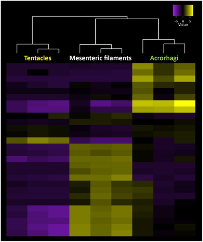
Toxins 2018, 10, 36                                                                                   5 of 15

inactivation. APETx2, from Anthopleura elegantissima, inhibits the acid-sensing ion channel ASIC3,
which is a proton-gated Na+ channel that has been implicated in pain transduction associated with
acidosis in inflamed or ischemic tissues [30]. However, this peptide also inhibits NaV 1.2 and NaV 1.8
channels, and to a lesser extent NaV 1.6 [31], as well as the human ether-a-go-go-related (hERG)
potassium channel (KV 11.1) [32]. APETx1 was identified initially as a gating modifier of the hERG
channel [33], but is also promiscuous, inhibiting mammalian NaV 1.2–NaV 1.6 and NaV 1.8 channels [31].
Recently, a new member of the APETx family, APETx4, was shown to have activity on a potential
anti-cancer target, the human ether-à-go-go channel (hEag1 or KV 10.1), but this peptide also inhibits
other KV and NaV channels [34]. The demonstrated lack of target specificity for members of the APETx
family limits their application as pharmacological tools, although analogues with specific mutations
have the potential to overcome this limitation [31]. Peptides from the sea anemone Heteractis crispa
(APHC1, APHC2 and APHC3) are active on TRPV1 receptors [35,36].
     The structures of APETx1, APETx2 and BDS-I (which acts on channels containing KV 3 subunits,
including KV 3.4 [37], but also modulates NaV channels [38]) are similar to those of the Na+ -channel
toxins such as AP-A although quite distinct from those of the ShK/BgK family of toxins (see below).
As noted previously [5], sea anemones use common structural scaffolds to create blockers for distinct
targets (AP-A, APETx1 and APETx2 act on VGSC, hERG and ASIC channels, respectively), while also
using different scaffolds (all-β in APETx1 vs. all-α in ShK) to block similar channels (hERG and
KV 1, respectively).
     Recent proteomic analyses of the venoms of Stichodactyla haddoni [39] and Aulactinia japonicus
(designated Cnidopus japonicas in the title of the paper) [40] identified many new toxin families,
a number of them with novel cysteine frameworks. A similar analysis of the mucus of Heteractis magnifica
revealed the presence of hundreds of peptides [41]. No doubt some of these new peptide families
will prove to have useful pharmacological properties and may eventually become therapeutic leads.
The next section focuses on a peptide for which this clearly is the case.

4. Potassium Channel Blockers from Sea Anemones: Therapeutic Leads for the Treatment of
Autoimmune Diseases
      The voltage-gated K+ channel KV 1.3 is involved in the activation of a sub-set of T lymphocytes
known as effector memory T (TEM ) cells as it regulates the membrane potential during activation by
allowing K+ efflux to counterbalance the influx of Ca2+ from intracellular stores and through CRAC
channels [42]. Blocking KV 1.3 channels in TEM cells blocks their activation and proliferation. As TEM
cells are key mediators of autoimmune diseases, KV 1.3 blockers are attractive leads as a new class of
therapeutic for these conditions [43,44].
      Several peptide toxins from scorpions were found to be potent blockers of KV 1.3 [45,46], but did
not progress to clinical trials for a variety of reasons, including the lack of desired specificity for this
channel over related KV 1 channels [47]. Around the same time, a novel peptide, ShK, was isolated
from the Caribbean sun anemone, Stichodactyla helianthus [48]. This 35-residue peptide was found to
be a potent competitive inhibitor of α-dendrotoxin binding to rat brain synaptosomes and blocked
K+ current in dorsal root ganglion cells. Its amino acid sequence [48] (Figure 4), disulfide bonding
pattern [49] and solution structure [50] were all very different from the scorpion toxins, although Lys22
and Tyr23 in ShK, the two key residues for KV 1.3 blockade, are spatially conserved in an arrangement
common to KV -channel blocking peptides from widely different species [51]. ShK has a very high
affinity (Ki ~10 pM) for KV 1.3 channels but also displays high pM affinity for KV 1.1, KV 1.4 and KV 1.6,
which are present in brain and cardiac tissues [52,53]. In order for this promising peptide toxin to
progress to clinical trials, therefore, more selective analogues had to be developed [54]. This eventually
led to ShK-186, which had a 100-fold improvement in selectivity for KV 1.3 over KV 1.1, KV 1.4 and
KV 1.6 [55].
      ShK and its analogues had already shown efficacy in animal models of human autoimmune
diseases such as multiple sclerosis and rheumatoid arthritis [56,57], and preclinical testing of ShK-186
Toxins 2018, 10, 36 6 of 15
yielded favourable results in both rats and monkeys [58]. Unexpectedly, ShK-186 was found to have a
long half-life at the site of sub-cutaneous injection, resulting in sustained high pM levels in plasma and
a prolonged therapeutic efficacy [58]. ShK-186, which is now known as dalazatide, completed Phase
1a and 1b trials in 2016. The Phase 1b trial in mild-to-moderate plaque psoriasis patients showed that
dalazatide was well tolerated and reduced psoriatic skin lesions [59]. It is expected to begin Phase 2a
trails in 2018. Dalazatide is being advanced as a treatment for various autoimmune diseases, including
inclusion body myositis, lupus, ANCA vasculitis, multiple sclerosis, psoriasis, psoriatic arthritis,
rheumatoid arthritis, type 1 diabetes and inflammatory bowel diseases [44,45]. New analogues of ShK
with
Toxinsgood selectivity
2018, 10, 36 for KV 1.3 over other KV 1 channels have also been developed [60–64]. 6 of 14
Figure 4.
Figure 4. (A)
(A)Amino
Aminoacid acidsequences
sequences of of
ShK ShK(UniProt
(UniProt entry P29187)
entry [48],[48],
P29187) BgKBgK (P29186) [65], [65],
(P29186) AsK
(Q9TWG1), also known as kaliseptine [66], and HmK (O16846) [67] with the
AsK (Q9TWG1), also known as kaliseptine [66], and HmK (O16846) [67] with the three disulfide three disulfide bonds
indicated.
bonds (B) Structures
indicated. of ShKof(pdb
(B) Structures ShKid 1ROO)
(pdb [50] and
id 1ROO) [50]BgK
and(pdb
BgK id 1BGK)
(pdb [51] are
id 1BGK) [51]shown, with
are shown,
backbones
with in lightblue
backbones and and
in lightblue disulfides in orange.
disulfides The The
in orange. sideside
chains of the
chains of Lys andand
the Lys TyrTyr
thatthat
constitute the
constitute
functional
the dyad
functional dyad[51][51]
in each peptide
in each areare
peptide shown
shown in in
blue and
blue andmagenta,
magenta,respectively.
respectively.ThisThisdyad
dyadis
isdisplayed
displayedonona ahelical
helicalscaffold
scaffoldininShK
ShK but
but aa β-sheet
β-sheet in the scorpion
scorpion toxins
toxins charybdotoxin
charybdotoxin and and
margatoxin(not
margatoxin (notshown).
shown).
Theprogress
The progressof ofShK
ShKanalogues
analoguestowards
towardsthe
theclinic
clinic has
has stimulated
stimulated interest
interest in
in this
this class
class of
of peptides,
peptides,
driven partly by the potential for finding additional members of this family of
driven partly by the potential for finding additional members of this family of peptides in sea
peptides in sea
anemones that might be selective for KV1.3 or the many other KV channels that are potential
anemones that might be selective for KV 1.3 or the many other KV channels that are potential therapeutic
therapeutic targets [45]. As a result, it has become apparent that the ‘ShK fold’, typified by ShK [50]
targets [45]. As a result, it has become apparent that the ‘ShK fold’, typified by ShK [50] and BgK [51],
and BgK [51], is widely distributed not only in sea anemones and other cnidarians [68–70], but also
is widely distributed not only in sea anemones and other cnidarians [68–70], but also throughout
throughout nature. The next section highlights this broad distribution.
nature. The next section highlights this broad distribution.
5. ShK:
5. ShK: A
A Privileged
Privileged Scaffold
Scaffold in
in Nature?
Nature?
In 2010,
In 2010, the
theSimple
SimpleModular
ModularArchitecture
Architecture Research
ResearchTool (SMART)
Tool (SMART)database (http://smart.embl-
database (http://smart.
heidelberg.de/) predicted the existence of a large superfamily of proteins
embl-heidelberg.de/) predicted the existence of a large superfamily of proteins that contain
that domains
contain
resembling
domains ShK or BgK,
resembling ShKwhich werewhich
or BgK, referred to collectively
were referred toascollectively
ShKT domains [71]. These
as ShKT domains
domains [71].
were distributed across nearly 400 proteins from both the plant and animal kingdoms, including
These domains were distributed across nearly 400 proteins from both the plant and animal kingdoms,
Viridiplantae, Arabidopsis thaliana, Oryza sativa, and green alga Ostreococcus sp.; Protozoa,
Cryptosporidium parvum; Cnidaria, sea anemones, hydra, and jellyfish; Echinodermata, sea urchin;
Mollusca, bivalve clams and oysters; Ciona, sea squirt Ciona intestinalis; Actinopterygii, zebrafish
Danio rerio and pufferfish Takifugu rubripes; Caenorhabditis, C. elegans and C. brigssae; Rhabditida,
rhabditid nematodes other than Caenorhabditis sp.; Ophidia, snakes; Xenopus, Xenopus tropicalis;
Aves, chicken Gallus gallus; Mammalia. Many of these proteins (~70) were metallopeptidases,Toxins 2018, 10, 36 7 of 15
including Viridiplantae, Arabidopsis thaliana, Oryza sativa, and green alga Ostreococcus sp.; Protozoa,
Cryptosporidium parvum; Cnidaria, sea anemones, hydra, and jellyfish; Echinodermata, sea urchin;
Mollusca, bivalve clams and oysters; Ciona, sea squirt Ciona intestinalis; Actinopterygii, zebrafish
Danio rerio and pufferfish Takifugu rubripes; Caenorhabditis, C. elegans and C. brigssae; Rhabditida,
rhabditid nematodes other than Caenorhabditis sp.; Ophidia, snakes; Xenopus, Xenopus tropicalis; Aves,
chicken Gallus gallus; Mammalia. Many of these proteins (~70) were metallopeptidases, whereas
others were prolyl-4-hydroxylases, tyrosinases, peroxidases, oxidoreductases, or proteins containing
epidermal growth factor-like domains, thrombospondin-type repeats, or trypsin-like serine protease
domains [71].
The only human protein containing a ShKT domain in the SMART data base was matrix
metalloprotease 23 (MMP23) [72,73]. A second human protein with an ShKT domain, microfibril
associated protein MFAP2, was not mentioned in the SMART database at that time [74].
The cysteine-rich secretory proteins (Crisp), which are found predominantly in the mammalian male
reproductive tract as well as in the venom of reptiles, are two-domain proteins, one domain of which
is an ShKT domain. For example, murine Tpx-1 (testis specific protein-1) contains two subdomains,
one of which has a similar fold to BgK and ShK [75]. The Tpx-1 Crisp domain inhibited the cardiac
ryanodine receptor (RyR2) and activated the skeletal RyR1.
By 2014, the SMART database identified 668 proteins that contained 1315 ShKT domains.
The largest family was found in worms, with 276 of those 668 proteins coming from
Caenorhabditis elegans, C. briggsae, Brugia malayi, B. pahangi, Ancylostoma ceylanicum, Schistosoma mansoni
and Toxocara canis [76]. This prompted an investigation of whether worm ShKTs share structural
similarity to ShK, block KV 1.3, and exhibit immunomodulatory activity. Based on phylogenetic analysis,
two worm peptides were selected for study: AcK1, a 51-residue peptide expressed in the anterior
secretory glands of both the dog-infecting hookworm Ancylostoma caninum and the human-infecting
hookworm Ancylostoma ceylanicum, and BmK1, the C-terminal domain of a metalloprotease from the
filarial worm Brugia malayi. These peptides proved to have helical structures closely resembling that of
ShK [76]. They also blocked KV 1.3 channels, selectively suppressed the function of TEM lymphocytes,
and inhibited delayed-type hypersensitivity. It was suggested that ShK-like worm peptides may be
among the active principles that contribute to the well-known protective effect of parasitic worms in
autoimmune diseases [76], although further work is required to confirm this hypothesis.
Recently, we have investigated several new ShK-like peptides identified in anemone
transcriptomes. One such peptide is AsK132958, from Anemonia sulcata, which had an ShKT
cysteine framework but was six amino acid residues shorter [77]. The disulfide connectivities and
structural scaffold were very similar to those of ShK, although the structure was more constrained.
However, AsK132958 showed no activity against grass shrimp, Artemia nauplii, or any of the KV
channels tested, owing partly to the absence of a functional Lys-Tyr dyad. It appears that Lys19,
which would be expected to occupy the pore of the channel, was not sufficiently accessible for
binding, and therefore that AsK132958 must have a distinct functional role that does not involve KV
channels. The evolutionary relationship between AsK132958 and other ShK-like amino acid sequences
is unclear. AsK132958 may represent the shortest peptide sequence that can support a stable ShKT
fold, although this remains to be confirmed.
Another peptide, identified in the transcriptome of an Oulactis species, was similar to BgK in
terms of amino acid sequence and three-dimensional structure, and furthermore contained a Lys-Tyr
dyad, but was inactive against KV channels tested to date (our unpublished results). These findings
highlight the likely diversity of functions supported by this versatile scaffold.
At the time of writing, the SMART database includes 3345 ShKT domains spread across
1797 proteins, a huge increase over the numbers documented in 2010 [71] and 2014 [76]. These domains
are found mainly in animals and plants, but also occur in fungi, viruses and undefined kingdoms.
With the dramatic expansion in the number of genomes and transcriptomes spanning all kingdoms,Toxins 2018, 10, 36 8 of 15
this number is set to increase rapidly. The next section discusses these developments in more detail
and outlines how we might begin to assess the functions of these domains.
6. New Methods for the Large-Scale Detection of Known and Novel Peptide Toxins
The large-scale identification of new peptide toxins, as well as the evolutionary analysis of
known peptide toxins, in sea anemones has been limited by a lack of genomic resources for many
species. This is changing rapidly, however, as transcriptome and genome assemblies are being
generated using next generation sequencing platforms for a number of sea anemone species [39,78–80].
Currently, the genome sequences of Nematostella vectensis [81] and Exaiptasia pallida are publicly
available, as well as transcriptomes of ~30 anemone species from a range of anemone superfamilies.
These genomic resources have enabled the in-depth characterization of gene families in sea anemones,
including immune gene families [82], and some detailed investigations of select toxin gene families
(e.g., actinoporins, which are cytolytic proteins [83]). These genomic resources can be used to study
the number, distribution and evolution of a range of candidate toxin gene families in anemones.
For example, we were able to identify 71 and 90 non-redundant open reading frames with ShKT
domains from previously-published, high-quality transcriptome assemblies for Calliactis polypus and
Actinia tenebrosa [82], respectively (our unpublished results). From these peptide sequences, we were
able to identify canonical ShK (Type I KTxs) toxin peptides with a single domain, as well as a many
proteins with multiple ShKT domains, and in some instances ShKT domains with other toxin domains.
Proteins with multiple ShKT domains have been identified previously in other anemone species,
including Megalactis griffithsi and Anemonia sulcata [79]. Further information about the expression
of toxin gene families can be derived by application of state-of-the-art transcriptomic based gene
expression studies across nematocyst-rich tissues in anemone species.
Gene expression analysis across morphological structures in sea anemones has shown that many
toxin genes are differentially expressed across tissues, presumably resulting in the production of
distinct venom combinations in different structures [79]. In fact, specific genes from toxin gene families
are expressed precisely in a region-specific pattern, with different gene copies expressed in specific
structures, such as tentacles, acrorhagi or mesenteric filaments, as exemplified for the venom Kunitz
peptides in Figure 5. This highlights that different genes from known toxin gene families show strong
regionalization in their expression patterns across nematocyst-rich tissues in a pattern consistent with
changes in nematocyst morphology and density (our unpublished results). Testing whether different
nematocyte populations are contributing directly to the changes in toxin gene expression across
tissues will require an examination of single-cell gene expression changes in nematocyte populations
isolated from specific tissues. The analysis of gene expression profiles from single cell populations
has improved greatly over recent years [84] and has led to a precise understanding of the function
of individual cell types in a range of species [85]. Determining single-cell venom gene expression
patterns in nematocyte populations will provide unprecedented insight into the evolution of venom
composition and how functionally different venoms can be produced by a single animal. While this
information is useful in understanding the evolution and composition of venoms, the identification of
peptide toxins with new modes of action will require proteomic analyses of the venom.understanding of the function of individual cell types in a range of species [85]. Determining single-
cell venom gene expression patterns in nematocyte populations will provide unprecedented insight
into the evolution of venom composition and how functionally different venoms can be produced by
a single animal. While this information is useful in understanding the evolution and composition of
venoms,
Toxins 2018,the
10, identification
36 of peptide toxins with new modes of action will require proteomic analyses
9 of 15
of the venom.
Figure 5. Gene
Figure 5. Geneexpression
expression patterns
patterns for for all gene
all gene copies
copies of theof the venom
venom Kunitz Kunitz geneacross
gene family family across
tentacles,
tentacles,
mesentericmesenteric filaments
filaments and acrorhagiandin acrorhagi inTissue-specific
A. tenebrosa. A. tenebrosa. Tissue-specific expression
expression can be observed can
amongbe
observed among copies across the three tissues, with significantly upregulated transcripts in
copies across the three tissues, with significantly upregulated transcripts in yellow and downregulatedyellow
and downregulated
transcripts in purpletranscripts in purple
(our unpublished (our unpublished data).
data).
High-throughput proteomic analyses of sea anemone venoms have been greatly accelerated
through the use of mass spectrometry [86] coupled with the availability of complete genome and
transcriptome sequences for sea anemones species. MALDI imaging [87,88] also offers the prospect
of mapping the tissue distributions of numerous peptides, and thus providing a clue to their
possible function in the anemone. Recent proteomic research has assayed venom composition in
the nematocytes of a few anemone species [69,86], revealing a high frequency of novel peptides
and proteins in nematocysts, and in some cases detecting peptides that had not been identified
in earlier proteomics studies [89–91]. Many of these represent novel candidate toxins and show
little overlap among species, highlighting the potential for proteogenomic approaches to identify
previously uncharacterized toxins. Another recent study used a combination of proteomic and
transcriptomic techniques to analyse the protein composition of milked venom from the sea anemone
Stichodactyla haddoni [39]; the milked venom contained 23 putative toxin families, 12 of which showed
no identity to any known peptide or protein in current databases. The identification of these
12 cysteine-rich, but previously unknown, putative toxins further emphasizes that a proteogenomic
approach can greatly accelerate the discovery of novel proteins with potential therapeutic applications.
Continual improvements in the efficiency of peptide synthesis and recombinant expression will
facilitate the production of such peptides for biological evaluation, although the challenge will remain
to define the native disulfide connectivities for novel sequences containing multiple cysteine residues.
New peptides need to be assessed against a range of targets, and in multiple functional assays, in order
to identify those with the potential to be new therapeutic leads or at least pharmacological tools.
For the most part, these assays will focus on mammalian targets, leaving open the intriguing but
often neglected question of what function(s) these peptides have in the anemones in which they are
identified. As noted above, MALDI imaging studies of peptide distribution in the anemone should
assist in identifying their endogenous functions.Toxins 2018, 10, 36 10 of 15
It is to be hoped that new bioinformatic approaches, such as weighted gene co-expression network
analysis, may provide preliminary information on the function of candidate peptides and proteins.
Weighted gene co-expression analysis categorizes genes into groups whose expression levels are highly
correlated across samples [92]. This approach has been used to identify gene clusters with functions in
development and lipid metabolism, among others [93,94]. In sea anemones, identifying gene clusters
with correlated expression levels that contain known toxins may offer a way to detect novel toxin
genes with similar or novel functions.
7. Conclusions
Sea anemones remain relatively under-explored as a source of peptide toxins, although that is
beginning to change as the transcriptomes and genomes of several species from different geographic
regions and different habitats are being investigated. Proteomics studies on sea anemones [39] are more
challenging than for other venomous species, where the venom apparatus can be readily milked or
dissected, but ongoing enhancements in the resolution and sensitivity of liquid chromatography-mass
spectrometry methods will enable characterization of the complete peptide repertoires of even limited
quantities of venom when it can be obtained.
The progress of an analogue of ShK through clinical trials for autoimmune diseases emphasizes
the promise of this scaffold and, more broadly, of sea anemone peptides. The abundance of ShKT
domains in nature and their distribution across different phyla pose questions about the diversity
of functions supported by this scaffold. Defining the relationships among sequence, structure and
function remains one of the exciting challenges of ongoing studies.
Acknowledgments: R.S.N. acknowledges fellowship support from the Australian National Health and Medical
Research Council. This work was supported in part by a grant from the Australian Research Council (LP150100621).
We thank Michela Mitchell and Joachim Surm for helpful comments on the manuscript.
Author Contributions: All authors contributed to writing the manuscript.
Conflicts of Interest: The authors declare no conflict of interest.
References
1. Beress, L. Biologically active compounds from coelenterates. Pure Appl. Chem. 1982, 54, 1981–1994. [CrossRef]
2. Norton, R.S. Structure and structure-function relationships of sea anemone proteins that interact with the
sodium channel. Toxicon 1991, 29, 1051–1084. [CrossRef]
3. Honma, T.; Shiomi, K. Peptide toxins in sea anemones: Structural and functional aspects. Mar. Biotechnol.
2006, 8, 1–10. [CrossRef] [PubMed]
4. Shiomi, K. Novel peptide toxins recently isolated from sea anemones. Toxicon 2009, 54, 1112–1118. [CrossRef]
[PubMed]
5. Norton, R.S. Structures of sea anemone toxins. Toxicon 2009, 54, 1075–1088. [CrossRef] [PubMed]
6. Norton, R.S. Sea anemone venom peptides. In Handbook of Biologically Active Peptides; Kastin, A.J., Ed.;
Elsevier (Academic): San Diego, CA, USA, 2013; pp. 430–436.
7. Jouiaei, M.; Yanagihara, A.A.; Madio, B.; Nevalainen, T.J.; Alewood, P.F.; Fry, B.G. Ancient venom systems:
A review on cnidaria toxins. Toxins 2015, 7, 2251–2271. [CrossRef] [PubMed]
8. Tibballs, J. Australian venomous jellyfish, envenomation syndromes, toxins and therapy. Toxicon 2006, 48,
830–859. [CrossRef] [PubMed]
9. Tibballs, J.; Li, R.; Tibballs, H.A.; Gershwin, L.A.; Winkel, K.D. Australian carybdeid jellyfish causing
“Irukandji syndrome”. Toxicon 2012, 59, 617–625. [CrossRef] [PubMed]
10. Mizuno, M.; Nishikawa, K.; Yuzawa, Y.; Kanie, T.; Mori, H.; Araki, Y.; Hotta, N.; Matsuo, S. Acute renal
failure after a sea anemone sting. Am. J. Kidney Dis. 2000, 36, E10. [CrossRef] [PubMed]
11. Mizuno, M. Envenomation by cnidarians and renal injuries. In The Cnidaria, Past, Present and Future: The World
of Medusa and Her Sisters; Goffredo, S., Dubinsky, Z., Eds.; Springer: Berlin, Germany, 2016; pp. 623–636.
12. Gracia Bara, M.T.; Iriarte, P.; Pineda, F. Allergy to Actinia equina and Anemona viridis. Allergy 2006, 61,
1151–1152. [CrossRef] [PubMed]Toxins 2018, 10, 36 11 of 15
13. Levy, S.; Masry, D.; Halstead, B.W. Report of stingings by the sea anemone Triactis producta Klunzinger from
Red Sea. Clin. Toxicol. 1970, 3, 637–643. [CrossRef] [PubMed]
14. Erhardt, H.; Knop, D. Corals: Indo-Pacific Field Guide. Gorgonians, Soft Corals, Stony Corals, Sea Anemones;
IKAN Unterwasserarchiv: ConchBooks, Frankfurt, Germany, 2005.
15. Mizuno, M.; Nozaki, M.; Morine, N.; Suzuki, N.; Nishikawa, K.; Morgan, B.P.; Matsuo, S. A protein toxin from
the sea anemone Phyllodiscus semoni targets the kidney and causes a severe renal injury with predominant
glomerular endothelial damage. Am. J. Pathol. 2007, 171, 402–414. [CrossRef] [PubMed]
16. Fautin, D.G. Structural diversity, systematics, and evolution of cnidae. Toxicon 2009, 54, 1054–1064. [CrossRef]
[PubMed]
17. Watson, G.M.; Hessinger, D.A. Cnidocyte mechanoreceptors are tuned to the movements of swimming prey
by chemoreceptors. Science 1989, 243, 1589–1591. [CrossRef] [PubMed]
18. Kass-Simon, G.; Scappaticci, A.A. The behavioral and developmental physiology of nematocysts. Can. J. Zool.
2002, 80, 1772–1794. [CrossRef]
19. Schama, R.; Mitchell, M.; Sole-Cava, A.M. Actinia ebhayiensis sp. nov., a new species of sea anemone
(Anthozoa: Actiniaria: Actiniidae) from South Africa. J. Mar. Biol. Assoc. UK 2012, 92, 885–894. [CrossRef]
20. Ayre, D.J. Inter-genotype aggression in the solitary sea anemone Actinia tenebrosa. Mar. Biol. 1982, 68, 199–205.
[CrossRef]
21. Reft, A.J. Understanding the Morphology and Distribution of Nematocysts in Sea Anemones and Their Relatives;
The Ohio State University: Columbus, OH, USA, 2012.
22. Macrander, J.; Brugler, M.R.; Daly, M. A RNA-seq approach to identify putative toxins from acrorhagi in
aggressive and non-aggressive Anthopleura elegantissima polyps. BMC Genom. 2015, 16, 221. [CrossRef]
[PubMed]
23. Frazao, B.; Vasconcelos, V.; Antunes, A. Sea anemone (Cnidaria, Anthozoa, Actiniaria) toxins: An overview.
Mar. Drugs 2012, 10, 1812–1851. [CrossRef] [PubMed]
24. Jungo, F.; Bairoch, A. Tox-Prot, the toxin protein annotation program of the Swiss-Prot protein knowledgebase.
Toxicon 2005, 45, 293–301. [CrossRef] [PubMed]
25. Reimer, N.S.; Yasunobu, C.L.; Yasunobu, K.T.; Norton, T.R. Amino acid sequence of the
Anthopleura xanthogrammica heart stimulant, anthopleurin-B. J. Biol. Chem. 1985, 260, 8690–8693. [PubMed]
26. Monks, S.A.; Pallaghy, P.K.; Scanlon, M.J.; Norton, R.S. Solution structure of the cardiostimulant polypeptide
anthopleurin-B and comparison with anthopleurin-A. Structure 1995, 3, 791–803. [CrossRef]
27. Schweitz, H.; Vincent, J.P.; Barhanin, J.; Frelin, C.; Linden, G.; Hugues, M.; Lazdunski, M. Purification and
pharmacological properties of eight sea anemone toxins from Anemonia sulcata, Anthopleura xanthogrammica,
Stoichactis giganteus, and Actinodendron plumosum. Biochemistry 1981, 20, 5245–5252. [CrossRef] [PubMed]
28. Kem, W.R.; Parten, B.; Pennington, M.W.; Price, D.A.; Dunn, B.M. Isolation, characterization, and amino acid
sequence of a polypeptide neurotoxin occurring in the sea anemone Stichodactyla helianthus. Biochemistry
1989, 28, 3483–3489. [CrossRef] [PubMed]
29. Cestele, S.; Catterall, W.A. Molecular mechanisms of neurotoxin action on voltage-gated sodium channels.
Biochimie 2000, 82, 883–892. [CrossRef]
30. Diochot, S.; Baron, A.; Rash, L.D.; Deval, E.; Escoubas, P.; Scarzello, S.; Salinas, M.; Lazdunski, M. A new sea
anemone peptide, APETx2, inhibits ASIC3, a major acid-sensitive channel in sensory neurons. EMBO J. 2004,
23, 1516–1525. [CrossRef] [PubMed]
31. Peigneur, S.; Beress, L.; Moller, C.; Mari, F.; Forssmann, W.G.; Tytgat, J. A natural point mutation changes both
target selectivity and mechanism of action of sea anemone toxins. FASEB J. 2012, 26, 5141–5151. [CrossRef]
[PubMed]
32. Jensen, J.E.; Cristofori-Armstrong, B.; Anangi, R.; Rosengren, K.J.; Lau, C.H.; Mobli, M.; Brust, A.;
Alewood, P.F.; King, G.F.; Rash, L.D. Understanding the molecular basis of toxin promiscuity: The analgesic
sea anemone peptide APETx2 interacts with acid-sensing ion channel 3 and hERG channels via overlapping
pharmacophores. J. Med. Chem. 2014, 57, 9195–9203. [CrossRef] [PubMed]
33. Zhang, M.; Liu, X.S.; Diochot, S.; Lazdunski, M.; Tseng, G.N. APETx1 from sea anemone
Anthopleura elegantissima is a gating modifier peptide toxin of the human ether-a-go-go-related potassium
channel. Mol. Pharmacol. 2007, 72, 259–268. [CrossRef] [PubMed]Toxins 2018, 10, 36 12 of 15
34. Moreels, L.; Peigneur, S.; Galan, D.T.; De Pauw, E.; Beress, L.; Waelkens, E.; Pardo, L.A.; Quinton, L.; Tytgat, J.
APETx4, a novel sea anemone toxin and a modulator of the cancer-relevant potassium channel KV 10.1.
Mar. Drugs 2017, 15, 287. [CrossRef] [PubMed]
35. Andreev, Y.A.; Kozlov, S.A.; Koshelev, S.G.; Ivanova, E.A.; Monastyrnaya, M.M.; Kozlovskaya, E.P.;
Grishin, E.V. Analgesic compound from sea anemone Heteractis crispa is the first polypeptide inhibitor
of vanilloid receptor 1 (TRPV1). J. Biol. Chem. 2008, 283, 23914–23921. [CrossRef] [PubMed]
36. Nikolaev, M.V.; Dorofeeva, N.A.; Komarova, M.S.; Korolkova, Y.V.; Andreev, Y.A.; Mosharova, I.V.;
Grishin, E.V.; Tikhonov, D.B.; Kozlov, S.A. TRPV1 activation power can switch an action mode for its
polypeptide ligands. PLoS ONE 2017, 12, e0177077. [CrossRef] [PubMed]
37. Yeung, S.Y.; Thompson, D.; Wang, Z.; Fedida, D.; Robertson, B. Modulation of Kv3 subfamily potassium
currents by the sea anemone toxin BDS: Significance for CNS and biophysical studies. J. Neurosci. 2005, 25,
8735–8745. [CrossRef] [PubMed]
38. Liu, P.; Jo, S.; Bean, B.P. Modulation of neuronal sodium channels by the sea anemone peptide BDS-I.
J. Neurophysiol. 2012, 107, 3155–3167. [CrossRef] [PubMed]
39. Madio, B.; Undheim, E.A.B.; King, G.F. Revisiting venom of the sea anemone Stichodactyla haddoni:
Omics techniques reveal the complete toxin arsenal of a well-studied sea anemone genus. J. Proteom.
2017, 166, 83–92. [CrossRef] [PubMed]
40. Babenko, V.V.; Mikov, A.N.; Manuvera, V.A.; Anikanov, N.A.; Kovalchuk, S.I.; Andreev, Y.A.; Logashina, Y.A.;
Kornilov, D.A.; Manolov, A.I.; Sanamyan, N.P.; et al. Identification of unusual peptides with new Cys
frameworks in the venom of the cold-water sea anemone Cnidopus japonicus. Sci. Rep. 2017, 7, 14534.
[CrossRef] [PubMed]
41. Sintsova, O.; Gladkikh, I.; Chausova, V.; Monastyrnaya, M.; Anastyuk, S.; Chernikov, O.; Yurchenko, E.;
Aminin, D.; Isaeva, M.; Leychenko, E.; et al. Peptide fingerprinting of the sea anemone Heteractis magnifica
mucus revealed neurotoxins, Kunitz-type proteinase inhibitors and a new β-defensin α-amylase inhibitor.
J. Proteom. 2018, 173, 12–21. [CrossRef] [PubMed]
42. Cahalan, M.D.; Chandy, K.G. The functional network of ion channels in T lymphocytes. Immunol. Rev. 2009,
231, 59–87. [CrossRef] [PubMed]
43. Chandy, K.G.; Wulff, H.; Beeton, C.; Pennington, M.; Gutman, G.A.; Cahalan, M.D. K+ channels as targets for
specific immunomodulation. Trends Pharmacol. Sci. 2004, 25, 280–289. [CrossRef] [PubMed]
44. Chandy, K.G.; Norton, R.S. Peptide blockers of Kv1.3 channels in T cells as therapeutics for autoimmune
disease. Curr. Opin. Chem. Biol. 2017, 38, 97–107. [CrossRef] [PubMed]
45. Norton, R.S.; Chandy, K.G. Venom-derived peptide inhibitors of voltage-gated potassium channels.
Neuropharmacology 2017, 127, 124–138. [CrossRef] [PubMed]
46. Pennington, M.W.; Czerwinski, A.; Norton, R.S. Peptide therapeutics from venom: Current status and
potential. Bioorg. Med. Chem. 2017. [CrossRef] [PubMed]
47. Bartok, A.; Toth, A.; Somodi, S.; Szanto, T.G.; Hajdu, P.; Panyi, G.; Varga, Z. Margatoxin is a non-selective
inhibitor of human Kv1.3 K+ channels. Toxicon 2014, 87, 6–16. [CrossRef] [PubMed]
48. Castañeda, O.; Sotolongo, V.; Amor, A.M.; Stocklin, R.; Anderson, A.J.; Harvey, A.L.; Engstrom, A.;
Wernstedt, C.; Karlsson, E. Characterization of a potassium channel toxin from the Caribbean Sea anemone
Stichodactyla helianthus. Toxicon 1995, 33, 603–613. [CrossRef]
49. Pohl, J.; Hubalek, F.; Byrnes, M.E.; Nielsen, K.R.; Woods, A.; Pennington, M.W. Assignment of
the three disulfide bonds in ShK toxin, a potent potassium channel blocker from the sea anemone
Stichodactyla helianthus. Lett. Pept. Sci. 1995, 1, 291–297. [CrossRef]
50. Tudor, J.E.; Pallaghy, P.K.; Pennington, M.W.; Norton, R.S. Solution structure of ShK toxin, a novel potassium
channel inhibitor from a sea anemone. Nat. Struct. Biol. 1996, 3, 317–320. [CrossRef] [PubMed]
51. Dauplais, M.; Lecoq, A.; Song, J.; Cotton, J.; Jamin, N.; Gilquin, B.; Roumestand, C.; Vita, C.; de Medeiros, C.L.;
Rowan, E.G.; et al. On the convergent evolution of animal toxins. Conservation of a diad of functional
residues in potassium channel-blocking toxins with unrelated structures. J. Biol. Chem. 1997, 272, 4302–4309.
[CrossRef] [PubMed]
52. Pennington, M.W.; Byrnes, M.E.; Zaydenberg, I.; Khaytin, I.; de Chastonay, J.; Krafte, D.S.; Hill, R.;
Mahnir, V.M.; Volberg, W.A.; Gorczyca, W.; et al. Chemical synthesis and characterization of ShK toxin:
A potent potassium channel inhibitor from a sea anemone. Int. J. Pept. Protein Res. 1995, 46, 354–358.
[CrossRef] [PubMed]Toxins 2018, 10, 36 13 of 15
53. Kalman, K.; Pennington, M.W.; Lanigan, M.D.; Nguyen, A.; Rauer, H.; Mahnir, V.; Paschetto, K.; Kem, W.R.;
Grissmer, S.; Gutman, G.A.; et al. ShK-Dap22, a potent Kv1.3-specific immunosuppressive polypeptide.
J. Biol. Chem. 1998, 273, 32697–32707. [CrossRef] [PubMed]
54. Chi, V.; Pennington, M.W.; Norton, R.S.; Tarcha, E.J.; Londono, L.M.; Sims-Fahey, B.; Upadhyay, S.K.;
Lakey, J.T.; Iadonato, S.; Wulff, H.; et al. Development of a sea anemone toxin as an immunomodulator for
therapy of autoimmune diseases. Toxicon 2012, 59, 529–546. [CrossRef] [PubMed]
55. Pennington, M.W.; Beeton, C.; Galea, C.A.; Smith, B.J.; Chi, V.; Monaghan, K.P.; Garcia, A.; Rangaraju, S.;
Giuffrida, A.; Plank, D.; et al. Engineering a stable and selective peptide blocker of the Kv1.3 channel in T
lymphocytes. Mol. Pharmacol. 2009, 75, 762–773. [CrossRef] [PubMed]
56. Beeton, C.; Wulff, H.; Barbaria, J.; Clot-Faybesse, O.; Pennington, M.; Bernard, D.; Cahalan, M.D.;
Chandy, K.G.; Beraud, E. Selective blockade of T lymphocyte K+ channels ameliorates experimental
autoimmune encephalomyelitis, a model for multiple sclerosis. Proc. Natl. Acad. Sci. USA 2001, 98,
13942–13947. [CrossRef] [PubMed]
57. Beeton, C.; Wulff, H.; Standifer, N.E.; Azam, P.; Mullen, K.M.; Pennington, M.W.; Kolski-Andreaco, A.; Wei, E.;
Grino, A.; Counts, D.R.; et al. Kv1.3 channels are a therapeutic target for T cell-mediated autoimmune
diseases. Proc. Natl. Acad. Sci. USA 2006, 103, 17414–17419. [CrossRef] [PubMed]
58. Tarcha, E.J.; Chi, V.; Munoz-Elias, E.J.; Bailey, D.; Londono, L.M.; Upadhyay, S.K.; Norton, K.; Banks, A.;
Tjong, I.; Nguyen, H.; et al. Durable pharmacological responses from the peptide drug ShK-186, a specific
Kv1.3 channel inhibitor that suppresses T cell mediators of autoimmune disease. J. Pharmacol. Exp. Ther.
2012, 342, 642–653. [CrossRef] [PubMed]
59. Tarcha, E.J.; Olsen, C.M.; Probst, P.; Peckham, D.; Munoz-Elias, E.J.; Kruger, J.G.; Iadonato, S.P. Safety
and pharmacodynamics of dalazatide, a Kv1.3 channel inhibitor, in the treatment of plaque psoriasis:
A randomized phase 1b trial. PLoS ONE 2017, 12, e0180762. [CrossRef] [PubMed]
60. Pennington, M.W.; Harunur Rashid, M.; Tajhya, R.B.; Beeton, C.; Kuyucak, S.; Norton, R.S. A C-terminally
amidated analogue of ShK is a potent and selective blocker of the voltage-gated potassium channel Kv1.3.
FEBS Lett. 2012, 586, 3996–4001. [CrossRef] [PubMed]
61. Rashid, M.H.; Heinzelmann, G.; Huq, R.; Tajhya, R.B.; Chang, S.C.; Chhabra, S.; Pennington, M.W.; Beeton, C.;
Norton, R.S.; Kuyucak, S. A potent and selective peptide blocker of the Kv1.3 channel: Prediction from
free-energy simulations and experimental confirmation. PLoS ONE 2013, 8, e78712. [CrossRef] [PubMed]
62. Chang, S.C.; Huq, R.; Chhabra, S.; Beeton, C.; Pennington, M.W.; Smith, B.J.; Norton, R.S. N-Terminally
extended analogues of the K+ channel toxin from Stichodactyla helianthus as potent and selective blockers of
the voltage-gated potassium channel Kv1.3. FEBS J. 2015, 282, 2247–2259. [CrossRef] [PubMed]
63. Pennington, M.W.; Chang, S.C.; Chauhan, S.; Huq, R.; Tajhya, R.B.; Chhabra, S.; Norton, R.S.; Beeton, C.
Development of highly selective Kv1.3-blocking peptides based on the sea anemone peptide ShK. Mar. Drugs
2015, 13, 529–542. [CrossRef] [PubMed]
64. Murray, J.K.; Qian, Y.X.; Liu, B.; Elliott, R.; Aral, J.; Park, C.; Zhang, X.; Stenkilsson, M.; Salyers, K.;
Rose, M.; et al. Pharmaceutical optimization of peptide toxins for ion channel targets: Potent, selective,
and long-lived antagonists of Kv1.3. J. Med. Chem. 2015, 58, 6784–6802. [CrossRef] [PubMed]
65. Cotton, J.; Crest, M.; Bouet, F.; Alessandri, N.; Gola, M.; Forest, E.; Karlsson, E.; Castaneda, O.; Harvey, A.L.;
Vita, C.; et al. A potassium-channel toxin from the sea anemone Bunodosoma granulifera, an inhibitor for
KV 1 channels. Revision of the amino acid sequence, disulfide-bridge assignment, chemical synthesis,
and biological activity. Eur. J. Biochem. 1997, 244, 192–202. [CrossRef] [PubMed]
66. Schweitz, H.; Bruhn, T.; Guillemare, E.; Moinier, D.; Lancelin, J.M.; Beress, L.; Lazdunski, M. Kalicludines
and kaliseptine. Two different classes of sea anemone toxins for voltage sensitive K+ channels. J. Biol. Chem.
1995, 270, 25121–25126. [CrossRef] [PubMed]
67. Gendeh, G.S.; Young, L.C.; de Medeiros, C.L.; Jeyaseelan, K.; Harvey, A.L.; Chung, M.C. A new potassium
channel toxin from the sea anemone Heteractis magnifica: Isolation, cDNA cloning, and functional expression.
Biochemistry 1997, 36, 11461–11471. [CrossRef] [PubMed]
68. Castañeda, O.; Harvey, A.L. Discovery and characterization of cnidarian peptide toxins that affect neuronal
potassium ion channels. Toxicon 2009, 54, 1119–1124. [CrossRef] [PubMed]
69. Rachamim, T.; Morgenstern, D.; Aharonovich, D.; Brekhman, V.; Lotan, T.; Sher, D. The dynamically evolving
nematocyst content of an anthozoan, a scyphozoan, and a hydrozoan. Mol. Biol. Evol. 2015, 32, 740–753.
[CrossRef] [PubMed]Toxins 2018, 10, 36 14 of 15
70. Ponce, D.; Brinkman, D.L.; Potriquet, J.; Mulvenna, J. Tentacle transcriptome and venom proteome of the
pacific sea nettle, Chrysaora fuscescens (Cnidaria: Scyphozoa). Toxins 2016, 8, 102. [CrossRef] [PubMed]
71. Rangaraju, S.; Khoo, K.K.; Feng, Z.P.; Crossley, G.; Nugent, D.; Khaytin, I.; Chi, V.; Pham, C.; Calabresi, P.;
Pennington, M.W.; et al. Potassium channel modulation by a toxin domain in matrix metalloprotease 23.
J. Biol. Chem. 2010, 285, 9124–9136. [CrossRef] [PubMed]
72. Page-McCaw, A.; Ewald, A.J.; Werb, Z. Matrix metalloproteinases and the regulation of tissue remodelling.
Nat. Rev. Mol. Cell Biol. 2007, 8, 221–233. [CrossRef] [PubMed]
73. Galea, C.A.; Nguyen, H.M.; George Chandy, K.; Smith, B.J.; Norton, R.S. Domain structure and function
of matrix metalloprotease 23 (MMP23): Role in potassium channel trafficking. Cell. Mol. Life Sci. 2014, 71,
1191–1210. [CrossRef] [PubMed]
74. Faraco, J.; Bashir, M.; Rosenbloom, J.; Francke, U. Characterization of the human gene for
microfibril-associated glycoprotein (MFAP2), assignment to chromosome 1p36.1-p35, and linkage to D1S170.
Genomics 1995, 25, 630–637. [CrossRef]
75. Gibbs, G.M.; Scanlon, M.J.; Swarbrick, J.; Curtis, S.; Gallant, E.; Dulhunty, A.F.; O’Bryan, M.K.
The cysteine-rich secretory protein domain of Tpx-1 is related to ion channel toxins and regulates ryanodine
receptor Ca2+ signaling. J. Biol. Chem. 2006, 281, 4156–4163. [CrossRef] [PubMed]
76. Chhabra, S.; Chang, S.C.; Nguyen, H.M.; Huq, R.; Tanner, M.R.; Londono, L.M.; Estrada, R.; Dhawan, V.;
Chauhan, S.; Upadhyay, S.K.; et al. Kv1.3 channel-blocking immunomodulatory peptides from parasitic
worms: Implications for autoimmune diseases. FASEB J. 2014, 28, 3952–3964. [CrossRef] [PubMed]
77. Krishnarjuna, B.; MacRaild, C.A.; Sunanda, P.; Morales, R.A.V.; Peigneur, S.; Macrander, J.; Yu, H.H.;
Daly, M.; Raghothama, S.; Dhawan, V.; et al. Structure, folding and stability of a minimal homologue from
Anemonia sulcata of the sea anemone potassium channel blocker ShK. Peptides 2018, 99, 169–178. [CrossRef]
[PubMed]
78. Baumgarten, S.; Simakov, O.; Esherick, L.Y.; Liew, Y.J.; Lehnert, E.M.; Michell, C.T.; Li, Y.; Hambleton, E.A.;
Guse, A.; Oates, M.E.; et al. The genome of Aiptasia, a sea anemone model for coral symbiosis.
Proc. Natl. Acad. Sci. USA 2015, 112, 11893–11898. [CrossRef] [PubMed]
79. Macrander, J.; Broe, M.; Daly, M. Tissue-specific venom composition and differential gene expression in sea
anemones. Genome Biol. Evol. 2016, 8, 2358–2375. [CrossRef] [PubMed]
80. Stewart, Z.K.; Pavasovic, A.; Hock, D.H.; Prentis, P.J. Transcriptomic investigation of wound healing and
regeneration in the cnidarian Calliactis polypus. Sci. Rep. 2017, 7, 41458. [CrossRef] [PubMed]
81. Putnam, N.H.; Srivastava, M.; Hellsten, U.; Dirks, B.; Chapman, J.; Salamov, A.; Terry, A.; Shapiro, H.;
Lindquist, E.; Kapitonov, V.V.; et al. Sea anemone genome reveals ancestral eumetazoan gene repertoire and
genomic organization. Science 2007, 317, 86–94. [CrossRef] [PubMed]
82. Van der Burg, C.A.; Prentis, P.J.; Surm, J.M.; Pavasovic, A. Insights into the innate immunome of actiniarians
using a comparative genomic approach. BMC Genom. 2016, 17, 850. [CrossRef] [PubMed]
83. Macrander, J.; Daly, M. Evolution of the cytolytic pore-forming proteins (actinoporins) in sea anemones.
Toxins 2016, 8, 368. [CrossRef] [PubMed]
84. Linnarsson, S.; Teichmann, S.A. Single-cell genomics: Coming of age. Genome Biol. 2016, 17, 97. [CrossRef]
[PubMed]
85. Gawad, C.; Koh, W.; Quake, S.R. Single-cell genome sequencing: Current state of the science. Nat. Rev. Genet.
2016, 17, 175–188. [CrossRef] [PubMed]
86. Moran, Y.; Praher, D.; Schlesinger, A.; Ayalon, A.; Tal, Y.; Technau, U. Analysis of soluble protein contents
from the nematocysts of a model sea anemone sheds light on venom evolution. Mar. Biotechnol. 2013, 15,
329–339. [CrossRef] [PubMed]
87. Mitchell, M.L.; Hamilton, B.R.; Madio, B.; Morales, R.A.V.; Tonkin-Hill, G.Q.; Papenfuss, A.T.; Purcell, A.W.;
King, G.F.; Undheim, E.A.B.; Norton, R.S. The use of imaging mass spectrometry to study peptide toxin
distribution in Australian sea anemones. Aust. J. Chem. 2017, 70, 1235–1237. [CrossRef]
88. Undheim, E.A.; Hamilton, B.R.; Kurniawan, N.D.; Bowlay, G.; Cribb, B.W.; Merritt, D.J.; Fry, B.G.; King, G.F.;
Venter, D.J. Production and packaging of a biological arsenal: Evolution of centipede venoms under
morphological constraint. Proc. Natl. Acad. Sci. USA 2015, 112, 4026–4031. [CrossRef] [PubMed]You can also read