Uncovering the Neuroprotective Mechanisms of Curcumin on Transthyretin Amyloidosis - MDPI
←
→
Page content transcription
If your browser does not render page correctly, please read the page content below
International Journal of
           Molecular Sciences

Review
Uncovering the Neuroprotective Mechanisms of
Curcumin on Transthyretin Amyloidosis
Nelson Ferreira 1 , Maria João Saraiva 2,3 and Maria Rosário Almeida 2,3,4, *
 1    Danish Research Institute of Translational Neuroscience, Nordic EMBL Partnership for Molecular Medicine,
      Department of Biomedicine, Aarhus University, 8000 Aarhus C, Denmark; nelson@biomed.au.dk
 2    Molecular Neurobiology Group, IBMC-Instituto de Biologia Molecular e Celular, Universidade do Porto,
      4200-135 Porto, Portugal; mjsaraiv@ibmc.up.pt
 3    Instituto de Investigação e Inovação em Saúde (I3S), Universidade do Porto, 4200-135 Porto, Portugal
 4    Molecular Biology Department, ICBAS-Instituto de Ciências Biomédicas Abel Salazar,
      Universidade do Porto, 4050-313 Porto, Portugal
 *    Correspondence: ralmeida@ibmc.up.pt; Tel.: +351 220 408 800

 Received: 15 February 2019; Accepted: 7 March 2019; Published: 14 March 2019

 Abstract: Transthyretin (TTR) amyloidoses (ATTR amyloidosis) are diseases associated with
 transthyretin (TTR) misfolding, aggregation and extracellular deposition in tissues as amyloid.
 Clinical manifestations of the disease are variable and include mainly polyneuropathy and/or
 cardiomyopathy. The reasons why TTR forms aggregates and amyloid are related with amino acid
 substitutions in the protein due to mutations, or with environmental alterations associated with aging,
 that make the protein more unstable and prone to aggregation. According to this model, several
 therapeutic approaches have been proposed for the diseases that range from stabilization of TTR,
 using chemical chaperones, to clearance of the aggregated protein deposited in tissues in the form of
 oligomers or small aggregates, by the action of disruptors or by activation of the immune system.
 Interestingly, different studies revealed that curcumin presents anti-amyloid properties, targeting
 multiple steps in the ATTR amyloidogenic cascade. The effects of curcumin on ATTR amyloidosis will
 be reviewed and discussed in the current work in order to contribute to knowledge of the molecular
 mechanisms involved in TTR amyloidosis and propose more efficient drugs for therapy.

 Keywords: curcumin; transthyretin; amyloidosis; protein aggregation; protein misfolding;
 drug discovery

1. General Introduction
      Transthyretin (TTR) is a plasma protein that functions mainly as a transporter for thyroid
hormones, in particular thyroxine (T4 ) and retinol (vitamin A) in complex with retinol binding
protein (RBP) [1]. TTR is also known to interact with other protein ligands and small molecules,
either natural or synthetic compounds. In plasma, TTR interacts with apolipoprotein AI (apo A-I) [2],
with the receptor of advanced glycation end-products (RAGE) [3] and with metallothionein [4]. In the
cerebrospinal fluid (CSF), TTR interacts with neuropeptide Y (NPY) [5] and with amyloid-β (Aβ)
peptide, indicating a neuroprotective role for TTR in the central nervous system [6,7].
      Concerning small ligands, TTR binds various types of compounds [8] besides T4 and retinol.
It binds pterins [9], halogenated polyphenols [10] and pharmacologic agents, such as some non-steroid
anti-inflammatory drugs (NSAIDS) [11] and natural polyphenols of plant origin [12–15].
      In humans and rodents, TTR is mainly synthetized by the liver and the choroid plexus of the
brain [16,17] and is secreted to the plasma and cerebrospinal fluid, respectively [18]. In minor amounts,
TTR is also synthesized in other tissues, such as the retinal pigmented epithelium, intestine, pancreas,
and meninges [19,20].

Int. J. Mol. Sci. 2019, 20, 1287; doi:10.3390/ijms20061287                         www.mdpi.com/journal/ijms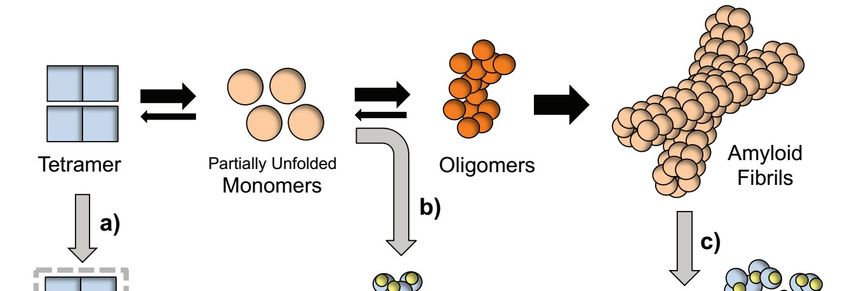
Int. J. Mol. Sci. 2019, 20, 1287                                                                   2 of 13

      At the molecular level, TTR is composed of four identical subunits of 127 amino acids forming
a tetramer [21,22]. Each polypeptide chain is organized in eight segments with a β-chain structure and
only a very small segment of alpha helix. The four monomers in the tetramer interact with each other
through non-covalent bonds, establishing a strong interaction between two monomers, forming dimers
that assemble as a tetramer originating a central hydrophobic channel limited by amino acids from
both dimers. This channel has two similar binding sites for thyroxine molecules [23]. The binding sites
can also accommodate other small TTR ligands that might occur in plasma as a result of metabolism,
diet origin, or even compounds administered for therapeutic purposes. For an extensive review of
TTR-ligand complex X-ray crystal structures, see a review by Pallaninathan et al. [24].
      The predominance of the β-chain structure in the polypeptide chains of the TTR tetramer, and its
organization as β-sheets contribute to the intrinsic amyloid potential of the protein, leading to
aggregation, fibril formation, and deposition under specific conditions, originating transthyretin
amyloidosis (ATTR amyloidosis).

2. ATTR Amyloidosis
      ATTR amyloidosis is a systemic amyloidosis of hereditary or non-hereditary origin. The hereditary
forms of the disease are due to mutations in the TTR gene that originate variants with a single amino
acid substitution [25,26] (Available online: amyloidosismutations.com). In the non-hereditary forms,
the main component of the amyloid fibrils is the wild type protein. In both cases, for different
reasons, namely amino acid alterations and/or environmental conditions, TTR becomes less stable
and dissociates into monomers that are partially unfolded and present a high tendency to aggregate
and form fibrils that deposit in the extracellular space. More than 120 TTR variants have been
described until now, related with different hereditary forms of ATTR (ATTRv). Though these are
mainly systemic forms of the disease, the most affected tissues or organs where amyloid gets deposited
are the peripheral nerves, gastro intestinal system, kidney, heart, carpal tunnel, eye, and in less cases
the meninges [27]. The non-hereditary form of the disease is mainly associated with cardiomyopathy
of aged people, over 80 years old, and the deposits are composed of wild type protein (ATTR wt) [28].
The most frequent TTR variant is TTR V30M that causes ATTRV30M amyloidosis (formerly designated
familial amyloid polyneuropathy (FAP)) [29]. The disease occurs in several foci in the world, the biggest
ones located in Portugal, Sweden, Japan, Brazil, Italy, France, and USA [30]. Concerning the hereditary
forms of the disease, TTR V122I is also a very frequent variant, in particular, in the Black American
population, being this variant related with a predominant involvement of the heart [31,32], now
designated as ATTR amyloidosis with cardiomyopathy [33].
      The clinical expression of the disease is highly heterogeneous in ATTR amyloidosis. In particular
the age of onset of the disease is variable for different variants and even for patients with the same
TTR variant, namely TTR V30M, in which the onset can vary from the 2nd to the 6th decade of
life [34,35]. Early onset cases are mainly characterized by predominant loss of small-diameter nerve
fibers, severe autonomic dysfunction, and cardiac conduction alterations, resulting in peripheral
neuropathy leading to loss of sensation, to pain and heat, lower and upper members muscle atrophy,
gastro-intestinal disturbances, and cardiomyopathy. In contrast, late onset TTR V30M patients
show loss of both small and large fibers, less severe polyneuropathy, mild autonomic dysfunction
and frequent cardiomegaly [36]. Among different TTR variants, there is also high variability of
predominance of polyneuropathy or cardiomyopathy as main clinical manifestations in ATTRv
amyloidosis (reviewed in Reference [26,37]).

3. Inhibitors of TTR Aggregation: Pharmacologic and Natural Inhibitors of TTR Amyloidosis
     Since plasma TTR is mainly synthesized by the liver, liver transplant has been one of the first
therapeutic approaches proposed and found effective for the disease [38]. However, as expected,
liver transplant is an invasive therapy, not suitable for all patients and with several limitations and
risks [39]. In addition, recently, it was found that after liver transplant, some patients develop TTR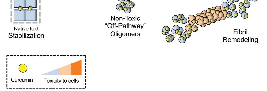
Int. J. Mol. Sci. 2019, 20, 1287                                                                     3 of 13

cardiomyopathy due to deposition of wild-type TTR in their heart [40–42]. This supports the need for
alternative therapeutic approaches that aim to stabilize TTR using small molecules that, by binding to
TTR, stabilize it and inhibit its aggregation and deposition [43]. The first evidence of TTR stabilization
through binding of small compounds came from the fact that when TTR is bound to T4 it is less
prone to aggregation. In addition, T4 binding sites in TTR are mostly unoccupied due to the high
TTR/T4 ratio in plasma, allowing TTR stabilization by binding of small compounds to TTR with high
affinity [44].
      Several nonsteroidal anti-inflammatory drugs (NSAIDs), have been known for a long time
to compete with T4 for the binding to TTR, such as salicylates, diclofenac, flufenamic acid and
diflunisal [45]. Among these, diflunisal was one of the most promising compounds due to its affinity
and specificity to bind TTR. In addition, several diflunisal derivatives have been synthetized to improve
its affinity and selectivity to bind TTR in plasma [46,47]. Diflunisal is still one of the compounds in
use for ATTR amyloidosis therapy in countries where Tafamidis has not yet been approved [48,49].
Tafamidis, diclorofenol benzoxazole carboxylic acid, is a more recent and widely-used drug that binds
to TTR and stabilizes it [50,51]. Tafamidis is highly safe and tolerable and has been found efficient in
slowing disease progression and preserving quality of life of TTR V30M patients [52]. Meanwhile,
other strategies for ATTR amyloidosis therapy have also been pursued, namely targeting different
steps in the cascade of amyloid formation, fibril disruption, and clearance [53]. An example of such
strategies is to use compounds that bind to TTR and block its polymerization or disrupt the amyloid
fibrils formed, such as molecular tweezers (CLR01) and doxycycline, respectively [54,55].

4. Natural Inhibitors—Polyphenols
     In the search for compounds of therapeutic interest, presenting very low toxicity and structural
similarities to other TTR ligands, several polyphenols of plant origin have been studied as inhibitors of
TTR amyloidogenesis. Some polyphenols were previously reported as inhibiting protein aggregation
and amyloid formation in neurodegenerative diseases, such as Alzheimer’s and Parkinson’s
disease [56]. One of the most studied polyphenols is resveratrol. In vitro studies using the AC16
cardiomyocyte cell line demonstrate that resveratrol is able to stabilize the native TTR tetramer,
preventing the formation of cytotoxic species and promoting aggregation of monomeric into non-toxic
species [12]. Furthermore, administration of resveratrol to Alzheimer’s disease (AD) mice revealed
an increase in TTR levels in plasma that does not result from higher expression of the protein, but,
instead, might be related with increased TTR stability and longer half-life in circulation [57]. However,
resveratrol seems to have not only these direct effects on TTR but also other properties namely those
involving protection against oxidation, which is difficult to discern.
     Other polyphenols, like nordihydroguaiaretic acid (NDGA), rosmarinic acid, caffeic acid and
epigallocatechin gallate (EGCG), have also been investigated in vitro for their interaction with
TTR [58–62]. Contrary to most polyphenols, EGCG did not compete with T4 for binding to TTR,
revealing that it binds at a different binding site in the molecule [62]. Indeed, the crystallographic
structure of the complex of TTR with EGCG revealed that it binds at different regions at the surface of
the molecule and not at the T4 binding sites [63]. In a subsequent structure–activity study, the galloyl
moiety has been highlighted as a key structural feature of EGCG by greatly enhancing its anti-amyloid
chaperone activity of TTR [61].
     When administered to a model mice expressing human TTR V30M, EGCG inhibited TTR
deposition in the gastrointestinal tract and in the dorsal root ganglia (DRG), the main sites of aggregated
TTR deposition in this animal model for the disease [64]. In addition, when administered to old mice,
EGCG treatment resulted in a decrease of TTR deposits in tissues, indicating a disruptive effect
on aggregated TTR deposits. A small pilot study with EGCG administration to human carriers of
amyloidogenic TTR mutations including TTR V30M revealed a reduction of myocardial mass in the case
of cardiomyopathy, indicating an inhibitory effect of EGCG on TTR amyloid fibril formation [65,66].
The reported studies show improvement in the cardiac function without increase of the patient’s
Int. J. Mol. Sci. 2019, 20, 1287 4 of 13
survival. The low toxicity and high tolerability to EGCG, confirmed in these studies, encourage
continuation of treatment with EGCG [67].
Among the polyphenols studied in vitro, curcumin revealed a particular behavior suggesting
different mechanism of inhibition of ATTR amyloidosis [58].
5. In Vitro Studies with Curcumin
5.1. Curcumin Binds to TTR and Increases Its Resistance to Dissociation
A decade ago, Pullakhandam and colleagues first reported curcumin interaction with TTR [68].
Using Scatchard analysis of fluorescence quenching, the authors showed that curcumin binds to
wild-type TTR with a molar ratio of 1.2:1 and Kd of 2.3 × 10⁻⁶ M [68]. In addition, curcumin was found
to dose-dependently displace 1-anilino-8-naphalene sulfonate (ANS) at pH 7.2 from TTR’s central
ligand-binding channel, to which various ligands are known to bind [68].
Shortly after, we further detailed the interaction between curcumin and TTR by unequivocally
showing that curcumin competed with radiolabeled T4 ([125 I]T4 ) for its binding to wild-type and
V30M mutant TTR, both in vitro and in whole human plasma [58]. These observations were later
corroborated by the crystal structures of TTR complexes with curcumin and also its degradation
product, ferulic acid, [15], and other curcumin-like compounds [69], showing that curcumin interacts
with Ser 117 and Lys15 and with Val 121 and Thr123 through a water molecule [15]. By filling the
largely unoccupied T4 binding pockets at the weaker dimer–dimer interface, curcumin increases
TTR tetramer resistance to dissociation in non-native monomers as shown by isoelectric focusing
(IEF) studies in semi-denaturing conditions (4 M urea) [58]. This, together with selective binding of
curcumin to TTR over other plasma proteins, resulted in a 25% increase of the tetramer/total TTR ratio
in plasma from controls and TTR V30M heterozygote carriers [58].
5.2. Curcumin Redirects TTR Aggregation into “Off-Pathway” Oligomers and Disaggregates Pre-Formed TTR
Amyloid Fibrils
Despite its inability to prevent the acid induced aggregation of TTR wild-type [68], we have
shown that curcumin robustly inhibits aggregation of the highly amyloidogenic Y78F variant under
physiological conditions (phosphate buffered saline, pH 7.4, 37 ◦ C) [58]. This supports the hypothesis
that protonation and isomerization of the phenolic and enolic hydroxyl groups of curcumin at low
pH might impair interaction with TTR [68]. Under transmission electron microscopy (TEM) and
dynamic light scattering (DLS), curcumin redirected the TTR Y78F amyloid formation pathway into
a monodispersed, highly stable population of “off-pathway” oligomers with approximately 80 nm in
hydrodynamic diameter (dH) [58]. In addition, we found that Schwann cells exposed to TTR Y78F
aggregates incubated with curcumin presented significantly reduced endoplasmic reticulum (ER)
stress and were protected from entering into the apoptotic signaling pathway [70], highlighting that
curcumin-induced oligomers are less toxic than untreated “on-pathway” aggregate intermediaries.
Moreover, dot–blot analysis of conditioned medium from Rat Schwannoma (RN22) cells expressing
TTR L55P incubated with curcumin revealed almost complete inhibition of TTR aggregation (90%). This
variant is associated with an aggressive form of ATTR amyloidosis, further supporting the protective
role of curcumin on the early stages of TTR aggregation, either by inhibiting tetramer dissociation
and/or redirecting pathological misfolding and aggregation into more innocuous counterparts [58].
Similar observations were later reported relative to different aggregation-prone proteins associated
with neurodegeneration, including Aβ [71], tau [72] and α-synuclein [73].
Beyond sharing many structural similarities with classical amyloid-binding dyes, such
as Thioflavin-S, Congo red, and crysamine-G, curcumin showed specific labeling of amyloid
deposits [74–76]. Although the precise atomic-detailed characteristics underlying the ability of
curcumin to break down β-sheet rich aggregates remain unclear, solid-state NMR studies haveInt. J. Mol. Sci. 2019, 20, 1287 5 of 13
highlighted the structural importance of the aromatic carbons adjacent to the methoxy and/or hydroxy
groups of curcumin in its binding with Aβ fibrils [77].
Overall, multiple lines of evidence favor the hypothesis that the non-specific modulatory role of
curcumin on amyloid formation and toxicity in vitro depends on aggregate-related conformational
structure rather than protein primary sequence.
6. In Vivo Studies with Curcumin
6.1. Curcumin Reduces TTR Load and Degrades Amyloid Deposits in Tissues
In recent years, an increasing amount of evidence supporting the anti-amyloidogenic role of
curcumin in different proteins prone to misfolding have paved the way to preclinical trials in transgenic
animal models [78].
With regard to ATTR amyloidosis, we have shown that chronically feeding young transgenic mice
for human TTR V30M with curcumin (2% w/w) results in micromolar steady-state levels of curcumin
in plasma (21.4 ± 3.6 µM) [79]. Selective competition of curcumin with T4 (42%) for the binding to TTR
in plasma significantly reduced tetramer dissociation into non-native monomeric intermediaries under
semi-dissociating conditions [79]. Beyond stabilizing TTR native fold, curcumin supplementation
alleviated TTR load and associated biomarkers in the gastrointestinal tract, the primary target organ in
this mouse model [79]. Dietary intake of curcumin was well-tolerated and non-toxic to animals and
the treatment did not interfere with TTR plasma levels in vivo [79].
In a later study, we evaluated the effect of curcumin in aged mice expressing the TTR V30M
variant on an Hsf-1 heterozygous background (hTTR V30M/Hsf), in which deposition of aggregated
TTR coexists with birefringent congophilic material in tissues. We found that curcumin intake not only
reduced non-fibrillar extracellular TTR burden in both gastrointestinal tract and dorsal root ganglia,
but also remodeled pre-existing congophilic amyloid material in tissues [70].
Our findings are in close alignment with recent observations made by others showing that
curcumin promotes remodeling of existing amyloid deposits and counteracts the formation of new
amyloid deposits, or even reduce the amount of remaining deposits [72,76,78,80].
6.2. Other Neuroprotective Mechanisms of Curcumin
Although we hypothesize that curcumin alleviates TTR extracellular burden most likely due
to its ability to directly interact and modify multiple partners of the TTR amyloid cascade, as
summarized in Figure 1, we speculate whether the pleiotropic therapeutic actions of curcumin [78]
might synergistically potentiate its efficacy in vivo.Int. J. Mol. Sci. 2019, 20, 1287 6 of 13
Int. J. Mol. Sci. 2019, 20, x FOR PEER REVIEW 6 of 13
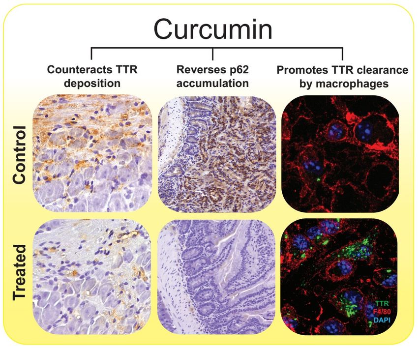
Figure
Figure1.1. Proposed
Proposed mechanism
mechanism for for TTR
TTR aggregation
aggregation pathway
pathwaymodulation
modulationby bycurcumin.
curcumin. Rate-limiting
Rate-limiting
tetramer
tetramer dissociation
dissociation ofof TTR
TTR into
into partially
partially unfolded
unfolded monomers
monomers precedes
precedes the the formation
formation of of toxic
toxic
oligomeric
oligomericintermediates
intermediatesthatthatevolve
evolveinto β-sheets enriched
intoβ-sheets enriched mature
maturefibrils.
fibrils. Curcumin
Curcumin modulates
modulatesTTR TTR
cascade by directly interacting with different binding partners: (a) Curcumin interaction
cascade by directly interacting with different binding partners: (a) Curcumin interaction with TTRwith TTR at the
at
Tthe
4 binding pockets stabilizes the tetrameric fold and blocks its dissociation into unfolded
T4 binding pockets stabilizes the tetrameric fold and blocks its dissociation into unfolded monomeric
species [15,58,68];
monomeric (b) Curcumin
species [15,58,68]; interaction
(b) Curcuminwith partially misfolded
interaction non-native
with partially monomersnon-native
misfolded redirects
TTR aggregation
monomers intoTTR
redirects “off-pathway”
aggregation unstructured oligomers
into “off-pathway” innocuous to
unstructured cells [58,68];
oligomers (c) Curcumin
innocuous to cells
breaks down and remodels β-sheet rich TTR fibrils in smaller amorphous aggregates
[58,68]; (c) Curcumin breaks down and remodels β-sheet rich TTR fibrils in smaller amorphous in in vitro [58]
and in vivo [70].
aggregates in in vitro [58] and in vivo [70].
Recently,
Recently,increasing
increasingrelevance
relevancehas hasbeen
beenattributed
attributedto toendothelial
endothelialabnormalities
abnormalitiesassociated
associated with
with
ATTRv amyloidosis and in particular ATTR V30M [81,82]. It has been suggested
ATTRv amyloidosis and in particular ATTR V30M [81,82]. It has been suggested that TTR variants that TTR variants
may
may affect
affect endothelial
endothelial cells
cells function
function even
even before
beforeamyloid
amyloidfibrilfibrilformation.
formation. Thus,
Thus, microangiopathy
microangiopathy
could play an important role in an initial lesion leading to organ damage [83].
could play an important role in an initial lesion leading to organ damage [83]. Interestingly, Interestingly, curcumin
curcumin
appears to improve endothelial cell function and, though its mechanisms of action
appears to improve endothelial cell function and, though its mechanisms of action are not completely are not completely
known,
known, itit seems
seems that
that by
by lowering
lowering the the expression
expression ofof pro-inflammatory
pro-inflammatory molecules,
molecules, and and byby reducing
reducing
levels
levels of reactive oxygen species, such as Nox-2 in endothelial cells, curcumin not only decreases
of reactive oxygen species, such as Nox-2 in endothelial cells, curcumin not only decreases
trans-endothelial
trans-endothelialmonocyte
monocytemigration,
migration,but butalso
alsomaintains
maintainsadequate
adequateNO NOlevels
levelsfor forthe
the proper
proper function
function
of cells [84].
of cells [84].
Accumulating
Accumulatingevidence
evidencehas haslinked
linked autophagy
autophagy impairment
impairment to neurodegeneration
to neurodegeneration andandneuronal cell
neuronal
death [85,86].
cell death Given Given
[85,86]. that stimulation of autophagy
that stimulation can potentially
of autophagy enhance degradation
can potentially of aggregation
enhance degradation of
prone-proteins, development of autophagy-inducing therapies, in which
aggregation prone-proteins, development of autophagy-inducing therapies, in which toxic toxic misfolded proteins are
misfolded
used as autophagy
proteins are used substrates,
as autophagy might be a valuable
substrates, mightpharmacological
be a valuableapproach for neurodegenerative
pharmacological approach for
diseases, including ATTR amyloidosis [85,86].
neurodegenerative diseases, including ATTR amyloidosis [85,86].
In
In preclinical
preclinical studies performed with
studies performed with TTR
TTRV30M
V30Mtransgenic
transgenicmice, mice,curcumin
curcuminhas hasbeen
been shown
shown to
to effectively reverse accumulation of p62, a key cargo receptor involved in
effectively reverse accumulation of p62, a key cargo receptor involved in selective autophagy, re- selective autophagy,
re-establishing
establishing thethe autophagicflux
autophagic fluxand
andmitigating
mitigatingapoptosis
apoptosis [87].[87]. Nevertheless,
Nevertheless, since
since curcumin
curcumin cancan
mediate crosstalk between different signaling pathways [88,89] it remains unclear
mediate crosstalk between different signaling pathways [88,89] it remains unclear to which extent to which extent
restoration of the autophagic flux in vivo occurs because: i) curcumin promotes autophagy or ii) itsInt. J. Mol. Sci. 2019, 20, 1287 7 of 13
restoration of the autophagic flux in vivo occurs because: (i) curcumin promotes autophagy or (ii) its
anti-amyloid activity prevents TTR “on-pathway” aggregation reaching a critical threshold beyond
which the autophagic machinery would be overwhelmed and irreversibly damaged.
In recent years, macrophage-mediated clearance of amyloid by a variety of phagocytic and
digestive mechanisms has been receiving increasing attention in the literature [90]. Several
small-molecules, including derivatives of curcumin, have been found to promote phagocytosis of
Aβ by macrophages [91–93]. Similarly, we have shown that pre-treatment of macrophages isolated
from aged FAP mice with physiologically achievable doses of curcumin, improves phagocytic uptake
and degradation of extracellular TTR aggregates, supporting that curcumin restores the inefficient
macrophage TTR clearance characteristic of pathological conditions [70].
7. Final Remarks
Several lines of evidence suggest that curcumin has neuroprotective properties in many
protein-misfolding disorders, including Alzheimer’s and Parkinson’s diseases and ATTR
amyloidosis [78]. Curcumin is a biologically well-tolerated polyphenol, with a long established
safety history [94]. According to JECFA (The Joint United Nations and World Health Organization
Expert Committee on Food Additives) and EFSA (European Food Safety Authority) guidelines,
the recommended allowable daily intake (ADI) amount of curcumin is 0–3 mg/kg body weight [94].
Nonetheless, some minor undesired side effects have been reported in a single dose escalation study
where healthy subjects were given increasing doses from 0.5 to 12 g of curcumin [95].
Despite its well-documented therapeutic efficacy, the poor absorption and rapid metabolism
of curcumin, has hindered its progress as a prospective pharmacological agent. To increase its
bioavailability, a wide array of novel formulations have been developed, including nanoparticles,
liposomes, micelles, and phospholipid complexes, which increase the bioavailability of curcumin by
providing longer circulation, enhanced permeability, and resistance to metabolic degradation and
excretion [78].
Presently, numerous disease-modifying targeted therapies for TTR amyloidosis are being
tested in human clinical trials, including TTR stabilizers (diflunisal, tafamidis), fibril disruptors
(doxycycline/TUDCA) and the most recent gene therapies to block TTR expression (small interference
RNAs (siRNAs) and antisense oligonucleotides therapy (ASOs)) [26,37,96,97]. Although development
of these strategies greatly improved the perspectives in ATTR amyloidosis, the complex nature of
the disease, in which several pathways are known to contribute to the pathology, prompts to seek
multi-stage interventions that not only block TTR synthesis and/or misfolding, but also suppress
inflammation and oxidative damage and enhance cellular protein degradation systems. Taken together,
the pleiotropic activities of curcumin provide multiple ways to tackle TTR pathophysiology, either
through direct interaction of curcumin with TTR, or indirect effects affecting signaling pathways
associated with TTR amyloid fibril formation and clearance. Accordingly, the works here reviewed,
and summarized in Figure 2, demonstrate interaction of curcumin with TTR through binding at the
thyroxine binding sites, resulting in TTR tetramer stabilization and consequent modulation of the TTR
misfolding cascade inhibiting aggregation and /or inducing formation of non-toxic aggregates. This
leads to restoring the autophagy flux and improving phagocytic uptake and clearance of extracellular
TTR. Curcumin also appears to directly induce disaggregation of TTR pre-formed fibrils and to promote
clearance of TTR aggregates through endocytose by fibroblasts and macrophages. Concomitant with
these effects, curcumin presents several non-specific effects counteracting common pathogenic events
in amyloidosis, such as oxidative stress, inflammation, apoptosis and extracellular matrix dysregulation
within a range of dosing with proven safety.Int. J. Mol. Sci. 2019, 20, 1287 8 of 13
Int. J. Mol. Sci. 2019, 20, x FOR PEER REVIEW 8 of 13
Figure
Figure2. 2.The
Thepleiotropic
pleiotropic effects
effects ofof curcumin
curcumin on the the molecular
molecular pathways
pathwaysassociated
associatedwith
with ATTR
ATTR
amyloidosis.
amyloidosis.Curcumin
Curcumin exerts
exerts neuroprotective effects on
neuroprotective effects on ATTR
ATTRamyloidosis
amyloidosisbybymodulating
modulating TTRTTR
abnormalaggregation
abnormal aggregationand andcounteracting
counteracting TTR TTR tissue
tissue deposition
deposition (left
(left panels, 20×magnification)
panels, 20× magnification)
immunohistochemistry
immunohistochemistry (IHC)
(IHC) analysis
analysis of TTR
of TTR in dorsal
in dorsal rootroot ganglia
ganglia (DRG) (DRG)
fromfrom
mice mice expressing
expressing human
human TTR V30M (hTTRV30M mice) treated with curcumin and age-matched
TTR V30M (hTTRV30M mice) treated with curcumin and age-matched controls [70]), re-establishing controls [70]), re-the
establishing
autophagic fluxthe
by autophagic
reversing p62 fluxaccumulation
by reversing(center
p62 accumulation
panels, 20×(center panels, 20×
magnification), magnification),
IHC analysis of p62,
in IHC analysissamples
duodenum of p62, in
fromduodenum
hTTRV30M samples
micefrom hTTRV30M
treated mice treated
with curcumin with curcumin
and age-matched and age-
controls [87])
andmatched
improvingcontrols [87]) and improving
the phagocytic uptake andthe phagocyticofuptake
degradation and degradation
extracellular of extracellular
TTR aggregates TTR
by macrophages
aggregates
(right panels, by63× macrophages (right
magnification), panels,
double 63× magnification), double
immunofluorescence labelingimmunofluorescence
for TTR, in green, andlabeling
F4/80,
for TTR, in green, and F4/80, in red, of primary macrophages from hTTRV30M mice
in red, of primary macrophages from hTTRV30M mice that were pre-incubated in presence of curcumin that were pre-
or incubated
its absencein(control),
presence of curcumin or its absence (control), before addition of TTR aggregates to cell
before addition of TTR aggregates to cell culture medium [70]). Nevertheless,
culture medium [70]). Nevertheless, other well-known neuroprotective properties of curcumin, such
other well-known neuroprotective properties of curcumin, such as its anti-inflammatory, anti-apoptotic,
as its anti-inflammatory, anti-apoptotic, and anti-oxidative activities [78,94], might potentiate its in
and anti-oxidative activities [78,94], might potentiate its in vivo effects.
vivo effects.
In conclusion, in this context, curcumin remains a promising scaffold for the development of
In conclusion, in this context, curcumin remains a promising scaffold for the development of
potent multi-stage disease-modifying drugs for the treatment of TTR amyloidosis.
potent multi-stage disease-modifying drugs for the treatment of TTR amyloidosis.
Funding:This
Funding: Thisresearch was funded
research was fundedbyby thethe European
European Regional
Regional Development
Development Fund (FEDER)
Fund (FEDER) throughthrough
the Nortethe
Norte Portugal Regional Operational Programme (NORTE 2020), under the PORTUGAL 2020
Portugal Regional Operational Programme (NORTE 2020), under the PORTUGAL 2020 Partnership Agreement, Partnership
Agreement, grant number Norte-01-0145-FEDER-000008—Porto Neurosciences and Neurologic Disease Research
grant number Norte-01-0145-FEDER-000008—Porto Neurosciences and Neurologic Disease Research Initiative
Initiative at I3S. Nelson Ferreira was a recipient of a Postdoctoral Fellowship R171-2014-591 from Lundbeck
at I3S. Nelson
foundation and byFerreira was aFoundation
Lundbeck recipient of agrant
Postdoctoral Fellowship
R248-2016-2518 for R171-2014-591 fromInstitute
Danish Research Lundbeckof foundation
Translational
and by Lundbeck Foundation grant R248-2016-2518 for Danish Research Institute
Neuroscience—DANDRITE, Nordic-EMBL Partnership for Molecular Medicine, Aarhus University, of Translational
Denmark.
Neuroscience—DANDRITE, Nordic-EMBL Partnership for Molecular Medicine, Aarhus University, Denmark.
Acknowledgments: The authors thank Cristina Teixeira (MSc) (from Molecular Neurobiology Group, IBMC
and I3S, University of Porto, Portugal) for the immunohistochemistry pictures of p62 in duodenum included in
Figure 2.
Conflicts of Interest: The authors declare no conflicts of interest.Int. J. Mol. Sci. 2019, 20, 1287 9 of 13
References
1. Raz, A.; Goodman, D.S. The interaction of thyroxine with human plasma prealbumin and with the
prealbumin-retinol-binding protein complex. J. Biol. Chem. 1969, 244, 3230–3237.
2. Sousa, M.M.; Berglund, L.; Saraiva, M.J. Transthyretin in high density lipoproteins: Association with
apolipoprotein A-I. J. Lipid Res. 2000, 41, 58–65.
3. Sousa, M.M.; Yan, S.D.; Stern, D.; Saraiva, M.J. Interaction of the receptor for advanced glycation end
products (RAGE) with transthyretin triggers nuclear transcription factor kB (NF-kB) activation. Lab. Investig.
2000, 80, 1101–1110. [CrossRef]
4. Gonçalves, I.; Quintela, T.; Baltazar, G.; Almeida, M.R.; Saraiva, M.J.M.; Santos, C.R. Transthyretin interacts
with metallothionein 2. Biochemistry 2008, 47, 2244–2251. [CrossRef]
5. Nunes, A.F.; Saraiva, M.J.; Sousa, M.M. Transthyretin knockouts are a new mouse model for increased
neuropeptide Y. FASEB J. 2006, 20, 166–168. [CrossRef]
6. Costa, R.; Gonçalves, A.; Saraiva, M.J.; Cardoso, I. Transthyretin binding to A-Beta peptide–impact on A-Beta
fibrillogenesis and toxicity. FEBS Lett. 2008, 582, 936–942. [CrossRef]
7. Liz, M.A.; Mar, F.M.; Franquinho, F.; Sousa, M.M. Aboard transthyretin: From transport to cleavage.
IUBMB Life 2010, 62, 429–435. [CrossRef]
8. Almeida, M.R.; Gales, L.; Damas, A.M.; Cardoso, I.; Saraiva, M.J. Small transthyretin (TTR) ligands as
possible therapeutic agents in TTR amyloidoses. Curr. Drug Targets CNS Neurol. Disord. 2005, 4, 587–596.
[CrossRef]
9. Ernström, U.; Pettersson, T.; Jörnvall, H. A yellow component associated with human transthyretin has
properties like a pterin derivative, 7,8-dihydropterin-6-carboxaldehyde. FEBS Lett. 1995, 360, 177–182.
[CrossRef]
10. Lans, M.C.; Klasson-Wehler, E.; Willemsen, M.; Meussen, E.; Safe, S.; Brouwer, A. Structure-dependent,
competitive interaction of hydroxy-polychlorobiphenyls, -dibenzo-p-dioxins and -dibenzofurans with
human transthyretin. Chem. Biol. Interact. 1993, 88, 7–21. [CrossRef]
11. Baures, P.W.; Oza, V.B.; Peterson, S.A.; Kelly, J.W. Synthesis and evaluation of inhibitors of transthyretin
amyloid formation based on the non-steroidal anti-inflammatory drug, flufenamic acid. Bioorg. Med. Chem.
1999, 7, 1339–1347. [CrossRef]
12. Bourgault, S.; Choi, S.; Buxbaum, J.N.; Kelly, J.W.; Price, J.L.; Reixach, N. Erratum to “Mechanisms of
transthyretin cardiomyocyte toxicity inhibition by resveratrol analogs” [Biochem. Biophys. Res. Commun.
410 (2011) 707–713]. Biochem. Biophys. Res. Commun. 2011, 412, 196. [CrossRef]
13. Trivella, D.B.B.; dos Reis, C.V.; Lima, L.M.T.R.; Foguel, D.; Polikarpov, I. Flavonoid interactions with
human transthyretin: Combined structural and thermodynamic analysis. J. Struct. Biol. 2012, 180, 143–153.
[CrossRef] [PubMed]
14. Yokoyama, T.; Kosaka, Y.; Mizuguchi, M. Inhibitory activities of propolis and its promising component,
caffeic acid phenethyl ester, against amyloidogenesis of human transthyretin. J. Med. Chem. 2014, 57,
8928–8935. [CrossRef]
15. Ciccone, L.; Tepshi, L.; Nencetti, S.; Stura, E.A. Transthyretin complexes with curcumin and bromo-estradiol:
Evaluation of solubilizing multicomponent mixtures. New Biotechnol. 2015, 32, 54–64. [CrossRef] [PubMed]
16. Aleshire, S.L.; Bradley, C.A.; Richardson, L.D.; Parl, F.F. Localization of human prealbumin in choroid plexus
epithelium. J. Histochem. Cytochem. 2017, 31, 608–612. [CrossRef]
17. Dickson, P.W.; Howlett, G.J.; Schreiber, G. Rat transthyretin (prealbumin). Molecular cloning, nucleotide
sequence, and gene expression in liver and brain. J. Biol. Chem. 1985, 260, 8214–8219. [PubMed]
18. Richardson, S.J. Cell and molecular biology of transthyretin and thyroid hormones. Int. Rev. Cytol. 2007, 258,
137–193. [PubMed]
19. Cavallaro, T.; Martone, R.L.; Dwork, A.J.; Schon, E.A.; Herbert, J. The retinal pigment epithelium is the
unique site of transthyretin synthesis in the rat eye. Investig. Ophthalmol. Vis. Sci. 1990, 31, 497–501.
20. Richardson, S.J. Evolutionary changes to transthyretin: Evolution of transthyretin biosynthesis. FEBS J. 2009,
276, 5342–5356. [CrossRef] [PubMed]
21. Blake, C.C.; Geisow, M.J.; Oatley, S.J.; Rérat, B.; Rérat, C. Structure of prealbumin: Secondary, tertiary and
quaternary interactions determined by Fourier refinement at 1.8 A. J. Mol. Biol. 1978, 121, 339–356. [CrossRef]Int. J. Mol. Sci. 2019, 20, 1287 10 of 13
22. Hamilton, J.A.; Steinrauf, L.K.; Braden, B.C.; Liepnieks, J.; Benson, M.D.; Holmgren, G.; Sandgren, O.;
Steen, L. The X-ray crystal structure refinements of normal human transthyretin and the amyloidogenic
Val-30–>Met variant to 1.7-A resolution. J. Biol. Chem. 1993, 268, 2416–2424. [PubMed]
23. Wojtczak, A. Crystal structure of rat transthyretin at 2.5 A resolution: First report on a unique tetrameric
structure. Acta Biochim. Pol. 1997, 44, 505–517.
24. Palaninathan, S.K. Nearly 200 X-ray crystal structures of transthyretin: What do they tell us about this
protein and the design of drugs for TTR amyloidoses? Curr. Med. Chem. 2012, 19, 2324–2342. [CrossRef]
[PubMed]
25. Benson, M.D.; Kincaid, J.C. The molecular biology and clinical features of amyloid neuropathy. Muscle Nerve
2007, 36, 411–423. [CrossRef]
26. Sekijima, Y. Transthyretin (ATTR) amyloidosis: Clinical spectrum, molecular pathogenesis and
disease-modifying treatments. J. Neurol. Neurosurg. Psychiatry 2015, 86, 1036–1043. [CrossRef]
27. Sousa, M.M.; Saraiva, M.J. Neurodegeneration in familial amyloid polyneuropathy: From pathology to
molecular signaling. Prog. Neurobiol. 2003, 71, 385–400. [CrossRef]
28. Patel, K.S.; Hawkins, P.N. Cardiac amyloidosis: Where are we today? J. Intern. Med. 2015, 278, 126–144.
[CrossRef]
29. Saraiva, M.J.; Birken, S.; Costa, P.P.; Goodman, D.S. Amyloid fibril protein in familial amyloidotic
polyneuropathy, Portuguese type. Definition of molecular abnormality in transthyretin (prealbumin).
J. Clin. Investig. 1984, 74, 104–119. [CrossRef]
30. Planté-Bordeneuve, V.; Kerschen, P. Transthyretin familial amyloid polyneuropathy. Handb. Clin. Neurol.
2013, 115, 643–658.
31. Jacobson, D.R.; Gorevic, P.D.; Buxbaum, J.N. A homozygous transthyretin variant associated with senile
systemic amyloidosis: Evidence for a late-onset disease of genetic etiology. Am. J. Hum. Genet. 1990, 47,
127–136.
32. Saraiva, M.J.; Sherman, W.; Marboe, C.; Figueira, A.; Costa, P.; de Freitas, A.F.; Gawinowicz, M.A. Cardiac
amyloidosis: Report of a patient heterozygous for the transthyretin isoleucine 122 variant. Scand. J. Immunol.
1990, 32, 341–346. [CrossRef]
33. Gertz, M.A.; Benson, M.D.; Dyck, P.J.; Grogan, M.; Coelho, T.; Cruz, M.; Berk, J.L.; Plante-Bordeneuve, V.;
Schmidt, H.H.-J.; Merlini, G. Diagnosis, Prognosis, and Therapy of Transthyretin Amyloidosis. J. Am.
Coll. Cardiol. 2015, 66, 2451–2466. [CrossRef]
34. Conceiçao, I.; Gonzalez-Duarte, A.; Obici, L.; Schmidt, H.H.-J.; Simoneau, D.; Ong, M.-L.; Amass, L.
“Red-flag” symptom clusters in transthyretin familial amyloid polyneuropathy. J. Peripher. Nerv. Syst.
2016, 21, 5–9. [CrossRef]
35. Ando, Y.; Coelho, T.; Berk, J.L.; Cruz, M.W.; Ericzon, B.-G.; Ikeda, S.-I.; Lewis, W.D.; Obici, L.;
Plante-Bordeneuve, V.; Rapezzi, C.; et al. Guideline of transthyretin-related hereditary amyloidosis for
clinicians. Orphanet J. Rare Dis. 2013, 8, 31. [CrossRef]
36. Koike, H.; Misu, K.; Sugiura, M.; Iijima, M.; Mori, K.; Yamamoto, M.; Hattori, N.; Mukai, E.; Ando, Y.;
Ikeda, S.; et al. Pathology of early- vs. late-onset TTR Met30 familial amyloid polyneuropathy. Neurology
2004, 63, 129–138. [CrossRef]
37. Plante-Bordeneuve, V. Transthyretin familial amyloid polyneuropathy: An update. J. Neurol. 2018, 265,
976–983. [CrossRef]
38. Holmgren, G.; Ericzon, B.G.; Groth, C.G.; Steen, L.; Suhr, O.; Andersen, O.; Wallin, B.G.; Seymour, A.;
Richardson, S.; Hawkins, P.N. Clinical improvement and amyloid regression after liver transplantation in
hereditary transthyretin amyloidosis. Lancet 1993, 341, 1113–1116. [CrossRef]
39. Carvalho, A.; Rocha, A.; Lobato, L. Liver transplantation in transthyretin amyloidosis: Issues and challenges.
Liver Transpl. 2015, 21, 282–292. [CrossRef]
40. Liepnieks, J.J.; Zhang, L.Q.; Benson, M.D. Progression of transthyretin amyloid neuropathy after liver
transplantation. Neurology 2010, 75, 324–327. [CrossRef]
41. Okamoto, S.; Zhao, Y.; Lindqvist, P.; Backman, C.; Ericzon, B.-G.; Wijayatunga, P.; Henein, M.Y.; Suhr, O.B.
Development of cardiomyopathy after liver transplantation in Swedish hereditary transthyretin amyloidosis
(ATTR) patients. Amyloid 2011, 18, 200–205. [CrossRef]Int. J. Mol. Sci. 2019, 20, 1287 11 of 13
42. Ericzon, B.-G.; Wilczek, H.E.; Larsson, M.; Wijayatunga, P.; Stangou, A.; Pena, J.R.; Furtado, E.; Barroso, E.;
Daniel, J.; Samuel, D.; et al. Liver Transplantation for Hereditary Transthyretin Amyloidosis: After 20 Years
Still the Best Therapeutic Alternative? Transplantation 2015, 99, 1847–1854. [CrossRef]
43. Miroy, G.J.; Lai, Z.; Lashuel, H.A.; Peterson, S.A.; Strang, C.; Kelly, J.W. Inhibiting transthyretin amyloid
fibril formation via protein stabilization. Proc. Natl. Acad. Sci. USA 1996, 93, 15051–15056. [CrossRef]
44. Johnson, S.M.; Wiseman, R.L.; Sekijima, Y.; Green, N.S.; Adamski-Werner, S.L.; Kelly, J.W. Native state
kinetic stabilization as a strategy to ameliorate protein misfolding diseases: A focus on the transthyretin
amyloidoses. Acc. Chem. Res. 2005, 38, 911–921. [CrossRef]
45. Munro, S.L.; Lim, C.F.; Hall, J.G.; Barlow, J.W.; Craik, D.J.; Topliss, D.J.; Stockigt, J.R. Drug competition for
thyroxine binding to transthyretin (prealbumin): Comparison with effects on thyroxine-binding globulin.
J. Clin. Endocrinol. Metab. 1989, 68, 1141–1147. [CrossRef]
46. Adamski-Werner, S.L.; Palaninathan, S.K.; Sacchettini, J.C.; Kelly, J.W. Diflunisal analogues stabilize the
native state of transthyretin. Potent inhibition of amyloidogenesis. J. Med. Chem. 2004, 47, 355–374.
[CrossRef]
47. Miller, S.R.; Sekijima, Y.; Kelly, J.W. Native state stabilization by NSAIDs inhibits transthyretin
amyloidogenesis from the most common familial disease variants. Lab. Investig. 2004, 84, 545–552. [CrossRef]
48. Tojo, K.; Sekijima, Y.; Kelly, J.W.; Ikeda, S.-I. Diflunisal stabilizes familial amyloid polyneuropathy-associated
transthyretin variant tetramers in serum against dissociation required for amyloidogenesis. Neurosci. Res.
2006, 56, 441–449. [CrossRef]
49. Sekijima, Y.; Dendle, M.A.; Kelly, J.W. Orally administered diflunisal stabilizes transthyretin against
dissociation required for amyloidogenesis. Amyloid 2006, 13, 236–249. [CrossRef]
50. Johnson, S.M.; Connelly, S.; Wilson, I.A.; Kelly, J.W. Biochemical and structural evaluation of highly
selective 2-arylbenzoxazole-based transthyretin amyloidogenesis inhibitors. J. Med. Chem. 2008, 51, 260–270.
[CrossRef]
51. Johnson, S.M.; Connelly, S.; Fearns, C.; Powers, E.T.; Kelly, J.W. The transthyretin amyloidoses: From
delineating the molecular mechanism of aggregation linked to pathology to a regulatory-agency-approved
drug. J. Mol. Biol. 2012, 421, 185–203. [CrossRef] [PubMed]
52. Coelho, T.; Maia, L.F.; da Silva, A.M.; Cruz, M.W.; Plante-Bordeneuve, V.; Suhr, O.B.; Conceiçao, I.;
Schmidt, H.H.-J.; Trigo, P.; Kelly, J.W.; et al. Long-term effects of tafamidis for the treatment of transthyretin
familial amyloid polyneuropathy. J. Neurol. 2013, 260, 2802–2814. [CrossRef]
53. Adams, D.; Cauquil, C.; Labeyrie, C.; Beaudonnet, G.; Algalarrondo, V.; Théaudin, M. TTR kinetic
stabilizers and TTR gene silencing: A new era in therapy for familial amyloidotic polyneuropathies.
Expert Opin. Pharmacother. 2016, 17, 791–802. [CrossRef] [PubMed]
54. Ferreira, N.; Pereira-Henriques, A.; Attar, A.; Klärner, F.-G.; Schrader, T.; Bitan, G.; Gales, L.; Saraiva, M.J.;
Almeida, M.R. Molecular tweezers targeting transthyretin amyloidosis. Neurotherapeutics 2014, 11, 450–461.
[CrossRef] [PubMed]
55. Galant, N.J.; Westermark, P.; Higaki, J.N.; Chakrabartty, A. Transthyretin amyloidosis: An under-recognized
neuropathy and cardiomyopathy. Clin. Sci. 2017, 131, 395–409. [CrossRef] [PubMed]
56. Ngoungoure, V.L.N.; Schluesener, J.; Moundipa, P.F.; Schluesener, H. Natural polyphenols binding to
amyloid: A broad class of compounds to treat different human amyloid diseases. Mol. Nutr. Food Res. 2014,
59, 8–20. [CrossRef] [PubMed]
57. Santos, L.M.; Rodrigues, D. Resveratrol Administration Increases Transthyretin Protein Levels, Ameliorating
AD Features: The Importance of Transthyretin Tetrameric Stability. Mol. Med. 2016, 22, 1. [CrossRef]
[PubMed]
58. Ferreira, N.; Saraiva, M.J.; Almeida, M.R. Natural polyphenols inhibit different steps of the process of
transthyretin (TTR) amyloid fibril formation. FEBS Lett. 2011, 585, 2424–2430. [CrossRef]
59. Florio, P.; Folli, C.; Cianci, M.; Del Rio, D.; Zanotti, G.; Berni, R. Transthyretin Binding Heterogeneity
and Anti-amyloidogenic Activity of Natural Polyphenols and Their Metabolites. J. Biol. Chem. 2015, 290,
29769–29780. [CrossRef]
60. Ortore, G.; Orlandini, E.; Braca, A.; Ciccone, L.; Rossello, A.; Martinelli, A.; Nencetti, S. Targeting Different
Transthyretin Binding Sites with Unusual Natural Compounds. ChemMedChem 2016, 11, 1865–1874.
[CrossRef]Int. J. Mol. Sci. 2019, 20, 1287 12 of 13
61. Ferreira, N.; Pereira-Henriques, A.; Almeida, M.R. Transthyretin chemical chaperoning by flavonoids:
Structure-activity insights towards the design of potent amyloidosis inhibitors. Biochem. Biophys. Rep. 2015,
3, 123–133. [CrossRef]
62. Ferreira, N.; Cardoso, I.; Domingues, M.R.; Vitorino, R.; Bastos, M.; Bai, G.; Saraiva, M.J.; Almeida, M.R.
Binding of epigallocatechin-3-gallate to transthyretin modulates its amyloidogenicity. FEBS Lett. 2009, 583,
3569–3576. [CrossRef]
63. Miyata, M.; Sato, T.; Kugimiya, M.; Sho, M.; Nakamura, T.; Ikemizu, S.; Chirifu, M.; Mizuguchi, M.;
Nabeshima, Y.; Suwa, Y.; et al. The crystal structure of the green tea polyphenol (−)-epigallocatechin
gallate-transthyretin complex reveals a novel binding site distinct from the thyroxine binding site.
Biochemistry 2010, 49, 6104–6114. [CrossRef]
64. Ferreira, N.; Saraiva, M.J.; Almeida, M.R. Epigallocatechin-3-gallate as a potential therapeutic drug for
TTR-related amyloidosis: “in vivo” evidence from FAP mice models. PLoS ONE 2012, 7, e29933. [CrossRef]
65. Kristen, A.V.; Lehrke, S.; Buss, S.; Mereles, D.; Steen, H.; Ehlermann, P.; Hardt, S.; Giannitsis, E.; Schreiner, R.;
Haberkorn, U.; et al. Green tea halts progression of cardiac transthyretin amyloidosis: An observational
report. Clin. Res. Cardiol. 2012, 101, 805–813. [CrossRef]
66. Aus dem Siepen, F.; Bauer, R.; Aurich, M.; Buss, S.J.; Steen, H.; Altland, K.; Katus, H.A.; Kristen, A.V. Green
tea extract as a treatment for patients with wild-type transthyretin amyloidosis: An observational study.
Drug Des. Dev. Ther. 2015, 9, 6319–6325. [CrossRef]
67. Cappelli, F.; Martone, R.; Taborchi, G.; Morini, S.; Bartolini, S.; Angelotti, P.; Farsetti, S.; Di Mario, C.;
Perfetto, F. Epigallocatechin-3-gallate tolerability and impact on survival in a cohort of patients with
transthyretin-related cardiac amyloidosis. A single-center retrospective study. Intern. Emerg. Med. 2018, 13,
873–880. [CrossRef]
68. Pullakhandam, R.; Srinivas, P.N.B.S.; Nair, M.K.; Reddy, G.B. Binding and stabilization of transthyretin by
curcumin. Arch. Biochem. Biophys. 2009, 485, 115–119. [CrossRef]
69. Polsinelli, I.; Nencetti, S.; Shepard, W.; Ciccone, L.; Orlandini, E.; Stura, E.A. A new crystal form of human
transthyretin obtained with a curcumin derived ligand. J. Struct. Biol. 2016, 194, 8–17. [CrossRef]
70. Ferreira, N.; Gonçalves, N.P.; Saraiva, M.J.; Almeida, M.R. Curcumin: A multi-target disease-modifying
agent for late-stage transthyretin amyloidosis. Sci. Rep. 2016, 6, 503. [CrossRef]
71. Thapa, A.; Jett, S.D.; Chi, E.Y. Curcumin Attenuates Amyloid-β Aggregate Toxicity and Modulates
Amyloid-β Aggregation Pathway. ACS Chem. Neurosci. 2015, 7, 56–68. [CrossRef] [PubMed]
72. Rane, J.S.; Bhaumik, P.; Panda, D. Curcumin Inhibits Tau Aggregation and Disintegrates Preformed Tau
Filaments in vitro. J. Alzheimer’s Dis. 2017, 60, 999–1014. [CrossRef] [PubMed]
73. Singh, P.K.; Kotia, V.; Ghosh, D.; Mohite, G.M.; Kumar, A.; Maji, S.K. Curcumin modulates α-synuclein
aggregation and toxicity. ACS Chem. Neurosci. 2013, 4, 393–407. [CrossRef] [PubMed]
74. Yang, F.; Lim, G.P.; Begum, A.N.; Ubeda, O.J.; Simmons, M.R.; Ambegaokar, S.S.; Chen, P.P.; Kayed, R.;
Glabe, C.G.; Frautschy, S.A.; et al. Curcumin inhibits formation of amyloid beta oligomers and fibrils, binds
plaques, and reduces amyloid in vivo. J. Biol. Chem. 2005, 280, 5892–5901. [CrossRef] [PubMed]
75. Garcia-Alloza, M.; Borrelli, L.A.; Rozkalne, A.; Hyman, B.T.; Bacskai, B.J. Curcumin labels amyloid pathology
in vivo, disrupts existing plaques, and partially restores distorted neurites in an Alzheimer mouse model.
J. Neurochem. 2007, 102, 1095–1104. [CrossRef] [PubMed]
76. Maiti, P.; Hall, T.C.; Paladugu, L.; Kolli, N.; Learman, C.; Rossignol, J.; Dunbar, G.L. A comparative study
of dietary curcumin, nanocurcumin, and other classical amyloid-binding dyes for labeling and imaging of
amyloid plaques in brain tissue of 5×-familial Alzheimer’s disease mice. Histochem. Cell Biol. 2016, 146,
609–625. [CrossRef] [PubMed]
77. Masuda, Y.; Fukuchi, M.; Yatagawa, T.; Tada, M.; Takeda, K.; Irie, K.; Akagi, K.-I.; Monobe, Y.; Imazawa, T.;
Takegoshi, K. Solid-state NMR analysis of interaction sites of curcumin and 42-residue amyloid β-protein
fibrils. Bioorg. Med. Chem. 2011, 19, 5967–5974. [CrossRef] [PubMed]
78. Maiti, P.; Dunbar, G.L. Use of Curcumin, a Natural Polyphenol for Targeting Molecular Pathways in Treating
Age-Related Neurodegenerative Diseases. Int. J. Mol. Sci. 2018, 19, 1637. [CrossRef] [PubMed]
79. Ferreira, N.; Santos, S.A.O.; Domingues, M.R.M.; Saraiva, M.J.; Almeida, M.R. Dietary curcumin
counteracts extracellular transthyretin deposition: Insights on the mechanism of amyloid inhibition.
Biochim. Biophys. Acta 2013, 1832, 39–45. [CrossRef]Int. J. Mol. Sci. 2019, 20, 1287 13 of 13
80. Chongtham, A.; Agrawal, N. Curcumin modulates cell death and is protective in Huntington’s disease
model. Sci. Rep. 2016, 6, 18736. [CrossRef]
81. Nunes, R.J.; de Oliveira, P.; Lages, A.; Becker, J.D.; Marcelino, P.; Barroso, E.; Perdigoto, R.; Kelly, J.W.;
Quintas, A.; Santos, S.C.R. Transthyretin proteins regulate angiogenesis by conferring different molecular
identities to endothelial cells. J. Biol. Chem. 2013, 288, 31752–31760. [CrossRef] [PubMed]
82. Koike, H.; Ikeda, S.; Takahashi, M.; Kawagashira, Y.; Iijima, M.; Misumi, Y.; Ando, Y.; Ikeda, S.-I.; Katsuno, M.;
Sobue, G. Schwann cell and endothelial cell damage in transthyretin familial amyloid polyneuropathy.
Neurology 2016, 87, 2220–2229. [CrossRef] [PubMed]
83. Koike, H.; Katsuno, M. Ultrastructure in Transthyretin Amyloidosis: From Pathophysiology to Therapeutic
Insights. Biomedicines 2019, 7, 11. [CrossRef]
84. Karimian, M.S.; Pirro, M.; Johnston, T.P.; Majeed, M.; Sahebkar, A. Curcumin and Endothelial Function:
Evidence and Mechanisms of Protective Effects. Curr. Pharm. Des. 2017, 23, 2462–2473. [CrossRef] [PubMed]
85. Rahman, M.A.; Rhim, H. Therapeutic implication of autophagy in neurodegenerative diseases. BMB Rep.
2017, 50, 345–354. [CrossRef] [PubMed]
86. Fujikake, N.; Shin, M.; Shimizu, S. Association Between Autophagy and Neurodegenerative Diseases.
Front. Neurosci. 2018, 12, 255. [CrossRef]
87. Teixeira, C.A.; Almeida, M.D.R.; Saraiva, M.J. Impairment of autophagy by TTR V30M aggregates: In vivo
reversal by TUDCA and curcumin. Clin. Sci. 2016, 130, 1665–1675. [CrossRef]
88. Rainey, N.; Motte, L.; Aggarwal, B.B.; Petit, P.X. Curcumin hormesis mediates a cross-talk between autophagy
and cell death. Cell Death Dis. 2015, 6, e2003. [CrossRef]
89. Moustapha, A.; Pérétout, P.A.; Rainey, N.E.; Sureau, F.; Geze, M.; Petit, J.-M.; Dewailly, E.; Slomianny, C.;
Petit, P.X. Curcumin induces crosstalk between autophagy and apoptosis mediated by calcium release from
the endoplasmic reticulum, lysosomal destabilization and mitochondrial events. Cell Death Discov. 2015, 1,
15017. [CrossRef]
90. Lai, A.Y.; McLaurin, J. Clearance of amyloid-β peptides by microglia and macrophages: The issue of what,
when and where. Future Neurol. 2012, 7, 165–176. [CrossRef]
91. Zhang, L.; Fiala, M.; Cashman, J.; Sayre, J.; Espinosa, A.; Mahanian, M.; Zaghi, J.; Badmaev, V.; Graves, M.C.;
Bernard, G.; et al. Curcuminoids enhance amyloid-β uptake by macrophages of Alzheimer’s disease patients.
J. Alzheimer’s Dis. 2006, 10, 1–7. [CrossRef]
92. Masoumi, A.; Goldenson, B.; Ghirmai, S.; Avagyan, H.; Zaghi, J.; Abel, K.; Zheng, X.; Espinosa-Jeffrey, A.;
Mahanian, M.; Liu, P.T.; et al. 1α,25-dihydroxyvitamin D3 Interacts with Curcuminoids to Stimulate
Amyloid-β Clearance by Macrophages of Alzheimer’s Disease Patients. J. Alzheimer’s Dis. 2009, 17, 703–717.
[CrossRef] [PubMed]
93. Fiala, M.; Mahanian, M.; Rosenthal, M.; Mizwicki, M.T.; Tse, E.; Cho, T.; Sayre, J.; Weitzman, R.; Porter, V.
MGAT3 mRNA: A Biomarker for Prognosis and Therapy of Alzheimer’s Disease by Vitamin D and
Curcuminoids. J. Alzheimer’s Dis. 2011, 25, 135–144. [CrossRef] [PubMed]
94. Hewlings, S.J.; Kalman, D.S. Curcumin: A Review of Its’ Effects on Human Health. Foods 2017, 6, 92.
[CrossRef] [PubMed]
95. Lao, C.D.; Ruffin, M.T.; Normolle, D.; Heath, D.D.; Murray, S.I.; Bailey, J.M.; Boggs, M.E.; Crowell, J.; Rock, C.L.;
Brenner, D.E. Dose escalation of a curcuminoid formulation. BMC Complement. Altern. Med. 2006, 6, 10.
[CrossRef]
96. Adams, D.; Gonzalez-Duarte, A.; O’Riordan, W.D.; Yang, C.-C.; Ueda, M.; Kristen, A.V.; Tournev, I.;
Schmidt, H.H.; Coelho, T.; Berk, J.L.; et al. Patisiran, an RNAi Therapeutic, for Hereditary Transthyretin
Amyloidosis. N. Engl. J. Med. 2018, 379, 11–21. [CrossRef] [PubMed]
97. Benson, M.D.; Waddington-Cruz, M.; Berk, J.L.; Polydefkis, M.; Dyck, P.J.; Wang, A.K.; Plante-Bordeneuve, V.;
Barroso, F.A.; Merlini, G.; Obici, L.; et al. Inotersen Treatment for Patients with Hereditary Transthyretin
Amyloidosis. N. Engl. J. Med. 2018, 379, 22–31. [CrossRef]
© 2019 by the authors. Licensee MDPI, Basel, Switzerland. This article is an open access
article distributed under the terms and conditions of the Creative Commons Attribution
(CC BY) license (http://creativecommons.org/licenses/by/4.0/).You can also read