ADAPTATION OF THE ENDEMIC CORONAVIRUSES HCOV-OC43 AND HCOV-229E TO THE HUMAN HOST - OXFORD ACADEMIC JOURNALS
←
→
Page content transcription
If your browser does not render page correctly, please read the page content below
Virus Evolution, 2021, 7(2), 1–11
                                                                                                                      DOI: https://doi.org/10.1093/ve/veab061
                                                                                                                 Advance access publication date: 24 June 2021
                                                                                                                                                Research Article

Adaptation of the endemic coronaviruses HCoV-OC43
and HCoV-229E to the human host
Diego Forni,1,*,† Rachele Cagliani,1 Federica Arrigoni,2 Martino Benvenuti,2 Alessandra Mozzi,1,‡ Uberto Pozzoli,1 Mario Clerici,3,4
Luca De Gioia,2 and Manuela Sironi1
1
  Scientific Institute IRCCS E. MEDEA, Bioinformatics, via don Luigi Monza, 23843 Bosisio Parini, Italy, 2 Department of Biotechnology and Biosciences, University of

                                                                                                                                                                        Downloaded from https://academic.oup.com/ve/article/7/2/veab061/6308720 by guest on 08 October 2021
Milan-Bicocca, Piazza della Scienza, Milan 20126, Italy, 3 Department of Physiopathology and Transplantation, University of Milan, via Francesco Sforza, Milan
20122, Italy and 4 Don C. Gnocchi Foundation ONLUS, IRCCS, Via Capecelatro, Milan 20148, Italy
†
  http://orcid.org/0000-0003-0670-7106
‡
  http://orcid.org/0000-0003-3911-1042
*Corresponding author: E-mail: diego.forni@lanostrafamiglia.it

Abstract
Four coronaviruses (HCoV-OC43, HCoV-HKU1, HCoV-NL63, and HCoV-229E) are endemic in human populations. All these viruses are
seasonal and generate short-term immunity. Like the highly pathogenic coronaviruses, the endemic coronaviruses have zoonotic
origins. Thus, understanding the evolutionary dynamics of these human viruses might provide insight into the future trajectories
of SARS-CoV-2 evolution. Because the zoonotic sources of HCoV-OC43 and HCoV-229E are known, we applied a population genetics–
phylogenetic approach to investigate which selective events accompanied the divergence of these viruses from the animal ones. Results
indicated that positive selection drove the evolution of some accessory proteins, as well as of the membrane proteins. However, the
spike proteins of both viruses and the hemagglutinin-esterase (HE) of HCoV-OC43 represented the major selection targets. Specifically,
for both viruses, most positively selected sites map to the receptor-binding domains (RBDs) and are polymorphic. Molecular dating
for the HCoV-229E spike protein indicated that RBD Classes I, II, III, and IV emerged 3–9 years apart. However, since the appearance of
Class V (with much higher binding affinity), around 25 years ago, limited genetic diversity accumulated in the RBD. These different time
intervals are not fully consistent with the hypothesis that HCoV-229E spike evolution was driven by antigenic drift. An alternative, not
mutually exclusive possibility is that strains with higher affinity for the cellular receptor have out-competed strains with lower affinity.
The evolution of the HCoV-OC43 spike protein was also suggested to undergo antigenic drift. However, we also found abundant signals
of positive selection in HE. Whereas such signals might result from antigenic drift, as well, previous data showing co-evolution of the
spike protein with HE suggest that optimization for human cell infection also drove the evolution of this virus. These data provide
insight into the possible trajectories of SARS-CoV-2 evolution, especially in case the virus should become endemic.

Key words: molecular evolution; positive selection; endemic coronavirus; molecular dating; antigenic drift; receptor binding

1. Introduction                                                                      Wong et al., 2020; Liu et al., 2020; Sironi et al., 2020). To date, more
                                                                                     than 132 million COVID-19 cases have been confirmed (https:/
Coronaviruses (order Nidovirales, family Coronaviridae, subfam-
                                                                                     /covid19.who.int/, as of 7 April 2021), suggesting that, until an
ily Coronavirinae) are a diverse group of positive-sense, single-
stranded RNA viruses with high zoonotic potential (Forni et al.,                     effective vaccination campaign is implemented, the virus will
2017; Cui, Li, and Shi, 2019; Ye et al., 2020). Three highly                         continue to circulate among people and, possibly, other animals
pathogenic coronaviruses are now known to infect humans (SARS-                       (Kissler et al., 2020; Olival et al., 2020; Oude Munnink et al., 2021).
CoV, MERS-CoV, and SARS-CoV-2). SARS-CoV and MERS-CoV had                               Before the emergence of these three highly pathogenic viruses,
their zoonotic origin in palm civets and camels, respectively                        coronaviruses were considered relatively harmless to humans. In
(Drosten et al., 2003; Zaki et al., 2012; Forni et al., 2017; Cui, Li,               fact, four other coronaviruses (HCoV-OC43, HCoV-HKU1, HCoV-
and Shi, 2019). Containment and surveillance strategies allowed                      NL63, and HCoV-229E), sometimes referred to as endemic or
the control of these viruses, which have never or only occa-                         ‘common cold coronaviruses’, have been circulating in human
sionally reappeared in human populations (Lipsitch et al., 2003).                    populations for decades, causing mild symptoms in most infected
SARS-CoV-2 was first recognized in China in late 2019 and is now                     individuals (Forni et al., 2017). All these viruses are seasonal and
recognized as the cause of COVID-19 (Zhu et al., 2020). Most likely,                 generate short-term immunity, with reinfections being common
the virus originated and evolved in bats, eventually spilling over to                within 1 year (Edridge et al., 2020; Galanti and Shaman, 2021).
humans, either directly or through an intermediate host (Killerby                       Like the highly pathogenic coronaviruses, the endemic coron-
et al., 2020; Zhou et al., 2020; Lam et al., 2020; Xiao et al., 2020;                aviruses have a zoonotic origin (Forni et al., 2017; Cui, Li, and Shi,

© The Author(s) 2021. Published by Oxford University Press.
This is an Open Access article distributed under the terms of the Creative Commons Attribution License (http://creativecommons.org/licenses/by/4.0/), which
permits unrestricted reuse, distribution, and reproduction in any medium, provided the original work is properly cited.
2     Virus Evolution

2019). Phylogenetic analyses indicated that bats most likely rep-        Lam, Ratmann, and Boni, 2018). Recombination events with a P
resent the ultimate animal reservoirs from which the HCoV-NL63           value  0.75 of having γ ≥ 1.
2. Materials and methods                                                     Sequence logos were generated using WebLogo (Crooks et al.,
2.1 Sequence selection and recombination                                 2004) (https://weblogo.berkeley.edu/, last accessed 28 May 2021).
analysis
Complete or almost complete genome sequences of HCoV-229E                2.3 Molecular modeling and epitope prediction
(n = 31) and HCoV-OC43 coronaviruses (n = 165) were downloaded           The structure of HCoV-229E RBD of Class I in complex with human
from the National Center for Biotechnology Information (NCBI)            aminopeptidase N (hANPEP) was retrieved from the Protein Data
database (http://www.ncbi.nlm.nih.gov/, last accessed 7 April            Bank (PDB ID: 6ATK). Such structure was also used as template
2021). Only sequences with known sampling dates were included            to model the interaction between the S protein RBD of HCoV-229E
in the analyses (Supplementary Table S1). The HCoV-OC43 Paris            camel ortholog and camel ANPEP (cANPEP), using the webserver
strain was excluded as its sampling date is uncertain (Vijgen et al.,    HOMCOS (Kawabata, 2016). The HOMCOS webserver performs
2005b). For both human coronaviruses, the reference sequences            blastp searches (Altschul et al., 1997) to look for complexes formed
of the closest phylogenetically related animal virus were also           by proteins, which are homologous to the query proteins. We
retrieved: camel alphacoronavirus (NC_028752) for HCoV-229E              then selected one of these complexes as a template and launched
and BCoV (NC_003045) for HCoV-OC43. These sequences were                 the program MODELLER (Šali and Blundell, 1993) that models the
used as outgroups in gammaMap analysis (see paragraph 2.2).              interaction between the query proteins with a script provided by
    All complete genome sequences with sampling year of BCoV             HOMCOS. On the basis of sequence similarity at the binding inter-
were also retrieved from NCBI (n = 92) (Supplementary Table S2           face, we chose structures 6U7E, 6U7F, and 6U7G as templates
and Supplementary Table S1).                                             to model the interaction of RBD Class I–II, Class IV, and Class
    The alignment of all viral open reading frames (ORFs) was ana-       V/VI, respectively, with hANPEP. The same templates have been
lyzed for evidence of recombination. In particular, we applied           used to model the sole RBDs using SWISS-MODEL (Arnold et al.,
five methods implemented in RDP4 (RDP, GENECONV, MaxChi,                 2006). These RBD structures were then used to map epitopes on
Chimera, and 3Seq) (Sawyer, 1989; Smith, 1992; Martin and                the molecular surface. Volume, Area, Dihedral Angle Reporter
Rybicki, 2000; Posada and Crandall, 2001; Martin et al., 2017;           (VADAR) 1.8 (Willard et al., 2003) was used to assess the accuracy
D. Forni et al.   3

of all models. VADAR uses a combination of more than fifteen spe-     sampling sizes >100 for all parameters. A maximum clade cred-
cific algorithms to calculate different parameters for each residue   ibility tree using TreeAnnotator (Bouckaert et al., 2014) was gen-
and for the overall protein structure. We used such parameters        erated and visualized with FigTree (http://tree.bio.ed.ac.uk/, last
to verify (1) the agreement of observed structural parameters         accessed 28 May 2021).
(such as φ and ψ dihedral and buried charges) of the newly
predicted structures with the expected values calculated on the
corresponding sequences and (2) the presence of a low number of       3. Results
packing defects. Structures were then analyzed using the software     3.1 Recent and ongoing evolution of HCoV-OC43
PyMOL (Schrödinger, 2017) that was also used to create protein       and HCoV-229E
figures.
                                                                      Coronaviruses have large and complex genomes which encode
    The structures of HCoV-OC43 HE and BCoV HE proteins have
                                                                      sixteen non-structural (nsps) and four structural proteins (spike,
also been retrieved from PDB (PDB IDs: 5N11 and 3CL5, respec-
                                                                      envelope, membrane, and nucleoprotein), as well as a variable
tively). Missing loops in the 3CL5 structure (such as β5–β6)
                                                                      number of accessory molecules. Embecoviruses (e.g. HCoV-OC43,
have been modeled using MODELLER. Epitope positions were
                                                                      HCoV-HKU1, and BCoV) encode an additional structural pro-

                                                                                                                                              Downloaded from https://academic.oup.com/ve/article/7/2/veab061/6308720 by guest on 08 October 2021
predicted using the BepiPred-2.0 method with default parame-
                                                                      tein, a hemagglutinin-esterase (HE), which serves as a receptor-
ters and accessed through the Immune Epitope Database (IEDB)
                                                                      destroying enzyme (de Groot, 2006; Forni et al., 2017). Analysis
server (http://tools.iedb.org/bcell/help/#Bepipred-2.0) (Jespersen
                                                                      of bat viruses indicated that SARS-CoV-2 required limited adap-
et al., 2017, last accessed 28 May 2021).
                                                                      tation to gain the ability to infect our species and to spread via
                                                                      human-to-human transmission (Cagliani et al., 2020; MacLean
2.4 Temporal signal and molecular dating                              et al., 2020). In analogy to SARS-CoV-2, human endemic coron-
Molecular dating of the HCoV-229E RBD was performed using             aviruses have zoonotic origins, and understanding their emer-
a set of ninety-five sequences (Supplementary Table S1). These        gence as human pathogens might provide insight into possible
included the ones deriving from complete genomes (N = 30, with        future dynamics of SARS-CoV-2 evolution. We thus focused on
the exclusion of one recombinant) plus sixty-five partial genomes.    HCoV-OC43 and HCoV-229E, as their zoonotic sources are rela-
These were selected from public repositories because they have        tively certain, to investigate the selective patterns acting on their
complete sequence information for the RBD and a known collec-         coding regions since the separation from bovine/camel viruses. To
                                                                      this aim, we first screened viral ORF alignments for the presence
tion date. These sequences do not necessarily represent complete
                                                                      of recombination events (see Methods), which were detected in
spike proteins. Recombination analysis was performed on this
                                                                      the spike proteins of both viruses. Recombinant sequences (one
extended data set using RDP4, as reported above. No significant
                                                                      for HCoV-229E and thirty three for HCoV-OC43) were removed
evidence of recombination was detected.
                                                                      from the data set. We next applied gammaMap (Wilson et al.,
    To evaluate whether the HCoV-229E RBD spike region carried
                                                                      2011), a method that combines analysis of within-population vari-
sufficient temporal signal, we calculated the correlation coeffi-
                                                                      ation and divergence from an outgroup to estimate codon-wise
cients (r) of regressions of root-to-tip genetic distances against
                                                                      selection coefficients (γ).
sequence sampling years (Murray et al., 2016). We applied a
                                                                          In line with data on several other viruses (Ho et al., 2011;
method that minimizes the residual mean squares of the mod-
                                                                      Cagliani et al., 2020), we found that most codons evolved under
els and calculated P values by performing clustered permutations
                                                                      strong to moderate purifying selection (γ < −5) (Supplementary
(1,000) of the sampling dates (Duchene et al., 2015; Murray et al.,
                                                                      Fig. S2). However, sites with robust evidence of positive selec-
2016). This method is robust to situations where the temporal
                                                                      tion (posterior probability > 0.75 of γ ≥ 1) could also be detected.
and the genetic structures are confounded (i.e. where closely
                                                                      The majority of these sites are located in a restricted number of
related sequences were preferentially sampled at the same time)
                                                                      proteins with mainly structural functions (Figs. 1A and 2A, and
(Duchene et al., 2015; Murray et al., 2016). HCoV-229E RBD spike
                                                                      Supplementary Fig. S3). In particular, for HCoV-229E and HCoV-
proteins showed evidence of temporal signal (Supplementary
                                                                      OC43, 64.4 per cent and 52.7 per cent of positively selected sites
Fig. S1).
                                                                      are located in the spike protein. Whereas most selected sites in
    To infer the best nucleotide substitution model, we run the
                                                                      the spike proteins and in HE are polymorphic in circulating viral
JmodelTest software (Posada and Crandall, 1998). Results indi-
                                                                      populations (suggesting ongoing selection), those located in other
cated the generalized time-reversible model as preferred, with
                                                                      regions are not (Supplementary Table S3). In line with previous
gamma distributed rate variation among sites and proportion of
                                                                      findings (Jo, Drosten, and Drexler, 2021; Kistler and Bedford, 2021),
invariable sites. The stepping-stone sampling method (Xie et al.,
                                                                      the positively selected sites in the spike proteins of both HCoV-
2011) implemented in the BEAST package was applied to select the
                                                                      OC43 and HCoV-229E are clustered within regions that interact
best-fit molecular clock and tree prior. We evaluated a strict and
                                                                      with the cellular receptors (9-O-acetylated sialoglycans, Sia-9-O
a lognormal relaxed clock with a constant size, an exponential
                                                                      and aminopeptidase N, ANPEP) and that were previously shown to
growth, or a coalescent Bayesian skyline tree prior and we com-
                                                                      modulate binding (Figs. 1 and 2). Thus, several positively selected
pared their corresponding marginal likelihoods. For each of the
                                                                      sites are located within the sialoglycan-binding site of the HCoV-
six models, we run 100 steps, 1,000,000 iterations each. A model
                                                                      OC43 spike protein. Specifically, Sites 29 and 259 are within the
is considered to be favored if the Bayes factor (BF) is more than
                                                                      binding pocket (Fig. 1B), whereas changes at Sites 22 and 24 in
two. The stepping-stone sampling showed that the model with a
                                                                      other embecoviruses largely affect binding affinity (Hulswit et al.,
BF > 2 compared to each of all other models was the lognormal
                                                                      2019). Similarly, the HE positively selected sites map to the lectin
relaxed clock with a coalescent constant tree prior.
                                                                      domain. Mutations at several positively selected sites determine
    Two final analyses were run for 50 million generations each,
                                                                      the loss of sialoglycan binding, which is thought to have con-
with 10 per cent burn-in, and sampled every 5,000 steps. The          tributed to the shift to the human host (Bakkers et al., 2017).
two runs were combined after checking for convergence with            Specifically, the N114 change (T114 in BCoV) creates a glycosy-
the Tracer tool (Rambaut et al., 2018) and for having effective       lation site that greatly decreases sugar binding (Bakkers et al.,
4 Virus Evolution
Downloaded from https://academic.oup.com/ve/article/7/2/veab061/6308720 by guest on 08 October 2021
Figure 1. Positive selection acting on HCoV-OC43. (A) A schematic representation of HCoV-OC43 ORFs is reported with indication of all positively
selected sites found by gammaMap. Sites with a frequency of the most common amino acidD. Forni et al. 5
2017). The Y184I substitution results in the loss of a hydrogen bond contribute to this increased affinity by changing loop conforma-
with the Sia-9-O ligand, whereas changes at Sites 177 and 178 tion and by establishing additional interactions (Supplementary
affect the conformation of the loop β5-β6, eventually decreas- Fig. S5). For instance, H407 in Classes V and VI forms an addi-
ing binding (Bakkers et al., 2017). Most likely, the same applies tional polar interaction with the spatially close D315 of hANPEP,
to changes at Positions 185 and 186 (Fig. 1C). With respect to and K408 in the same classes intercepts the E291 backbone in the
HCoV-229E, most positively selected sites in the S protein map to receptor (Fig. 3A).
the three loops that contact human ANPEP (hANPEP) (see para- Variations in the RBD loops, which progressively emerged over
graph 3.2) (Wong et al., 2017; Li et al., 2019). Overall, these data the last 50 years (Fig. 3C), were previously proposed to derive from
indicate that gammaMap reliably identified relevant selection immune selection (Wong et al., 2017; Jo, Drosten, and Drexler,
signatures. 2021; Kistler and Bedford, 2021). Inspection of the IEDB database
We next analyzed polymorphic positively selected sites in the revealed that no experimental epitope for the spike protein of
S and HE proteins by grouping viruses collected in 10-year inter- HCoV-229E has been described. We thus used the sequences
vals (with the first interval spanning a longer period to include of RBDs belonging to different classes to predict epitope posi-
early samples). Although the number of sequences in each inter- tions using BepiPred-2. Results indicated that epitopes do differ
Downloaded from https://academic.oup.com/ve/article/7/2/veab061/6308720 by guest on 08 October 2021
val differs, the amount of observed polymorphisms does not among RBD classes (Supplementary Table S4) and map to differ-
seem to be related to the sample size. Overall, the binning into ent structural regions (Fig. 3B) (data for HCoV-OC43 are shown in
time intervals suggests that the evolution of HCoV-OC43 and Supplementary Fig. S6). This is in line with the observation that
HCoV-229E is ongoing and that new amino acid combinations antibodies against Classes I and IV show no cross-neutralization
have progressively emerged (Figs. 1D and 2C). Indeed, the amino and that HCoV-229E is undergoing antigenic drift (Wong et al.,
acid status at the positively selected sites broadly corresponds 2017; Li et al., 2019; Eguia et al., 2021). Nonetheless, the hypoth-
to the RBD classes of HCoV-229E and to HCoV-OC43 genotypes. esis that antigenic drift is the only driver of S protein evolution is
Interestingly, analysis of the RBD region in ninety-two BCoV difficult to reconcile with the evidence that reinfection with HCoV-
sequences revealed limited variability with no clear temporal pat- 229E is common and humoral immunity is short-lived (Edridge
tern (Supplementary Fig. S4). The same comparison could not be et al., 2020; Galanti and Shaman, 2021).
performed for camelid viruses as most of them were sampled in To clarify these issues, we used an extended set of spike
2014–2015. protein sequences (n = 95) to date the temporal emergence of
RBD classes. The spike protein data set had a robust temporal
signal (Supplementary Fig. S1), allowing application of molec-
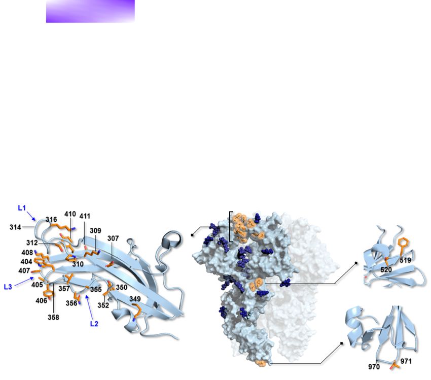
3.2 Evolution of HCoV-229E optimized receptor ular dating approaches. Results indicated that Classes II, III,
binding and IV, which have about twofold higher affinity than Class I,
Because, in analogy to SARS-CoV-2, HCoV-229E binds a protein emerged 3–9 years apart (Fig. 3C). However, since the appear-
receptor, we further investigated the positively selected sites in ance of Class V (with much higher affinity) about 25 years ago,
the spike protein. The specificity of HCoV-229E for hANPEP was no RBD class emerged for 10 years (Fig. 3C). In fact, Class VI split
previously ascribed to an extended tandem of H-bonds involving from Class V about 15 years ago and the two classes show very
the 314–320 segment of RBD Loop 1 and the 287–292 portion of a similar sequence and binding properties. Thus, little variation
surface-exposed β-strand hANPEP Domain II (Wong et al., 2017). seems to have accumulated approximately in the last 25 years.
Most of these interactions involve backbone atoms, reducing the These different time intervals are not fully consistent with anti-
dependency on sequence variations. In fact, the camel alphacoro- genic drift, which is expected to result in a more regular emer-
navirus can use hANPEP as a receptor (Corman et al., 2016). It was, gence of antigenic variants. An alternative, not mutually exclu-
however, suggested that changes in loop regions might accom- sive possibility is that strains with higher affinity for the cellu-
modate species-specific differences among ANPEP orthologs and lar receptor have out-competed strains with lower affinity and
optimize receptor-binding affinity (Li et al., 2019). We thus com- that HCoV-229E has evolved to optimize binding to the cellular
pared the HCoV-229E RBD crystal structure and the corresponding receptor.
model for camel alphacoronavirus (Fig. 3A). We also modeled
camel ANPEP (cANPEP) based on the structure of the human
ortholog. Overall, cANPEP features fewer charged residues at the
4. Discussion
interface than the human protein. In particular, T287 and I314 Zoonotic diseases have been constantly emerging during human
are replaced by D288 and D315 in the human receptor, whereas history, accounting for a large number of epidemics and pan-
G291 is replaced by K292. Analysis of the contact interface indi- demics, as well as for an enormous health burden. The endemic
cated that the Positively Selected Sites 316 (R or K, depending on coronaviruses usually cause very mild symptoms, at least in
RBD class), 407 (S in Class I and H in Classes V and VI), and 408 immunocompetent individuals, and can hardly be regarded as
(K in Classes I, V, and VI) contribute to additional interactions pathogens of concern. We however mention that, because they
with the human protein than those established by the camel virus have now circulated in (and adapted to) human populations for
(Fig. 3A). These are made possible by the presence of the charged decades or centuries, it cannot be excluded that they were once
residues in the human receptor. Overall, these observations sug- more pathogenic than they are now. Although we cannot go back
gest that HCoV-229E can interact with hANPEP more efficiently in time and infer the original phenotype of endemic coronaviruses,
than the camel virus and that positively selected sites contribute nor can we have a full picture of their ancestral genetic diver-
to increased affinity. sity, analysis of their evolution is potentially very informative to
Previous investigations showed that the affinity of the six understand the future trajectories of SARS-CoV-2 and of coron-
RBD classes of HCoV-229E for hANPEP varies in a range of Kd aviruses in general. Analysis of bat coronaviruses indicated that,
from ∼430 nM (Class I) to ∼30 nM (Classes V and VI) (Fig. 3C) in analogy to SARS-CoV, SARS-CoV-2 required limited adaptation
(Wong et al., 2017). In particular, a strong increase in affinity is to gain the ability to infect and spread in our species (Cagliani
observed for Classes V and VI. Some of the positively selected sites et al., 2020; MacLean et al., 2020). As HCoV-OC43 and HCoV-229E6 Virus Evolution
Downloaded from https://academic.oup.com/ve/article/7/2/veab061/6308720 by guest on 08 October 2021
Figure 2. Positive selection acting on HCoV-229E. (A) A schematic representation of HCoV-229E ORFs is reported with indication of all positively
selected sites found by gammaMap. Sites with a frequency of the most common amino acidD. Forni et al. 7
Downloaded from https://academic.oup.com/ve/article/7/2/veab061/6308720 by guest on 08 October 2021
Figure 3. Molecular evolution of HCoV-229E spike protein. (A) Atomic details of the interactions between HCoV-229E (six RBD classes, blue shades) and
the corresponding model for camel alphacoronavirus (pink) with both hANPEP (gray) and cANPEP (light green). RBDs-hANPEP conserved interaction
pattern, involving Loop 1, is shown in the upper-left panel. Relevant structural differences among the various RBDs and the details on their
interactions at the binding interface are represented in the other three panels. Positively selected sites are underlined. N atoms are colored in blue and
O atoms are in red. Salt bridges and H-bonds are represented by dashed black lines. (B) Epitopes mapped on the spike protein RBDs of three different
HCoV-E229 classes. The RBDs are in different shades of blue, whereas the epitopes are in red. Labels 1, 2, and 3 refer to Loop 1, Loop 2, and Loop 3.
(C) Timescaled maximum clade credibility tree of the spike protein RBD. Branch lengths represent the evolutionary time measured by the grids
corresponding to the timescale shown at the tree base (in years). For internal nodes, 95 per cent credible interval bars are shown and black dots
indicate a posterior probability >0.80 for that node. Tip nodes are colored based on the figure legend, where the Kd of all six RBD classes for hANPEP
interaction calculated by Wong et al., (2017) is also reported.
Our results indicate that the spike protein and other structural several coronaviruses, including HCoV-OC43, in addition to their
proteins of both viruses represented the major targets of selec- role in virion maturation, are capable of antagonizing interferon
tion. An interesting exception is the strong signature of selection responses (Yang et al., 2013; Siu et al., 2014; Beidas and Chehadeh,
we observed for HCoV-OC43 ORF5 (also known as ns12.9). The 2018). Overall, these data suggest that positively selected sites
encoded protein functions as a viroporin and its deletion reduces in these proteins might contribute to fine-tuning the interaction
viral replication, inflammatory response, and virulence in mouse between coronaviruses and human immune responses.
models (Zhang et al., 2015). Positive selection also drove the evo- Clearly, the spike protein and HE in the case of HCoV-OC43 have
lution of the membrane proteins of both viruses, as well as of the a major interest as targets of selection, as they represent major
envelope protein of HCoV-OC43. This latter, besides having struc- determinants of host range and infectivity (Forni et al., 2017; Cui,
tural roles, acts as a viroporin and represents a neurovirulence Li, and Shi, 2019). Most selected sites were found to be located in
factor (Stodola et al., 2018). Likewise, the membrane proteins of the RBDs of the spike proteins, as well as in the lectin domain of8 Virus Evolution
HE. However, additional sites mapped to other regions of the spike drift. However, these observations do not imply that immune
proteins and were mostly fixed in frequency. These include three escape is the only driver of HCoV-229E evolution. In fact, grow-
sites in the heptad repeat region of the spike protein of HCoV-229E ing evidence suggests that the humoral immune response against
and one site in the fusion peptide of HCoV-OC43 (Figs 1. and 2). endemic coronaviruses wanes in a few months (Kiyuka et al., 2018;
Notably, the heptad repeat region was previously described as a Edridge et al., 2020; Galanti and Shaman, 2021). As a consequence,
major target of selection in MERS-CoV and related camel viruses natural reinfection is common between 6 and 105 months (Edridge
(Cotten et al., 2014; Forni et al., 2015) and variants within this et al., 2020). Thus, it is unclear whether the antibody response can
region and/or the fusion peptide were shown to modulate viral be regarded as a strong selective pressure for these viruses. Our
tropism and host range in several viruses, including animal coro- dating of the emergence of HCoV-229E RBD classes indicates an
naviruses (de Haan et al., 2006; Yamada et al., 2009). It is also initial rapid turnover of Classes I–IV followed by a 10-year time
worth mentioning that, in line with our data, a previous analy- during which no variant turned up after the emergence of Class
sis that focused on the spike proteins detected positive selection V. Class V and the closely related Class VI RBDs differ in binding
for both HCoV-OC43 and HCoV-229E, although the sites did not affinity from the other classes by almost an order of magnitude
exactly correspond to the ones we describe herein (Jo, Drosten, (Wong et al., 2017). Since the emergence of these high-affinity
Downloaded from https://academic.oup.com/ve/article/7/2/veab061/6308720 by guest on 08 October 2021
and Drexler, 2021). The reason for this is that different methodolo- classes, the HCoV-229E spike proteins have accumulated fewer
gies were applied to search for selection signatures. Specifically, changes compared to earlier time periods. These patterns are not
we used a method that jointly uses divergence (from the out- readily explained by the antigenic drift hypothesis, which pre-
group) and genetic diversity (within the sampled human viruses) dicts a more regular emergence of spike variants. Thus, together
to detect selection events that occurred since the separation from with the remarkable seasonality of endemic coronaviruses, these
the bovine or camel viruses. As a consequence, the selected sites patterns suggest that selection has also been acting to optimize
detected by gammaMap can be either fixed or polymorphic in cir- binding to the cellular receptor and that strains with increasing
culating human strains. Conversely, Jo and coworkers did not affinity have replaced those with lower binding ability and, possi-
include outgroup information and used methods that detected bly, lower infectivity. In this respect, it is also worth mentioning
sites with dN/dS significantly higher than one in the sampled that even Class V and VI RBDs have much lower affinity for hAN-
population of human viruses (Jo, Drosten, and Drexler, 2021). PEP (Kd ∼30 nM) than most other human coronaviruses for their
Coronaviruses can use very different cellular receptors and respective cellular receptors (Kd in the range of 1–5 nM for SARS-
their spike proteins display a remarkable ability to adapt to dif- CoV-2, SARS-CoV, and HCoV-NL63) (Wu et al., 2009; Yi et al., 2020).
ferent cellular receptors (Forni et al., 2017). Embecoviruses such It is thus possible that optimization for receptor binding played a
as HCoV-OC43, HCoV-HKU1, and BCoV attach to 9-O-acetylated relevant role for HCoV-229E evolution.
sialoglycans via the spike protein, with HE acting as a receptor- We also found signals of positive selection in the lectin domain
destroying enzyme (de Groot, 2006; Hulswit et al., 2019). Con- of HE. Whereas such signals might also result from antigenic
versely, HCoV-229E and HCoV-NL63 use a protein receptor (Forni drift (Jo, Drosten, and Drexler, 2021; Kistler and Bedford, 2021),
et al., 2017). Biochemical and crystallographic analyses indicated previous data showing co-evolution of the spike protein with HE
that, since the shift to the human host, the spike and HE proteins (Bakkers et al., 2017; Lang et al., 2020) suggest that optimization
of HCoV-OC43 have co-evolved to optimize the balance between for human cell infection contributed to the evolution of this virus.
binding and release from sialoglycans in human airways (Bakkers In this respect, it is interesting to note that very limited variation
et al., 2017; Lang et al., 2020). We confirm herein the previously with no temporal pattern was evident in the RBD region of BCoV
observed emergence of spike and HE variants over time and the sequences sampled over 34 years. This suggests that, if antigenic
replacement of earlier variants with the more recent ones (Fig. 1). drive occurs in humans, it does not in cattle.
However, the relative binding affinity of HE and spike variants The observations above are not meant to imply that immune
have not been extensively investigated, yet. This fact, the com- escape played no role in the evolution of HCoV-229E and HCoV-
plex interplay between the two proteins and the poor knowledge OC43 and, most likely, distinct coronaviruses are subject to
of the structure of 9-O-acetylated sialoglycoconjugates that are diverse selective pressures. For instance, Kistler and Bedford
effectively bound in the human respiratory tract make it impos- detected no evidence of antigenic drift for HCoV-NL63 (Kistler
sible to analyze in detail affinity changes over time. Conversely, and Bedford, 2021). Clearly, gaining insight into the evolution
binding assays have shown that different classes of the HCoV- of the other human coronaviruses has relevance for our under-
229E spike protein RBD have very different binding affinities for standing of SARS-CoV-2. Recent work has indicated that the spike
hANPEP. The appearance of variants with increased affinity has protein of SARS-CoV-2 can tolerate a substantial number of sub-
clearly occurred progressively in time (Fig. 3C), as a result of pos- stitutions, with some of them even increasing receptor binding
itive selection (Fig. 3A). On one hand, these data suggest that (Starr et al., 2020). The N501Y substitution in the RBD is one
HCoV-OC43 and HCoV-229E have been adapting to optimize recep- such variant and it is shared by three of the recently emerged
tor engagement and spread in human populations. On the other SARS-CoV-2 lineages (B.1.1.7, P.1, and B.1.351), which also carry a
hand, the evolution of the spike proteins of endemic coronaviruses number of additional replacements in the spike (https://www.gov.
has been interpreted in terms of antigenic drift (Wong et al., 2017; uk/government/publications/investigation-of-novel-sars-cov-2-
Li et al., 2019; Eguia et al., 2021; Jo, Drosten, and Drexler, 2021; variant-variant-of-concern-20201201, last accessed 28 May 2021)
Kistler and Bedford, 2021). Indeed, it was previously demon- (Faria et al., 2021; Tegally et al., 2021). The initial characterization
strated that antibodies raised against HCoV-229E Class I RBD do of these lineages has indicated that B.1.1.7 is more transmissi-
not neutralize viruses with RBDs belonging to different classes ble than previous lineages (Leung et al., 2021), but seems to have
(Wong et al., 2017; Li et al., 2019). Along the same lines, Eguia similar antigenic properties as the prototypic strain (Xie et al.,
and coworkers showed that human sera collected in the 1980s 2021; Muik et al., 2021; Wang et al., 2021; Hoffmann et al., 2021).
and 1990s have low neutralizing activity against the spike proteins Conversely, B.1.351 and P.1, both carrying the E484K substitu-
from HCoV-229E strains isolated years later (Eguia et al., 2021). tion in the RBD, have been associated with cases of reinfection
This is a clear indication that HCoV-229E has undergone antigenic (Resende et al., 2020; Kuzmina et al., 2021; Nonaka et al., 2021;D. Forni et al. 9
Naveca et al., 2021) and evasion of naturally elicited or vaccine- Cotten, M. et al. (2014) ‘Spread, Circulation, and Evolution of the Mid-
elicited antibody responses (Wang et al., 2021; Hoffmann et al., dle East Respiratory Syndrome Coronavirus’, mBio, 5: e01062-13.
2021; Zhou et al., 2021; Jangra et al., 2021; Li et al., 2021). Albeit Crooks, G. E. et al. (2004) ‘WebLogo: A Sequence Logo Generator’,
very preliminary, these observations suggest that SARS-CoV-2 can Genome Research, 14: 1188–90.
adapt to elude previous immunity. Notably, the mass deploy- Cui, J., Li, F., and Shi, Z. L. (2019) ‘Origin and Evolution of Pathogenic
ment of vaccines against SARS-CoV-2 will subject the virus to Coronaviruses’, Nature Reviews: Microbiology, 17: 181–92.
a selective pressure that the endemic coronaviruses have never de Groot, R. J. (2006) ‘Structure, Function and Evolution of the
experienced. Hemagglutinin-Esterase Proteins of Corona- and Toroviruses’,
Glycoconjugate Journal, 23: 59–72.
Data availability de Haan, C. A. et al. (2006) ‘Cooperative Involvement of the S1 and
S2 Subunits of the Murine Coronavirus Spike Protein in Recep-
Sequences were retrieved from the NCBI (http://www.ncbi.
tor Binding and Extended Host Range’, Journal of Virology, 80:
nlm.nih.gov/, last accessed 7 April 2021) database. Lists of all
10909–18.
accession IDs are reported in Supplementary Table S1 and Sup-
Drosten, C. et al. (2003) ‘Identification of a Novel Coronavirus in
plementary Table S2.
Downloaded from https://academic.oup.com/ve/article/7/2/veab061/6308720 by guest on 08 October 2021
Patients with Severe Acute Respiratory Syndrome’, New England
Journal of Medicine, 348: 1967–76.
Supplementary data Duchene, S. et al. (2015) ‘The Performance of the Date-
Supplementary data is available at Virus Evolution online. Randomization Test in Phylogenetic Analyses of Time-Structured
Virus Data’, Molecular Biology and Evolution, 32: 1895–906.
Funding Edridge, A. W. D. et al. (2020) ‘Seasonal Coronavirus Protective
Immunity Is Short-lasting’, Nature Medicine, 26: 1691–3.
This work was supported by the Italian Ministry of Health (‘Ricerca
Eguia, R. et al. (2021) ‘A Human Coronavirus Evolves Antigenically to
Corrente 2019-2020’ to M.S. and ‘Ricerca Corrente 2018–2020’ to
Escape Antibody Immunity’, PLOS Pathogens, 17: e1009453.
D.F.), by Fondazione Cariplo (grant CORONA, no. 2020-1353), and
Faria, N. R. et al. (2021) ‘Genomics and Epidemiology of a Novel SARS-
by Regione Lombardia (Bando Progetti Ricerca Covid 19 - CUP
CoV-2 Lineage in Manaus, Brazil’. Science, 372: 815–21.
H44I20000470002).
Forni, D. et al. (2015) ‘The Heptad Repeat Region Is a Major Selection
Target in MERS-CoV and Related Coronaviruses’, Scientific Reports,
Conflict of interest: None declared.
5: 14480.
—— et al. (2017) ‘Molecular Evolution of Human Coronavirus
References Genomes’, Trends in Microbiology, 25: 35–48.
Al-Khannaq, M. N. et al. (2016) ‘Molecular Epidemiology and Evo- Galanti, M., and Shaman, J. (2021) ‘Direct Observation of Repeated
lutionary Histories of Human Coronavirus OC43 and HKU1 Infections with Endemic Coronaviruses’, The Journal of Infectious
among Patients with Upper Respiratory Tract Infections in Kuala Diseases, 223: 409–15.
Lumpur, Malaysia’, Virology Journal, 13: 33. Ho, S. Y. et al. (2011) ‘Time-dependent Rates of Molecular Evolution’,
Altschul, S. F. et al. (1997) ‘Gapped BLAST and PSI-BLAST: A New Molecular Ecology, 20: 3087–101.
Generation of Protein Database Search Programs’, Nucleic Acids Hoffmann, M. et al. (2021) ‘SARS-CoV-2 Variants B.1.351 and P.1
Research, 25: 3389–402. Escape from Neutralizing Antibodies’, Cell, 184: 2384–93.e12.
Arnold, K. et al. (2006) ‘The SWISS-MODEL Workspace: A Web- Hulswit, R. J. G. et al. (2019) ‘Human Coronaviruses OC43 and HKU1
based Environment for Protein Structure Homology Modelling’, Bind to 9-O-Acetylated Sialic Acids via a Conserved Receptor-
Bioinformatics, 22: 195–201. Binding Site in Spike Protein Domain A’, Proceedings of the National
Bakkers, M. J. et al. (2017) ‘Betacoronavirus Adaptation to Humans Academy of Sciences of the United States of America, 116: 2681–90.
Involved Progressive Loss of Hemagglutinin-Esterase Lectin Activ- Huynh, J. et al. (2012) ‘Evidence Supporting a Zoonotic Origin of
ity’, Cell Host and Microbe, 21: 356–66. Human Coronavirus Strain NL63’, Journal of Virology, 86: 12816–25.
Beidas, M., and Chehadeh, W. (2018) ‘Effect of Human Coronavirus Jangra, S. et al. (2021) ‘The E484K Mutation in the SARS-CoV-2 Spike
OC43 Structural and Accessory Proteins on the Transcriptional Protein Reduces but Does Not Abolish Neutralizing Activity of
Activation of Antiviral Response Elements’, Intervirology, 61: 30–5. Human Convalescent and Post-Vaccination Sera’, medRxiv.
Bidokhti, M. R. M. et al. (2013) ‘Evolutionary Dynamics of Bovine Coro- Jespersen, M. C. et al. (2017) ‘BepiPred-2.0: Improving Sequence-
naviruses: Natural Selection Pattern of the Spike Gene Implies Based B-cell Epitope Prediction Using Conformational Epitopes’,
Adaptive Evolution of the Strains’, Journal of General Virology, 94: Nucleic Acids Research, 45: W24–9.
2036–49. Jo, W. K., Drosten, C., and Drexler, J. F. (2021) ‘The Evolutionary
Bouckaert, R. et al. (2014) ‘BEAST 2: A Software Platform for Bayesian Dynamics of Endemic Human Coronaviruses’, Virus Evolution, 7:
Evolutionary Analysis’, PLoS Computational Biology, 10: e1003537. veab020.
Cagliani, R. et al. (2020) ‘Computational Inference of Selection Under- Katoh, K., and Standley, D. M. (2013) ‘MAFFT Multiple Sequence
lying the Evolution of the Novel Coronavirus, SARS-CoV-2’, Journal Alignment Software Version 7: Improvements in Performance and
of Virology, 94: e00411–20. Usability’, Molecular Biology and Evolution, 30: 772–80.
Corman, V. M. et al. (2015) ‘Evidence for an Ancestral Association Kawabata, T. (2016) ‘HOMCOS: An Updated Server to Search and
of Human Coronavirus 229E with Bats’, Journal of Virology, 89: Model Complex 3D Structures’, Journal of Structural and Functional
11858–70. Genomics, 17: 83–99.
—— et al. (2016) ‘Link of a Ubiquitous Human Coronavirus to Killerby, M. E. et al. (2020) ‘Middle East Respiratory Syndrome Coro-
Dromedary Camels’, Proceedings of the National Academy of Sciences navirus Transmission’, Emerging Infectious Diseases, 26: 191–8.
of the United States of America, 113: 9864–9. Kissler, S. M. et al. (2020) ‘Projecting the Transmission Dynamics
—— et al. (2018) ‘Hosts and Sources of Endemic Human Coron- of SARS-CoV-2 through the Postpandemic Period’, Science, 368:
aviruses’, Advances in Virus Research, 100: 163–88. 860–8.10 Virus Evolution
Kistler, K. E., and Bedford, T. (2021) ‘Evidence for Adaptive Evolution —— (2001) ‘Evaluation of Methods for Detecting Recombination
in the Receptor-Binding Domain of Seasonal Coronaviruses OC43 from DNA Sequences: Computer Simulations’, Proceedings of the
and 229e’, eLife, 10: e64509. National Academy of Sciences of the United States of America, 98:
Kiyuka, P. K. et al. (2018) ‘Human Coronavirus NL63 Molecular Epi- 13757–62.
demiology and Evolutionary Patterns in Rural Coastal Kenya’, The Rambaut, A. et al. (2018) ‘Posterior Summarization in Bayesian Phy-
Journal of Infectious Diseases, 217: 1728–39. logenetics Using Tracer 1.7’, Systematic Biology, 67: 901–4.
Kuzmina, A. et al. (2021) ‘SARS-CoV-2 Spike Variants Exhibit Differ- Resende, P. C. et al. (2020), Spike E484K Mutation in the First SARS-
ential Infectivity and Neutralization Resistance to Convalescent CoV-2 Reinfection Case Confirmed in Brazil. accessed 28 May 2021.
rithmic Complexity for the 3SEQ Recombination Detection Algo- Šali, A., and Blundell, T. L. (1993) ‘Comparative Protein Modelling by
rithm’, Molecular Biology and Evolution, 35: 247–51. Satisfaction of Spatial Restraints’, Journal of Molecular Biology, 234:
Lam, T. T. et al. (2020) ‘Identification of 2019-nCoV Related Coro- 779–815.
naviruses in Malayan Pangolins in Southern China’, BioRxiv. Sawyer, S. (1989) ‘Statistical Tests for Detecting Gene Conversion’,
Downloaded from https://academic.oup.com/ve/article/7/2/veab061/6308720 by guest on 08 October 2021
02.13.945485. Molecular Biology and Evolution, 6: 526–38.
Lang, Y. et al. (2020) ‘Coronavirus Hemagglutinin-esterase and Spike Schrödinger, L. (2017) The PyMOL Molecular Graphics System, Version 2.0.
Proteins Coevolve for Functional Balance and Optimal Virion Schrödinger, LLC.
Avidity’, Proceedings of the National Academy of Sciences of the United Sironi, M. et al. (2020) ‘Editors of Infection, Genetics and Evo-
States of America, 117: 25759–70. lution. SARS-CoV-2 and COVID-19: A Genetic, Epidemiological,
Leung, K. et al. (2021) ‘Early Transmissibility Assessment of the and Evolutionary Perspective’, Infection, Genetics and Evolution, 84:
N501Y Mutant Strains of SARS-CoV-2 in the United Kingdom, 104384.
October to November 2020’, Eurosurveillance, 26: 2002106. Siu, K. et al. (2014) ‘Suppression of Innate Antiviral Response by
Li, Q. et al. (2021) ‘SARS-CoV-2 501Y.V2 Variants Lack Higher Infec- Severe Acute Respiratory Syndrome Coronavirus M Protein Is
tivity but Do Have Immune Escape’, Cell, 184: 2362–71.e9. Mediated through the First Transmembrane Domain’, Cellular and
Li, Z. et al. (2019) ‘The Human Coronavirus HCoV-229E S-protein Molecular Immunology, 11: 141–9.
Structure and Receptor Binding’, eLife, 8: e51230. Smith, J. M. (1992) ‘Analyzing the Mosaic Structure of Genes’, Journal
Lipsitch, M. et al. (2003) ‘Transmission Dynamics and Control of of Molecular Evolution, 34: 126–9.
Severe Acute Respiratory Syndrome’, Science, 300: 1966–70. Starr, T. N. et al. (2020) ‘Deep Mutational Scanning of SARS-CoV-
Liu, P. et al. (2020) ‘Are Pangolins the Intermediate Host of the 2019 2 Receptor Binding Domain Reveals Constraints on Folding and
Novel Coronavirus (SARS-CoVV-2)?’ PLoS Pathogens, 16: e1008421. ACE2 Binding’, Cell, 182: 1295–310.e20.
MacLean, O. A. et al. (2020) ‘Evidence of Significant Natural Selection Stodola, J. K. et al. (2018) ‘The OC43 Human Coronavirus Envelope
in the Evolution of SARS-CoV-2 in Bats, Not Humans’, bioRxiv. Protein Is Critical for Infectious Virus Production and Propagation
Martin, D., and Rybicki, E. (2000) ‘RDP: Detection of Recombination in Neuronal Cells and Is a Determinant of Neurovirulence and
amongst Aligned Sequences’, Bioinformatics, 16: 562–3. CNS Pathology’, Virology, 515: 134–49.
Martin, D. P. et al. (2017) ‘Detecting and Analyzing Genetic Recombi- Tao, Y. et al. (2017) ‘Surveillance of Bat Coronaviruses in Kenya Iden-
nation Using RDP4’, Methods in Molecular Biology (Clifton, NJ), 1525: tifies Relatives of Human Coronaviruses NL63 and 229E and Their
433–60. Recombination History’, Journal of Virology, 91: e01953–16.
Muik, A. et al. (2021) ‘Neutralization of SARS-CoV-2 Lineage B.1.1.7 Tegally, H. et al. (2021) ‘Detection of a SARS-CoV-2 Variant of Concern
Pseudovirus by BNT162b2 Vaccine-Elicited Human Sera’, Science, in South Africa’, Nature, 592: 438–43.
371: 1152–3. Vijgen, L. et al. (2006) ‘Evolutionary History of the Closely
Murray, G. G. et al. (2016) ‘The Effect of Genetic Structure on Molec- Related Group 2 Coronaviruses: Porcine Hemagglutinating
ular Dating and Tests for Temporal Signal’, Methods in Ecology and Encephalomyelitis Virus, Bovine Coronavirus, and Human Coro-
Evolution, 7: 80–9. navirus OC43’, Journal of Virology, 80: 7270–4.
Naveca, F. et al. (2021), SARS-CoV-2 Reinfection by the New Variant of —— et al. (2005a) ‘Complete Genomic Sequence of Human Coro-
Concern (VOC) P. 1 in Amazonas, Brazil. accessed 28 May 2021. ogy, 79: 1595–604.
Nonaka, C. K. V. et al. (2021) ‘Genomic Evidence of a SARS-CoV-2 —— et al. (2005b) ‘Genetic Variability of Human Respiratory Coron-
Reinfection Case with E484K Spike Mutation in Brazil’, Emerging avirus OC43’, Journal of Virology, 79: 3223–4.
Infectious Diseases, 27: 1522–4. Walls, A. C. et al. (2016) ‘Cryo-Electron Microscopy Structure of a
Olival, K. J. et al. (2020) ‘Possibility for Reverse Zoonotic Transmission Coronavirus Spike Glycoprotein Trimer’, Nature, 531: 114–7.
of SARS-CoV-2 to Free-ranging Wildlife: A Case Study of Bats’, PLoS Wang, P. et al. (2021) ‘Antibody Resistance of SARS-CoV-2 Variants
Pathogens, 16: e1008758. B.1.351 and B.1.1.7’, Nature, 593: 130–5.
Oude Munnink, B. B. et al. (2021) ‘Transmission of SARS-CoV-2 on Wernersson, R., and Pedersen, A. G. (2003) ‘RevTrans: Multiple Align-
Mink Farms between Humans and Mink and Back to Humans’, ment of Coding DNA from Aligned Amino Acid Sequences’, Nucleic
Science, 371: 172–7. Acids Research, 31: 3537–9.
Pfefferle, S. et al. (2009) ‘Distant Relatives of Severe Acute Res- Willard, L. et al. (2003) ‘VADAR: A Web Server for Quantitative Eval-
piratory Syndrome Coronavirus and Close Relatives of Human uation of Protein Structure Quality’, Nucleic Acids Research, 31:
Coronavirus 229E in Bats, Ghana’, Emerging Infectious Diseases, 15: 3316–9.
1377–84. Wilson, D. J. et al. (2011) ‘A Population Genetics-Phylogenetics
Posada, D., and Crandall, K. A. (1998) ‘MODELTEST: Testing the Model Approach to Inferring Natural Selection in Coding Sequences’,
of DNA Substitution’, Bioinformatics, 14: 817–8. PLoS Genetics, 7: e1002395.D. Forni et al. 11
Wong, A. H. M. et al. (2017) ‘Receptor-Binding Loops in Alphacoro- Coronavirus (MERS-CoV) Are Potent Interferon Antagonists’, Pro-
navirus Adaptation and Evolution’, Nature Communications, 8: tein and Cell, 4: 951–61.
1735. Ye, Z. W. et al. (2020) ‘Zoonotic Origins of Human Coronaviruses’,
Wong, M. C. et al. (2020) ‘Evidence of Recombination in Coronaviruses International Journal of Biological Sciences, 16: 1686–97.
Implicating Pangolin Origins of nCoV-2019’, BioRxiv. 02.07.939207. Yi, C. et al. (2020) ‘Key Residues of the Receptor Binding Motif in
Wu, K. et al. (2009) ‘Crystal Structure of NL63 Respiratory Coro- the Spike Protein of SARS-CoV-2 That Interact with ACE2 and
navirus Receptor-Binding Domain Complexed with its Human Neutralizing Antibodies’, Cellular and Molecular Immunology, 17:
Receptor’, Proceedings of the National Academy of Sciences of the 621–30.
United States of America, 106: 19970–4. Zaki, A. M. et al. (2012) ‘Isolation of a Novel Coronavirus from a Man
Xiao, K. et al. (2020) ‘Isolation of SARS-CoV-2-Related Coronavirus with Pneumonia in Saudi Arabia’, New England Journal of Medicine,
from Malayan Pangolins’, Nature, 583: 286–9. 367: 1814–20.
Xie, W. et al. (2011) ‘Improving Marginal Likelihood Estimation for Zhang, R. et al. (2015) ‘The Ns12.9 Accessory Protein of Human Coro-
Bayesian Phylogenetic Model Selection’, Systematic Biology, 60: navirus OC43 Is a Viroporin Involved in Virion Morphogenesis and
150–60. Pathogenesis’, Journal of Virology, 89: 11383–95.
Downloaded from https://academic.oup.com/ve/article/7/2/veab061/6308720 by guest on 08 October 2021
Xie, X. et al. (2021) ‘Neutralization of SARS-CoV-2 Spike 69/70 Dele- Zhou, D. et al. (2021) ‘Evidence of Escape of SARS-CoV-2 Vari-
tion, E484K and N501Y Variants by BNT162b2 Vaccine-Elicited ant B.1.351 from Natural and Vaccine-Induced Sera’, Cell, 184:
Sera’, Nature Medicine, 27: 620–1. 2348–61.e6.
Yamada, Y. et al. (2009) ‘Acquisition of Cell-cell Fusion Activity Zhou, P. et al. (2020) ‘A Pneumonia Outbreak Associated with A New
by Amino Acid Substitutions in Spike Protein Determines the Coronavirus of Probable Bat Origin’, Nature, 579: 270–3.
Infectivity of a Coronavirus in Cultured Cells’, PLoS One, 4: e6130. Zhu, N. et al. (2020) ‘China Novel Coronavirus Investigating and
Yang, Y. et al. (2013) ‘The Structural and Accessory Proteins M, ORF Research Team. A Novel Coronavirus from Patients with Pneumo-
4a, ORF 4b, and ORF 5 of Middle East Respiratory Syndrome nia in China, 2019’, New England Journal of Medicine, 382: 727–33.You can also read