Exosome-Depleted Excretory-Secretory Products of the Fourth-Stage Larval Angiostrongylus cantonensis Promotes Alternative Activation of ...
←
→
Page content transcription
If your browser does not render page correctly, please read the page content below
ORIGINAL RESEARCH
                                                                                                                                               published: 23 July 2021
                                                                                                                                     doi: 10.3389/fimmu.2021.685984

                                            Exosome-Depleted Excretory-
                                            Secretory Products of the Fourth-
                                            Stage Larval Angiostrongylus
                                            cantonensis Promotes Alternative
                                            Activation of Macrophages Through
                                            Metabolic Reprogramming by the
                                            PI3K-Akt Pathway
                          Edited by:
                                            Shuo Wan 1,2,3,4†, Xiaoqiang Sun 2,5†, Wenyan Tang 6, Lifu Wang 1,2,3, Zhongdao Wu 1,2,3
                      Diana Boraschi,
     Shenzhen Institutes of Advanced
                                            and Xi Sun 1,2,3*
           Technology (CAS), China          1 Department of Parasitology of Zhongshan School of Medicine, Sun Yat-sen University, Guangzhou, China, 2 Key Laboratory

                        Reviewed by:        of Tropical Disease Control (SYSU), Ministry of Education, Guangzhou, China, 3 Provincial Engineering Technology Research
                       Nadia Lampiasi,      Center for Biological Vector Control, Guangzhou, China, 4 The First Affiliated Hospital, Jinan University, Guangzhou, China,
Institute of Biomedicine and Molecular      5 Zhongshan School of Mathematics, Sun Yat-Sen University, Guangzhou, China, 6 Department of Neonatology, Guangzhou

   Immunology Alberto Monroy (IBIM),        Women and Children’s Medical Center, Guangzhou Medical University, Guangzhou, China
                                    Italy
                          Paola Italiani,
National Research Council (CNR), Italy      Angiostrongylus cantonensis (AC), which parasitizes in the brain of the non-permissive
                  *Correspondence:          host, such as mouse and human, is an etiologic agent of eosinophilic meningitis.
                               Xi Sun       Excretory-secretory (ES) products play an important role in the interaction between
              sunxi2@mail.sysu.edu.cn
     †
                                            parasites and hosts’ immune responses. Inflammatory macrophages are responsible
      These authors have contributed
                 equally to this work       for eosinophilic meningitis induced by AC, and the soluble antigens of Angiostrongylus
                                            cantonensis fourth stage larva (AC L4), a mimic of dead AC L4, aggravate eosinophilic
                   Specialty section:       meningitis in AC-infected mice model via promoting alternative activation of macrophages.
         This article was submitted to
                Cytokines and Soluble
                                            In this study, we investigated the key molecules in the ES products of AC L4 on
               Mediators in Immunity,       macrophages and observed the relationship between metabolic reprogramming and
               a section of the journal
                                            the PI3K-Akt pathway. First, a co-culture system of macrophage and AC L4 was
              Frontiers in Immunology
                                            established to define the role of AC L4 ES products on macrophage polarization. Then,
           Received: 26 March 2021
            Accepted: 07 July 2021          AC L4 exosome and exosome-depleted excretory-secretory products (exofree) were
            Published: 23 July 2021         separated from AC L4 ES products using differential centrifugation, and their distinct roles
                            Citation:       on macrophage polarization were confirmed using qPCR and ELISA experiments.
Wan S, Sun X, Tang W, Wang L, Wu Z
and Sun X (2021) Exosome-Depleted
                                            Moreover, AC L4 exofree induced alternative activation of macrophages, which is
 Excretory-Secretory Products of the        partially associated with metabolic reprogramming by the PI3K-Akt pathway. Next,
 Fourth-Stage Larval Angiostrongylus
                                            lectin blot and deglycosylation assay were done, suggesting the key role of N-linked
   cantonensis Promotes Alternative
 Activation of Macrophages Through          glycoproteins in exofree. Then, glycoproteomic analysis of exofree and RNA-seq analysis
        Metabolic Reprogramming by          of exofree-treated macrophage were performed. Bi-layer PPI network analysis based on
               the PI3K-Akt Pathway.
         Front. Immunol. 12:685984.
                                            these results identified macrophage-related protein Hexa as a key molecule in inducing
   doi: 10.3389/fimmu.2021.685984           alternative activation of macrophages. Our results indicate a great value for research of

Frontiers in Immunology | www.frontiersin.org                                      1                                           July 2021 | Volume 12 | Article 685984
Wan et al.                                                                                                Exofree Promote Macrophage Alternative Activation

                                          helminth-derived immunoregulatory molecules, which might contribute to drug
                                          development for immune-related diseases.
                                          Keywords: Angiostrongylus cantonensis, exosome-depleted excretory-secretory products, N-linked glycoproteins,
                                          macrophage polarization, mechanism

INTRODUCTION                                                                    autoimmune encephalomyelitis (20). Thus, identification of the
                                                                                important compound or protein that has the function of
Angiostronglyiasis is a serious foodborne parasitic disease caused              modulating host immune responses is of great significance for
by infection of AC, which parasitizes in the brain of the non-                  understanding the interaction of parasite and host.
permissive host mouse and human and leads to serious                                Recently, the functions of exosomes in parasite-host interactions
eosinophilic meningitis; however, the underlying mechanism                      are given considerable attention. Exosomes are nanosized
remains poorly understood. Microglia/macrophages are major                      membrane-bound extracellular vesicles with a diameter of 50–200
immune cells involved in directing host inflammatory and                         nm which are released from most cell types (21). They contain
immune processes, which are extremely important in helminth                     different biomolecules, including proteins, microRNAs, lipids,
infections. Microglia, brain-resident macrophages, are activated in             glycans, etc., performing biological functions, particularly in cell-
AC-infected mice; however, minocycline (1), a specific inhibitor of              to-cell communication. Recently, studies have found that exosomes
microglia activation, failed to reverse the brain inflammation. In               can be used by parasites to deliver molecules and modulate host
our previous results, Chi3l3, an eosinophil-related protein, was                immune response (22).
found to increase sharply after AC infection, and monocyte-                         Our previous study suggested that the soluble antigens (sAg) of
derived alternative macrophages were confirmed as the main                       AC L4, a mimic of dead AC L4, aggravate eosinophilic meningitis in
source of Chi3l3 (2), which indicated that M2 macrophage may                    the AC-infected mice model, via a Chi3l3-IL-13 positive feedback
be involved in the pathogenesis of Angiostrongyliasis.                          loop (2). However, the key compound or protein in the ES products
    It is widely assumed that helminth could modulate host                      of live AC L4 is not clear. In this study, the ES products of live AC
immune responses through its ES products, including proteins,                   L4 were separated into exosomes and exofree. Interestingly, our
glycans (3, 4), and exosomes (5–7). Fasciola hepatica–derived                   results suggested that AC L4 exofree, but not exosome, could induce
Fh12 and Fh15 could significantly promote M2 polarization of                     a significant M2 polarization of macrophages through metabolic
macrophages and inhibit TLR2-, TLR5-, TLR8-induced M1                           reprogramming by the PI3K-Akt pathway. Considering that exofree
polarization of macrophages (8). Acanthocheilonema viteae–                      of N-linked glycosylated analogs failed to induce M2 polarization of
derived AvCystatin was shown to induce IL-10 production of                      macrophages, mass spectrometry analysis was performed to identify
macrophages and inhibit T cell proliferation and restore allergic               proteins with N-glycosylation sites in AC L4 exofree. Proteins with
airway inflammation (9). Moreover, AvCystatin could                              N-glycosylation sites in AC L4 exofree were identified using mass
significantly upregulate PD-L1 and PD-L2 levels in macrophages                   spectrometry analysis. Furthermore, RNA-seq analysis of exofree-
and protect against mucosal inflammation (10). MIF homologs in                   treated macrophages was performed. Bi-layer PPI network analysis
Brugia malayi could promote IL-4-induced macrophage                             based on the results above supports that macrophage-related
alternative polarization through upregulating IL-4Ra (11).                      protein Hexa in exofree is a key molecule in inducing alternative
While Trichinella spiralis rTsP53 could induce significant                       activation of macrophages.
alternative activation of macrophages via STAT6 but not                             Collectively, our results suggest that AC L4 might modulate
IL-4Ra in vitro (12). Fasciola hepatica–derived FhHDM-1 was                     macrophage function through its ES products. Especially, N-linked
shown to bind directly to LPS and protect mice against LPS-                     glycoproteins from exosome-depleted ES products of AC L4 could
induced inflammation by reducing inflammatory mediators from                      induce significant alternative activation of macrophages through
macrophages (13). S. japonicum extracellular vesicle miR-125b                   metabolic reprogramming by the PI3K-Akt pathway. Our results
and bantam miRNAs could be internalized by macrophages and                      support that Hexa, a macrophage-related protein, might be the key
modulate cell proliferation and TNF-a production, which                         molecule in exofree in inducing macrophage polarization, which
contributes to parasite survival (14). Meanwhile, numerous                      might play a key role in directing host CNS inflammation in AC
studies have highlighted the importance of glycans as their                     infection. The results of this study indicate a great value for research
interactions with the host immune system, as they were found                    of helminth-derived immunoregulatory molecules, which might
on the surfaces of helminths and within their ES products (15, 16),             contribute to drug development for immune-related diseases.
which might interact with host glycan-binding proteins, such as
C-type lectin receptors and galectins, shaping innate and adaptive
immune responses upon infection. High-mannose-type glycans
were identified in Taenia solium metacestodes (17). Glycan
                                                                                MATERIALS AND METHODS
determinants of Trichuris suis soluble products modulate LPS-                   The Animal Studies
induced activation of human DCs (18). SEA and Omega-1 from                      Male Sprague Dawley (SD) Rat (specific pathogen-free, SPF)
Schistosoma manosoni were also proved to drive DCs for Th2                      aged 6 weeks (weighing 100–120 g) were purchased from the
induction (19) and modulated the progression of experimental                    Guangdong Medical Laboratory Animal Center. The animal

Frontiers in Immunology | www.frontiersin.org                               2                                       July 2021 | Volume 12 | Article 685984
Wan et al.                                                                                         Exofree Promote Macrophage Alternative Activation

studies were approved by the Medical Research Ethics                      Na2S2O3 was added to the oxidized solutions to quench the
Committee of Sun Yat-sen University and conformed to the                  oxidation reaction. The solution was added to the prewashed
Chinese National Institute of Health Guide for the Care (No               hydrazide resins. The coupling reaction was carried out with
SYSU-IACUC-2019-535). The rats were group-housed in                       gentle shaking at 4°C overnight. Then, 1,000 units of PNGase F
ventilated cages in a temperature-control room (25°C) and                 in PBS were added to the resins, and the released supernatant was
were fed standard mouse chow. Each SD rat was infected with               collected into a dialysis bag. The supernatant was dialyzed in PBS
600 AC L3 larvae (the third stage larvae) to collect the AC L4 (the       for 6 h overnight at 4°C. The supernatant was concentrated and
fourth stage larvae) for in vitro studies.                                stored as deglycosylated AC L4 exofree. The same volume of
                                                                          exofree and deglycosylated AC exofree was used for BMDM
Preparation of Macrophages                                                functional assay in Figures 5L–M.
and Reagents
Bone marrow leukocytes were resuspended at 1.5×10⁷ cells/10 ml            Functional Experiments of Macrophages
in macrophage media (DMEM high glucose with 10% heat-                     BMDMs and freshly isolated live AC L4 (10 worms per well) was
inactivated (56°C, 30 min) FBS, 100 U/ml of penicillin-                   co-cultured for 12 h, and Chi3l3 level in the culture medium was
streptomycin, supplemented with 20 ng/ml M-CSF) and then                  measured using ELISA method. IL-13 working concentration
plated in 10 cm non-TC treated culture dishes. BMDMs were                 was 10 ng/ml in this experiment. OCR and ECAR were
cultured for 7 days, with a media change on D3. BMDMs were                monitored consecutively with a Seahorse Bioscience
detached from the dish using warm 0.05% trypsin and cultured              extracellular flux analyzer (XF24, Seahorse Bioscience) as
in tissue culture plates for functional assay. The following              described previously. Briefly, BMDMs were cultured in 24-well
reagents and working concentration were used in this study:               seahorse cell culture plate, 15,000–25,000 cells per well in 0.5 ml
recombinant murine M-CSF (20 ng/ml, 315-02;                               medium (2), stimulated with PBS, LPS (0.05 mg/ml), IL-13 (10 ng/
Peprotech), recombinant mouse IL-4 (10 ng/ml; 214-14-5,                   ml), AC L4 exosome (25 mg/ml), and AC L4 exofree (44 mg/ml)
Peprotech), recombinant mouse IL-13 (10 ng/ml; 413-ML-025/                for 24 h in a 37°C incubator. Then, cells were immersed in 500 ml
CF, R&D Systems), Ly294002 (10 mM, S1737-1mg, Beyotime),                  specified medium following two wash steps with specified
Wortmannin (100 nM, S1952, Beyotime), GW9662 (1 mg/ml,                    medium and incubated in an incubator without CO2 for 1 h
S2915, selleckchem), Leflunomide (100 mM, S1247, selleckchem),             before the measurements. The OCR and ECAR were then
Affi-Gel Hz Immunoaffinity Kit (#1536060, Biorad), PNGase F                 measured in a typical 8-min cycle of mix (2–4 min), dwell
(500 units/ml, P0704S, New England Biolabs). For the BMDM                 (2 min), and measure (2–4 min) as recommended by Seahorse
functional experiment, AC L4 exosomes working concentration               Bioscience. Then 1 mM oligomycin, 1.0 mM FCCP, 0.5 mM
was 25 mg/ml, and AC L4 exofree working concentration was 4.4,            rotenone + 0.5 mM antimycin, 10 mM glucose, 1.0 mM
44, and 440 mg/ml. LPS working concentration was 0.05 mg/ml.              oligomycin, and 50 mM 2-deoxyglucose (2-DG) were used in
sAg working concentration was 25 mg/ml. IL-4/IL-13 working                this experiment. For qPCR and ELISA assay, BMDMs were
concentration was 10 ng/ml.                                               cultured in 12-well cell culture plate, 106 cells per well in 0.5 ml
                                                                          medium, stimulated with IL-13 (10 ng/ml), IL-4 (10 ng/ml), AC
Isolation of AC L4 Exofree and                                            L4 exofree (4.4, 44, 440 mg/ml) for 24 h. For CCK8 assay, BMDMs
AC L4 Exosome                                                             were cultured in 96-well tissue culture plate, 5 × 10⁴ cells per well
AC L4 were collected from rat brains at 21 days post-infection            in 0.2 ml medium, stimulated with IL-13 (10 ng/ml), AC L4
and cultured in concentration up to 200 worms/ml with serum-              exofree (4.4, 44, 440 mg/ml) for 6, 12, 24, 48, and 72h. Especially,
free media [DMEM high glucose, 100 U/ml of penicillin-                    to define the key molecule from exofree, ultracentrifugal filters
streptomycin] in vitro in 37°C. The ES products were collected            were used for exofree fractionation, and the equal volume (200 ml)
every day for a maximum of 1 week. AC L4 exosome and exofree              of fractionated AC L4 exofree was used for BMDM stimulation
(extracellular vesicle–deleted exosome) were purified by                   (24 h) and the qPCR analysis. Oligomycin (1 mM, 495455-10MG,
differential centrifugation according to the recommended                  Sigma-Aldrich), FCCP (50 nM, C2920-10MG, Sigma-Aldrich), 2-
protocol (23). Briefly, larvae and pellets were removed by low-            DG (10 mM, D8375-10MG, Sigma-Aldrich) were used in this
speed spinning at 300 × g (10 min at 4°C), followed by 2,000 × g          experiment. All the experiments consist of quadruplicate
for 10 min at 4°C, 10,000 × g for 30 min at 4°C to deplete residual       biological replicates.
debris, and then 120,000 × g for 120 min at 4°C to gather AC L4
exofree and AC L4 exosome. AC L4 exosomes were resuspended                Exosome Characterization and Exosome
with PBS and stored at 4°C for short-term (1~7 days) or −80°C             Uptake Assay
for long-term storage. AC L4 exofree without N−glycosylated               The concentration of exosomes (number/ml) and size
analogs was obtained as previously described (24). Briefly, AC L4          distribution (in nanometer) of AC L4 were analyzed using
exofree was first desalted with spin desalting column after                IZON qNano particle analyzer (Izon Science Ltd.) equipped
incubating in boiling water for 10 min. Then, the desalted                with fast video capture and Nanoparticle Tracking Analysis
solutions were resuspended with oxidation buffer. Next, 50                software. The samples were captured for 60 s at room
mM NaIO4 was added to the solutions, and the reactions were               temperature. Each sample was measured at least three times.
kept in the dark at room temperature for 1 h. Then, 100 mM                Isolated exosomes from ES products of AC L4 were placed on a

Frontiers in Immunology | www.frontiersin.org                         3                                      July 2021 | Volume 12 | Article 685984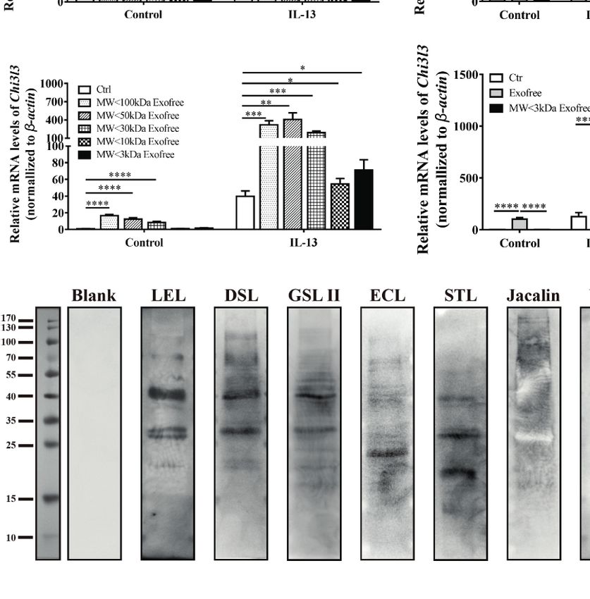
Wan et al.                                                                                     Exofree Promote Macrophage Alternative Activation

formvar-coated copper grid and settled for 2 min. The grids were        between worm proteins and mouse genes, we developed a bi-
washed with deionized water. Then, the sample was contrasted            layer network method by integrating the RNA-seq data and the
by adding an aqueous solution of 1% phosphotungstic acid                proteomics data. First, a gene-gene network was constructed for
for 2 min, followed by a rinse with deionized water. The grid           the TCGs using LASSO regression model based on the RNA-seq
was visualized using Hitachi H7650 transmission electron                expression data. A dynamic network entropy (DNE) index (26)
microscope (Hitachi-Science & Technology). For exosome                  was calculated for each node to measure the importance of genes
uptake assay, BMDM cells were incubated with PBS or with                in the constructed network by taking into account dynamic gene
5 mg/ml PKH26-labeled exosomes for 60 min. Subsequently,                interactions. Subsequently, a protein-gene network was
cells were fixed with 4% paraformaldehyde, permeabilized with            constructed for worm-proteins with molecular weight less than
1% Triton X-100, blocked with 5% BSA, and stained with Alexa            50 kDa and TCGs of exofree-, IL-4-, and exofree+IL-4-treated
Fluor Phalloidin-488 as well as DAPI, followed by a confocal            BMDMs by using String database (https://string-db.org/).
microscope analysis using Nikon C2. Scale bar=20 mm.                    Finally, a bi-layer network for worm-protein and mouse genes
                                                                        was built by integrating the above two networks. Cytoscape
Protein and mRNA Analysis                                               software was used to visualize the constructed networks. GO
For SDS-PAGE analysis, 30 mg of protein sample was loaded on            biological process significantly enriched TCGs in exofree-treated
the SDS-PAGE gel and run in SDS-PAGE running buffer. Then,              BMDMs were identified using R package clusterprofiler (v.3.8.1).
the protein gels were stained with the coomassie blue staining or
silver staining method, and the image was acquired and                  Statistical Methods
evaluated by SmartGelTM (Sagecreation Inc., China). Protein             All analyses were performed using GraphPad Prism 6.0
molecular weight standards were obtained from Bio-Rad                   (GraphPad Software, Inc. USA) or SPSS v22 (IBM, USA)
(Thermo Scientific, USA). Lectin blot analysis was performed             software. Statistical analyses of data were performed using
as described previously (25). The following reagents from               Student’s independent samples t-test. Data are expressed as
VectorLaboratories were used in this experiment: Lectin Kit             arithmetic mean ± SD, and statistical significance is defined as
III, Biotinylated (Cat. No: BK-3000), VECTASTAIN® Elite®                P < 0.05 (two-sided).
ABC HRP Kit (Peroxidase, Standard) (Cat. No: PK-6100),
Carbo-Free Blocking Solution (10×Concentrate). Protein
Deglycosylation assay was performed using Affi-Gel Hz
Immunoaffinity Kit (Biorad, #1536060) as described previously
                                                                        RESULTS
(24). And PNGase F was used to remove almost all N-linked               Excretory-Secretory Products of AC L4
oligosaccharides from glycoproteins. For mRNA analysis, qRT-
                                                                        Promote M2 Polarization of Macrophages
PCR was carried out using RevertAid™ FirstStrand cDNA
                                                                        A co-culture system of BMDMs and freshly isolated live AC L4
Synthesis Kit (Thermo Fisher Scientific, USA) according to the
                                                                        (10 worms per well) was established as shown (Figure 1A), and
manufacturer’s protocol. Specific gene expression was quantified
                                                                        Chi3l3 protein level was measured after 12 h. We observed a
with SYBR® Premix Ex Taq™ (Tli RNaseH Plus) (RR420A)
                                                                        significant increase in the protein level of Chi3l3 in BMDMs
using the Roche LightCycler® 480 real-time PCR platform. The
                                                                        treated with IL-13 and 10 AC L4. In addition, a striking increase
following amplification primers (Sangon Biotech) were used (5′
                                                                        of Chi3l3 level was observed when BMDMs were treated with 10
to 3′) (Supplementary Table 1).
                                                                        AC L4 in the presence of IL-13 (Figure 1B), suggesting that the
                                                                        ES products of AC L4 could significantly promote macrophage
RNA-Seq and Proteomics Analysis                                         M2 polarization. These results are consistent with our previous
Proteomics analysis of exofree (10~20 kDa fractions) and
                                                                        finding in BMDMs treated with AC L4 sAg (2, 27).
exosomes was performed by Jingjie PTM Biolab (Hangzhou,
China). For each category of proteins, InterPro database (a
resource that provides functional analysis of protein sequences         Isolation and Characterization of Exosome
by classifying them into families and predicting the presence of        and Exofree From AC L4 Excretory-
domains and important sites) was researched, and a two-tailed           Secretory Products
fisher’s exact test was employed to test the enrichment of the           In order to determine the key components in inducing the
identified protein against all proteins. Protein domains with a          alternative activation of macrophages, exosomes and exofree
corrected p-value < 0.05 were considered significant. Soft motif-x       were isolated from the ES product of AC L4 using modified
was used to analyze the model of sequences constituted with             differential centrifugation (Figure 2A). Transmission electron
amino acids in specific positions of modify-21-mers (10 amino            microscopy (TEM) (Figure 2B) and IZON qNano particle
acids upstream and downstream of the site) in all protein               analyzer (Figure 2C) were used to examine the size and
sequences. And all the database protein sequences were used as          morphology of AC L4 exosome. The electron micrographs of
background database parameters, other parameters with default.          the exosomes revealed rounded structures with a size from 60 to
BMDMs were treated with PBS, exofree, IL-4, or exofree+IL-4             200 nm, as previously shown (23), consistent with the results of
for 6, 12, 24h, respectively, and the RNA-seq analysis was              IZON qNano particle analyzer.
performed by Novogene Bioinformatics Technology                             It has been proven that helminth-derived microRNAs or
Cooperation (Beijing, China). To predict potential interactions         proteins-containing exosomes could be internalized by

Frontiers in Immunology | www.frontiersin.org                       4                                    July 2021 | Volume 12 | Article 685984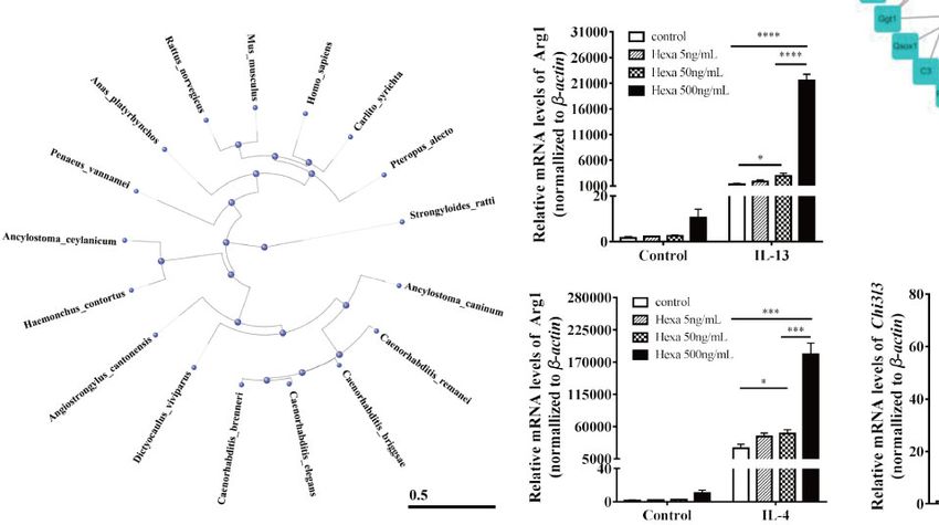
Wan et al.                                                                                                          Exofree Promote Macrophage Alternative Activation

                                A                                                       B

 FIGURE 1 | Excretory-secretory products in AC L4 induce M2 polarization of macrophage. (A) Diagram of co-culture system with 10 AC L4 above the membrane
 and BMDMs below the membrane. (B) ELISA analysis of Chi3l3 of BMDMs in the presence of 10 AC L4, IL-13, and IL-13+10 AC L4 for 24 h. Data information:
 *P
Wan et al.                                                                                                         Exofree Promote Macrophage Alternative Activation

upregulates Arg1 (Figure 3A) mRNA level in BMDMs initiated                            exosome is different from that induced by LPS (29). Intriguingly,
by IL-4 but does not significantly influence Arg1 and Chi3l3                            AC L4 exofree elicits a dose-dependent Arg1 (Figure 3H) and
mRNA level in BMDMs stimulated with AC L4 exosome alone                               Chi3l3 (Figure 3I), M2-specific genes initiated by IL-13, and a
(Figure 3B), implying that AC L4 exosome might not                                    dose-dependent suppression of Nos2 (Figure 3J), an M1-specific
significantly affect M2 polarization of macrophages. An                                gene induced by LPS in macrophages. RNA-seq results also
increase in inflammatory cytokines, such as Nos2 (Figure 3C),                          suggested that macrophage M2 signature genes, including
Il6 (Figure 3D), Il1b (Figure 3E), and Il12b (Figure 3F), was also                    Cd163, Chil3, Arg1, Nos2, Clec10a, Retnla, Pdcd1lg2, are
observed in BMDMs treated with AC L4 exosome, even in the                             sharply increased in IL-4, IL-4, and AC L4 exofree co-treated
presence of IL-4 or IL-13, suggesting that it could induce a                          group (Figure 3K) at 6, 12, and 24 h. However, cytokines,
remarkable M1 polarization of macrophages. However, Il10                              chemokines, and secreted mediators associated with
mRNA level (Figure 3G) was undetectable in BMDMs,                                     macrophage polarization were not significantly changed
suggesting M1 polarization of macrophage induced by AC L4                             (Supplementary Figure 2). Still, a low level of IL-10 in all the

                  A                             B                            C                              D

                  E                             F                            G

                  H                                          I                                          J

                   K                                                          L                                    M

 FIGURE 3 | AC L4 exofree could promote a M2 polarization, while AC L4 exosomes induce M1 polarization of macrophage. (A–G) Macrophage activation markers
 Arg1 (A), Chi3l3 (B), Nos2 (C), and related interleukins Il6 (D), Il1b (E), Il12b (F), and Il10 (G) expression level were measured in the presence of AC L4 exosome,
 IL-4, IL-4+exosome, IL-13, and IL-13+exosome for 24 h using qPCR. The working concentration of AC L4 exosome is 25 mg/ml. The working concentration of IL-13
 and IL-4 is 10 ng/ml. (H–J) Macrophage activation markers Arg1 (H), Chi3l3 (I), Nos2 (J) were measured in the presence of exofree, IL-4, IL-4+exofree, IL-13, and
 IL-13+exofree for 24 h using qPCR. (K) Signature gene expression profile of macrophage in the presence of PBS (ctr), free (AC L4 exofree), IL-4, com (IL-4+AC L4
 exofree), and 6, 12, and 24 h represent 6, 12, and 24 h after the treatment, respectively. The working concentration of AC L4 exofree is 440 mg/ml. The working
 concentration of IL-13/IL-4 is 10 ng/ml. The working concentration of LPS is 0.05 mg/ml. The horizontal axis represents genes, and vertical coordinates represent
 types of treatment. (L, M) BMDMs were stimulated with PBS, AC L4 exofree, IL-4, IL-4+ AC L4 exofree, IL-13, IL-13+ AC L4 exofree in 0~24 h The culture medium
 was discarded. BMDMs were then washed with PBS and restimulated in 24~48 h as shown. And Chi3l3 protein level of 24~48 h culture medium was measured
 using ELISA. The working concentration of AC L4 exofree is 440 mg/ml. The working concentration of IL-13/IL-4 is 10 ng/ml. The detailed experiment information
 refer to Supplementary Table 2. Data information: *P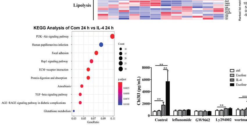
Wan et al.                                                                                            Exofree Promote Macrophage Alternative Activation

groups suggests that M2 polarization of macrophage induced by                macrophage was severely affected whether glycolysis or OXPHOS
AC L4 exofree was not caused by hemoglobin or IL-10 (30, 31).                was inhibited (Supplementary Figures 3E, F).
   To further determine the role of AC L4 exofree in inducing                    To define the key signal pathway, GO analysis was performed
macrophage polarization, BMDMs were pretreated in 0~24 h.                    based on the RNA-seq results of BMDMs treated with AC L4
BMDM culture medium was discarded. BMDMs were washed                         exofree+IL-4 and IL-4 for 24 h. And PI3K-Akt pathway was
with PBS for three times, followed by indicated treatments in                identified as the top enriched signaling pathway (Figure 4L). The
24~48 h. Finally, the 24~48 h-culture medium was collected, and              upregulated genes are as follows (Figures 4J, K).
the Chi3l3 protein level was measured by ELISA (Figures 3L, M).                  The results showed that when BMDMs were treated with
For the detailed experiment information, refer to                            leflunomide, GW9662, PI3K-specific inhibitor (Ly294002 and
Supplementary Table 2. When compared with “PBS”-                             wortmannin), Chi3l3 level in IL-4/IL-13-treated BMDMs showed
treatment (0~24 h) BMDMs, IL-4 treatment (24~48 h) could                     no difference from Chi3l3 level in PBS-treated BMDMs, exofree-
induce a significant upregulation of Chi3l3 level in “exofree”-               treated BMDMs, and “IL-4/IL-13+exofree”-treated BMDMs,
treated (0~24 h) BMDMs (Figure 3L). Intriguingly, in “exofree”               suggesting JAK-STAT pathway and PPARg pathway are sufficient
pretreated (0~24 h) BMDMs, “IL-4+exofree” treatment (24~48                   for M2 polarization of macrophages. Interestingly, in wortmannin-
h) produced a comparable amount of Chi3l3 as “IL-4” treatment                treated BMDMs, Chi3l3 level in “exofree+IL-4,” “exofree+IL-13”
(24~48 h) (Figure 3L). Similarly, IL-4 treatment (24~48 h) could             group is a little bit higher than that in “IL-4,” “IL-13” group
induce a significant upregulation of Chi3l3 level when BMDMs                  respectively, suggesting that exofree-induced Chi3l3 expression
were treated with “IL-4+exofree”-treated (0~24 h) (Figure 3L),               might be partially associated with PI3K signaling (Figures 4M, N).
compared with “IL-4”-treatment (0~24 h) BMDMs. Intriguingly,                 These results demonstrate that AC L4 exosomes and exofree
in “IL-4+exofree” pretreated (0~24 h) BMDMs, “IL-4+exofree”                  could differently affect macrophage activation through metabolic
treatment (24~48 h) produced a comparable amount of Chi3l3 as                reprogramming. And PI3K-Akt pathway might play a vital role
“IL-4” treatment (24~48 h) (Figure 3L). However, these                       in AC L4 exofree–induced M2 polarization of macrophages.
phenotypes above were not observed in BMDMs stimulated
with IL-13 (Figure 3M). Collectively, these results indicated                Glycosylated Analogs From AC L4 Exofree
that AC L4 exofree might act as a switch in promoting M2                     Could Be the Key Ingredient in Enhancing
polarization of macrophages. And it could be of great value to               Alternative Activation of Macrophage
explore the key molecules in AC L4 exofree.                                  Accumulating findings have suggested the predominance of M1
                                                                             macrophages in autoimmune diseases, such as inflammatory bowel
Metabolic Reprogramming by the PI3K-                                         disease and systemic lupus erythematosus (32). Considering
Akt Pathway Plays a Vital Role in AC L4                                      macrophages are critical mediators of the immune responses,
Exofree–Induced M2 Polarization of                                           and AC L4 exofree could significantly inhibit pro-inflammatory
Macrophages                                                                  M1 macrophages and promote M2 anti-inflammatory
To functionally validate whether metabolism is affected in BMDMs             macrophages, thus defining the key ingredient might contribute
treated with AC L4 exosomes or AC L4 exofree, BMDMs were                     to drug development for immune-related diseases. To identify the
treated with AC L4 exosome and AC L4 exofree for 24 h, and the               key components of AC L4 exofree, it was then fractionated by size
oxygen consumption rate (OCR) and extracellular acidification rate            (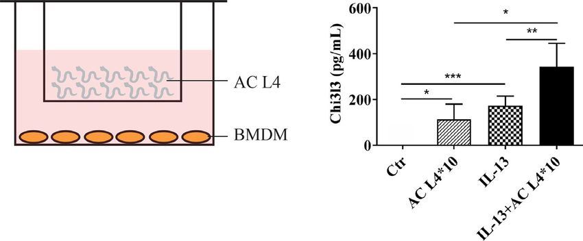
Wan et al.                                                                                                           Exofree Promote Macrophage Alternative Activation

                   A                                       B                           C                        G

                   D                                       E                           F                        H

                    I                                                                      J                                  K

                   L

                                                          M                                    N

 FIGURE 4 | Metabolic Reprogramming by the PI3K-Akt pathway plays a vital role in AC L4 exofree–induced M2 polarization of macrophages. (A–C) BMDMs were
 cultured for 24 h in medium alone or treated with LPS, exofree, exosome, and then the ECAR (A) was monitored using the Seahorse Bioscience extracellular flux
 analyzer in real time. Dotted lines indicate incubation of cells with the indicated compounds. Basal ECAR (B) and Max ECAR (C) of BMDMs were calculated.
 *P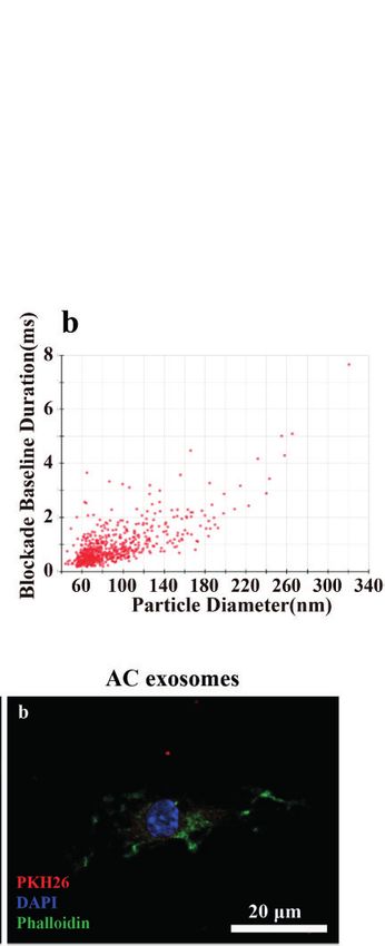
Wan et al.                                                                                                             Exofree Promote Macrophage Alternative Activation

                         A                                                C                                  E

                         B                                                D                                  F

                         G                                                I                                  K

                         H                                                J                                  L

                         N                                                                                   M

 FIGURE 5 | Chemical and physical property analysis of AC L4 exofree suggested glycoproteins as the key components in AC L4 exofree–induced M2 polarization of
 macrophage. (A, B) qPCR analysis of Arg1 (A), Chi3l3 (B) mRNA level of BMDMs in the presence of PBS, AC L4 exofree, AC L4 exofree (50°C water bath, 15min), AC
 L4 exofree (100°C water bath, 15min), IL-13, AC L4 exofree+IL-13, AC L4 exofree (50°C water bath, 15min)+IL-13, AC L4 exofree (100°C water bath, 15 min)+IL-13 at
 24 h was performed. (C, D) qPCR analysis of Arg1 (C), Chi3l3 (D) mRNA level of BMDMs in the presence of PBS, AC L4 exofree, AC L4 exofree (repeated freeze-thaw
 cycles for four times) at 24 h was performed. (E, F) qPCR analysis of Arg1 (E), Chi3l3 (F) mRNA level of BMDMs in the presence of PBS, AC L4 exofree, AC L4 exofree
 (100 mg/ml proteinase K treatment: 58°C water bath, 2 h; proteinase K inactivation: 100°C water bath, 15 min) at 24 h was performed. (G, H) qPCR analysis of Arg1
 (G), Chi3l3 (H) mRNA level of BMDMs in the presence of PBS, AC L4 exofree, AC L4 exofree (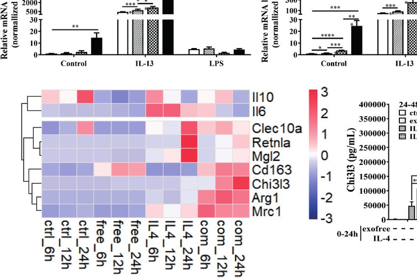
Wan et al. Exofree Promote Macrophage Alternative Activation
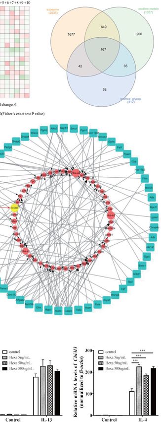
by coomassie blue staining (Figure 6A). Protein mass spectrum polarization of macrophages (36). Interestingly, M2 polarization
analysis of AC L4 exofree (10~20 kDa) was performed, and 312 of macrophages and worm burden could be significantly
N-linked glycoproteins were identified (Figures 6B, C). Amino inhibited using antibody-mediated EV uptake block (36).
acid composition heatmap (Figure 6B) suggested that serine (S) Leishmania major exosomes were reported to play a role in the
and threonine (T) were significantly enriched at position +2 N- establishment of parasite infection (37). Injection of
glycosylation site, indicating that N glycoproteins derived from Trypanosoma cruzi extracellular vesicles resulted in a
AC L4 exofree are conserved, with a consensus N-glycosylation significant reduction of iNOS production and an increase of
“sequon” (AsnXxxSer/Thr/Cys, where Xxx can be any amino parasitism in internal organs in Trypanosoma cruzi–infected
acid except proline) (35). Compared with protein mass mice (38). However, in some studies, exosomes play a role in
spectrometry analysis of AC L4 exosome, 35 potential M2- generating immunity against parasitic infections. Toxoplasma
related protein was selected. gondii–derived exosomes were reported to induce protective
RNA-seq analysis of BMDMs with different treatments at immunity by upregulating IFN-g, TNF-a, and IL-12 in
different time points was performed (Supplementary macrophages (39). Adult Schistosoma japonicum–derived
Figure 5A). Significant temporally changing genes (TCGs) on exosome-like vesicles were reported to induce macrophage
BMDMs were selected accordingly (2), which could be classified polarization to an M1 phenotype (40). Confusingly, M1
into six groups based on their expression profiles macrophages are favorable to kill schistosomula by producing
(Supplementary Figure 5B). GO biological process NO (41, 42), preventing hepatic fibrosis.
enrichment analysis of exofree-induced TCGs of BMDMs Differential centrifugation is a recommended method to
(Figure 6E) showed that the genes with the time-increasing separate exosomes and exosome-depleted ES products.
pattern were mainly enriched in nuclear division. By integrating Exosomes are small membrane-bound vesicles and have
RNA-seq expression data and proteomics data, we developed a proven to be stable, which could mediate long-range
bi-layer network approach (Figure 6D) to predict important communication with distant targets (43).
genes and potential interactions between AC proteins and mouse It has been reported that proteins in exosome-like vesicles of
genes. The bi-layer network (Figure 6F) prioritized Chi3l3, a Schistosoma japonicum are significantly different from soluble
marker of M2 type macrophage, as an important gene and worm antigenic preparations, and debris, extracellular vesicle
suggested that Hexa protein in AC L4 exofree is functionally (EV) from ES products (40). Similarly, EV content is significantly
related to M2 macrophage marker Chi3l3. Phylogenetic tree of different from that in EV-depleted ES products and ES of H.
Hexa protein in 17 species including Homo sapiens and M. polygyrus (36). However, rare studies are focused on the
musculus was drawn using a constraint-based alignment tool exosome-depleted component, which also might play a more
(Figure 6G). Amino acid sequence alignment of AC and human vital role than exosomes in tissue hemostasis.
Hexa protein was performed using ESPript 3 (Supplementary In this study, exosomes and exosome-depleted ES products,
Figure 6). To directly test whether Hexa was the main driver of exofree, were obtained, and their distinct roles in regulating
this effect, BMDMs were treated with recombinant human Hexa, macrophage polarization were firstly confirmed. AC L4 exosome
as the high sequence consistency with AC Hexa, and the results could induce a significant M1, not M2, polarization of
showed a dose-dependent increase of Arg1, but not Chi3l3 macrophages. This might partially clarify the reason why
expression (Figures 6H–K) in BMDMs. These results microglia (44), brain-resident macrophages, are classically
demonstrate that AC L4 exofree might promote alternative activated in AC infection. However, AC L4 exofree, heat and
activation of macrophage through Hexa. protease-stable, could significantly promote M2 polarization
induced by IL-4/IL-13, while inhibiting M1 polarization of
macrophage induced by LPS. These results strongly cue that
DISCUSSION when studying the function of parasitic exosomes, we should also
pay attention to other components within excretory/
Macrophages are key participants in AC-host interaction. secretory products.
Previous studies have shown that AC L4-derived soluble Lectin blot and deglycosylation assay reinforce the important
antigen, without any IL-4, IL-13, IL-33, and IL-6 analogs role of glycosylated products of AC L4 exofree on macrophage
(unpublished data), could promote M2 polarization of polarization (Figure 5M). Therefore, N-glycoproteins in AC L4
macrophage in a PPARg-dependent manner (27). However, exofree were identified using mass spectrometry–based
the important compound or protein in AC-derived soluble glycoproteomics (24). Then, a PPI network analysis was
antigen is not clear. performed based on AC L4 exofree-derived N-glycoproteins,
During the recent few years, exosomes and other extracellular and BMDMs differentially expressed genes regulated by AC L4
vesicles have been isolated and characterized in all known exofree, and Hexa was predicted to be the key molecule in M2
pathogen classes, including parasites. The research on parasite- macrophage polarization. Hexosaminidase, a lysosomal enzyme
derived exosomes has expanded dramatically. Parasite-derived involved in the breakdown of gangliosides, consists of a and/or b
exosomes are capable of modulating immune responses through subunit, which is encoded by Hexa gene and Hexb gene,
regulating the functions of macrophages. Extracellular vesicles of respectively, forming three isozymes—hexosaminidase A (a/b
Heligmosomoides polygyrus could suppress both M1 and M2 heterodimer), hexosaminidase B (b/b homodimer), and
Frontiers in Immunology | www.frontiersin.org 10 July 2021 | Volume 12 | Article 685984Wan et al. Exofree Promote Macrophage Alternative Activation
A B C
D E F
G H
I J K
FIGURE 6 | AC-derived Hexa act as a key ingredient in promoting M2 activation of macrophages. (A) Diagram of isolation of 10~20 kDa-AC L4 exofree using
ultrafiltration tubes, and the efficiency was confirmed using western blot. (B) Heatmap showed the relative frequencies of amino acids between the asparagine (Asn)
position of N-linked glycoproteins in AC L4 exofree. Enrichment level of the amino acid at indicate position is shown in different colors. Red means high enrichment
level, and green means low enrichment level. (C) Venn diagram of AC L4 exosome, AC L4 exofree proteins identified using mass spectrum analysis. (D) Diagram of
Bi-layer PPI network analysis using AC L4 exofree proteins and RNA-seq data of BMDMs. (E) GO enrichment analysis (Biological process) of exofree-induced TCGs
of BMDMs (Supplementary Figure 5B). (F) A protein-gene network was constructed for worm proteins with molecular weight less than 50kDa and TCGs of
exofree-, IL-4-, and exofree+IL-4-treated BMDMs (Supplementary Figure 5A), based on the LC-MS/MS and RNA-seq data. (G) Phylogene tree of Hexa protein in
17 species. (H, J) qPCR analysis of mRNA level of Arg1 (H) and Chi3l3 (J) were measured in the presence of PBS, Hexa 5 ng/ml, Hexa 50 ng/ml, Hexa 500 ng/ml,
IL-13 10 ng/ml, IL-13+Hexa 5 ng/ml, IL-13+Hexa 50 ng/ml, IL-13+Hexa 500 ng/ml for 24 h. (I, K) qPCR analysis of mRNA level of Arg1 (I) and Chi3l3 (K) were
measured in the presence of PBS, Hexa 5 ng/ml, Hexa 50 ng/ml, Hexa 500 ng/ml, IL-4 10 ng/ml, IL-4+Hexa 5 ng/ml, IL-4+Hexa 50 ng/ml, IL-4+Hexa 500 ng/ml for
24 h. Data information: *PWan et al. Exofree Promote Macrophage Alternative Activation
hexosaminidase S (a/a homodimer). Hexb, a peptidoglycan DATA AVAILABILITY STATEMENT
hydrolase, could either kill mycobacteria directly or restrict its
growth in an IFNg-dependent manner (45). Functional assay of The datasets presented in this study can be found in online
AC Hexa was performed using human Hexa protein, an analog repositories, further inquiries can be directed to the
recombinant protein produced by E. coli, suggesting a role of corresponding author. Proteomics data of AC L4 exosomes and
Hexa in promoting M2 polarization of macrophage induced by AC L4 exofree have been deposited to the ProteomeXchange
IL-4/IL-13. However, Arg1, but not Chi3l3, in BMDMs could be Consortium via the PRIDE (54) partner repository with the
upregulated in a Hexa dose-dependent manner. dataset identifier PXD025218. RNA-seq data of BMDMs were
Intriguingly, M2 macrophage polarization induced by Hexa deposited in the Gene Expression Omnibus (GEO) datasets under
seems unrelated to its enzymic activity, as Hexa protein displayed reference number GSE175497. The code used for network analysis
a single-phase inactivation curve at 48°C (46), and Hexb protein in this study is available at https://github.com/SunXQlab/
behaves as heat-stable enzyme activities at 55°C (47). A comparative Hexa-network.
functional study of Hexa and Hexb in macrophage polarization
could be carried out in the future. However, the coding sequences of
AC Hexa/Hexb still remain to be identified. As proteins produced
from E. coli are lacking of sugar chain, which is of great importance ETHICS STATEMENT
for protein antigenicity (33) and stability (48), eukaryotic protein
expression systems could be used in our study. Previous studies The animal study was reviewed and approved by the medical
have shown that N-glycans, but not O-glycans or fucose-containing research ethics committee of Sun Yat-sen University (SYSU-
glycans, from AC L4 seem essential for immune recognition (34); IACUC-2019-B535).
thus, a-L-Fucosidase, Endo H, and O-glycosidase might help
exclude the possible role of O-glycosylation or fucosylation in
this process.
Energy metabolism has been proven to be an mTOR-dependent AUTHOR CONTRIBUTIONS
cellular process, and inhibition of mTOR could induce a switch of
XS, ZW, and SW conceived the concept, contributed to
energy metabolism from mitochondrial oxidative phosphorylation
experimental designs. SW, WT, and LW performed the study and
to aerobic glycolysis (46). Inhibition of mTORC1 could significantly
the experiments. XQS conducted RNA-seq and proteomics analysis.
upregulate M1-associated cytokines level (49), and both mTORC1
SW and XS wrote the manuscript. All authors contributed to the
(50) and mTORC2 (51) seem critical for M2 macrophages. Based
article and approved the submitted version.
on our previous study on macrophage and KEGG results of
BMDMs stimulated with AC L4 exofree, we supposed PI3K/Akt,
an upstream modulator of the mTORC1 signal pathway, might play
an important role in macrophage M2 polarization induced by AC
L4 exofree. Expectedly, class I PI3K inhibitor Ly294002 and
FUNDING
wortmannin successfully induced a sharp decrease of Chi3l3 This project was supported by the Science and Technology
protein level in BMDMs treated with “AC L4 exofree+IL-4” and Planning Project of Guangdong Province (2016A020219004),
“IL-4.” However, Chi3l3 protein level in BMDMs treated with “AC the National Key R&D Program of China (2020YFC1200100),
L4 exofree+IL-4” was significantly higher than IL-4-treated group in Natural Science Foundation of Guangdong Province (No
the presence of 100 nM wortmannin, but not in 10 mM Ly294002. 2019A1515012068, 2021A1515010976), the Pearl River Nova
These results suggested that PI3K/Akt pathway might be partially Program of Guangzhou (No. 201710010030), 111 Project
associated with AC L4 exofree–induced M2 polarization of (Grant No. B12003). Shuo Wan was supported by the Project
BMDMs. This might be because of their different inhibitory fund ed by China Postdoctoral Science Foundation
effects on class II PI3K, and 10 mM LY294002 has a partial (2021M691236). Xiaoqiang Sun was supported by the National
inhibitory effect on PI3K-C2b (52), while 100 nM wortmannin is Natural Science Foundation of China (11871070), the
not sufficient to inhibit PI3K-C2a activity (53). Therefore, a deeper Guangdong Basic and Applied Basic Research Foundation
understanding of the signaling pathways in this process is needed. (2020B151502120), the Fundamental Research Funds for the
In this study, we present evidence in favor of a key role for N- Central Universities (20ykzd20), and Guangdong Key Field R&D
linked glycoprotein in AC L4 exofree, a component free of Plan (2019B020228001).
exosomes isolated from ES products of AC L4, in promoting M2
macrophage polarization. And metabolic reprogramming by the
PI3K-Akt pathway might play an important role in this process.
Hexa, a macrophage-related N-linked glycoprotein, might play a SUPPLEMENTARY MATERIAL
key role in directing host CNS inflammation in AC infection. The
results of this study indicate a great value in studying ES products of The Supplementary Material for this article can be found online
AC L4, as worm-derived immunoregulatory molecules might at: https://www.frontiersin.org/articles/10.3389/fimmu.2021.
contribute to drug development for immune-related diseases. 685984/full#supplementary-material
Frontiers in Immunology | www.frontiersin.org 12 July 2021 | Volume 12 | Article 685984Wan et al. Exofree Promote Macrophage Alternative Activation
18. Klaver EJ, Kuijk LM, Laan LC, Kringel H, van Vliet SJ, Bouma G, et al. Trichuris
REFERENCES Suis-Induced Modulation of Human Dendritic Cell Function is Glycan-Mediated.
Int J Parasitol (2013) 43:191–200. doi: 10.1016/j.ijpara.2012.10.021
1. Zhao J, Lv Z, Wang F, Wei J, Zhang Q, Li S, et al. Ym1, an Eosinophilic
19. Everts B, Hussaarts L, Driessen NN, Meevissen MH, Schramm G, van der
Chemotactic Factor, Participates in the Brain Inflammation Induced by
Ham AJ, et al. Schistosome-Derived Omega-1 Drives Th2 Polarization by
Angiostrongylus Cantonensis in Mice. Parasitol Res (2013) 112:2689–95.
Suppressing Protein Synthesis Following Internalization by the Mannose
doi: 10.1007/s00436-013-3436-x
Receptor. J Exp Med (2012) 209:1753–67. doi: 10.1084/jem.20111381
2. Wan S, Sun X, Wu F, Yu Z, Wang L, Lin D, et al. Chi3l3: A Potential Key
20. Zheng X, Hu X, Zhou G, Lu Z, Qiu W, Bao J, et al. Soluble Egg Antigen From
Orchestrator of Eosinophil Recruitment in Meningitis Induced by
Schistosoma Japonicum Modulates the Progression of Chronic Progressive
Angiostrongylus Cantonensis. J Neuroinflamm (2018) 15:31. doi: 10.1186/
Experimental Autoimmune Encephalomyelitis via Th2-Shift Response.
s12974-018-1071-2
J Neuroimmunol (2008) 194:107–14. doi: 10.1016/j.jneuroim.2007.12.001
3. Rodriguez E, Noya V, Cervi L, Chiribao ML, Brossard N, Chiale C, et al.
21. Kowal J, Arras G, Colombo M, Jouve M, Morath JP, Primdal-Bengtson B, et al.
Glycans From Fasciola Hepatica Modulate the Host Immune Response and
Proteomic Comparison Defines Novel Markers to Characterize
TLR-Induced Maturation of Dendritic Cells. PloS Negl Trop Dis (2015) 9:
Heterogeneous Populations of Extracellular Vesicle Subtypes. Proc Natl
e0004234. doi: 10.1371/journal.pntd.0004234
Acad Sci USA (2016) 113:E968–77. doi: 10.1073/pnas.1521230113
4. Prasanphanich NS, Luyai AE, Song X, Heimburg-Molinaro J, Mandalasi M,
22. Nawaz M, Malik MI, Hameed M, Zhou J. Research Progress on the
Mickum M, et al. Immunization With Recombinantly Expressed Glycan Antigens
Composition and Function of Parasite-Derived Exosomes. Acta Trop (2019)
From Schistosoma Mansoni Induces Glycan-Specific Antibodies Against the
196:30–6. doi: 10.1016/j.actatropica.2019.05.004
Parasite. Glycobiology (2014) 24:619–37. doi: 10.1093/glycob/cwu027
23. Thery C, Amigorena S, Raposo G, Clayton A. Isolation and Characterization
5. Eichenberger RM, Talukder MH, Field MA, Wangchuk P, Giacomin P,
of Exosomes From Cell Culture Supernatants and Biological Fluids. Curr
Loukas A, et al. Characterization of Trichuris Muris Secreted Proteins and
Protoc Cell Biol (2006) 3. doi: 10.1002/0471143030.cb0322s30
Extracellular Vesicles Provides New Insights Into Host-Parasite
24. Zhu J, Sun Z, Cheng K, Chen R, Ye M, Xu B, et al. Comprehensive Mapping of
Communication. J Extracell Vesicles (2018) 7:1428004. doi: 10.1080/
Protein N-Glycosylation in Human Liver by Combining Hydrophilic
20013078.2018.1428004
Interaction Chromatography and Hydrazide Chemistry. J Proteome Res
6. Buck AH, Coakley G, Simbari F, McSorley HJ, Quintana JF, Le Bihan T, et al.
(2014) 13:1713–21. doi: 10.1021/pr401200h
Exosomes Secreted by Nematode Parasites Transfer Small RNAs to
25. Sato T. Lectin-Probed Western Blot Analysis. Methods Mol Biol (2014)
Mammalian Cells and Modulate Innate Immunity. Nat Commun (2014)
1200:93–100. doi: 10.1007/978-1-4939-1292-6_8
5:5488. doi: 10.1038/ncomms6488
26. Zhang J, Zhu W, Wang Q, Gu J, Huang LF, Sun X. Differential Regulatory
7. Soulat D, Bogdan C. Function of Macrophage and Parasite Phosphatases in
Network-Based Quantification and Prioritization of Key Genes Underlying
Leishmaniasis. Front Immunol (2017) 8:1838. doi: 10.3389/fimmu.2017.01838
Cancer Drug Resistance Based on Time-Course RNA-Seq Data. PloS Comput
8. Ramos-Benitez MJ, Ruiz-Jimenez C, Aguayo V, Espino AM. Recombinant
Biol (2019) 15:e1007435. doi: 10.1371/journal.pcbi.1007435
Fasciola Hepatica Fatty Acid Binding Protein Suppresses Toll-Like Receptor
27. Wu F, Wei J, Liu Z, Zeng X, Yu Z, Lv Z, et al. Soluble Antigen Derived From
Stimulation in Response to Multiple Bacterial Ligands. Sci Rep (2017) 7:5455.
IV Larva of Angiostrongylus Cantonensis Promotes Chitinase-Like Protein 3
doi: 10.1038/s41598-017-05735-w
(Chil3) Expression Induced by Interleukin-13. Parasitol Res (2016) 115:3737–
9. Danilowicz-Luebert E, Steinfelder S, Kuhl AA, Drozdenko G, Lucius R, Worm
46. doi: 10.1007/s00436-016-5135-x
M, et al. A Nematode Immunomodulator Suppresses Grass Pollen-Specific
28. Silva VO, Maia MM, Torrecilhas AC, Taniwaki NN, Namiyama GM, Oliveira KC,
Allergic Responses by Controlling Excessive Th2 Inflammation. Int J Parasitol
et al. Extracellular Vesicles Isolated From Toxoplasma Gondii Induce Host
(2013) 43:201–10. doi: 10.1016/j.ijpara.2012.10.014
Immune Response. Parasite Immunol (2018) 40:e12571. doi: 10.1111/pim.12571
10. Ziegler T, Rausch S, Steinfelder S, Klotz C, Hepworth MR, Kuhl AA, et al. A
29. Iyer SS, Ghaffari AA, Cheng G. Lipopolysaccharide-Mediated IL-10
Novel Regulatory Macrophage Induced by a Helminth Molecule Instructs IL-
Transcriptional Regulation Requires Sequential Induction of Type I IFNs
10 in CD4+ T Cells and Protects Against Mucosal Inflammation. J Immunol
and IL-27 in Macrophages. J Immunol (2010) 185:6599–607. doi: 10.4049/
(2015) 194:1555–64. doi: 10.4049/jimmunol.1401217
jimmunol.1002041
11. Prieto-Lafuente L, Gregory WF, Allen JE, Maizels RM. MIF Homologues
30. Etzerodt A, Moestrup SK. CD163 and Inflammation: Biological, Diagnostic,
From a Filarial Nematode Parasite Synergize With IL-4 to Induce Alternative
and Therapeutic Aspects. Antioxid Redox Signaling (2013) 18:2352–63. doi:
Activation of Host Macrophages. J Leukoc Biol (2009) 85:844–54. doi:
10.1089/ars.2012.4834
10.1189/jlb.0808459
31. Philippidis P, Mason JC, Evans BJ, Nadra I, Taylor KM, Haskard DO, et al.
12. Du L, Wei H, Li L, Shan H, Yu Y, Wang Y, et al. Regulation of Recombinant
Hemoglobin Scavenger Receptor CD163 Mediates Interleukin-10 Release and
Trichinella Spiralis 53-kDa Protein (Rtsp53) on Alternatively Activated
Heme Oxygenase-1 Synthesis: Antiinflammatory Monocyte-Macrophage
Macrophages via STAT6 But Not IL-4ralpha In Vitro. Cell Immunol (2014)
Responses In Vitro, in Resolving Skin Blisters In Vivo, and After
288:1–7. doi: 10.1016/j.cellimm.2014.01.010
Cardiopulmonary Bypass Surgery. Circ Res (2004) 94:119–26. doi: 10.1161/
13. Robinson MW, Donnelly S, Hutchinson AT, To J, Taylor NL, Norton RS, et al.
01.RES.0000109414.78907.F9
A Family of Helminth Molecules That Modulate Innate Cell Responses via
32. Italiani P, Boraschi D. From Monocytes to M1/M2 Macrophages:
Molecular Mimicry of Host Antimicrobial Peptides. PloS Pathog (2011) 7:
Phenotypical vs Function Differentiation. Front Immunol (2014) 5:514. doi:
e1002042. doi: 10.1371/journal.ppat.1002042
10.3389/fimmu.2014.00514
14. Liu J, Zhu L, Wang J, Qiu L, Chen Y, Davis RE, et al. Schistosoma Japonicum
33. Morassutti AL, Levert K, Perelygin A, da Silva AJ, Wilkins P, Graeff-Teixeira
Extracellular Vesicle miRNA Cargo Regulates Host Macrophage Functions
C. The 31-kDa Antigen of Angiostrongylus Cantonensis Comprises Distinct
Facilitating Parasitism. PloS Pathog (2019) 15:e1007817. doi: 10.1371/
Antigenic Glycoproteins. Vector Borne Zoonotic Dis (Larchmont NY) (2012)
journal.ppat.1007817
12:961–8. doi: 10.1089/vbz.2011.0957
15. Nyame AK, Lewis FA, Doughty BL, Correa-Oliveira R, Cummings RD.
34. Verı́ssimo CM, Morassutti AL, von Itzstein M, Sutov G, Hartley-Tassell L,
Immunity to Schistosomiasis: Glycans are Potential Antigenic Targets for
McAtamney S, et al. Characterization of the N-Glycans of Female
Immune Intervention. Exp Parasitol (2003) 104:1–13. doi: 10.1016/S0014-
Angiostrongylus Cantonensis Worms. Exp Parasitol (2016) 166:137–43. doi:
4894(03)00110-3
10.1016/j.exppara.2016.04.012
16. Khoo KH, Dell A. Glycoconjugates From Parasitic Helminths: Structure
35. Medzihradszky KF. Characterization of Site-Specific N-Glycosylation.
Diversity and Immunobiological Implications. Adv Exp Med Biol (2001)
Methods Mol Biol (2008) 446:293–316. doi: 10.1007/978-1-60327-084-7_21
491:185–205. doi: 10.1007/978-1-4615-1267-7_14
36. Coakley G, McCaskill JL, Borger JG, Simbari F, Robertson E, Millar M, et al.
17. Restrepo BI, Obregon-Henao A, Mesa M, Gil DL, Ortiz BL, Mejia JS, et al.
Extracellular Vesicles From a Helminth Parasite Suppress Macrophage
Characterisation of the Carbohydrate Components of Taenia Solium
Activation and Constitute an Effective Vaccine for Protective Immunity.
Metacestode Glycoprotein Antigens. Int J Parasitol (2000) 30:689–96. doi:
Cell Rep (2017) 19:1545–57. doi: 10.1016/j.celrep.2017.05.001
10.1016/S0020-7519(00)00057-6
Frontiers in Immunology | www.frontiersin.org 13 July 2021 | Volume 12 | Article 685984Wan et al. Exofree Promote Macrophage Alternative Activation
37. Hassani K, Shio MT, Martel C, Faubert D, Olivier M. Absence of 49. Weichhart T, Säemann MD. The Multiple Facets of mTOR in Immunity.
Metalloprotease GP63 Alters the Protein Content of Leishmania Exosomes. Trends Immunol (2009) 30:218–26. doi: 10.1016/j.it.2009.02.002
PloS One (2014) 9:e95007. doi: 10.1371/journal.pone.0095007 50. Covarrubias AJ, Aksoylar HI, Yu J, Snyder NW, Worth AJ, Iyer SS, et al. Akt-
38. Trocoli Torrecilhas AC, Tonelli RR, Pavanelli WR, da Silva JS, Schumacher RI, Mtorc1 Signaling Regulates Acly to Integrate Metabolic Input to Control of
de Souza W, et al. Trypanosoma Cruzi: Parasite Shed Vesicles Increase Heart Macrophage Activation. eLife (2016) 5:e11612. doi: 10.7554/eLife.11612
Parasitism and Generate an Intense Inflammatory Response. Microbes Infect 51. Hallowell RW, Collins SL, Craig JM, Zhang Y, Oh M, Illei PB, et al. Mtorc2
(2009) 11:29–39. doi: 10.1016/j.micinf.2008.10.003 Signalling Regulates M2 Macrophage Differentiation in Response to Helminth
39. Li Y, Liu Y, Xiu F, Wang J, Cong H, He S, et al. Characterization of Exosomes Infection and Adaptive Thermogenesis. Nat Commun (2017) 8:14208. doi:
Derived From Toxoplasma Gondii and Their Functions in Modulating Immune 10.1038/ncomms14208
Responses. Int J Nanomed (2018) 13:467–77. doi: 10.2147/IJN.S151110 52. Maffucci T, Cooke FT, Foster FM, Traer CJ, Fry MJ, Falasca M. Class II
40. Wang L, Li Z, Shen J, Liu Z, Liang J, Wu X, et al. Exosome-Like Vesicles Phosphoinositide 3-Kinase Defines a Novel Signaling Pathway in Cell
Derived by Schistosoma Japonicum Adult Worms Mediates M1 Type Migration. J Cell Biol (2005) 169:789–99. doi: 10.1083/jcb.200408005
Immune- Activity of Macrophage. Parasitol Res (2015) 114:1865–73. doi: 53. Domin J, Gaidarov I, Smith ME, Keen JH, Waterfield MD. The Class II
10.1007/s00436-015-4373-7 Phosphoinositide 3-Kinase PI3K-C2alpha is Concentrated in the Trans-Golgi
41. Ahmed SF, Oswald IP, Caspar P, Hieny S, Keefer L, Sher A, et al. Network and Present in Clathrin-Coated Vesicles. J Biol Chem (2000)
Developmental Differences Determine Larval Susceptibility to Nitric Oxide- 275:11943–50. doi: 10.1074/jbc.275.16.11943
Mediated Killing in a Murine Model of Vaccination Against Schistosoma 54. Perez-Riverol Y, Csordas A, Bai J, Bernal-Llinares M, Hewapathirana S,
Mansoni. Infect Immun (1997) 65:219–26. doi: 10.1128/iai.65.1.219-226.1997 Kundu DJ, et al. The PRIDE Database and Related Tools and Resources in
42. Shen J, Lai DH, Wilson RA, Chen YF, Wang LF, Yu ZL, et al. Nitric Oxide 2019: Improving Support for Quantification Data. Nucleic Acids Res (2019) 47:
Blocks the Development of the Human Parasite Schistosoma Japonicum. Proc D442–d450. doi: 10.1093/nar/gky1106
Natl Acad Sci USA (2017) 114:10214–9. doi: 10.1073/pnas.1708578114
43. Zhao Z, Zlokovic BV. Remote Control of BBB: A Tale of Exosomes and Conflict of Interest: The authors declare that the research was conducted in the
microRNA. Cell Res (2017) 27:849–50. doi: 10.1038/cr.2017.71 absence of any commercial or financial relationships that could be construed as a
44. Wei J, Wu F, He A, Zeng X, Ouyang LS, Liu MS, et al. Microglia Activation: potential conflict of interest.
One of the Checkpoints in the CNS Inflammation Caused by Angiostrongylus
Cantonensis Infection in Rodent Model. Parasitol Res (2015) 114:3247–54. Publisher’s Note: All claims expressed in this article are solely those of the authors
doi: 10.1007/s00436-015-4541-9 and do not necessarily represent those of their affiliated organizations, or those of
45. Koo IC, Ohol YM, Wu P, Morisaki JH, Cox JS, Brown EJ. Role for Lysosomal the publisher, the editors and the reviewers. Any product that may be evaluated in
Enzyme Beta-Hexosaminidase in the Control of Mycobacteria Infection. Proc this article, or claim that may be made by its manufacturer, is not guaranteed or
Natl Acad Sci USA (2008) 105:710–5. doi: 10.1073/pnas.0708110105 endorsed by the publisher.
46. Schieke SM, Phillips D, McCoy JPJr., Aponte AM, Shen RF, Balaban RS, et al.
The Mammalian Target of Rapamycin (mTOR) Pathway Regulates
Mitochondrial Oxygen Consumption and Oxidative Capacity. J Biol Chem Copyright © 2021 Wan, Sun, Tang, Wang, Wu and Sun. This is an open-access article
(2006) 281:27643–52. doi: 10.1074/jbc.M603536200 distributed under the terms of the Creative Commons Attribution License (CC BY).
47. Hechtman P, Rowlands A. Apparent Hexosaminidase B Deficiency in Two The use, distribution or reproduction in other forums is permitted, provided the
Healthy Members of a Pedigree. Am J Hum Genet (1979) 31:428–38. original author(s) and the copyright owner(s) are credited and that the original
48. Jayaprakash NG, Surolia A. Role of Glycosylation in Nucleating Protein Folding publication in this journal is cited, in accordance with accepted academic practice. No
and Stability. Biochem J (2017) 474:2333–47. doi: 10.1042/BCJ20170111 use, distribution or reproduction is permitted which does not comply with these terms.
Frontiers in Immunology | www.frontiersin.org 14 July 2021 | Volume 12 | Article 685984You can also read