PREVALENT, PROTECTIVE, AND CONVERGENT IGG RECOGNITION OF SARS-COV-2 NON-RBD SPIKE EPITOPES - SCIENCE
←
→
Page content transcription
If your browser does not render page correctly, please read the page content below
REPORTS

                                                                                                                                   Cite as: W. N. Voss et al., Science
                                                                                                                                    10.1126/science.abg5268 (2021).

Prevalent, protective, and convergent IgG recognition of
SARS-CoV-2 non-RBD spike epitopes
William N. Voss1, Yixuan J. Hou2#, Nicole V. Johnson1#, George Delidakis3, Jin Eyun Kim4, Kamyab
Javanmardi1, Andrew P. Horton1, Foteini Bartzoka1, Chelsea J. Paresi5, Yuri Tanno3, Chia-Wei Chou1, Shawn A.
Abbasi6, Whitney Pickens1, Katia George1, Daniel R. Boutz1,7, Dalton M. Towers3, Jonathan R. McDaniel8,
Daniel Billick1, Jule Goike1, Lori Rowe9,10, Dhwani Batra9, Jan Pohl9, Justin Lee9, Shivaprakash Gangappa11,
Suryaprakash Sambhara11, Michelle Gadush12, Nianshuang Wang1, Maria D. Person12, Brent L. Iverson5, Jimmy
D. Gollihar1,7,13, John Dye6, Andrew Herbert6, Ilya J. Finkelstein1, Ralph S. Baric2,14, Jason S. McLellan1, George
Georgiou1,3,4,15, Jason J. Lavinder1,3*, Gregory C. Ippolito1,13,15*
1Department of   Molecular Biosciences, The University of Texas at Austin, Austin, TX, USA. 2Department of Epidemiology, University of North Carolina at Chapel Hill, Chapel
Hill, NC, USA. 3Department of Chemical Engineering, The University of Texas at Austin, Austin, TX, USA. 4Department of Biomedical Engineering, The University of Texas at

                                                                                                                                                                               Downloaded from http://science.sciencemag.org/ on May 20, 2021
Austin, Austin, TX, USA. 5Department of Chemistry, The University of Texas at Austin, Austin, TX, USA. 6U.S. Army Medical Research Institute of Infectious Diseases,
Frederick, MD, USA. 7CCDC Army Research Laboratory-South, The University of Texas at Austin, Austin, TX, USA. 8Biomedicine Design, Pfizer, Cambridge, MA, USA.
9Biotechnology Core Facility Branch, Division of Scientific Resources, National Center for Emerging and Zoonotic Infectious Diseases, Centers for Disease Control and

Prevention, Atlanta, GA, USA. 10Tulane National Primate Research Center Department of Microbiology 18703 Three Rivers Road Covington, LA, USA. 11Immunology and
Pathogenesis Branch, Influenza Division, National Center for Immunization and Respiratory Diseases, Centers for Disease Control and Prevention, Atlanta, GA, USA.
12Center for Biomedical Research Support, The University of Texas at Austin, Austin, TX, USA. 13Department of Pathology and Genomic Medicine, Houston Methodist

Research Institute, Houston Methodist Hospital, Houston, TX, USA. 14Department of Microbiology and Immunology, University of North Carolina at Chapel Hill, Chapel Hill,
NC, USA. 15Department of Oncology, Dell Medical School, The University of Texas at Austin, Austin, TX, USA.
#These authors contributed equally to this work.
*Corresponding author. Email: jlavinder@utexas.edu (J.J.L.); gci@utexas.edu (G.C.I.)

The molecular composition and binding epitopes of the immunoglobulin G (IgG) antibodies that circulate in
blood plasma following SARS-CoV-2 infection are unknown. Proteomic deconvolution of the IgG repertoire
to the spike glycoprotein in convalescent subjects revealed that the response is directed predominantly
(>80%) against epitopes residing outside the receptor-binding domain (RBD). In one subject, just four IgG
lineages accounted for 93.5% of the response, including an N-terminal domain (NTD)-directed antibody
that was protective against lethal viral challenge. Genetic, structural, and functional characterization of a
multi-donor class of “public” antibodies revealed an NTD epitope that is recurrently mutated among
emerging SARS-CoV-2 variants of concern. These data show that “public” NTD-directed and other non-RBD
plasma antibodies are prevalent and have implications for SARS-CoV-2 protection and antibody escape.

The SARS-CoV-2 spike ectodomain (S-ECD) folds into a mul-                                manifested with plasma virus-neutralization titers in the low-
tidomain architecture (1, 2) and includes the RBD, which is                              est quartile (P1 and P3), the second highest quartile (P2), or
essential for viral infectivity, and the structurally adjacent                           the highest quartile (P4) compared to a larger cohort (table
NTD, which plays an uncertain role. Humoral immunity to                                  S1 and fig. S1). The lineage composition and relative abun-
the spike (S) surface glycoprotein can correlate with protec-                            dance of constituent IgG antibodies comprising the plasma
tion, (3) and it is the primary antigenic target for most vac-                           response to either intact stabilized S-ECD (S-2P (1)) or RBD
cines and monoclonal antibodies (mAbs). That the B cell                                  was determined using the Ig-Seq pipeline (13, 14, 20) that in-
repertoire can recognize multiple spike epitopes is supported                            tegrates analytical proteomics of affinity purified IgG frac-
by extensive single-cell cloning campaigns (4–9). However,                               tions with peripheral B cell antibody variable region
the identity, abundance, and clonality of the IgG plasma an-                             repertoires (BCR-Seq).
tibody repertoire and the epitopes it may target are not                                     IgG lineages detected by Ig-Seq in the S-ECD fraction but
known (10–12). Divergence between the two repertoires is bi-                             absent from the RBD fraction were deemed to be reactive
ologically plausible (13–17) and the evidence in COVID-19 in-                            with spike epitopes outside the RBD. In subject P3, we de-
cludes a paradoxical disconnect between virus-neutralizing                               tected six IgG lineages that bound to S-ECD (Fig. 1A). Four of
IgG titers and RBD-specific B cell immunity (6, 11, 18, 19).                             these (Lin.1 to Lin.4) accounted for 93.5% abundance of the
   To analyze the IgG repertoire, blood was collected during                             total plasma IgG S-ECD response and exhibited extensive in-
early convalescence from four seroconverted study subjects                               tralineage diversity (fig. S2) indicative of clonal expansion
(P1–P4) who experienced mild COVID-19 disease that                                       and selection. Notably, the top three lineages (Lin.1 to Lin.3;

First release: 4 May 2021                                                 www.sciencemag.org                 (Page numbers not final at time of first release) 1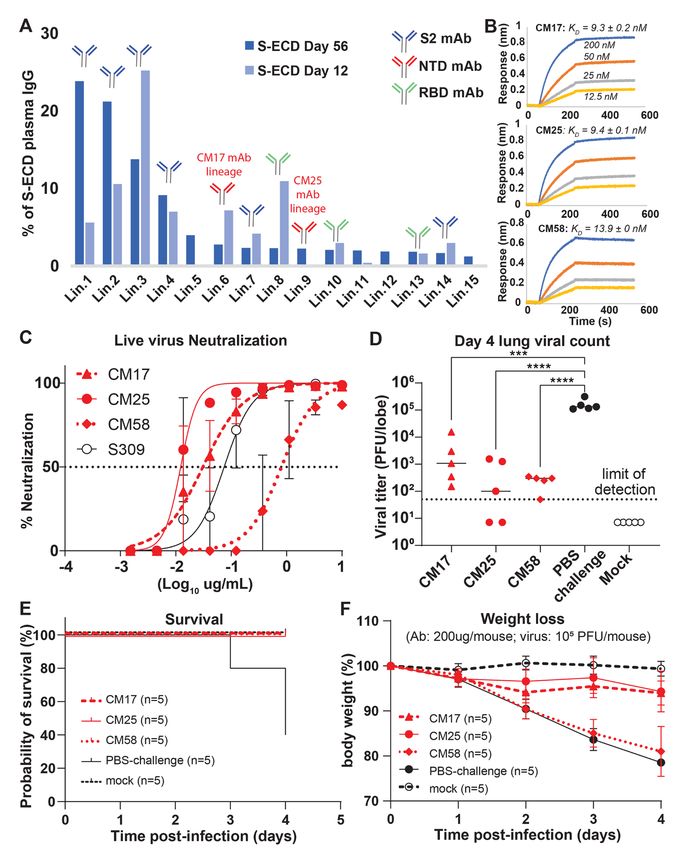
>85% abundance) all bound to non-RBD epitopes (S2 subunit           targeting lineages each comprising ~2.5% of the response at
or NTD). Bulk serology ELISAs recapitulated the Ig-Seq result       day 56 (Ig-Seq Lin.6 and Lin.9) (Fig. 2A), were both encoded
and demonstrated similarly high levels of non-RBD-binding           by unmutated or near-germline IGHV1-24. We found an ad-
IgG (P>0.05) (Fig. 1B), confirming that RBD-binding plasma          ditional NTD-targeting unmutated IGHV1-24 plasma mAb
antibodies comprise only a minor proportion of all spike-           (CM58) in subject P4. CM17, CM25 and CM58 bound S-ECD
binding IgG in naturally infected individuals (21). In all four     with similar single-digit nM affinity (Fig. 2B and table S2)
subjects, the detected plasma IgG repertoire to S-ECD was ol-       and all three potently neutralized SARS-CoV-2 virus, with IC50
igoclonal, comprising only 6–22 lineages, with the top-ranked       values of 0.01–0.81 μg/ml comparable to S309 anti-RBD con-
lineage comprising 15 to 50% total abundance. On average,           trol (25) (Fig. 2C, fig. S6, and table S2). For all three mAbs,
84% of the anti-S-ECD plasma IgG repertoire bound to                pre-administration in the MA10 mouse model resulted in sig-
epitopes outside the RBD (Fig. 1C), a finding consistent with       nificantly reduced lung viral titers post-infection with 105
data from single B cell analyses (22), and the most abundant        PFU (Fig. 2D; P85% of the IgG plasma lin-              CM58, and 1-68 but did not compete with the other three
eages to S-ECD; Fig. 1A) showed the most robust protection          IGHV1-24 NTD mAbs.
and lung viral titers below the limit of detection (LOD) in             To better understand the IGHV1-24 interactions with the
high viral load challenge (104 PFU).                                spike NTD, we determined a cryo-EM structure of CM25 Fabs
    Subject P2, with ~10-fold higher neutralizing titer com-        bound to trimeric S-ECD (Fig. 4A and figs. S7 and S8). Fo-
pared to subject P3 (fig. S1 and table S1), displayed a more        cused refinement of the CM25-NTD interface resulted in a
polyclonal IgG response (Fig. 2A), with 12/15 lineages (>80%        3.5-Å reconstruction that revealed a heavy-chain–dominant
total abundance) in the anti-S-ECD repertoire recognizing           mode of binding, with substantial contacts mediated by in-
non-RBD epitopes. Conspicuously, as with P3, the most abun-         teractions between the three CDRs and the N3 and N5 loops
dant S-ECD-directed plasma antibodies target the S2 subunit,        of the NTD (Fig. 4B). The light chain contributes only 11% (86
with the four topmost lineages (68% total abundance) bind-          Å2) of the total CM25 binding interface, mainly through a
ing to S2. MAbs CM25 and CM17, representative of two NTD-           stacked hydrophobic interaction between CDR-L2 Tyr55 and

First release: 4 May 2021                               www.sciencemag.org       (Page numbers not final at time of first release)   2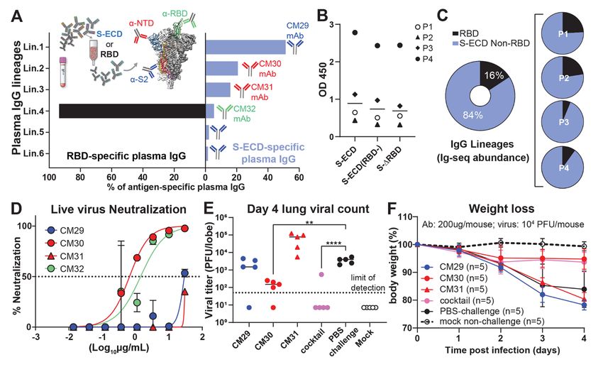
Pro251 within the N5 loop. Unique germline IGHV1-24 resi-        REFERENCES AND NOTES
dues contribute 20% (149 Å2) of the total binding interface.     1. D. Wrapp, N. Wang, K. S. Corbett, J. A. Goldsmith, C.-L. Hsieh, O. Abiona, B. S.
                                                                      Graham, J. S. McLellan, Cryo-EM structure of the 2019-nCoV spike in the
CDR-H1 interacts extensively through hydrogen bonds and               prefusion        conformation.        Science       367,       1260–1263        (2020).
contacts between hydrophobic residues, including a salt               doi:10.1126/science.abb2507 Medline
bridge formed between the conserved Glu36 residue and the        2. A. C. Walls, Y.-J. Park, M. A. Tortorici, A. Wall, A. T. McGuire, D. Veesler, Structure,
N5 loop residue Arg246 (Fig. 4C). The common IGHV1-24                 Function, and Antigenicity of the SARS-CoV-2 Spike Glycoprotein. Cell 181, 281–
                                                                      292.e6 (2020). doi:10.1016/j.cell.2020.02.058 Medline
Phe56 residue in CDR-H2 forms a pi-cation interaction with       3. K. McMahan, J. Yu, N. B. Mercado, C. Loos, L. H. Tostanoski, A. Chandrashekar, J.
Lys147 in the N3 loop (Fig. 4C). CM25 contains a 14-amino-            Liu, L. Peter, C. Atyeo, A. Zhu, E. A. Bondzie, G. Dagotto, M. S. Gebre, C. Jacob-
acid CDR-H3 loop that contributes 35% (261 Å2) of the total           Dolan, Z. Li, F. Nampanya, S. Patel, L. Pessaint, A. Van Ry, K. Blade, J. Yalley-
interface, including the AV aliphatic motif found in all but          Ogunro, M. Cabus, R. Brown, A. Cook, E. Teow, H. Andersen, M. G. Lewis, D. A.
                                                                      Lauffenburger, G. Alter, D. H. Barouch, Correlates of protection against SARS-
one of the convergent IGHV1-24 NTD-binding mAbs. Ala109               CoV-2 in rhesus macaques. Nature 590, 630–634 (2021). doi:10.1038/s41586-
and Val110 are buried at the interface in a binding pocket            020-03041-6 Medline
framed by the N3 and N5 loops. A comparison of CM25 with         4. X. Chi, R. Yan, J. Zhang, G. Zhang, Y. Zhang, M. Hao, Z. Zhang, P. Fan, Y. Dong, Y.
an extant structure of an IGHV1-24 NTD-binding antibody               Yang, Z. Chen, Y. Guo, J. Zhang, Y. Li, X. Song, Y. Chen, L. Xia, L. Fu, L. Hou, J. Xu,
                                                                      C. Yu, J. Li, Q. Zhou, W. Chen, A neutralizing human antibody binds to the N-
isolated by B cell cloning, 4A8 (4), revealed that the AV di-         terminal domain of the Spike protein of SARS-CoV-2. Science 369, 650–655

                                                                                                                                                                  Downloaded from http://science.sciencemag.org/ on May 20, 2021
peptide interaction is structurally conserved, and the 21             (2020). doi:10.1126/science.abc6952 Medline
amino-acid CDR-H3 of 4A8 extends along the outside of the        5. L. Liu, P. Wang, M. S. Nair, J. Yu, M. Rapp, Q. Wang, Y. Luo, J. F.-W. Chan, V. Sahi, A.
NTD, contributing three additional contacts and 46% (415 Å2)          Figueroa, X. V. Guo, G. Cerutti, J. Bimela, J. Gorman, T. Zhou, Z. Chen, K.-Y. Yuen,
                                                                      P. D. Kwong, J. G. Sodroski, M. T. Yin, Z. Sheng, Y. Huang, L. Shapiro, D. D. Ho,
of the total binding interface (Fig. 4D). Both structures show        Potent neutralizing antibodies against multiple epitopes on SARS-CoV-2 spike.
extensive contacts between the heavy chain of the Fabs and            Nature 584, 450–456 (2020). doi:10.1038/s41586-020-2571-7 Medline
the NTD N3 and N5 loops. The Glu36-Arg246 salt bridge and        6. D. F. Robbiani, C. Gaebler, F. Muecksch, J. C. C. Lorenzi, Z. Wang, A. Cho, M.
an identical CDR-H2 contact between Phe56 and Lys147 are              Agudelo, C. O. Barnes, A. Gazumyan, S. Finkin, T. Hägglöf, T. Y. Oliveira, C. Viant,
                                                                      A. Hurley, H.-H. Hoffmann, K. G. Millard, R. G. Kost, M. Cipolla, K. Gordon, F.
conserved in the 4A8-NTD interface.                                   Bianchini, S. T. Chen, V. Ramos, R. Patel, J. Dizon, I. Shimeliovich, P. Mendoza, H.
    SARS-CoV-2 variants of concern contain mutations in the           Hartweger, L. Nogueira, M. Pack, J. Horowitz, F. Schmidt, Y. Weisblum, E.
NTD N3 and N5 loops, including Y144/Y145Δ and K147E (UK               Michailidis, A. W. Ashbrook, E. Waltari, J. E. Pak, K. E. Huey-Tubman, N. Koranda,
                                                                      P. R. Hoffman, A. P. West Jr., C. M. Rice, T. Hatziioannou, P. J. Bjorkman, P. D.
lineage B.1.1.7), W152C (California B.1.429), and 242-244Δ or
                                                                      Bieniasz, M. Caskey, M. C. Nussenzweig, Convergent antibody responses to
R246I (South Africa B.1.351). Alanine substitutions at several        SARS-CoV-2 in convalescent individuals. Nature 584, 437–442 (2020).
of these positions ablated binding or reduced affinity more           doi:10.1038/s41586-020-2456-9 Medline
than fivefold by public IGHV1-24 antibodies as exemplified       7. P. J. M. Brouwer, T. G. Caniels, K. van der Straten, J. L. Snitselaar, Y. Aldon, S.
                                                                      Bangaru, J. L. Torres, N. M. A. Okba, M. Claireaux, G. Kerster, A. E. H. Bentlage, M.
by 4A8, CM17, and CM25 (Fig. 4E and fig. S9), a result con-
                                                                      M. van Haaren, D. Guerra, J. A. Burger, E. E. Schermer, K. D. Verheul, N. van der
sistent with the CM25-NTD and 4A8-NTD structures. Addi-               Velde, A. van der Kooi, J. van Schooten, M. J. van Breemen, T. P. L. Bijl, K. Sliepen,
tionally, we confirmed that an engineered N3-N5 double-               A. Aartse, R. Derking, I. Bontjer, N. A. Kootstra, W. J. Wiersinga, G. Vidarsson, B. L.
mutant and native B.1.351 (28) both evade neutralization by           Haagmans, A. B. Ward, G. J. de Bree, R. W. Sanders, M. J. van Gils, Potent
                                                                      neutralizing antibodies from COVID-19 patients define multiple targets of
mAbs CM25 and 4A8 (Fig. 4F). Thus, mutations in SARS-CoV-
                                                                      vulnerability. Science 369, 643–650 (2020). doi:10.1126/science.abc5902
2 variants confer escape from public neutralizing anti-NTD            Medline
antibodies.                                                      8. S. J. Zost, P. Gilchuk, J. B. Case, E. Binshtein, R. E. Chen, J. P. Nkolola, A. Schäfer, J.
    In conclusion, we find that the convalescent plasma IgG           X. Reidy, A. Trivette, R. S. Nargi, R. E. Sutton, N. Suryadevara, D. R. Martinez, L. E.
                                                                      Williamson, E. C. Chen, T. Jones, S. Day, L. Myers, A. O. Hassan, N. M. Kafai, E. S.
response to SARS-CoV-2 is oligoclonal and directed over-
                                                                      Winkler, J. M. Fox, S. Shrihari, B. K. Mueller, J. Meiler, A. Chandrashekar, N. B.
whelmingly toward non-RBD epitopes in the S-ECD. This in-             Mercado, J. J. Steinhardt, K. Ren, Y.-M. Loo, N. L. Kallewaard, B. T. McCune, S. P.
cludes public, near-germline, and potently neutralizing               Keeler, M. J. Holtzman, D. H. Barouch, L. E. Gralinski, R. S. Baric, L. B. Thackray,
antibodies against the NTD. The degree to which public anti-          M. S. Diamond, R. H. Carnahan, J. E. Crowe Jr., Potently neutralizing and
                                                                      protective human antibodies against SARS-CoV-2. Nature 584, 443–449 (2020).
NTD antibodies contribute to protection is likely related to
                                                                      doi:10.1038/s41586-020-2548-6 Medline
their relative levels in plasma, which can be dominant in        9. A. Z. Wec, D. Wrapp, A. S. Herbert, D. P. Maurer, D. Haslwanter, M. Sakharkar, R. K.
some individuals. Our finding that mutations present in cir-          Jangra, M. E. Dieterle, A. Lilov, D. Huang, L. V. Tse, N. V. Johnson, C.-L. Hsieh, N.
culating SARS-CoV-2 variants can impair or ablate binding             Wang, J. H. Nett, E. Champney, I. Burnina, M. Brown, S. Lin, M. Sinclair, C.
                                                                      Johnson, S. Pudi, R. Bortz 3rd, A. S. Wirchnianski, E. Laudermilch, C. Florez, J. M.
and neutralization by public anti-NTD antibodies may con-
                                                                      Fels, C. M. O’Brien, B. S. Graham, D. Nemazee, D. R. Burton, R. S. Baric, J. E. Voss,
stitute a mechanism of viral escape in a subset of the popula-        K. Chandran, J. M. Dye, J. S. McLellan, L. M. Walker, Broad neutralization of SARS-
tion. Numerous other NTD mutations—which overlap with                 related viruses by human monoclonal antibodies. Science 369, 731–736 (2020).
the structural epitope recognized by the public IGHV1-24 an-          doi:10.1126/science.abc7424 Medline
                                                                 10. T. J. Ripperger, J. L. Uhrlaub, M. Watanabe, R. Wong, Y. Castaneda, H. A. Pizzato,
tibody class—have been described in additional circulating
                                                                      M. R. Thompson, C. Bradshaw, C. C. Weinkauf, C. Bime, H. L. Erickson, K. Knox, B.
variants, in laboratory escape mutants, and in immunocom-             Bixby, S. Parthasarathy, S. Chaudhary, B. Natt, E. Cristan, T. El Aini, F. Rischard,
promised patients (12, 29–33).                                        J. Campion, M. Chopra, M. Insel, A. Sam, J. L. Knepler, A. P. Capaldi, C. M. Spier,

First release: 4 May 2021                            www.sciencemag.org             (Page numbers not final at time of first release)                        3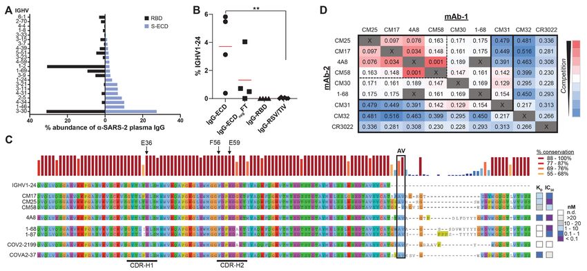
M. D. Dake, T. Edwards, M. E. Kaplan, S. J. Scott, C. Hypes, J. Mosier, D. T. Harris,        M. Yuan, D. M. Smith, D. Nemazee, J. R. Teijaro, J. E. Voss, I. A. Wilson, R. Andrabi,
     B. J. LaFleur, R. Sprissler, J. Nikolich-Žugich, D. Bhattacharya, Orthogonal SARS-           B. Briney, E. Landais, D. Sok, J. G. Jardine, D. R. Burton, Isolation of potent SARS-
     CoV-2 Serological Assays Enable Surveillance of Low-Prevalence Communities                   CoV-2 neutralizing antibodies and protection from disease in a small animal
     and Reveal Durable Humoral Immunity. Immunity 53, 925–933.e4 (2020).                         model. Science 369, 956–963 (2020). doi:10.1126/science.abc7520 Medline
     doi:10.1016/j.immuni.2020.10.004 Medline                                                 23. S. R. Leist, K. H. Dinnon 3rd, A. Schäfer, L. V. Tse, K. Okuda, Y. J. Hou, A. West, C.
11. J. A. Juno, H.-X. Tan, W. S. Lee, A. Reynaldi, H. G. Kelly, K. Wragg, R. Esterbauer, H.       E. Edwards, W. Sanders, E. J. Fritch, K. L. Gully, T. Scobey, A. J. Brown, T. P.
     E. Kent, C. J. Batten, F. L. Mordant, N. A. Gherardin, P. Pymm, M. H. Dietrich, N. E.        Sheahan, N. J. Moorman, R. C. Boucher, L. E. Gralinski, S. A. Montgomery, R. S.
     Scott, W.-H. Tham, D. I. Godfrey, K. Subbarao, M. P. Davenport, S. J. Kent, A. K.            Baric, A Mouse-Adapted SARS-CoV-2 Induces Acute Lung Injury and Mortality in
     Wheatley, Humoral and circulating follicular helper T cell responses in recovered            Standard        Laboratory     Mice.      Cell    183,     1070–1085.e12         (2020).
     patients with COVID-19. Nat. Med. 26, 1428–1434 (2020). doi:10.1038/s41591-                  doi:10.1016/j.cell.2020.09.050 Medline
     020-0995-0 Medline                                                                       24. K. H. Dinnon 3rd, S. R. Leist, A. Schäfer, C. E. Edwards, D. R. Martinez, S. A.
12. Y. Weisblum, F. Schmidt, F. Zhang, J. DaSilva, D. Poston, J. C. C. Lorenzi, F.                Montgomery, A. West, B. L. Yount Jr., Y. J. Hou, L. E. Adams, K. L. Gully, A. J.
     Muecksch, M. Rutkowska, H.-H. Hoffmann, E. Michailidis, C. Gaebler, M. Agudelo,              Brown, E. Huang, M. D. Bryant, I. C. Choong, J. S. Glenn, L. E. Gralinski, T. P.
     A. Cho, Z. Wang, A. Gazumyan, M. Cipolla, L. Luchsinger, C. D. Hillyer, M. Caskey,           Sheahan, R. S. Baric, A mouse-adapted model of SARS-CoV-2 to test COVID-19
     D. F. Robbiani, C. M. Rice, M. C. Nussenzweig, T. Hatziioannou, P. D. Bieniasz,              countermeasures. Nature 586, 560–566 (2020). doi:10.1038/s41586-020-
     Escape from neutralizing antibodies by SARS-CoV-2 spike protein variants. eLife              2708-8 Medline
     9, e61312 (2020). doi:10.7554/eLife.61312 Medline                                        25. D. Pinto, Y.-J. Park, M. Beltramello, A. C. Walls, M. A. Tortorici, S. Bianchi, S. Jaconi,
13. J. J. Lavinder, Y. Wine, C. Giesecke, G. C. Ippolito, A. P. Horton, O. I. Lungu, K. H.        K. Culap, F. Zatta, A. De Marco, A. Peter, B. Guarino, R. Spreafico, E. Cameroni, J.
     Hoi, B. J. DeKosky, E. M. Murrin, M. M. Wirth, A. D. Ellington, T. Dörner, E. M.             B. Case, R. E. Chen, C. Havenar-Daughton, G. Snell, A. Telenti, H. W. Virgin, A.

                                                                                                                                                                                               Downloaded from http://science.sciencemag.org/ on May 20, 2021
     Marcotte, D. R. Boutz, G. Georgiou, Identification and characterization of the               Lanzavecchia, M. S. Diamond, K. Fink, D. Veesler, D. Corti, Cross-neutralization of
     constituent human serum antibodies elicited by vaccination. Proc. Natl. Acad. Sci.           SARS-CoV-2 by a human monoclonal SARS-CoV antibody. Nature 583, 290–295
     U.S.A. 111, 2259–2264 (2014). doi:10.1073/pnas.1317793111 Medline                            (2020). doi:10.1038/s41586-020-2349-y Medline
14. J. J. Lavinder, A. P. Horton, G. Georgiou, G. C. Ippolito, Next-generation sequencing     26. S. C. A. Nielsen, F. Yang, K. J. L. Jackson, R. A. Hoh, K. Röltgen, G. H. Jean, B. A.
     and protein mass spectrometry for the comprehensive analysis of human cellular               Stevens, J.-Y. Lee, A. Rustagi, A. J. Rogers, A. E. Powell, M. Hunter, J. Najeeb, A. R.
     and serum antibody repertoires. Curr. Opin. Chem. Biol. 24, 112–120 (2015).                  Otrelo-Cardoso, K. E. Yost, B. Daniel, K. C. Nadeau, H. Y. Chang, A. T. Satpathy, T.
     doi:10.1016/j.cbpa.2014.11.007 Medline                                                       S. Jardetzky, P. S. Kim, T. T. Wang, B. A. Pinsky, C. A. Blish, S. D. Boyd, Human B
15. W. E. Purtha, T. F. Tedder, S. Johnson, D. Bhattacharya, M. S. Diamond, Memory B              Cell Clonal Expansion and Convergent Antibody Responses to SARS-CoV-2. Cell
     cells, but not long-lived plasma cells, possess antigen specificities for viral escape       Host Microbe 28, 516–525.e5 (2020). doi:10.1016/j.chom.2020.09.002 Medline
     mutants. J. Exp. Med. 208, 2599–2606 (2011). doi:10.1084/jem.20110740                    27. S. D. Boyd, B. A. Gaëta, K. J. Jackson, A. Z. Fire, E. L. Marshall, J. D. Merker, J. M.
     Medline                                                                                      Maniar, L. N. Zhang, B. Sahaf, C. D. Jones, B. B. Simen, B. Hanczaruk, K. D. Nguyen,
16. K. G. Smith, A. Light, G. J. Nossal, D. M. Tarlinton, The extent of affinity maturation       K. C. Nadeau, M. Egholm, D. B. Miklos, J. L. Zehnder, A. M. Collins, Individual
     differs between the memory and antibody-forming cell compartments in the                     variation in the germline Ig gene repertoire inferred from variable region gene
     primary immune response. EMBO J. 16, 2996–3006 (1997).                                       rearrangements.           J.     Immunol.         184,       6986–6992            (2010).
     doi:10.1093/emboj/16.11.2996 Medline                                                         doi:10.4049/jimmunol.1000445 Medline
17. C. O. Barnes, A. P. West Jr., K. E. Huey-Tubman, M. A. G. Hoffmann, N. G. Sharaf,         28. H. Tegally, E. Wilkinson, M. Giovanetti, A. Iranzadeh, V. Fonseca, J. Giandhari, D.
     P. R. Hoffman, N. Koranda, H. B. Gristick, C. Gaebler, F. Muecksch, J. C. C. Lorenzi,        Doolabh, S. Pillay, E. J. San, N. Msomi, K. Mlisana, A. von Gottberg, S. Walaza, M.
     S. Finkin, T. Hägglöf, A. Hurley, K. G. Millard, Y. Weisblum, F. Schmidt, T.                 Allam, A. Ismail, T. Mohale, A. J. Glass, S. Engelbrecht, G. Van Zyl, W. Preiser, F.
     Hatziioannou, P. D. Bieniasz, M. Caskey, D. F. Robbiani, M. C. Nussenzweig, P. J.            Petruccione, A. Sigal, D. Hardie, G. Marais, N. Hsiao, S. Korsman, M.-A. Davies, L.
     Bjorkman, Structures of Human Antibodies Bound to SARS-CoV-2 Spike Reveal                    Tyers, I. Mudau, D. York, C. Maslo, D. Goedhals, S. Abrahams, O. Laguda-Akingba,
     Common Epitopes and Recurrent Features of Antibodies. Cell 182, 828–842.e16                  A. Alisoltani-Dehkordi, A. Godzik, C. K. Wibmer, B. T. Sewell, J. Lourenço, L. C. J.
     (2020). doi:10.1016/j.cell.2020.06.025 Medline                                               Alcantara, S. L. Kosakovsky Pond, S. Weaver, D. Martin, R. J. Lessells, J. N.
18. L. L. Luchsinger, B. P. Ransegnola, D. K. Jin, F. Muecksch, Y. Weisblum, W. Bao, P.           Bhiman, C. Williamson, T. de Oliveira, Detection of a SARS-CoV-2 variant of
     J. George, M. Rodriguez, N. Tricoche, F. Schmidt, C. Gao, S. Jawahar, M. Pal, E.             concern in South Africa. Nature 592, 438–443 (2021). doi:10.1038/s41586-021-
     Schnall, H. Zhang, D. Strauss, K. Yazdanbakhsh, C. D. Hillyer, P. D. Bieniasz, T.            03402-9
     Hatziioannou, Serological Assays Estimate Highly Variable SARS-CoV-2                     29. K. R. McCarthy, L. J. Rennick, S. Nambulli, L. R. Robinson-McCarthy, W. G. Bain, G.
     Neutralizing Antibody Activity in Recovered COVID-19 Patients. J. Clin. Microbiol.           Haidar, W. P. Duprex, Recurrent deletions in the SARS-CoV-2 spike glycoprotein
     58, e02005-20 (2020). doi:10.1128/JCM.02005-20 Medline                                       drive      antibody        escape.      Science       371,      1139–1142         (2021).
19. F. Wu, M. Liu, A. Wang, L. Lu, Q. Wang, C. Gu, J. Chen, Y. Wu, S. Xia, Y. Ling, Y.            doi:10.1126/science.abf6950 Medline
     Zhang, J. Xun, R. Zhang, Y. Xie, S. Jiang, T. Zhu, H. Lu, Y. Wen, J. Huang, Evaluating   30. B. Choi, M. C. Choudhary, J. Regan, J. A. Sparks, R. F. Padera, X. Qiu, I. H. Solomon,
     the Association of Clinical Characteristics With Neutralizing Antibody Levels in             H.-H. Kuo, J. Boucau, K. Bowman, U. D. Adhikari, M. L. Winkler, A. A. Mueller, T. Y.-
     Patients Who Have Recovered From Mild COVID-19 in Shanghai, China. JAMA                      T. Hsu, M. Desjardins, L. R. Baden, B. T. Chan, B. D. Walker, M. Lichterfeld, M. Brigl,
     Intern. Med. 180, 1356–1362 (2020). doi:10.1001/jamainternmed.2020.4616                      D. S. Kwon, S. Kanjilal, E. T. Richardson, A. H. Jonsson, G. Alter, A. K. Barczak, W.
     Medline                                                                                      P. Hanage, X. G. Yu, G. D. Gaiha, M. S. Seaman, M. Cernadas, J. Z. Li, Persistence
20. B. J. DeKosky, T. Kojima, A. Rodin, W. Charab, G. C. Ippolito, A. D. Ellington, G.            and Evolution of SARS-CoV-2 in an Immunocompromised Host. N. Engl. J. Med.
     Georgiou, In-depth determination and analysis of the human paired heavy- and                 383, 2291–2293 (2020). doi:10.1056/NEJMc2031364 Medline
     light-chain antibody repertoire. Nat. Med. 21, 86–91 (2015). doi:10.1038/nm.3743         31. V. A. Avanzato, M. J. Matson, S. N. Seifert, R. Pryce, B. N. Williamson, S. L. Anzick,
     Medline                                                                                      K. Barbian, S. D. Judson, E. R. Fischer, C. Martens, T. A. Bowden, E. de Wit, F. X.
21. A. J. Greaney, A. N. Loes, K. H. D. Crawford, T. N. Starr, K. D. Malone, H. Y. Chu, J.        Riedo, V. J. Munster, Case Study: Prolonged Infectious SARS-CoV-2 Shedding
     D. Bloom, Comprehensive mapping of mutations in the SARS-CoV-2 receptor-                     from an Asymptomatic Immunocompromised Individual with Cancer. Cell 183,
     binding domain that affect recognition by polyclonal human plasma antibodies.                1901–1912.e9 (2020). doi:10.1016/j.cell.2020.10.049 Medline
     Cell Host Microbe 29, 463–476.e6 (2021). doi:10.1016/j.chom.2021.02.003                  32. P. C. Resende et al., The ongoing evolution of variants of concern and interest of
     Medline                                                                                      SARS-CoV-2 in Brazil revealed by convergent indels in the amino (N)-terminal
22. T. F. Rogers, F. Zhao, D. Huang, N. Beutler, A. Burns, W. T. He, O. Limbo, C. Smith,          domain of the Spike protein. medRxiv 2021.03.19.21253946 [Preprint]. 20 March
     G. Song, J. Woehl, L. Yang, R. K. Abbott, S. Callaghan, E. Garcia, J. Hurtado, M.            2021. https://doi.org/10.1101/2021.03.19.21253946.
     Parren, L. Peng, S. Ramirez, J. Ricketts, M. J. Ricciardi, S. A. Rawlings, N. C. Wu,     33. E. Andreano et al., SARS-CoV-2 escape in vitro from a highly neutralizing COVID-

First release: 4 May 2021                                                     www.sciencemag.org                 (Page numbers not final at time of first release)                        4
19 convalescent plasma. bioRxiv 2020.12.28.424451 [Preprint]. 28 December 48. E. F. Pettersen, T. D. Goddard, C. C. Huang, E. C. Meng, G. S. Couch, T. I. Croll, J.
2020. https://doi.org/10.1101/2020.12.28.424451. H. Morris, T. E. Ferrin, UCSF ChimeraX: Structure visualization for researchers,
34. C. L. Hsieh, J. A. Goldsmith, J. M. Schaub, A. M. DiVenere, H.-C. Kuo, K. educators, and developers. Protein Sci. 30, 70–82 (2021). doi:10.1002/pro.3943
Javanmardi, K. C. Le, D. Wrapp, A. G. Lee, Y. Liu, C.-W. Chou, P. O. Byrne, C. K. Medline
Hjorth, N. V. Johnson, J. Ludes-Meyers, A. W. Nguyen, J. Park, N. Wang, D. 49. C. Michael, W. Mona, Y. Choonhan, COSMIC2 – A Science Gateway for Cryo-
Amengor, J. J. Lavinder, G. C. Ippolito, J. A. Maynard, I. J. Finkelstein, J. S. Electron Microscopy (2017).
McLellan, Structure-based design of prefusion-stabilized SARS-CoV-2 spikes. 50. J. Dunbar, K. Krawczyk, J. Leem, C. Marks, J. Nowak, C. Regep, G. Georges, S.
Science 369, 1501–1505 (2020). doi:10.1126/science.abd0826 Medline Kelm, B. Popovic, C. M. Deane, SAbPred: A structure-based antibody prediction
35. R. Henderson, R. J. Edwards, K. Mansouri, K. Janowska, V. Stalls, S. M. C. Gobeil, server. Nucleic Acids Res. 44 , W474–W478 (2016). doi:10.1093/nar/gkw361
M. Kopp, D. Li, R. Parks, A. L. Hsu, M. J. Borgnia, B. F. Haynes, P. Acharya, Medline
Controlling the SARS-CoV-2 spike glycoprotein conformation. Nat. Struct. Mol. 51. E. F. Pettersen, T. D. Goddard, C. C. Huang, G. S. Couch, D. M. Greenblatt, E. C.
Biol. 27, 925–933 (2020). doi:10.1038/s41594-020-0479-4 Medline Meng, T. E. Ferrin, UCSF Chimera—A visualization system for exploratory
36. E. Salazar, K. K. Perez, M. Ashraf, J. Chen, B. Castillo, P. A. Christensen, T. Eubank, research and analysis. J. Comput. Chem. 25, 1605–1612 (2004).
D. W. Bernard, T. N. Eagar, S. W. Long, S. Subedi, R. J. Olsen, C. Leveque, M. R. doi:10.1002/jcc.20084 Medline
Schwartz, M. Dey, C. Chavez-East, J. Rogers, A. Shehabeldin, D. Joseph, G. 52. P. Emsley, K. Cowtan, Coot: Model-building tools for molecular graphics. Acta
Williams, K. Thomas, F. Masud, C. Talley, K. G. Dlouhy, B. V. Lopez, C. Hampton, J. Crystallogr. D Biol. Crystallogr. 60, 2126–2132 (2004).
Lavinder, J. D. Gollihar, A. C. Maranhao, G. C. Ippolito, M. O. Saavedra, C. C. Cantu, doi:10.1107/S0907444904019158 Medline
P. Yerramilli, L. Pruitt, J. M. Musser, Treatment of Coronavirus Disease 2019 53. P. D. Adams, R. W. Grosse-Kunstleve, L.-W. Hung, T. R. Ioerger, A. J. McCoy, N. W.
(COVID-19) Patients with Convalescent Plasma. Am. J. Pathol. 190, 1680–1690 Moriarty, R. J. Read, J. C. Sacchettini, N. K. Sauter, T. C. Terwilliger, PHENIX:
Downloaded from http://science.sciencemag.org/ on May 20, 2021
(2020). doi:10.1016/j.ajpath.2020.05.014 Medline Building new software for automated crystallographic structure determination.
37. G. C. Ippolito, K. H. Hoi, S. T. Reddy, S. M. Carroll, X. Ge, T. Rogosch, M. Zemlin, L. Acta Crystallogr. D Biol. Crystallogr. 58, 1948–1954 (2002).
D. Shultz, A. D. Ellington, C. L. Vandenberg, G. Georgiou, Antibody repertoires in doi:10.1107/S0907444902016657 Medline
humanized NOD-scid-IL2Rγ(null) mice and human B cells reveals human-like 54. T. I. Croll, ISOLDE: A physically realistic environment for model building into low-
diversification and tolerance checkpoints in the mouse. PLOS ONE 7, e35497 resolution electron-density maps. Acta Crystallogr. D Struct. Biol. 74, 519–530
(2012). doi:10.1371/journal.pone.0035497 Medline (2018). doi:10.1107/S2059798318002425 Medline
38. J. R. McDaniel, B. J. DeKosky, H. Tanno, A. D. Ellington, G. Georgiou, Ultra-high-
throughput sequencing of the immune receptor repertoire from millions of ACKNOWLEDGMENTS
lymphocytes. Nat. Protoc. 11, 429–442 (2016). doi:10.1038/nprot.2016.024 We are indebted to our study subjects for providing the blood samples required for
Medline this study. We wish to thank Drs. G. Fenves, D. Jaffee, and A. Matouschek for
39. A. M. Bolger, M. Lohse, B. Usadel, Trimmomatic: A flexible trimmer for Illumina their support. The authors are grateful for the administrative expertise of E. K.
sequence data. Bioinformatics 30, 2114–2120 (2014). Miller, to The LaMontagne Center for Infectious Disease, for the university’s core
doi:10.1093/bioinformatics/btu170 Medline facilities, and to Dr. C.-L. Hsieh for providing reagents and advice. Funding:
40. D. A. Bolotin, S. Poslavsky, I. Mitrophanov, M. Shugay, I. Z. Mamedov, E. V. Funding for USAMRIID was provided through the CARES Act with programmatic
Putintseva, D. M. Chudakov, MiXCR: Software for comprehensive adaptive oversight from the Military Infectious Diseases Research Program–project
immunity profiling. Nat. Methods 12, 380–381 (2015). doi:10.1038/nmeth.3364 14066041. Opinions, conclusions, interpretations, and recommendations are
Medline those of the authors and are not necessarily endorsed by the U.S. Army. The
41. R. C. Edgar, Search and clustering orders of magnitude faster than BLAST. mention of trade names or commercial products does not constitute
Bioinformatics 26, 2460–2461 (2010). doi:10.1093/bioinformatics/btq461 endorsement or recommendation for use by the Department of the Army or the
Medline Department of Defense. The findings and conclusions in this report are those of
42. M. P. Lefranc, G. Lefranc, Immunoglobulins or Antibodies: IMGT® Bridging Genes, the authors and do not necessarily represent the views of Centers for Disease
Structures and Functions. Biomedicines 8, 319 (2020). Control and Prevention. Molecular graphics and analyses performed with UCSF
doi:10.3390/biomedicines8090319 Medline Chimera, developed by the Resource for Biocomputing, Visualization, and
43. Y. H. Ching, T. K. Ghosh, S. J. Cross, E. A. Packham, L. Honeyman, S. Loughna, T. Informatics at the University of California, San Francisco, with support from NIH
E. Robinson, A. M. Dearlove, G. Ribas, A. J. Bonser, N. R. Thomas, A. J. Scotter, L. P41-GM103311. The Sauer Structural Biology Laboratory is supported by the
S. D. Caves, G. P. Tyrrell, R. A. Newbury-Ecob, A. Munnich, D. Bonnet, J. D. Brook, University of Texas College of Natural Sciences and by award RR160023 from
Mutation in myosin heavy chain 6 causes atrial septal defect. Nat. Genet. 37, 423– the Cancer Prevention and Research Institute of Texas (CPRIT). This research
428 (2005). doi:10.1038/ng1526 Medline was funded in part by the Clayton Foundation (C.J.P., B.L.I., G.G.); a National
44. K. Javanmardi et al., Rapid characterization of spike variants via mammalian cell Institutes of Health (NIH)/National Institute of Allergy and Infectious Diseases
surface display. bioRxiv 2021.03.30.437622 [Preprint]. 30 March 2021. (NIAID) grant awarded to J.S.M. (R01-AI127521) as well as Welch Foundation
https://doi.org/10.1101/2021.03.30.437622. Grant No. F-0003-19620604; NIH NCI COVID-19 SeroNet grant U54-CA260543
45. Y. J. Hou, K. Okuda, C. E. Edwards, D. R. Martinez, T. Asakura, K. H. Dinnon 3rd, T. (R.S.B.); UT System Proteomics Network pilot funding (J.J.L, M.D.P.), and in part
Kato, R. E. Lee, B. L. Yount, T. M. Mascenik, G. Chen, K. N. Olivier, A. Ghio, L. V. Tse, with federal funds under a contract from the NIH NIAID, Contract Number
S. R. Leist, L. E. Gralinski, A. Schäfer, H. Dang, R. Gilmore, S. Nakano, L. Sun, M. L. 75N93019C00050 (G.G., J.J.L., G.C.I.). Author contributions:
Fulcher, A. Livraghi-Butrico, N. I. Nicely, M. Cameron, C. Cameron, D. J. Kelvin, A. Conceptualization: W.N.V., G.G., J.J.L. and G.C.I.; Methodology: W.N.V., Y.J.H.,
de Silva, D. M. Margolis, A. Markmann, L. Bartelt, R. Zumwalt, F. J. Martinez, S. P. N.V.J., J.E.K., G.D., A.P.H., B.L.I., M.D.P., J.D., A.H., R.S.B., J.S.M., G.G., J.J.L., and
Salvatore, A. Borczuk, P. R. Tata, V. Sontake, A. Kimple, I. Jaspers, W. K. O’Neal, G.C.I.; Investigation: W.N.V., Y.J.H., N.V.J., J.E.K., G.D., A.P.H., F.B., C.P., Y.T.,
S. H. Randell, R. C. Boucher, R. S. Baric, SARS-CoV-2 Reverse Genetics Reveals a S.A.A., W.P., K.G., D.R.B., D.M.T., J.G., D.B., M.G., J.J.L., and G.C.I.; Data Analysis
Variable Infection Gradient in the Respiratory Tract. Cell 182, 429–446.e14 and Interpretation: W.N.V., Y.J.H., N.V.J., J.E.K., G.D., A.P.H., S.A.A., W.P., D.R.B.,
(2020). doi:10.1016/j.cell.2020.05.042 Medline J.R.M., L.R., D.B., J.L., J.P., S.G., S.S., A.H., J.D.G., R.S.B., J.S.M., G.G., J.J.L., and
46. D. Tegunov, P. Cramer, Real-time cryo-electron microscopy data preprocessing G.C.I.; Data Curation: W.N.V., J.Y.H., N.V.J., J.E.K., G.D., J.J.L., and G.C.I.; Writing:
with Warp. Nat. Methods 16, 1146–1152 (2019). doi:10.1038/s41592-019-0580-y Original Draft, W.N.V., N.J.V., J.J.L., and G.C.I.; Writing: Review & Editing: W.N.V.,
Medline Y.J.H., N.J.V., B.L.I., R.S.B., J.S.M., G.G., J.J.L., and G.C.I.; Funding: J.D.G., R.S.B.,
47. A. Punjani, J. L. Rubinstein, D. J. Fleet, M. A. Brubaker, cryoSPARC: Algorithms for J.S.M., G.G., and G.C.I. Competing interests: A patent application submitted by
rapid unsupervised cryo-EM structure determination. Nat. Methods 14, 290–296 The University of Texas Board of Regents is pending for anti-SARS-CoV-2
(2017). doi:10.1038/nmeth.4169 Medline monoclonal antibodies described in the manuscript (W.N.V., J.D.G., J.S.M., G.G.,
First release: 4 May 2021 www.sciencemag.org (Page numbers not final at time of first release) 5J.J.L., and G.C.I.). Data and materials availability: FASTQ VH and VH:VL
sequence files have been deposited in the NCBI Sequence Read Archive with
accession numbers PRJNA422864. The monoclonal antibodies have been
deposited in GenBank (https://www.ncbi.nlm.nih.gov/genbank/) with accession
numbers MZ049539 to MZ049552. Coordinates for the CM25 Fab in complex
with trimeric spike ectodomain have been deposited to the Protein Data Bank as
PDBID:7M8J. Cryo-EM maps have been deposited to the Electron Microscopy
Data Bank under accession code EMD-23717. These structural data are
presented in Fig. 4, table S4, and figs. S7-S8. All other data are available in the
main text or the supplementary materials. This work is licensed under a Creative
Commons Attribution 4.0 International (CC BY 4.0) license, which permits
unrestricted use, distribution, and reproduction in any medium, provided the
original work is properly cited. To view a copy of this license, visit
https://creativecommons.org/licenses/by/4.0/. This license does not apply to
figures/photos/artwork or other content included in the article that is credited
to a third party; obtain authorization from the rights holder before using such
material.
SUPPLEMENTARY MATERIALS
Downloaded from http://science.sciencemag.org/ on May 20, 2021
science.sciencemag.org/cgi/content/full/science.abg5268/DC1
Materials and Methods
Figs. S1 to S9
Tables S1 to S5
References (34–54)
MDAR Reproducibility Checklist
12 January 2021; accepted 29 April 2021
Published online 4 May 2021
10.1126/science.abg5268
First release: 4 May 2021 www.sciencemag.org (Page numbers not final at time of first release) 6Downloaded from http://science.sciencemag.org/ on May 20, 2021
Fig. 1. Most plasma IgG antibodies bind non-RBD spike epitopes such as the NTD. (A) Affinity-purification
using spike S-ECD (1) or RBD for subject P3. Plasma IgG lineage identities, binding specificity, and relative
abundance were mapped via Ig-seq proteomics (14), facilitating recombinant plasma mAb characterization;
anti-RBD (green); anti-S2 (blue); anti-NTD (red). (B) IgG ELISA binding (1:150 plasma dilution) to S-ECD alone,
or in the presence of 50 μg/ml of RBD (S-ECD(RBD-)) or S-∆RBD deletion mutant. (C) Quantitative Ig-seq
determination of anti-RBD and non-RBD IgG mAb abundance in early convalescent plasmas across four
subjects. (D) Authentic virus neutralization (in duplicate) of the four most abundant plasma IgGs (CM29, CM30,
CM31, CM32) from plasma lineages Lin.1, Lin.2, Lin.3, Lin.4 in subject P3. (E and F) Prophylactic protection of
12-month-old BALB/c mice (n=5 per group) against lethal challenge with high dose (104 PFU) mouse-adapted
(MA10) SARS-CoV-2. Cocktail of non-RBD mAbs (200 μg per mouse) at 2:1:1 ratio reflecting their relative
plasma abundance. **PDownloaded from http://science.sciencemag.org/ on May 20, 2021
Fig. 2. Protective spike NTD-targeting antibodies are prevalent in COVID-19 convalescent plasma. (A)
Temporal Ig-seq dynamics of the anti-S-ECD IgG repertoire at days 12 and 56 post-symptom onset. (B) Biolayer
interferometry (BLI) sensorgrams to S-ECD ligand of anti-NTD mAbs CM17, CM25 (subject P2), and CM58
(subject P4). (C) In vitro live virus neutralization (performed in duplicate). (D-F) In vivo prophylactic protection
of 12-month-old BALB/c mice (n=5 per group) against high dose intranasal challenge (105 PFU) of mouse-
adapted (MA10) SARS-CoV-2. ***PDownloaded from http://science.sciencemag.org/ on May 20, 2021
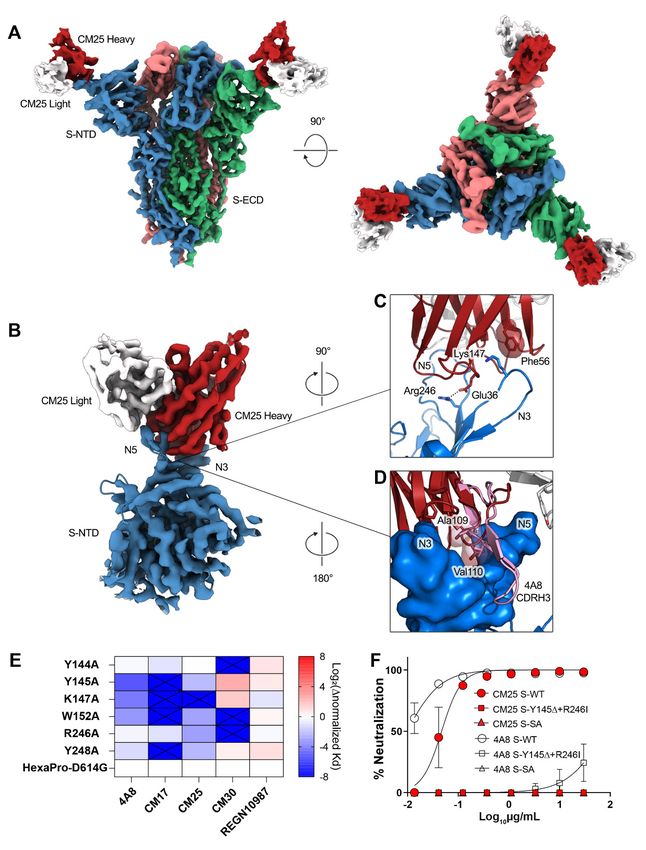
Fig. 3. Genetic basis of a shared, or public, class of IGHV1-24 plasma antibodies targeting the spike NTD.
(A) IGHV usage of plasma antibodies in all subjects (n=4). (B) Comparative IGHV1-24 usage of anti-S-ECD (IgG-
ECD) and anti-RBD (IgG-RBD) plasma antibodies, or in depleted S-ECD affinity column flow through (IgG-
ECDnegFT) in all subjects (n=4). IgG-RSV/TIV: IgG specific to respiratory syncytial virus (RSV) or trivalent
influenza vaccine hemagglutinin HA1 (TIV) in healthy controls post-vaccination (n=6). **PFig. 4. Structural basis of public IGHV1-24 plasma antibodies, NTD mutations, and antibody Downloaded from http://science.sciencemag.org/ on May 20, 2021
escape. (A) Side and top views of the structure of CM25 Fab bound to S-ECD shown as cryo-EM
density. (B) Focused refinement density revealing a VH-dominant mode of binding, with substantial
contacts mediated by interactions between the three CDRs and the N3 and N5 loops of the NTD. (C)
CDR-H1 interaction includes a salt bridge formed between the uniquely encoded Glu36 residue and
the N5 loop residue Arg246; Phe56 unique residue in CDR-H2 forms a pi-cation interaction with
Lys147 in the N3 loop. (D) The AV dipeptide interaction with the N3 and N5 loops of the NTD is
structurally conserved between mAbs CM25 (red) and 4A8 (pink). (E) Normalized shift (Log2) in
binding KD, as measured by differential BLI affinities for single Ala mutants and parental D614G spike
protein. (F) Authentic virus neutralization of CM25 and 4A8 against WT, double S-N3/N5 loop
mutants, and South Africa (SA) B.1.351 viral variant.
First release: 4 May 2021 www.sciencemag.org (Page numbers not final at time of first release) 10Prevalent, protective, and convergent IgG recognition of SARS-CoV-2 non-RBD spike epitopes
William N. Voss, Yixuan J. Hou, Nicole V. Johnson, George Delidakis, Jin Eyun Kim, Kamyab Javanmardi, Andrew P. Horton,
Foteini Bartzoka, Chelsea J. Paresi, Yuri Tanno, Chia-Wei Chou, Shawn A. Abbasi, Whitney Pickens, Katia George, Daniel R.
Boutz, Dalton M. Towers, Jonathan R. McDaniel, Daniel Billick, Jule Goike, Lori Rowe, Dhwani Batra, Jan Pohl, Justin Lee,
Shivaprakash Gangappa, Suryaprakash Sambhara, Michelle Gadush, Nianshuang Wang, Maria D. Person, Brent L. Iverson, Jimmy
D. Gollihar, John Dye, Andrew Herbert, Ilya J. Finkelstein, Ralph S. Baric, Jason S. McLellan, George Georgiou, Jason J. Lavinder
and Gregory C. Ippolito
published online May 4, 2021
Downloaded from http://science.sciencemag.org/ on May 20, 2021
ARTICLE TOOLS http://science.sciencemag.org/content/early/2021/05/03/science.abg5268
SUPPLEMENTARY http://science.sciencemag.org/content/suppl/2021/05/03/science.abg5268.DC1
MATERIALS
RELATED http://stm.sciencemag.org/content/scitransmed/13/590/eabf7517.full
CONTENT
http://stm.sciencemag.org/content/scitransmed/13/578/eabd6990.full
http://stm.sciencemag.org/content/scitransmed/13/577/eabd2223.full
http://stm.sciencemag.org/content/scitransmed/13/577/eabf1555.full
REFERENCES This article cites 53 articles, 15 of which you can access for free
http://science.sciencemag.org/content/early/2021/05/03/science.abg5268#BIBL
PERMISSIONS http://www.sciencemag.org/help/reprints-and-permissions
Use of this article is subject to the Terms of Service
Science (print ISSN 0036-8075; online ISSN 1095-9203) is published by the American Association for the Advancement of
Science, 1200 New York Avenue NW, Washington, DC 20005. The title Science is a registered trademark of AAAS.
Copyright © 2021 The Authors, some rights reserved; exclusive licensee American Association for the Advancement of Science.
No claim to original U.S. Government Works. Distributed under a Creative Commons Attribution License 4.0 (CC BY).You can also read