Extracellular and Intracellular Angiotensin II Regulate the Automaticity of Developing Cardiomyocytes via Different Signaling Pathways - Frontiers
←
→
Page content transcription
If your browser does not render page correctly, please read the page content below
ORIGINAL RESEARCH
                                                                                                                                               published: 25 August 2021
                                                                                                                                         doi: 10.3389/fmolb.2021.699827

                                            Extracellular and Intracellular
                                            Angiotensin II Regulate the
                                            Automaticity of Developing
                                            Cardiomyocytes via Different
                                            Signaling Pathways
                                            Zenghua Qi 1,2, Tao Wang 3, Xiangmao Chen 4, Chun Kit Wong 1, Qianqian Ding 1,
                                            Heinrich Sauer 5, Zhi-Feng Chen 2, Cheng Long 4, Xiaoqiang Yao 6, Zongwei Cai 3 and
                                            Suk Ying Tsang 1,7,8*
                                            1
                                             School of Life Sciences, The Chinese University of Hong Kong, Shatin, Hong Kong, SAR China, 2Institute of Environmental Health
                          Edited by:
                                            and Pollution Control, School of Environmental Science and Engineering, Guangdong University of Technology, Guangzhou,
                         Xiao-Yu Liu,
                                            China, 3State Key Laboratory of Environmental and Biological Analysis, Department of Chemistry, Hong Kong Baptist University,
  Southern University of Science and
                                            Kowloon, Hong Kong, SAR China, 4School of Life Sciences, South China Normal University, Guangzhou, China, 5Department of
                 Technology, China
                                            Physiology, Justus Liebig University Giessen, Giessen, Germany, 6School of Biomedical Sciences, The Chinese University of
                        Reviewed by:        Hong Kong, Shatin, Hong Kong, SAR China, 7Key Laboratory for Regenerative Medicine, Ministry of Education, The Chinese
                                 Jun Lu,    University of Hong Kong, Shatin, Hong Kong, SAR China, 8State Key Laboratory of Agrobiotechnology, The Chinese University of
       Guilin Medical University, China     Hong Kong, Shatin, Hong Kong, SAR China
                    Domenico Cautela,
 Experimental Station for the Industry of
the Essential oils and Citrus products,     Angiotensin II (Ang II) plays an important role in regulating various physiological processes.
                                    Italy   However, little is known about the existence of intracellular Ang II (iAng II), whether iAng II
                   *Correspondence:         would regulate the automaticity of early differentiating cardiomyocytes, and the underlying
                       Suk Ying Tsang
               fayetsang@cuhk.edu.hk
                                            mechanism involved. Here, iAng II was detected by immunocytochemistry and ultra-high
                                            performance liquid chromatography combined with electrospray ionization triple
                    Specialty section:      quadrupole tandem mass spectrometry in mouse embryonic stem cell–derived
         This article was submitted to
                                            cardiomyocytes (mESC-CMs) and neonatal rat ventricular myocytes. Expression of
                 Molecular Diagnostics
                     and Therapeutics,      AT1R-YFP in mESC-CMs revealed that Ang II type 1 receptors were located on the
                a section of the journal    surface membrane, while immunostaining of Ang II type 2 receptors (AT2R) revealed that
    Frontiers in Molecular Biosciences
                                            AT2R were predominately located on the nucleus and the sarcoplasmic reticulum. While
            Received: 24 April 2021
            Accepted: 27 July 2021          extracellular Ang II increased spontaneous action potentials (APs), dual patch clamping
          Published: 25 August 2021         revealed that intracellular delivery of Ang II or AT2R activator C21 decreased spontaneous
                             Citation:      APs. Interestingly, iAng II was found to decrease the caffeine-induced increase in
   Qi Z, Wang T, Chen X, Wong CK,
                                            spontaneous APs and caffeine-induced calcium release, suggesting that iAng II
Ding Q, Sauer H, Chen Z-F, Long C,
  Yao X, Cai Z and Tsang SY (2021)          decreased spontaneous APs via the AT2R- and ryanodine receptor–mediated
         Extracellular and Intracellular    pathways. This is the first study that provides evidence of the presence and function
           Angiotensin II Regulate the
           Automaticity of Developing       of iAng II in regulating the automaticity behavior of ESC-CMs and may therefore shed light
         Cardiomyocytes via Different       on the role of iAng II in fate determination.
                  Signaling Pathways.
        Front. Mol. Biosci. 8:699827.       Keywords: developing cardiomyocytes, angiotensin II, angiotensin II receptor, spontaneous action potential,
    doi: 10.3389/fmolb.2021.699827          calcium

Frontiers in Molecular Biosciences | www.frontiersin.org                            1                                            August 2021 | Volume 8 | Article 699827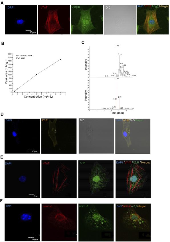
Qi et al.                                                                                                 Ang II Regulates Automaticity of Cardiomyocytes

INTRODUCTION                                                                   whether iAng II exerts any effect in early developing CMs,
                                                                               which, unlike adult CMs, uniquely display automaticity, and
The renin–angiotensin system (RAS) plays a central role in the                 the signaling pathways involved.
regulation of water balance and blood pressure; in addition, it also               Automaticity is a fundamental physiological feature of the
has an important role in the cardiovascular system (Harada et al.,             pacemaker cells in the heart; it is characterized by the existence of
1999). Angiotensin II (Ang II) is the key bioactive molecule of                spontaneous phase 4 diastolic depolarization (DD) in an action
RAS; it is produced either systemically or locally via the                     potential (AP). It is now recognized by numerous researchers in
proteolytic processing of angiotensinogen to angiotensin I                     the pacemaker field that the two clocks (the membrane clock and
(Ang I) by renin, followed by the subsequent conversion of                     the calcium clock) function in a coupled system synergistically to
Ang I to Ang II by angiotensin converting enzyme (ACE) or                      ensure the automaticity (Lakatta et al., 2010; Monfredi et al.,
chymase (Danser et al., 1999; Paul et al., 2006). Ang II, the major            2013). The membrane clock is attributed by an interplay of a
effector of RAS, was found to directly affect the contractility and            number of ion channels, pumps, and exchangers, such as the
the metabolism of adult cardiomyocytes (CMs) and is responsible                L-type Ca2+ channel, T-type Ca2+ channel, delayed rectifier K+
for hypertrophy (Baker et al., 1992). Two distinct Ang II receptor             channel, hyperpolarization-activated cyclic nucleotide-gated
subtypes, namely, the Ang II type 1 receptor (AT1R) and the Ang                channels, sodium–calcium exchanger, and Na+/K+-ATPase.
II type 2 receptor (AT2R), have been identified. Both AT1R and                  Besides the membrane clock, spontaneous calcium release
AT2R belong to the G protein–coupled receptor (GPCR)                           from the sarcoplasmic reticulum (SR) is another determinant
superfamily, but they are found to induce different cellular                   of automaticity. Local calcium releases (LCRs) are the elementary
signaling pathways (Akazawa et al., 2013; Karnik et al., 2015).                SR calcium releases mediated by the calcium release unit (CRU),
Numerous studies have demonstrated that AT1R activation can                    which is composed mainly of ryanodine receptor isoform 2
lead to disease states including hypertension, cardiac arrhythmia,             (RyR2) but also IP3 receptor (IP3R) (Cheng and Lederer,
stroke, diabetic nephropathy, and metabolic disorders (de                      2008). LCRs can occur spontaneously in CMs independent of
Gasparo et al., 2000; Zaman et al., 2002; Thomas and                           the calcium influx through the L-type Ca2+ channel, as LCRs
Mendelsohn, 2003). AT1R-null mice appear to show a lower                       continue even when the L-type Ca2+ channel is blocked
risk of cardiovascular disease (Harada et al., 1999). The most                 pharmacologically (Cheng and Lederer, 2008). Calcium release
classical pathway of AT1R is dependent on heterotrimeric G                     from the SR and calcium refill into the SR are regarded as the
proteins. AT1R-Gq/11-phospholipase Cβ (PLCβ) coupling leads                    calcium clock.
to the production of inositol trisphosphate (IP3) and                              Apart from its role in maintaining the LCR, another important
diacylglycerol (DAG) (Yusuf et al., 2000), which can activate                  role of RyR2 is its contribution to Ca2+-induced Ca2+ release
the inositol 1,4,5-trisphosphate receptor (IP3R) and the transient             (CICR). Upon the arrival of AP, plasma membrane
receptor potential (TRP) canonical 3 and 6 (TRPC3 and TRPC6)                   depolarization opens L-type Ca2+ channels; the calcium influx
channels, respectively, to increase the concentration of cytosolic             via L-type Ca2+ channels would in turn trigger the opening of
Ca2+ ([Ca2+]i) (Onohara et al., 2006). Although AT2R shares                    RyR2, leading to CICR. This calcium increase is the link for
∼34% amino acid sequence homology with AT1R, AT2R is                           excitation–contraction coupling in CMs.
markedly different from AT1R in terms of tissue-specific                            While it has been known for a long time that calcium is the
expression, signaling mechanisms, receptor function                            activator of RyR2 and that ryanodine is a widely used
regulation, and pharmacological properties (Jones et al., 2008;                pharmacological blocker of RyR2, the physiological inhibitory
Karnik et al., 2015). According to previous studies, AT2R can                  pathway of RyR2 has not been clearly identified. Whether there
attenuate the detrimental effects of AT1R to protect the heart                 would be any upstream signaling pathway which would
from disease development (Padia and Carey, 2013; Inuzuka et al.,               eventually lead to the inhibition of RyR2, either directly or
2016). AT2R has been proven to be a GPCR, with all the classical               indirectly, is unexplored.
motifs and signature residues of a GPCR (Lokuta et al., 1994;                      While some recent studies revealed the function of iAng II in
Gallinat et al., 1999; Asada et al., 2018). However, it is still unclear       the cardiovascular system (De Mello, 2015; Tadevosyan et al.,
whether AT2R can activate a classical G protein signaling                      2015; Tadevosyan et al., 2017), it remains unclear whether iAng II
pathway.                                                                       could modulate the automaticity of developing CMs [embryonic
    AT1R and AT2R were traditionally thought to be presented on                stem cell–derived CMs (ESC-CMs) and neonatal rat ventricular
the plasma membrane (George et al., 2010; Savoia et al., 2011).                myocytes (NRVMs)]. All in all, the existence and the function of
However, Ang II was also shown to bind to the nuclear membrane                 iAng II and AT2R in automatically firing CMs are unknown.
and nuclei of CMs (Baker et al., 1992), suggesting the existence of            Because of the pivotal role of RyR2 in these CMs, it would be
intracellular Ang II (iAng II) receptors. In addition, previous                important to determine if iAng II, upon the activation of AT2R, if
studies have shown that AT2R was strongly detected in the                      any, would act via RyR2 to exert its effect. The aims of this study
perinuclear region or in the nuclei of CMs (Senbonmatsu                        were 1) to investigate if iAng II and its receptors are present in
et al., 2003; Tadevosyan et al., 2010), hinting that iAng II may               developing CMs, 2) to investigate if extracellular and iAng II
act in an intracrine manner through AT2R. Although there were                  regulate the spontaneous APs of developing CMs and exert their
previous studies showing that exogenous application of Ang II                  effects differentially, and 3) to elucidate the signaling pathways
increases the contraction amplitude and frequency of CMs                       through which iAng II regulates spontaneous APs of
(Sedan et al., 2008; Lagerqvist et al., 2011), it is unclear                   developing CMs.

Frontiers in Molecular Biosciences | www.frontiersin.org                   2                                     August 2021 | Volume 8 | Article 699827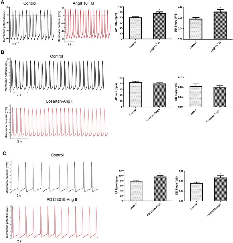
Qi et al.                                                                                             Ang II Regulates Automaticity of Cardiomyocytes

MATERIALS AND METHODS                                                      of determination r2 for validation was found to be 0.9993. The
                                                                           detection limit and the lower limit of quantification were
                                                                           determined to be 0.1 and 0.3 ng/ml, respectively. The detailed
Mouse Embryonic Stem Cell Culture and                                      procedures for the quantification of iAng II levels in NRVMs by
Differentiation of Mouse Embryonic Stem                                    UHPLC-ESI-MS/MS are described in “Supplementary Materials
Cells                                                                      and Methods.”
Culture of mESC cell line D3 (ATCC, Manassas, VA,
United States, 28 passages) and differentiation of mESCs using             Electrophysiology
the hanging drop method were performed as we have previously               For single-cell patch clamp recording, glass coverslips containing
described (Ng et al., 2010; Wong et al., 2012; Law et al., 2013; Qi        single cells were placed onto a recording chamber with
et al., 2016). A full description of the methods is available in           temperature control (33°C) and perfused with Tyrode’s
“Supplementary Materials and Methods.”                                     solution. The pipette solution referred to the components in
                                                                           the previous articles (Muller et al., 1997; Qi et al., 2016). Action
                                                                           potentials were recorded using an Axopatch 200B amplifier
Isolation of Neonatal Rat Ventricular                                      (Molecular Devices, Sunnyvale, CA, United States) and
Cardiomyocytes                                                             pCLAMP 10.4 software (Molecular Devices), using rupture
NRVMs were isolated from neonatal Sprague-Dawley rats (1- to               whole-cell patch-clamp in current-clamp configurations.
2-day-old) using a previously described method (Wollert et al.,               To investigate the effect of iAng II on the automaticity of
1996). Briefly, the rat hearts were dissected out and digested with         CMs, we developed a dual patch clamp method which was
trypsin. The dissociated cells were then suspended in DMEM/F12             performed as follows: one electrode was designated as the
with GlutaMAX (Gibco, Grand Island, NY, United States)                     recording electrode, and the other electrode was designated as
supplemented with 10% horse serum (Gibco), 5% FBS                          the drug delivery electrode. GΩ seals were acquired with both
(Gibco), and 50 μg/ml gentamicin (Gibco). The isolated cells,              electrodes on the same cell. The cell membrane patched by the
which were still heterogeneous, were then plated to cultureware in         recording electrode was first broken without breaking the
an incubator for 60 min to allow the removal of cardiac fibroblasts         membrane patched by the drug delivery electrode. Baseline
as they would selectively adhere to the cultureware. Nonadhesive           APs of CMs were recorded for 3 min. Thereafter, the cell
NRVMs which were still in suspension were transferred and                  membrane patched by the drug electrode was ruptured to
plated on glass coverslips pre-coated with Matrigel (Corning,              allow drug delivery into cells and APs of CMs were
Corning, NY, United States) for further culture.                           simultaneously recorded by the recording electrode. The
    Quantification of iAng II levels in NRVMs by ultra-high                 bath solution and pipette solution of the recording
performance liquid chromatography combined with                            electrode were the same as those in single patch clamp.
electrospray ionization triple quadrupole tandem mass                      Pipette solution of the drug delivery electrode was
spectrometry (UHPLC-ESI-MS/MS) was carried out.                            composed of pipette solution and drugs. Clampfit 10.4
    NRVMs (2 × 10⁶ cells) from 10 neonatal rats were lysed in ice-         (Molecular Devices) and GraphPad Prism 5 (GraphPad
cold RIPA buffer freshly supplemented with a protease inhibitor            Prism Software, La Jolla, CA, United States) were used to
cocktail and placed on ice for 15 min and centrifuged at 16,000 g          analyze the recorded data.
at 4°C for 20 min. The supernatant (300 μl) that contained the
protein was stored at −80°C as the next procedure sample. Protein          Measurement of [Ca2+]i
concentration was determined using Bradford assay with bovine              For detecting the effect of iAng II on the [Ca2+]i, 5 µM Fluo-4
serum albumin (BSA) as the standard. For the analysis of iAng II,          (Invitrogen) was loaded into the single mESC-CMs. Thereafter,
a sample (300 μl) was subsequently extracted using C18 Sep-Pak             whole-cell patch-clamp was performed with pipette solution
Vac 3cc cartridges (200 mg) (Waters, Milford, MA,                          containing Ang II and bath solution that was Tyrode’s
United States), purified by immunoaffinity chromatography                    solution. After the establishment of the current-clamp mode,
with the immobilization of the anti–Ang II antibody on                     the bath solution was gently changed to Ca2+-free solution.
CNBr-activated Sepharose 4B cartridges (GE Healthcare,                     Subsequently, cells were imaged using a CCD camera
Uppsala, Sweden), and analyzed using an Ultimate 3000                      (Photometrics, Tucson, AZ, United States). Images were
UHPLC system coupled with a Thermo TSQ mass                                acquired at a frequency of 35 Hz using a MetaFluor/
spectrometer (Thermo Fisher Scientific, Waltham, MA,                        MetaMorph Imaging System (Molecular Devices).
United States). The double-charged parent ions of the native
Ang II were m/z 523.8 ± 0.1 (Supplementary Figure S1A). The                Statistical Analysis
structure of Ang II was confirmed by two kinds of MRM                       Each experiment was repeated at least three times, and the data
transitions, which were m/z 523.8 ± 0.1 → m/z 263.1 ± 0.1                  were shown as mean ± SEM. Unpaired Student’s t-test and
and m/z 523.8 ± 0.1 → m/z 784.4 ± 0.1 (Supplementary Figures               analysis of variance (ANOVA) were applied to take the
S1B,C). The former fragment ion was the dominant ion with the              statistical significance between two groups and three groups or
highest intensity, so that it was chosen as the quantitative ion,          more, respectively. p < 0.05 was considered to be statistically
while the latter one was used as the qualitative ion. The coefficient       significant.

Frontiers in Molecular Biosciences | www.frontiersin.org               3                                     August 2021 | Volume 8 | Article 699827
Qi et al.                                                                                                               Ang II Regulates Automaticity of Cardiomyocytes

  FIGURE 1 | Ang II, AT1R, and AT2R were detected in mESC-CMs and NRVMs. (A) Representative images showing the immunocytochemistry of iAng II in
  mESC-CMs. Cells were immunostained for cTnT (red), Ang II (green), and the nucleus with DAPI (blue). The results indicated that Ang II was present in mESC-CMs. (B)
  Linear calibration curve of Ang II. (C) Analysis of the NRVM samples by UHPLC-ESI-MS/MS. The extracted ion chromatograms of the two kinds of MRM. The upper one
  is the 523.8 ± 0.1 m/z →263.1 ± 0.1 m/z; the lower one is the 523.8 ± 0.1 m/z →784.4 ± 0.1 m/z. (D) mESC-CMs were transduced with the vector carrying
  AT1R-YFP and were stained with DAPI (blue). AT1R-YFP signals were detected on the plasma membrane. (E, F) mESC-CMs were stained with DAPI (blue), anti-AT2R
  (green), and (E) anti-cTnT (red) or (F) anti-SERCA2 (red). Merged images revealed extensive co-localization of AT2R with the nucleus and the SERCA2, suggesting that
  AT2R was located predominately on the nucleus and the SR. Scale bars represent 10 μm.

Frontiers in Molecular Biosciences | www.frontiersin.org                            4                                           August 2021 | Volume 8 | Article 699827
Qi et al.                                                                                        Ang II Regulates Automaticity of Cardiomyocytes

RESULTS                                                               Extracellular Ang II Regulated Action
iAng II Was Detected in Developing                                    Potentials in a Transient Manner Through
Cardiomyocytes                                                        the Ang II Type 1 Receptor–Dependent
The iAng II was detected by immunocytochemistry and our               Pathway
newly developed method by UHPLC-ESI-MS/MS in mESC-                    The effect of exogenously applied Ang II on APs of mESC-CMs was
CMs and NRVMs, respectively. First, we examined whether               first examined. When fluorescence-labeled Ang II was applied to
Ang II is present in mESC-CMs by immunostaining, followed             mESC-CMs, Ang II was not observed to enter mESC-CMs even after
by confocal fluorescence microscopy. Positive staining with            being applied for 36 h (data not shown). The results hinted that
cardiac tropinin T (cTnT), a CM-specific marker, confirmed              exogenous Ang II may act on receptors located on the cell surface
that the cells were CMs. Ang II was detected in the cytoplasm         plasma membrane. Next, Ang II was applied to mESC-CMs to
of mESC-CMs (Figure 1A). Furthermore, the result of                   determine its effect on APs. Ang II (10–7 M) significantly and rapidly
UHPLC-ESI-MS/MS analysis revealed that Ang II could be                increased the AP rate and the DD slope of APs (Figure 2A), while it
detected in NRVMs, and the concentration of Ang II in the             did not affect the action potential duration at 50% repolarization
NRVM sample was 0.79 ng/ml (Figures 1B,C). The linearity              (APD50) and maximum diastolic depolarization (MDP) of APs in
range of the native Ang II was determined. The six-point              mESC-CMs (data not shown). These changes in the AP rate and the
calibration curve of Ang II showed a reliable reproducibility         DD slope were transient (lasting 100–150 s after the addition of Ang
in the concentration range from 0.3 to 10 ng/ml. The                  II) and reversible, and they reappeared upon exposure to higher
calibration curve was prepared. The area of the peak was              concentrations of Ang II. Furthermore, the changes in APs in
proportional to the concentration of the analyte. The                 response to Ang II (10–7 M) in the presence of AT1R blocker
coefficient of determination r2 for validation was found to            losartan (50 μM) or AT2R blocker PD123319 (10 μM) were
be 0.9993 (Figure 1B). The mean recovery rate was found to be         examined. Preincubation of losartan attenuated the effects of Ang
37.2 ± 1.4%, and the recovery rate was sufficient to quantify          II on APs (Figure 2B), while preincubation of PD123319 did not
Ang II in the determined calibration range. The detection limit       affect the Ang II–induced response (Figure 2C). Similarly,
was determined to be 0.1 ng/ml. The lower limit of                    preincubation of losartan attenuated the effects of Ang II on
quantification was determined to be 0.3 ng/ml. After the               calcium transients (CaTs) (Supplementary Figures S3), while
correction by the loss of each procedure, the level of iAng           preincubation of PD123319 did not affect the Ang II–induced
II in NRVMs was calculated to be 0.63 pmol/mg.                        response (data not shown). These results suggested that AT1R,
                                                                      which is present on the cell surface plasma membrane of mESC-
Developing Cardiomyocytes Expressed                                   CMs, plays a critical role in mediating the transient effect of
                                                                      extracellular Ang II on APs and CaTs of mESC-CMs.
Ang II Type 1 Receptor on the Cell Surface
Membrane and Expressed Ang II Type 2
                                                                      Dual Current Patch Clamp Is an
Receptor Predominately on the Nucleus
                                                                      Advantageous Approach to Investigate the
and the Sarcoplasmic Reticulum
To examine the subcellular localization of AT1R and AT2R,             Effect of Intracellular Delivery of Drugs on
immunostaining followed by confocal microscopy was used.              the Action Potentials of Cardiomyocytes
AT1R and AT2R were found to be present in mESC-CMs.                   Next, we confirmed and showed that the formation of a patch by
Since conventional antibodies against AT1R have been                  the glass electrode, followed by subsequent breakage of the
reported to be nonspecific, we constructed recombinant                 membrane, could be employed to effectively deliver drugs into
adenoviruses to express the AT1R fluorescent fusion protein            CMs. Ang II-FITC could be successfully delivered into mESC-
(AT1R-YFP) to probe the subcellular localization of AT1R.             CMs using the glass electrode as shown by the appearance of the
AT1R-YFP was mainly expressed on the cell surface                     green fluorescence signal inside the cell without leakage into the
membrane of mESC-CMs (Figure 1D). On the other hand,                  extracellular space (Figure 3A). Moreover, Fluo-4-IM, a
immunostaining of AT2R revealed that AT2R was expressed in            membrane-impermeant Ca2+ indicator, could also be
a unique, web-like pattern (Figures 1E,F). Multiple antibodies        successfully delivered into mESC-CMs and generated high
against AT2R were used and similar expression pattern of AT2R         fluorescence intensity by binding intracellular Ca2+ (Figure 3B).
was obtained. Co-staining with the nuclear stain DAPI and with           Although a single glass electrode could simultaneously deliver
antibodies against SERCA2 (a cardiac-specific isoform of               drugs and record APs, basal APs before drug delivery (i.e., before
SERCA) revealed that AT2R was located on the nucleus                  rupture by whole-cell patch-clamp configuration) cannot be
(Figures 1E,F) and the SR (Figure 1F). In addition, the               obtained. To circumvent this limitation, we developed a dual
distribution of AT1R and AT2R in NRVMs was also                       current patch clamp method (Figure 3C). It is known that
investigated (Supplementary Figures S2A,B). The results were          intracellular cyclic AMP (cAMP) increases the rate of AP
consistent with the expression patterns of AT1R and AT2R in           generation in the cardiac pacemaker cells. Therefore, cAMP
mESC-CMs. Western blot analysis in NRVMs confirmed the                 was selected as a positive control to assess whether the dual
quality of the AT2R antibody used in our study (Supplementary         current patch clamp is an efficient and reliable method to
Figures S2C).                                                         investigate the effect of intracellular delivery of drugs on the

Frontiers in Molecular Biosciences | www.frontiersin.org          5                                     August 2021 | Volume 8 | Article 699827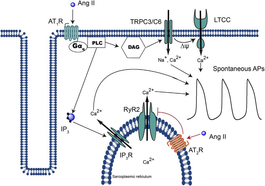
Qi et al.                                                                                                              Ang II Regulates Automaticity of Cardiomyocytes

  FIGURE 2 | Application of exogenous Ang II increased APs transiently, and the effect could be attenuated by the AT1R blocker but not the AT2R blocker. (A) Ang II
  increased the APs of mESC-CMs in a transient manner. Raw traces of spontaneous APs at the basal level and upon treatment with 10–7 M Ang II in mESC-CMs (left
  panel). Summarized data on the AP rate and the DD slope of APs (right panel). (B) AT1R blocker attenuated Ang II–induced changes in APs. Raw traces of
  spontaneous APs at the basal level and upon treatment with 50 μM losartan, followed by the subsequent application of 10–7 M Ang II in mESC-CMs (left panel).
  Summarized data on the AP rate and the DD slope of APs at the basal level or upon treatment with 50 μM losartan, followed by 10–7 M Ang II (right panel). Losartan
  attenuated Ang II–induced changes in the parameters of APs. (C) AT2R blocker did not affect Ang II–induced changes in APs. Raw traces of spontaneous APs at the
  basal level and upon treatment with 10 μM PD123319, followed by the subsequent application of 10–7 M Ang II in mESC-CMs (left panel). Summarized data on the AP
  rate and the DD slope of APs at the basal level or upon treatment with 10 μM PD123319, followed by 10–7 M Ang II (right panel). PD123319 did not affect Ang
  II–induced changes in the parameters of APs. Values are mean ± SEM of 4–6 independent experiments. *p < 0.05 vs. control group.

Frontiers in Molecular Biosciences | www.frontiersin.org                           6                                          August 2021 | Volume 8 | Article 699827
Qi et al. Ang II Regulates Automaticity of Cardiomyocytes
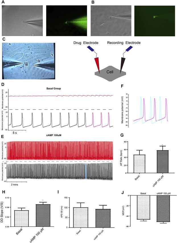
FIGURE 3 | Dual current patch clamp is an advantageous approach to investigate the effect of intracellular delivery of drugs on the APs of mESC-CMs reliably. (A)
Ang II-FITC can be successfully delivered into mESC-CMs using the glass electrode as shown by the appearance of the green fluorescence signal inside the cell. (B)
Calcium indicator Fluo-4-IM can be delivered into mESC-CMs without a leak from the glass electrode. (C) Bright view of a dual current patch clamp experiment of mESC-
CMs (left panel). A schematic diagram on the dual patch clamp experimental configuration (right panel). In dual patch clamp, AP measurement will first be made
using the “recording electrode” [as shown by black traces in (D) and (E); on the other hand, red traces in (D) and (E) represent recording in the “drug electrode”]. Before
(Continued )
Frontiers in Molecular Biosciences | www.frontiersin.org 7 August 2021 | Volume 8 | Article 699827Qi et al. Ang II Regulates Automaticity of Cardiomyocytes
FIGURE 3 | the membrane breakage by the “drug electrode”, the recording in the “recording electrode” serves as the “basal” level recording. After membrane breakage
of the “drug electrode” and thereby the delivery of the drug to the intracellular environment, the AP measured in the “recording electrode” represents the effect of the drug.
(D, E) cAMP was used as a positive control to show the utilization of dual patch clamp. (D) Representative trace showing the basal APs in mESC-CMs (before membrane
breakage by the “drug electrode”). Upper panel represents recording in the “drug electrode” while the lower panel represents recording in the “recording electrode”. (E)
Representative trace showing the APs after intracellular cAMP delivery (after membrane breakage by the “drug electrode”). Upper panel represents recording in the “drug
electrode” while the lower panel represents recording in the “recording electrode”. (F) Traces labeled as pink in (D) (represents APs before intracellular cAMP delivery)
and as blue in (E) (represents APs after intracellular cAMP delivery) are overlaid for comparison. (G–J) Summarized data on the (G) AP rate, (H) DD slope, (I) APD 50, and
(J) MDP of APs upon treatment with cAMP (100 µM). cAMP increased the AP rate and the DD slope of APs. Values are mean ± SEM of 6–10 independent experiments.
*p < 0.05 vs. control group.
APs of CMs. As expected, cAMP markedly increased the AP rate investigate whether RyR2 contributes to the effects of iAng II on APs,
and the DD slope without affecting the APD50 and MDP (Figures we examined the changes in APs in response to the RyR2 activator
3D–J). Thus, our results showed that dual current patch clamp is caffeine in the presence of iAng II (10–7 M).
an advantageous approach to investigating the effect of Application of iAng II partly attenuated the effects of caffeine on
intracellular delivery of drugs on the APs of CMs. the automaticity of mESC-CMs (Figures 6A–F). Furthermore, in
the absence of external Ca2+, iAng II (10–7 M) reduced the caffeine-
Intracellular Ang II Decreased the Action induced Ca2+ release in mESC-CMs (Figures 6G–I). These results
suggested that iAng II negatively regulates the automaticity of
Potentials of Developing Cardiomyocytes mESC-CMs through inhibiting the activity of RyR2.
Through the Ang II Type 2
Receptor–Dependent Pathway
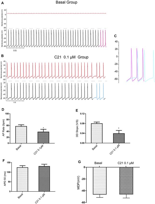
Using dual current patch clamp, Ang II was delivered DISCUSSION
intracellularly into mESC-CMs. iAng II (10–7 M) decreased the
pacemaker activity of mESC-CMs (Figures 4A–C). iAng II The present study provides significant new insights into the role of
reduced the AP rate (Figure 4D) and decreased the DD slope extracellular and intracellular Ang II in the regulation of automaticity
(Figure 4E) without affecting the APD50 (Figure 4F) and MDP of CMs. Our main findings include the following: 1) iAng II could be
(Figure 4G); intracellular delivery of the solvent did not exert any detected in both NRVMs and mESC-CMs, 2) AT1R is located on the
effect (Supplementary Figures S4). Unexpectedly, intracellular plasma membrane, while AT2R is predominately located on the
delivery of AT1R blocker losartan (50 μM) and AT2R blocker nucleus and the SR of developing CMs, 3) extracellular Ang II
PD123319 (10 μM) alone did not affect the APs of mESC-CMs increases APs in a transient manner through the AT1R-dependent
(Supplementary Figures S5, S6), hinting that endogenously pathway, 4) iAng II decreases APs in a persistent manner through the
produced Ang II in an unstimulated condition may not be AT2R-dependent pathway, and 5) iAng II regulates the automaticity
present in significant amounts to activate the AT1R- or of developing CMs by decreasing the activity of RyR2.
AT2R-dependent pathway. Interestingly, intracellular delivery
of AT2R activator C21 (0.1 µM) (Wan et al., 2004)
significantly decreased the APs of mESC-CMs (Figure 5), Intracellular Ang II Can Be Detected in
indicating that activation of AT2R would decrease APs. Similar Developing Cardiomyocytes
results were obtained when NRVMs were used (Supplementary Increasing evidence revealed that Ang II may function as an
Figures S7, S8). Since a decent amount of iAng II and the intracellular peptide to activate intracellular/nuclear receptors and
activation of intracellular AT2R would similarly decrease the subsequently activates downstream signaling effectors independent
APs of developing CMs, our results suggested that iAng II of cell surface receptors in CMs. However, previous studies focused
activates intracellular AT2R, leading to a decrease in APs in CMs. on the iAng II function in CMs were restricted to nonspecific
detection approaches because of the lack of the specific Ang II
Effect of Intracellular Ang II on the antibody and its low concentration in CMs. In our study, we first
Automaticity of Mouse Embryonic Stem built up a detection system (including sample collection, purification,
concentration, and detection by LC-MS) to detect and to quantify
Cell–Derived Cardiomyocytes Was the iAng II in developing CMs. Our results showed that the
Associated With the Activity of Ryanodine concentration of iAng II in NRVMs was 0.63 pmol/mg, which
Receptor Isoform 2 was similar to that detected by ELISA in rat CMs (Singh et al., 2008).
The data presented so far suggested that the iAng II signaling Previous reports suggested that Ang II can be produced
pathway is different from the signaling pathway of extracellular endogenously in different cell types, including adult CMs
Ang II and indicated that iAng II exerts its effect predominantly (Baker et al., 1992; Malhotra et al., 1999; Paul et al., 2006;
through AT2R. Since AT2R is mainly located on the SR, we Singh et al., 2007; Tsai et al., 2008). However, whether Ang II
speculated that iAng II may decrease the APs by regulating the can be produced locally in early developing CMs and, if yes, what
SR Ca2+ release channel RyR2. Caffeine can trigger Ca2+ release by its role is in regulating the unique characteristic automaticity of
reducing the threshold for luminal Ca2+ activation of RyR2, which is early developing CMs and sinoatrial cells are unknown. Our
usually used to examine the activity of RyR2 (Kong et al., 2008). To results revealed that endogenous Ang II is present in mESC-CMs.
Frontiers in Molecular Biosciences | www.frontiersin.org 8 August 2021 | Volume 8 | Article 699827Qi et al. Ang II Regulates Automaticity of Cardiomyocytes FIGURE 4 | iAng II decreased the APs of mESC-CMs in a persistent manner. (A) Dual current patch clamp of APs of mESC-CMs without intracellular drug delivery (i.e., before membrane breakage by the “drug electrode”). Upper panel represents recording in the “drug electrode” while the lower panel represents recording in the “recording electrode.” (B) Dual current patch clamp of APs of mESC-CMs with intracellular delivery of Ang II (i.e., after membrane breakage by the “drug electrode”). Upper panel represents recording in the “drug electrode” while the lower panel represents recording in the “recording electrode.” (C) Traces labeled as pink in (A) (represents APs before iAng II delivery) and as blue in (B) (represents APs after iAng II delivery) are overlaid for comparison. (D–G) Summarized data on the (D) AP rate, (E) DD slope, (F) APD 50, and (G) MDP of APs upon treatment with iAng II. Values are mean ± SEM of 6–10 independent experiments. *p < 0.05 vs. control group. Frontiers in Molecular Biosciences | www.frontiersin.org 9 August 2021 | Volume 8 | Article 699827
Qi et al.                                                                                                                      Ang II Regulates Automaticity of Cardiomyocytes

  FIGURE 5 | Intracellular delivery of AT2R activator C21 decreased the APs of mESC-CMs. (A) Dual current patch clamp of APs of mESC-CMs without intracellular
  drug delivery (i.e., before membrane breakage by the “drug electrode”). Upper panel represents recording in the “drug electrode” while the lower panel represents
  recording in the “recording electrode.” (B) Dual current patch clamp of APs of mESC-CMs with intracellular delivery of C21 (i.e., after membrane breakage by the “drug
  electrode”). Upper panel represents recording in the “drug electrode” while the lower panel represents recording in the “recording electrode.” (C) Traces labeled as
  pink in (A) (represents APs before intracellular C21 delivery) and as blue in (B) (represents APs after intracellular intracellular C21 delivery) are overlaid for comparison.
  (D–G) Summarized data on the (D) AP rate, (E) DD slope, (F) APD50, and (G) MDP of APs upon treatment with intracellular C21. Values are mean ± SEM of 6–10
  independent experiments. *p < 0.05 vs. control group.

Frontiers in Molecular Biosciences | www.frontiersin.org                                10                                              August 2021 | Volume 8 | Article 699827
Qi et al.                                                                                                              Ang II Regulates Automaticity of Cardiomyocytes

  FIGURE 6 | iAng II attenuated caffeine-induced changes of APs and caffeine-induced Ca2+ release in mESC-CMs. (A) Raw traces of spontaneous APs at the basal
  level and upon treatment with caffeine (5 μM) in mESC-CMs. (B) Raw traces of spontaneous APs upon treatment with iAng II (10–7 M) followed by the subsequent
  application of caffeine in mESC-CMs. (C–F) Summarized data on the (C) AP rate, (D) DD slope, (E) APD50, and (F) MDP upon treatment with caffeine (5 μM) alone or
  upon treatment with iAng II (10–7 M) followed by caffeine (5 μM). (G) Raw traces of changes in Fluo-4 fluorescence at the basal level and upon treatment with
  caffeine (5 μM) in mESC-CMs. (H) Raw traces of changes in Fluo-4 fluorescence with caffeine in the presence of iAng II (10–7 M) in mESC-CMs. (I) Summarized data on
  the effects of iAng II (10–7 M) on the caffeine-induced Ca2+ release in mESC-CMs when external Ca2+ was absent. Values are mean ± SEM of 6–10 independent
  experiments. *p < 0.05 vs. control group.

Frontiers in Molecular Biosciences | www.frontiersin.org                          11                                          August 2021 | Volume 8 | Article 699827
Qi et al.                                                                                                    Ang II Regulates Automaticity of Cardiomyocytes

This endogenous Ang II is unlikely to be attributed to the entry of              signaling mechanism (George et al., 2010; Savoia et al., 2011)
extracellular Ang II, as the uptake of exogenously applied                       (Figure 7). On the contrary, the role of iAng II, the identity of
fluorescently labeled Ang II by mESC-CMs was not observed                         AT2R, and the signaling pathway through which iAng II/AT2R
after 36 h of application. This result indicated that mESC-CMs                   mediate their effect are still under extensive investigation.
can probably synthesize their own Ang II.                                            Several previous reports have shown that iAng II implemented its
    In addition to synthesizing their own Ang II, the two distinct               effects via AT2R (Tadevosyan et al., 2015; Zhao et al., 2015). In CMs,
subtypes of Ang II receptors, AT1R and AT2R, were both found to be               iAng II has been shown to interact with nuclear AT2R and regulate
present in the developing CMs; AT1R is present on the cell surface               RNA synthesis, cell proliferation, and collagen secretion (Singh et al.,
membrane, while AT2R is present on the nucleus and the SR. After                 2008). Our results suggest that iAng II would act through AT2R and
decades of research, many components of the RAS, including                       negatively regulate automaticity on a much shorter timescale.
angiotensinogen, angiotensin converting enzyme, Ang II, and                          On the other hand, our results indicated that iAng II negatively
Ang II receptors, have been detected in the whole heart where                    regulates RyR2 and controls APs in CMs. Since iAng II also
myocytes and non-myocytes are present (Serneri et al., 2001; van                 concomitantly mediates its effect via AT2R, it is reasonable to
Kats et al., 2001; Danser, 2003). However, it is unclear whether these           speculate that binding of iAng II to AT2R would decrease the
components are synthesized locally in the CMs. For instance, the                 activity of RyR2. However, whether iAng II/AT2R can directly
origin of renin, which is required for the generation of local Ang II, is        couple to RyR2 and downregulate its activity, or whether further
still unclear. Therefore, the exact synthesis mechanism for the iAng             downstream signaling is involved, is unknown. Interestingly, AT2R
II in CMs has remained an unanswered question. We speculate that                 was first described as a thiol-potentiated Ang II receptor in 1989.
the local Ang II production in developing CMs may be the result of a             Previous studies reported that AT2R may exist in the nuclear
combination of RAS component uptake from the extracellular                       membrane and the mitochondrial inner membrane. However, in
environment, local synthesis of the RAS components, and in-site                  comparison to AT1R, the functions of AT2R are relatively
synthesis of iAng II. On the other hand, while it was previously                 unexplored. Some studies reported that AT2R can increase nitric
technically challenging to detect genuine iAng II due to the                     oxide (NO) release, reduce vascular tone, and attenuate ischemia
potentially nonspecific method and its extremely low intracellular                reperfusion injury in the myocardium (Mendoza-Torres et al., 2018;
level, the UHPLC-ESI-MS/MS method developed in our current                       Paz Ocaranza et al., 2020). NO has a function to diminish the
study has solidly proven the existence of iAng II in developing CMs.             opening probability of RyR2, which is a mechanism that could be
                                                                                 cardioprotective (Lim et al., 2008). Thus, the pathway AT2R-NO-
Functional Intracellular Ang II–Ang II Type 2                                    RyR2 may be a potential mechanism by which AT2R inhibits RyR2.
Receptor–Ryanodine Receptor Isoform 2                                            Further studies would be needed to investigate how AT2R can
Pathway Is Present in Cardiomyocytes,                                            modulate the activity of RyR2.
                                                                                     While at a physiological level, it has been known for a long
Counteracting the Effect of Extracellular                                        time that an increase in the intracellular Ca2+ level potentiates the
Ang II in Regulating the Automaticity of                                         activity of RyR2 (in the classical Ca2+-induced Ca2+ release
Cardiomyocytes                                                                   mechanism) and that at the pharmacological level, the
With components of local RAS being present, Ang II shall regulate                activators and inhibitors of RyR2 are well studied, knowledge
some of the functions of these developing CMs. Previous studies                  about physiological inhibitors of RyR2 remains scarce. In our
have shown that extracellular Ang II increased the amplitude of                  present study, we found that iAng II, probably by activating AT2R
CaTs and the contraction amplitude in paced ESC-CMs or in                        at the SR, decreased the Ca2+ release from RyR2. However,
embryoid bodies containing CMs (Sedan et al., 2008; Lagerqvist                   whether iAng II/AT2R can directly couple to RyR2 and
et al., 2011). Automaticity is the unique feature of developing CMs.             downregulate its activity, or whether further downstream
Our study showed that extracellular Ang II increased the AP rate and             signaling is involved, is unknown. Further study shall
the DD slope of the developing CMs through the AT1R-dependent                    concentrate on elucidating the mechanism behind this
pathway. Importantly, our study revealed that iAng II that is                    potentially important signaling pathway (Figure 7).
produced endogenously would exert the opposite effect on                             Some previous reports have documented that activation of
spontaneous APs; iAng II decreased both the AP rate and the                      AT1R and AT2R would lead to the opposite effect in the
DD slope of APs. Interestingly, our results showed that                          cardiovascular system (Jones et al., 2008; Padia and Carey,
intracellular delivery of AT2R activator C21 exerted a similar                   2013; Lagatta et al., 2018); however, there is no study clearly
effect to that of iAng II, suggesting that iAng II acts through                  documenting whether extracellular Ang II and iAng II would
AT2R. In addition, iAng II attenuated the effect of the activation               exert an opposing effect or not. Our study is the first study clearly
of RyR2 and decreased the Ca2+ released from the RyR2. Previous                  demonstrating that extracellular Ang II and iAng II activate AT1R
studies on automaticity of sinoatrial nodal cells showed that a                  and AT2R, respectively, to exert opposing cellular functions.
decrease in RyR2 activity would decrease the AP rate (Rigg et al.,
2000). Our novel results suggest that iAng II decreases the activity of
RyR2, leading to the decrease in APs.                                            CONCLUSION
    AT1R is a typical seven-transmembrane GPCR. It was widely
reported that the majority of extracellular Ang II–evoked cellular               In conclusion, our study has invented a novel UHPLC-ESI-MS/MS
responses are mediated through the activation of the “classical”                 method to solidly reveal the existence of iAng II in developing CMs.

Frontiers in Molecular Biosciences | www.frontiersin.org                    12                                      August 2021 | Volume 8 | Article 699827
Qi et al. Ang II Regulates Automaticity of Cardiomyocytes
FIGURE 7 | Schematic representation of the different signaling pathways that extracellular Ang II and iAng II utilize to regulate the automaticity of CMs. Extracellular
Ang II, upon binding to AT1R, leads to the production of DAG and IP3, which open TRPC3/C6 and subsequently LTCC and IP3R, respectively, and increases the
automaticity of CMs. On the other hand, binding of iAng II to AT2R decreases the automaticity of CMs by reducing the activity of RyR2.
Our study has also shown that AT1R is located on the plasma investigation; HS, Z-FC, CL, XY, and SYT: resources; ZQ, TW,
membrane, while AT2R is predominately located on the nucleus and XC, CW, and QD: data curation; ZQ and SYT: writing—original
the SR of developing CMs. In addition, our study revealed that draft preparation; ZQ and SYT: writing—review and editing; HS,
extracellular Ang II regulates APs through AT1R in a transient Z-FC, CL, XY, and SYT: supervision; ZQ and SYT: project
manner. Importantly, by employing the novel and technically administration; ZQ, XY, and SYT: funding acquisition. All
challenging dual patch clamp technique and the calcium imaging authors have read and agreed to the published version of the
technique, we clearly showed that iAng II negatively regulates the manuscript.
automaticity of developing CMs through the AT2R-RyR2 pathway.
The present investigation has uncovered the vital role of iAng II and
provided the potential mechanisms of how Ang II regulates cardiac FUNDING
automaticity.
This work was supported by the General Research Fund (474913
and 14176817) from the University Grants Committee (UGC) of
DATA AVAILABILITY STATEMENT the Hong Kong SAR, the National Natural Science Foundation of
China (21806025), the Germany/Hong Kong Joint Research
The original contributions presented in the study are included in Scheme (G-CUHK409/13) from the German Academic
the article/Supplementary Material; further inquiries can be Exchange Service (DAAD) and the UGC of the Hong Kong
directed to the corresponding author. SAR, and the Innovative Technology Fund of the Innovation
Technology Commission: Funding Support to State Key
Laboratory of Agrobiotechnology. The APC was funded by the
ETHICS STATEMENT School of Life Sciences, the Chinese University of Hong Kong. ZQ
and QD were supported by the postgraduate studentship from
The animal study was reviewed and approved by the Animal the CUHK.
Ethics Committee, the Chinese University of Hong Kong.
SUPPLEMENTARY MATERIAL
AUTHOR CONTRIBUTIONS
The Supplementary Material for this article can be found online at:
ZQ and SYT: conceptualization; ZQ, TW, XC, CW, and QD: https://www.frontiersin.org/articles/10.3389/fmolb.2021.699827/
methodology; ZQ: formal analysis; ZQ, TW, XC, CW, and QD: full#supplementary-material
Frontiers in Molecular Biosciences | www.frontiersin.org 13 August 2021 | Volume 8 | Article 699827Qi et al. Ang II Regulates Automaticity of Cardiomyocytes
REFERENCES Myocardial Infarction on Cardiac Differentiation of Embryonic Stem Cells.
Int. J. Cardiol. 168 (4), 3458–3472. doi:10.1016/j.ijcard.2013.04.178
Lim, G., Venetucci, L., Eisner, D. A., and Casadei, B. (2008). Does Nitric Oxide
Akazawa, H., Yano, M., Yabumoto, C., Kudo-Sakamoto, Y., and Komuro, I. (2013). Modulate Cardiac Ryanodine Receptor Function? Implications for Excitation-
Angiotensin II Type 1 and Type 2 Receptor-Induced Cell Signaling. Cpd 19 Contraction Coupling. Cardiovasc. Res. 77 (2), 256–264. doi:10.1093/cvr/
(17), 2988–2995. doi:10.2174/1381612811319170003 cvm012
Asada, H., Horita, S., Hirata, K., Shiroishi, M., Shiimura, Y., Iwanari, H., et al. Lokuta, A. J., Cooper, C., Gaa, S. T., Wang, H. E., and Rogers, T. B. (1994).
(2018). Crystal Structure of the Human Angiotensin II Type 2 Receptor Bound Angiotensin II Stimulates the Release of Phospholipid-Derived Second
to an Angiotensin II Analog. Nat. Struct. Mol. Biol. 25 (7), 570–576. Messengers through Multiple Receptor Subtypes in Heart Cells. J. Biol.
doi:10.1038/s41594-018-0079-8 Chem. 269 (7), 4832–4838. doi:10.1016/S0021-9258(17)37619-6
Baker, K. M., Booz, G. W., and Dostal, D. E. (1992). Cardiac Actions of Angiotensin Malhotra, R., Sadoshima, J., Brosius, F. C., and Izumo, S. (1999). Mechanical
II: Role of an Intracardiac Renin-Angiotensin System. Annu. Rev. Physiol. 54, Stretch and Angiotensin II Differentially Upregulate the Renin-Angiotensin
227–241. doi:10.1146/annurev.ph.54.030192.001303 System in Cardiac Myocytes In Vitro. Circ. Res. 85 (2), 137–146. doi:10.1161/
Cheng, H., and Lederer, W. J. (2008). Calcium sparks. Physiol. Rev. 88 (4), 01.Res.85.2.137
1491–1545. doi:10.1152/physrev.00030.2007 Mendoza-Torres, E., Riquelme, J. A., Vielma, A., Sagredo, A. R., Gabrielli, L.,
Danser, A. H. (2003). Local Renin-Angiotensin Systems: the Unanswered Bravo-Sagua, R., et al. (2018). Protection of the Myocardium against Ischemia/
Questions. Int. J. Biochem. Cel Biol 35 (6), 759–768. doi:10.1016/s1357- reperfusion Injury by Angiotensin-(1-9) through an AT2R and Akt-dependent
2725(02)00178-4 Mechanism. Pharmacol. Res. 135, 112–121. doi:10.1016/j.phrs.2018.07.022
Danser, A., Saris, J. J., Schuijt, M. P., and van Kats, J. P. (1999). Is There a Local Monfredi, O., Maltsev, V. A., and Lakatta, E. G. (2013). Modern Concepts
Renin-Angiotensin System in the Heart?. Cardiovasc. Res. 44 (2), 252–265. Concerning the Origin of the Heartbeat. Physiology 28 (2), 74–92.
doi:10.1016/S0008-6363(99)00202-3 doi:10.1152/physiol.00054.2012
de Gasparo, M., Catt, K. J., Inagami, T., Wright, J. W., and Unger, T. (2000). Muller, J., Corodimas, K. P., Fridel, Z., and LeDoux, J. E. (1997). Functional
International union of Pharmacology. XXIII. The Angiotensin II Receptors. Inactivation of the Lateral and Basal Nuclei of the Amygdala by Muscimol
Pharmacol. Rev. 52, 415–472. Infusion Prevents Fear Conditioning to an Explicit Conditioned Stimulus and
De Mello, W. C. (2015). Intracellular Angiotensin II Disrupts Chemical to Contextual Stimuli. Behav. Neurosci. 111 (4), 683–691. doi:10.1037/0735-
Communication and Impairs Metabolic Cooperation between Cardiac 7044.111.4.683
Myocytes. Peptides 72, 57–60. doi:10.1016/j.peptides.2015.04.001 Ng, S.-Y., Chin, C.-H., Lau, Y.-T., Luo, J., Wong, C.-K., Bian, Z.-X., et al. (2010).
Gallinat, S., Busche, S., Schütze, S., Krönke, M., and Unger, T. (1999). AT2 Receptor Role of Voltage-Gated Potassium Channels in the Fate Determination of
Stimulation Induces Generation of Ceramides in PC12W Cells. Febs Lett. 443 Embryonic Stem Cells. J. Cel. Physiol. 224 (1), a–n. doi:10.1002/jcp.22113
(1), 75–79. doi:10.1016/s0014-5793(98)01675-5 Onohara, N., Nishida, M., Inoue, R., Kobayashi, H., Sumimoto, H., Sato, Y., et al.
George, A. J., Thomas, W. G., and Hannan, R. D. (2010). The Renin-Angiotensin (2006). TRPC3 and TRPC6 Are Essential for Angiotensin II-Induced Cardiac
System and Cancer: Old Dog, New Tricks. Nat. Rev. Cancer 10 (11), 745–759. Hypertrophy. Embo J. 25 (22), 5305–5316. doi:10.1038/sj.emboj.7601417
doi:10.1038/nrc2945 Padia, S. H., and Carey, R. M. (2013). AT2 Receptors: Beneficial Counter-
Harada, K., Sugaya, T., Murakami, K., Yazaki, Y., and Komuro, I. (1999). regulatory Role in Cardiovascular and Renal Function. Pflugers Arch. - Eur.
Angiotensin II Type 1A Receptor Knockout Mice Display Less Left J. Physiol. 465 (1), 99–110. doi:10.1007/s00424-012-1146-3
Ventricular Remodeling and Improved Survival after Myocardial Infarction. Paul, M., Poyan Mehr, A., and Kreutz, R. (2006). Physiology of Local Renin-
Circulation 100 (20), 2093–2099. doi:10.1161/01.Cir.100.20.2093 Angiotensin Systems. Physiol. Rev. 86 (3), 747–803. doi:10.1152/
Inuzuka, T., Fujioka, Y., Tsuda, M., Fujioka, M., Satoh, A. O., Horiuchi, K., et al. physrev.00036.2005
(2016). Attenuation of Ligand-Induced Activation of Angiotensin II Type 1 Paz Ocaranza, M., Riquelme, J. A., García, L., Jalil, J. E., Chiong, M., Santos, R. A. S.,
Receptor Signaling by the Type 2 Receptor via Protein Kinase C. Sci. Rep. 6. et al. (2020). Counter-regulatory Renin-Angiotensin System in Cardiovascular
doi:10.1038/srep21613 Disease. Nat. Rev. Cardiol. 17 (2), 116–129. doi:10.1038/s41569-019-0244-8
Jones, E. S., Vinh, A., McCarthy, C. A., Gaspari, T. A., and Widdop, R. E. (2008). Qi, Z., Wong, C. K., Suen, C. H., Wang, J., Long, C., Sauer, H., et al. (2016). TRPC3
AT2 Receptors: Functional Relevance in Cardiovascular Disease. Pharmacol. Regulates the Automaticity of Embryonic Stem Cell-Derived Cardiomyocytes.
Ther. 120 (3), 292–316. doi:10.1016/j.pharmthera.2008.08.009 Int. J. Cardiol. 203, 169–181. doi:10.1016/j.ijcard.2015.10.018
Karnik, S. S., Unal, H., Kemp, J. R., Tirupula, K. C., Eguchi, S., Vanderheyden, P. M. Rigg, L., Heath, B. M., Cui, Y., and Terrar, D. A. (2000). Localisation and
L., et al. (2015). International Union of Basic and Clinical Pharmacology. XCIX. Functional Significance of Ryanodine Receptors during β-adrenoceptor
Angiotensin Receptors: Interpreters of Pathophysiological Angiotensinergic Stimulation in the guinea-pig Sino-Atrial Node. Cardiovasc. Res. 48 (2),
Stimuli. Pharmacol. Rev. 67 (4), 754–819. doi:10.1124/pr.114.010454 254–264. doi:10.1016/s0008-6363(00)00153-x
Kong, H., Jones, P. P., Koop, A., Zhang, L., Duff, H. J., and Chen, S. R. W. (2008). Savoia, C., Burger, D., Nishigaki, N., Montezano, A., and Touyz, R. M. (2011).
Caffeine Induces Ca2+ Release by Reducing the Threshold for Luminal Ca2+ Angiotensin II and the Vascular Phenotype in Hypertension. Expert Rev. Mol.
Activation of the Ryanodine Receptor. Biochem. J. 414 (3), 441–452. Med. 13. doi:10.1017/S1462399411001815
doi:10.1042/BJ20080489 Sedan, O., Dolnikov, K., Zeevi-Levin, N., Leibovich, N., Amit, M., Itskovitz-Eldor,
Lagatta, D. C., Kuntze, L. B., Ferreira-Junior, N. C., and Resstel, L. B. M. (2018). J., et al. (2008). 1,4,5-Inositol Trisphosphate-Operated Intracellular Ca2+Stores
Medial Prefrontal Cortex TRPV1 and CB1 Receptors Modulate Cardiac and Angiotensin-II/Endothelin-1 Signaling Pathway Are Functional in Human
Baroreflex Activity by Regulating the NMDA Receptor/nitric Oxide Embryonic Stem Cell-Derived Cardiomyocytes. Stem Cells 26 (12), 3130–3138.
Pathway. Pflugers Arch. - Eur. J. Physiol. 470 (10), 1521–1542. doi:10.1007/ doi:10.1634/stemcells.2008-0777
s00424-018-2149-5 Senbonmatsu, T., Saito, T., Landon, E. J., Watanabe, O., Price, E., Roberts, R. L.,
Lagerqvist, E. L., Finnin, B. A., Pouton, C. W., and Haynes, J. M. (2011). et al. (2003). A Novel Angiotensin II Type 2 Receptor Signaling Pathway:
Endothelin-1 and Angiotensin II Modulate Rate and Contraction Amplitude Possible Role in Cardiac Hypertrophy. EMBO J. 22 (24), 6471–6482.
in a Subpopulation of Mouse Embryonic Stem Cell-Derived Cardiomyocyte- doi:10.1093/emboj/cdg637
Containing Bodies. Stem Cel Res. 6 (1), 23–33. doi:10.1016/j.scr.2010.09.001 Serneri, G. G. N., Boddi, M., Cecioni, I., Vanni, S., Coppo, M., Papa, M. L., et al.
Lakatta, E. G., Maltsev, V. A., and Vinogradova, T. M. (2010). A Coupled SYSTEM (2001). Cardiac Angiotensin II Formation in the Clinical Course of Heart
of Intracellular Ca 2+ Clocks and Surface Membrane Voltage Clocks Controls Failure and its Relationship with Left Ventricular Function. Circ. Res. 88 (9),
the Timekeeping Mechanism of the Heart’s Pacemaker. Circ. Res. 106 (4), 961–968. doi:10.1161/hh0901.089882
659–673. doi:10.1161/CIRCRESAHA.109.206078 Singh, V. P., Le, B., Bhat, V. B., Baker, K. M., and Kumar, R. (2007). High-glucose-
Law, S. K., Leung, C. S.-L., Yau, K. L., Tse, C. L., Wong, C. K., Leung, F. P., et al. induced Regulation of Intracellular ANG II Synthesis and Nuclear
(2013). Regulation of Multiple Transcription Factors by Reactive Oxygen Redistribution in Cardiac Myocytes. Am. J. Physiology-Heart Circulatory
Species and Effects of Pro-inflammatory Cytokines Released during Physiol. 293 (2), H939–H948. doi:10.1152/ajpheart.00391.2007
Frontiers in Molecular Biosciences | www.frontiersin.org 14 August 2021 | Volume 8 | Article 699827Qi et al. Ang II Regulates Automaticity of Cardiomyocytes Singh, V. P., Le, B., Khode, R., Baker, K. M., and Kumar, R. (2008). Intracellular Wong, C.-K., So, W.-Y., Law, S.-K., Leung, F.-P., Yau, K.-L., Yao, X., et al. (2012). Angiotensin II Production in Diabetic Rats Is Correlated with Cardiomyocyte Estrogen Controls Embryonic Stem Cell Proliferation via Store-Operated Apoptosis, Oxidative Stress, and Cardiac Fibrosis. Diabetes 57 (12), 3297–3306. Calcium Entry and the Nuclear Factor of Activated T-Cells (NFAT). J. Cel. doi:10.2337/db08-0805 Physiol. 227 (6), 2519–2530. doi:10.1002/jcp.22990 Tadevosyan, A., Létourneau, M., Folch, B., Doucet, N., Villeneuve, L. R., Mamarbachi, A. Yusuf, S., Yusuf, S., Sleight, P., Pogue, J., Bosch, J., Davies, R., et al. (2000). Effects of M., et al. (2015). Photoreleasable Ligands to Study Intracrine Angiotensin II an Angiotensin-Converting-Enzyme Inhibitor, Ramipril, on Cardiovascular Signalling. J. Physiol. 593 (3), 521–539. doi:10.1113/jphysiol.2014.279109 Events in High-Risk Patients. N. Engl. J. Med. 342 (3), 145–153. doi:10.1056/ Tadevosyan, A., Maguy, A., Villeneuve, L. R., Babin, J., Bonnefoy, A., Allen, B. G., nejm200001203420301 et al. (2010). Nuclear-delimited Angiotensin Receptor-Mediated Signaling Zaman, M. A., Oparil, S., and Calhoun, D. A. (2002). Drugs Targeting the Renin- Regulates Cardiomyocyte Gene Expression. J. Biol. Chem. 285 (29), Angiotensin-Aldosterone System. Nat. Rev. Drug Discov. 1 (8), 621–636. 22338–22349. doi:10.1074/jbc.M110.121749 doi:10.1038/nrd873 Tadevosyan, A., Xiao, J., Surinkaew, S., Naud, P., Merlen, C., Harada, M., et al. Zhao, Y., Lützen, U., Fritsch, J., Zuhayra, M., Schütze, S., Steckelings, U. M., et al. (2017). Intracellular Angiotensin-II Interacts with Nuclear Angiotensin (2015). Activation of Intracellular Angiotensin AT2 Receptors Induces Rapid Receptors in Cardiac Fibroblasts and Regulates RNA Synthesis, Cell Cell Death in Human Uterine Leiomyosarcoma Cells. Clin. Sci. 128 (9), Proliferation, and Collagen Secretion. Jaha 6 (4). doi:10.1161/jaha.116.004965 567–578. doi:10.1042/cs20140627 Thomas, W. G., and Mendelsohn, F. A. O. (2003). Angiotensin Receptors: Form and Function and Distribution. Int. J. Biochem. Cel Biol. 35 (6), 774–779. Conflict of Interest: The authors declare that the research was conducted in the doi:10.1016/s1357-2725(02)00263-7 absence of any commercial or financial relationships that could be construed as a Tsai, C.-T., Lai, L.-P., Hwang, J.-J., Chen, W.-P., Chiang, F.-T., Hsu, K.-L., et al. potential conflict of interest. (2008). Renin-angiotensin System Component Expression in the HL-1 Atrial Cell Line and in a Pig Model of Atrial Fibrillation. J. Hypertens. 26 (3), 570–582. Publisher’s Note: All claims expressed in this article are solely those of the authors doi:10.1097/HJH.0b013e3282f34a4a and do not necessarily represent those of their affiliated organizations, or those of van Kats, J. P., Methot, D., Paradis, P., Silversides, D. W., and Reudelhuber, T. L. the publisher, the editors, and the reviewers. Any product that may be evaluated in (2001). Use of a Biological Peptide Pump to Study Chronic Peptide Hormone this article, or claim that may be made by its manufacturer, is not guaranteed or Action in Transgenic Mice. J. Biol. Chem. 276 (47), 44012–44017. doi:10.1074/ endorsed by the publisher. jbc.M106132200 Wan, Y., Wallinder, C., Plouffe, B., Beaudry, H., Mahalingam, A. K., Wu, X., et al. Copyright © 2021 Qi, Wang, Chen, Wong, Ding, Sauer, Chen, Long, Yao, Cai and (2004). Design, Synthesis, and Biological Evaluation of the First Selective Tsang. This is an open-access article distributed under the terms of the Creative Nonpeptide AT2 Receptor Agonist. J. Med. Chem. 47 (24), 5995–6008. Commons Attribution License (CC BY). The use, distribution or reproduction in doi:10.1021/jm049715t other forums is permitted, provided the original author(s) and the copyright owner(s) Wollert, K. C., Taga, T., Saito, M., Narazaki, M., Kishimoto, T., Glembotski, C. C., are credited and that the original publication in this journal is cited, in accordance et al. (1996). Cardiotrophin-1 Activates a Distinct Form of Cardiac Muscle Cell with accepted academic practice. No use, distribution or reproduction is permitted Hypertrophy. J. Biol. Chem. 271 (16), 9535–9545. doi:10.1074/jbc.271.16.9535 which does not comply with these terms. Frontiers in Molecular Biosciences | www.frontiersin.org 15 August 2021 | Volume 8 | Article 699827
You can also read