Long-Term Protection Elicited by a DNA Vaccine Candidate Expressing the prM-E Antigen of Dengue Virus Serotype 3 in Mice - Frontiers
←
→
Page content transcription
If your browser does not render page correctly, please read the page content below
ORIGINAL RESEARCH
                                                                                                                                                   published: 17 March 2020
                                                                                                                                             doi: 10.3389/fcimb.2020.00087

                                              Long-Term Protection Elicited by a
                                              DNA Vaccine Candidate Expressing
                                              the prM-E Antigen of Dengue Virus
                                              Serotype 3 in Mice
                                              Kaihao Feng 1 , Xiaoyan Zheng 2 , Ran Wang 1 , Na Gao 1 , Dongying Fan 1 , Ziyang Sheng 1 ,
                                              Hongning Zhou 3 , Hui Chen 1* and Jing An 1,4*
                                              1
                                               Department of Microbiology and Parasitology, School of Basic Medical Sciences, Capital Medical University, Beijing, China,
                                              2
                                               Beijing Tropical Medicine Research Institute, Beijing Friendship Hospital, Capital Medical University, Beijing, China, 3 Yunnan
                                              Provincial Key Laboratory of Vector-borne Disease Control and Research, Yunnan Institute of Parasitic Diseases, Pu’er,
                                              China, 4 Center of Epilepsy, Beijing Institute for Brain Disorders, Beijing, China

                                              Dengue virus (DENV) is the causative agent of dengue, and its incidence has increased
                                              30-fold in the past five decades. Among the four cocirculating serotypes, DENV3 is
                                              associated with an increased number of severe infections and has become widespread.
                          Edited by:
                                              Vaccination is the mainstay of prevention in reducing disease burden. Previously, the
                       Gong Cheng,            protective efficacy of DNA vaccine candidates toward DENV1, 2, and 4 was confirmed
            Tsinghua University, China
                                              in mice. In this study, a DNA vaccine candidate (pVAX1-D3ME) expressing the prM and
                         Reviewed by:
                                              E proteins of DENV3 was constructed, and then the immunogenicity and protection
                          Jianfeng Dai,
            Soochow University, China         were assessed in mice to further develop a tetravalent dengue vaccine. Moreover, the
                            Yongfen Xu,       cross-reactive immune responses against the other three serotypes were investigated.
           Institut Pasteur of Shanghai
                           (CAS), China
                                              The results showed that three doses of 50 µg of pVAX1-D3ME were sufficient to
                 *Correspondence:
                                              induce strong antigen-specific T cell responses and robust and consistent neutralizing
                          Hui Chen            antibodies. Additionally, immunization with pVAX1-D3ME offered protective immunity
            chenhuicxh@ccmu.edu.cn
                                              against not only DENV3 but also the other three serotypes, which could be observed
                           Jing An
                anjing@ccmu.edu.cn            even after 12 months. This study shows great promise for the further evaluation
                                              of a dengue tetravalent DNA vaccine candidate in large animal models, including
                    Specialty section:
                                              non-human primates.
          This article was submitted to
                         Virus and Host,      Keywords: dengue virus, DNA vaccine, prM-E, immunization, cross-protection
                 a section of the journal
      Frontiers in Cellular and Infection
                            Microbiology
                                              INTRODUCTION
       Received: 06 December 2019
        Accepted: 19 February 2020            Dengue virus (DENV) is the cause of dengue, which is widespread in tropical and subtropical
         Published: 17 March 2020             countries and affects 390 million people annually (Kularatne, 2015). The incidence of dengue
                              Citation:       infection has increased rapidly over the last 50 years along with the geographic expansion of
    Feng K, Zheng X, Wang R, Gao N,           the disease (Kraemer et al., 2019). It is estimated that Asia bore 70% of the disease burden
Fan D, Sheng Z, Zhou H, Chen H and            (Bhatt et al., 2013).
    An J (2020) Long-Term Protection
                                                 DENV belongs to the genus Flavivirus of the family Flaviviridae. Serologically, DENV can
 Elicited by a DNA Vaccine Candidate
       Expressing the prM-E Antigen of
                                              be subclassed into four distinct but closely related serotypes: DENV1, DENV2, DENV3, and
     Dengue Virus Serotype 3 in Mice.         DENV4. Infection with any one serotype presents with similar and indistinguishable clinical
  Front. Cell. Infect. Microbiol. 10:87.      manifestations that range from asymptomatic, undifferentiated fever to dengue fever, and more
       doi: 10.3389/fcimb.2020.00087          severe life-threatening diseases, including dengue hemorrhagic fever and dengue shock syndrome.

Frontiers in Cellular and Infection Microbiology | www.frontiersin.org                1                                                  March 2020 | Volume 10 | Article 87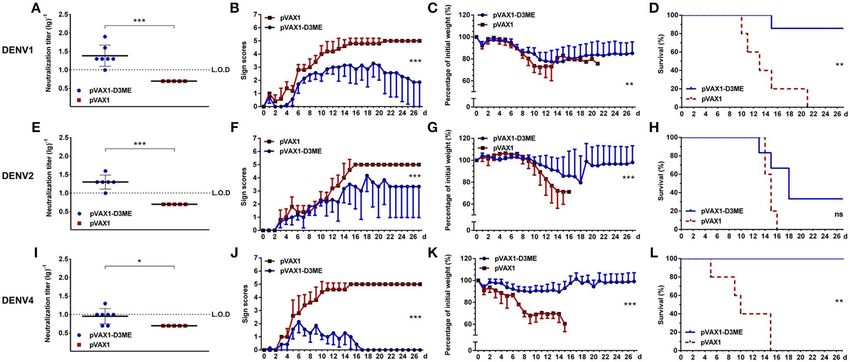
Feng et al.                                                                                                          A DNA Vaccine Against DENV3

    It should be noted that DENV3 from Southeast Asia area                   which is the unique America FDA-approved vector in clinical
causes the greatest percentage of severe cases in primary infection          trial. The breadth of the humoral and cellular immune responses
(Soo et al., 2016) and that DENV3 genotype III has been                      elicited by the vaccine candidate was investigated. Moreover, its
associated with a widespread global distribution of dengue fever             short-term and long-term protective efficacies against DENV3
(Tan et al., 2018). During 2016, a change in the circulating                 as well as the other three DENV serotypes were evaluated in a
serotype occurred, leading to dominance of DENV3 in India                    mouse model. The results indicated that three doses of pVAX1-
(Parveen et al., 2019). In 2017–2018, in Dhaka, Bangladesh, the              D3ME via in vivo electroporation induced effective humoral
largest dengue outbreak with a high frequency of severe dengue               and cellular immune responses and significantly protected mice
cases and high fatality could be traced back to the reemergence of           against lethal DENV3 challenge. Moreover, immunization with
DENV3 (Shirin et al., 2019).                                                 pVAX1-D3ME also provided cross-reactive protection against
    In China, DENV3 was the first serotype documented in                     DENV1, DENV2, or DENV4 challenge. This work will be
Guangdong in 1978 and was later isolated in Zhejiang in 2009                 of particular importance for the ongoing effort to develop a
and in Yunnan in 2013, including from severe cases (Lai et al.,              tetravalent dengue vaccine.
2015). In 2013, the first dengue outbreak in central China was
reported in Henan Province in the northern temperate regions
and was characterized by DENV3 predominance. The northward                   MATERIALS AND METHODS
shift of the dengue epidemic region is a warning that the endemic
range has expanded geographically in China (Huang et al., 2014;              Animals and Ethics Statement
Lai et al., 2015). In addition, reemerging DENV3 necessitates                Adult BALB/c mice were purchased from Vital River Laboratory
immediate public health attention.                                           Animal Technology Co., Ltd. (Beijing, China). One-day-old
    Currently, there is no widely used effective, specific                   BALB/c mice used for the neutralization assay were produced
prevention, and therapy against this severe disease. To resolve              by adult female mice. The animals were maintained in
this significant, international public health problem, considerable          specific-pathogen-free environments. The protocol for animal
effort has been directed toward the development of safe dengue               experiments was approved by the Institutional Animal Care and
vaccines. A number of candidates have been reported, including               Use Committee of Chinese Capital Medical University (ethics
inactivated vaccines, live-attenuated vaccines, DNA vaccines,                approval number AEEI-2015-066). In vitro and in vivo infections
and subunit protein vaccines (Eckels and Putnak, 2003;                       were performed in the biosafety level 2 laboratory. All animal
Whitehead, 2016; Bustos-Arriaga et al., 2018; Manoff et al.,                 experiments were performed under diethyl ether anesthesia. All
2019; Prompetchara et al., 2019; Wang et al., 2019). Of great                efforts were made to minimize suffering.
concern, the first and only licensed dengue vaccine, Dengvaxia,
had been approved for use in endemic areas owing to its higher               Cells and Viruses
efficacy among participants vaccinated at age ≥9 years (Ferguson             Vero cells (ATCC CRL-1586) were cultured in minimum
et al., 2016; Henein et al., 2017); however, a post-hoc analysis of          essential medium with 5% fetal bovine serum and 1% penicillin–
safety and efficacy reported a higher risk of severe dengue attack           streptomycin solution at 37◦ C with 5% CO2 . Aedes albopictus
and hospitalization in vaccinated persons who had not been                   C6/36 cells (ATCC CRL-1660) were grown in RPMI 1640
exposed to dengue (Sridhar et al., 2018). There continues to be              medium supplemented with 10% fetal bovine serum at 28◦ C.
a strong and urgent public health need for effective preventive                 The Hawaii strain of DENV1, the H87 strain of DENV3, and
interventions against dengue.                                                the H241 strain of DENV4 were provided by the Guangdong
    DENV has a single-stranded, positive-sense RNA genome                    Provincial CDC. The Tr1751 strain of DENV2 was isolated from
containing a single open reading frame that encodes three                    a patient with dengue fever. Viruses were propagated in C6/36
structural (capsid, C; premembrane, prM; and envelope, E) and                cells and titered in Vero cells by plaque forming assay.
seven non-structural (NS1, NS2A, NS2B, NS3, NS4A, NS4B, and                     Purified DENV3 particles were harvested from DENV3-
NS5) proteins. Exposed on the surface of mature DENV particles,              infected C6/36 cells and concentrated by 8% polyethylene glycol
the E glycoprotein protein is rich in immunological epitopes and             precipitation. Then, DENV3 particles were purified from clarified
contributes to the generation of effective protective immunity               extracts by ultracentrifugation.
(Putnak et al., 2003; Lin et al., 2012). The E protein assembly
is dependent on the prM protein expression (Oliveira et al.,
2017). Therefore, both the prM and E proteins have become                    Construction and Identification of
major antigen targets for vaccine design and development against             pVAX1-D3ME
DENVs and other flaviviruses (Guirakhoo et al., 2001; Mellado-               The prM and E regions of the H87 strain of DENV3 (GenBank
Sanchez et al., 2005; Osorio et al., 2014).                                  accession number AB609590, nucleotides 365–2,413 bp) were
    In our previous work, DNA vaccine candidates encoding the                amplified by PCR using the forward primer (5′ -CGG ATC GCT
prM and E proteins of DENV1 (Zheng et al., 2017), DENV2                      AGC ATG GCG ATG CTG AGC ATT ATC AAC-3′ ) and reverse
(Chen et al., 2016), or DENV4 (Sheng et al., 2019) could protect             primer (5′ -CAC ACA GGA TCC TTA AGC TTG CAC CAC
mice from lethal corresponding virus challenge. In this study,               GGC TCC CAG ATA-3′ ). Subsequently, the amplified fragment
another DNA vaccine, pVAX1-D3ME expressing the prM and E                     was subcloned into the pVAX1 vector (Invitrogen, USA) using
proteins of DENV3, was constructed in the same vector pVAX1,                 Nhe I and BamH I restriction sites, and the recombinant plasmid

Frontiers in Cellular and Infection Microbiology | www.frontiersin.org   2                                       March 2020 | Volume 10 | Article 87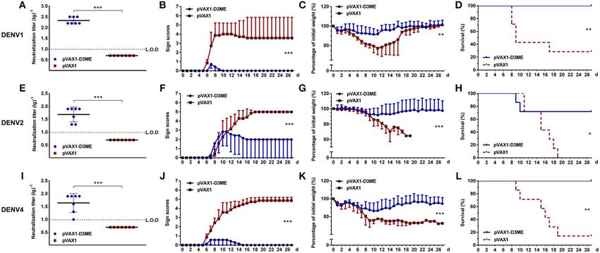
Feng et al.                                                                                                           A DNA Vaccine Against DENV3

was named pVAX1-D3ME, which was confirmed by double-                         added to a confluent monolayer of Vero cells in a 24-well plate
enzyme digestion and DNA sequencing. The pVAX1 vector                        and incubated at 37◦ C for another hour under gentle rocking
served as the negative control.                                              every 15 min. After washing, infected Vero cells were overlaid
                                                                             with medium containing 1.05% methylcellulose, followed by
Indirect Immunofluorescence (IFA) Staining                                   incubation at 37◦ C for 5–8 days. Finally, plaques were visualized
pVAX1-D3ME or pVAX1 was transfected into Vero cells                          by crystal violet diluent and counted. The reciprocal of the
using Lipofectamine 2000 (Thermo Fisher Scientific, USA).                    highest dilution that yielded a 50% reduction in the average
Transfected cells were fixed with 4% paraformaldehyde,                       number of plaques compared with the virus control wells was
permeabilized with 0.2% Triton X-100 in PBS, and blocked                     calculated as the neutralization titer (PRNT50 ).
with 1% bovine serum albumin. DENV3-infected mouse serum
(1:100) and FITC-conjugated goat anti-mouse IgG (1:200,
Immunotech, France) were used as the primary and secondary                   Enzyme-Linked Immunospot (ELISPOT)
antibodies, respectively. The cells were examined and imaged                 Assay
under a fluorescence microscope (Olympus BX61, Japan).                       The cytokines IFN-γ and IL-4 secreted by splenocytes were
                                                                             captured by antibodies and visualized as spots based on a
Western Blotting                                                             colorimetric reaction according to the manufacturer’s (BD, USA)
pVAX1-D3ME or pVAX1 was transfected into Vero cells                          recommendations. In brief, 96-well-filtration plates (Millipore,
using Lipofectamine 2000 (Thermo Fisher Scientific, USA).                    USA) were coated with the capture antibodies at 4◦ C overnight
Seventy-two hours after transfection, the supernatant and                    and then blocked with 1% bovine serum albumin at 37◦ C for
lysate were collected and fractionated by 12% SDS-PAGE and                   2 h. Splenocytes isolated from immunized mice were aliquoted
transferred to a polyvinylidene difluoride membrane according                at 5 × 10⁵ cells/well and stimulated with purified DENV3
to the manufacturer’s recommendation (Millipore, USA). The                   particles at a concentration of 5 µg/well at 37◦ C for 48 h. After
membrane was blocked with 5% bovine serum albumin for                        incubation with a biotinylated detection antibody, the spots
2 h and incubated with anti-flavivirus E protein monoclonal                  were visualized by adding streptavidin-HRP and AEC substrate
antibody (1:50, D1-4G2-4-15 hybridoma supernatant, ATCC                      and finally numerated automatically using an ELISPOT analyzer
HB-112) at 4◦ C overnight, and then probed with goat anti-                   (CTL, USA). Splenocytes cocultured with concanavalin A served
mouse HRP secondary antibody (1:4,000, abcam, China) for                     as a positive control, and those cultured with RPMI 1640 medium
1 h. Blots were developed with chemiluminescent HRP substrate                served as a negative control.
peroxide solution mix (Millipore, USA) and visualized with Li-
Cor Odyssey CLx imaging system.
                                                                             Flow Cytometry Analysis
Immunization                                                                 All antibodies for flow cytometry were purchased from BD
Six-week-old female BALB/c mice were randomly separated into                 Biosciences, USA. A total of 2 × 10⁶ splenic lymphocytes
two groups. A total of 50 µg of pVAX1-D3ME or pVAX1 was                      were blocked with rat anti-mouse CD16/CD32 monoclonal
injected into the quadriceps muscle, followed by six electric                antibody for 30 min. For surface staining, all cells were
pulses (36 V, 10 ms) using a gene delivery device (Terasa                    stained with anti-CD3e-FITC, anti-CD8-APC-H7, anti-CD44-
Healthcare Sci-Tech, China). The mice were boosted twice at                  APC, and anti-CD62L-PE at a standard dilution. However, for
three-week intervals with the inoculation. The immunization and              intracellular cytokine staining, the splenocytes were stimulated
sample collection schedule are shown in Figure 1A.                           with 1 µg of purified DENV3 particles for 24 h, and
                                                                             brefeldin A (10 µg/ml, Sigma, USA) was added at the
Immunohistochemistry (IHC) Staining                                          end of the six-hour incubation. After staining with anti-
The thigh muscles were isolated one week after the final                     CD3e-FITC, anti-CD8-APC-H7, and anti-CD11a-APC, the
immunization. Paraffin sections were processed by conventional               splenocytes were fixed and permeabilized using the BD
techniques as described previously (Chen et al., 2016).                      Cytofix/Cytoperm kit and stained with anti-IFN-γ-PE. Finally,
Convalescent serum derived from a DENV3-infected mouse                       the stained cells were read on a DxFLEX flow cytometer
was used at a 1:200 dilution as the primary antibody. HRP-                   (Beckman Coulter, USA) and analyzed by CytExpert software
conjugated goat anti-mouse IgG (Santa Cruz Biotechnology,                    (version 1.2).
USA) was diluted 1:2,000 and used as the secondary antibody.

Plaque Reduction Neutralization Test                                         In vitro Neutralizing and Passive Protective
(PRNT)                                                                       Effects of Immune Sera on Neonatal Mice
The neutralizing antibody (NAb) titer against DENV1–4 was                    After heat inactivation, pooled sera isolated from the DNA-
determined by PRNT. Serum samples were heated at 56◦ C for                   immunized mice were mixed with live DENV3 followed by
30 min to inactivate complement and then serially diluted from               incubation at 37◦ C for 1 h. Subsequently, 10 µl of the sera–
1:10 to 1:1,280. Next, 100 µl of diluted sera was mixed with                 virus mixture containing 100 PFU of DENV3 was gently injected
100 µl of DENV suspension containing 50 plaque-forming units                 intracranially into one-day-old neonatal mice. The body weight
(PFU) and then incubated at 37◦ C for 1 h. The mixture was                   and survival rate were monitored daily for 14 days.

Frontiers in Cellular and Infection Microbiology | www.frontiersin.org   3                                        March 2020 | Volume 10 | Article 87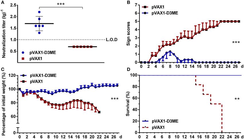
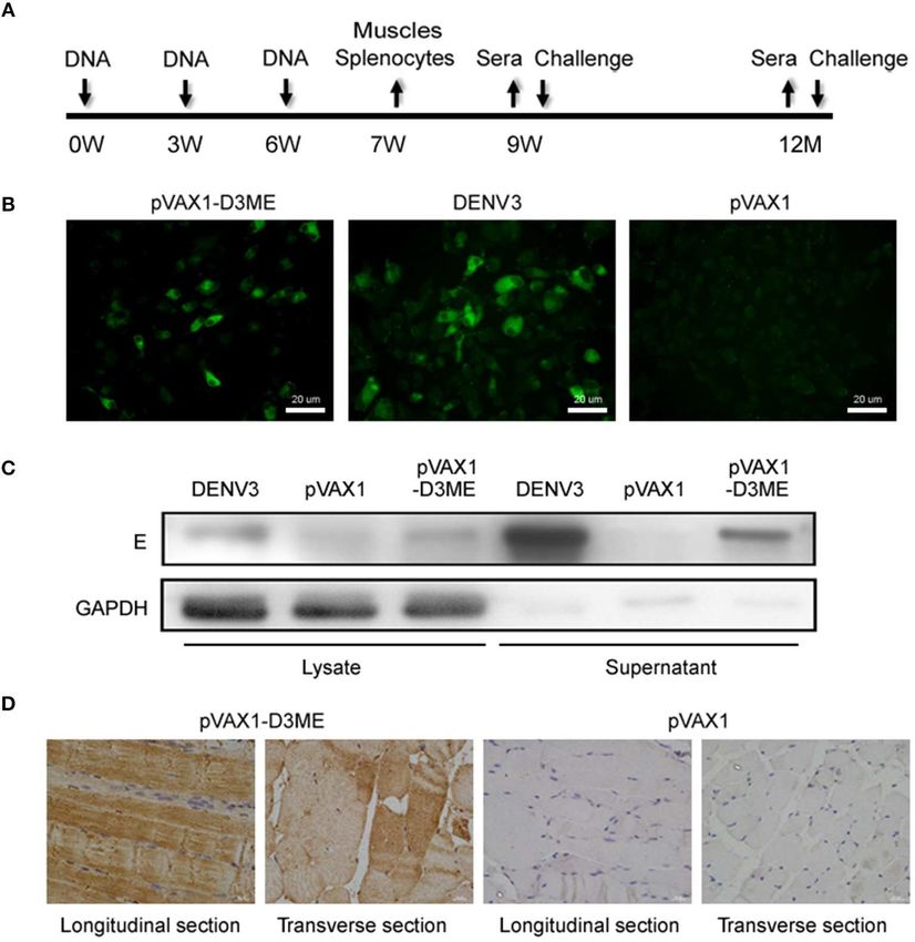
Feng et al.                                                                                                                              A DNA Vaccine Against DENV3

 FIGURE 1 | The immunization and sample collection schedule, and in vitro and in vivo expression of the prM-E proteins. (A) Diagram of immunization, sample
 collection and challenge in BALB/c mice. The mice were vaccinated at weeks 0, 3, and 6. Splenocytes and muscle tissues were obtained at the seventh week, and
 sera were collected at the ninth week, the sixth month, and the 12th month. The mice were challenged with DENVs at the ninth week and the 12th month. (B) In vitro
 expression of the prM-E proteins in plasmid-transfected Vero cells detected by IFA. DENV3-infected Vero cells served as the positive control. (C) The cell lysate and
 the supernatant of plasmid-transfected Vero cells were collected at 72 h after transfection and in vitro expression of the E protein was detected by Western blotting.
 DENV3-infected Vero cells served as the positive control. GAPDH served as the loading control. (D) In vivo expression of the prM-E protein at the injection site in
 muscle tissues detected by IHC.

Active Protection Against DENV1–4 in                                                   challenge). The survival rate was reported as the percentage
DNA-Immunized Mice                                                                     of survivors.
Mice were challenged intracerebrally with a lethal dose
of DENV1, DENV2, DENV3, or DENV4. Pathological                                         Statistical Analysis
symptoms and body weight were monitored daily for 27                                   Statistical analysis was conducted with SPSS 17.0 or GraphPad
days. Pathological symptoms were recorded as the mean clinical                         Prism 6 software. Geometric mean titers (GMTs) of NAbs
sign scores: 0 = healthy; 1 = ruffled hair or hunchbacked                              were calculated as log-transformed reciprocal titers. Weight
appearance; 2 = asthenia, wasting, or bradykinesia; 3 =                                changes and clinical sign scores were analyzed by repeated
forelimb or hindlimb weakness; 4 = paralysis or moribundity;                           measures analysis of variance. Kaplan–Meier survival curves
and 5 = death. Body weight changes were reported as                                    were plotted and evaluated statistically by the log-rank test.
percentages compared to those on day 0. The percentage was                             The data of PRNT, ELISPOT, and flow cytometry analysis were
determined by 100% × (weight after challenge)/(weight before                           compared using one-way analysis of variance. p < 0.05 were

Frontiers in Cellular and Infection Microbiology | www.frontiersin.org             4                                                 March 2020 | Volume 10 | Article 87
Feng et al. A DNA Vaccine Against DENV3
considered statistically significant: ∗ p < 0.05; ∗∗ p < 0.01; and Second, the protective efficacy of NAbs in the collected
∗∗∗ p < 0.001. sera was evaluated in a neonatal mouse model. As shown in
Figures 2B,C, the suckling mice that received sera from the
pVAX1-D3ME group grew steadily with increasing body weights.
RESULTS The average body weight was 6.95 ± 0.42 g at day 11. In contrast,
the control suckling mice showed a slow increase in body weight,
In vitro and in vivo Expression of the prM-E and the average value was only 4.99 ± 1.11 g at day 11. By
Protein the end of the observation period, all of the suckling mice in
To test the in vitro expression of the recombinant plasmid the control group were dead, but 100% of the suckling mice
in eukaryotic cells, Vero cells were transfected with in the pVAX1-D3ME group survived. Statistical analysis of the
pVAX1-D3ME or pVAX1, and the prM-E proteins were overall body weight and survival rate showed that there were
examined by IFA. As shown in Figure 1B, cells transfected significant differences between the two groups (∗∗ p < 0.01 and
with pVAX1-D3ME exhibited intense green fluorescence ∗∗∗ p < 0.001). Overall, pVAX1-D3ME-immunized sera provided
signals, and the transfection rate was more than 50%. In suckling mice effective neutralization against DENV3 challenge
contrast, no specific fluorescence signal was observed in and, to some extent, inhibited or reduced viral pathogenicity and
pVAX1-transfected cells. Furthermore, the expression of the delayed disease progression in suckling mice.
recombinant plasmid was confirmed by Western blotting
with the antibody 4G2, which recognizes the E antigen.
Specific expression was detected not only in the lysate of
DENV3-Specific Cellular Immune
pVAX1-D3ME-transfected cells but also in the supernatant Response
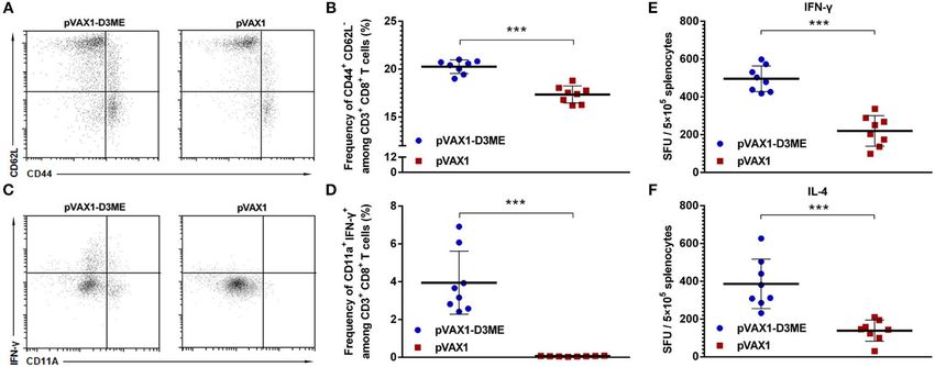
(Figure 1C). As well-known, once T cells were activated, the expression of
Moreover, to confirm the in vivo expression of the CD44 increased and that of CD62L decreased. CD44+ and
recombinant plasmid, mice were inoculated with plasmids three CD62L− were used to mark the effective memory T cells in
times at 3-week intervals, and the quadriceps femoris muscles this study. As shown in Figures 3A,B, after vaccination, the
were collected 1 week after the last immunization and tested by percentage of CD44+ CD62L− cells within CD8+ T cells in
IHC. As shown in Figure 1D, in both the longitudinal and the the pVAX1-D3ME group was significantly higher than that in
transverse sections, the specific expression of the target protein the pVAX1 group (∗∗∗ p < 0.001). This result indicated that the
was detected in the pVAX1-D3ME-inoculated muscle tissue, pVAX1-D3ME vaccination effectively improved the activation of
confirming that pVAX1-D3ME was effectively expressed in vivo effector memory T cells.
and could be used in subsequent experiments. Similarly, the memory T cells generated after vaccination
might play an essential role in long-term protection. Memory
T cells can rapidly gain effector function to kill infected cells
DENV3-Specific Humoral Immune and/or secrete IFN-γ to inhibit the replication of viruses (Kaech
Response et al., 2002; Zellweger et al., 2015). Murine CD11a+ CD8+
First, to evaluate the DENV3-specific antibody response T cells are capable of mounting vigorous recall responses
triggered by pVAX1-D3ME, sera were collected from mice three upon a secondary antigenic challenge (Rai et al., 2009).
weeks after the last immunization. The neutralization titers were Therefore, combining the analysis with CD11a, a surface
assessed using in vitro PRNT. As expected, the sera of control marker of antigen-experienced CD8+ T cells, we used flow
mice failed to neutralize DENV3. In contrast, the PRNT50 GMT cytometry to analyze changes in the percentage of memory
of pVAX1-D3ME-immunized sera was 1:538.17 (Figure 2A), CD8+ T cells expressing IFN-γ and CD11a in immunized
suggesting a robust humoral immune response against DENV3. mice. As shown in Figures 3C,D, after gating on CD3+ CD8+
FIGURE 2 | Humoral immune response against DENV3 in mouse sera. Sera were collected at the ninth week after the immunization. (A) Endpoint titers (n = 8) of
DENV3-specific NAbs were detected by PRNT50 and recorded as GMT ± SD. The limit of detection (L.O.D.) depicted as a dotted line represents the lowest dilution
that the experiment could detect. (B,C) In vitro neutralizing activity and passive protective effect of immunized sera on suckling mice (n = 13 in the pVAX1-D3ME
group, n = 10 in the pVAX1 group). The pooled sera mixed with live DENV3 were transferred into 1-day-old suckling mice. The mice were monitored daily for 14 days.
(B) Body weight change from day 0. (C) The survival rate is shown as the percentage of survivors. **p < 0.01; ***p < 0.001.
Frontiers in Cellular and Infection Microbiology | www.frontiersin.org 5 March 2020 | Volume 10 | Article 87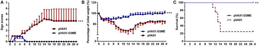
Feng et al.                                                                                                                           A DNA Vaccine Against DENV3

 FIGURE 3 | Cellular immune response against DENV3 in mouse splenocytes. Splenocytes were isolated 1 week after the final immunization. (A–D) The
 antigen-experienced CD8+ T cell response was assayed by flow cytometry (n = 8). (A) Representative images of CD44+ CD62L− T cells (gated on CD3e+ CD8+ T
 cells). (B) Quantification of the frequency of CD44+ CD62L− CD8+ T cells. The data are expressed as the mean percentage ± SD from three independent
 experiments. (C) Representative images of CD11a+ IFN-γ+ T cells (gated on CD3e+ CD8+ T cells). (D) Quantification of the frequency of CD11a+ IFN-γ+ CD8+ T
 cells. The data are expressed as the mean percentage ± SD from three independent experiments. (E,F) Splenocyte-secreted cytokines detected by ELISPOT assay
 (n = 8). The number of cytokine-positive cells is recorded as the mean spot-forming unit (SFU)/5 × 105 splenocytes ± SD. ***p < 0.001.

 FIGURE 4 | Short-term active protective immunity against DENV3 challenge (n = 8). Mice were challenged with DENV3 at the ninth week after the immunization and
 monitored daily for 27 days. (A) Pathological symptoms recorded as the mean clinical sign scores. (B) Percentage of body weight from day 0. (C) Survival rate shown
 as the percentage of survivors. Results are representative of three independent experiments. **p < 0.01; ***p < 0.001.

T cells, an average 3.95 ± 1.56% proportion of CD11a+                                Active Protection Against DENV3
IFN-γ+ cells was found in the pVAX1-D3ME group, which                                Challenge
was ∼60-fold higher than that in the pVAX1 group (∗∗∗ p                              Three weeks after the final vaccination, mice were challenged
< 0.001).                                                                            with a lethal dose of DENV3. As shown in Figure 4, obvious
   Taken together, these results indicated that pVAX1-D3ME                           illness signs appeared on the sixth day after challenge in
induced a rapid and strong antigen-specific CD8+ T cell                              the control group treated with pVAX1 (Figure 4A), and
immune response.                                                                     the clinical scores progressively increased. Moreover, body
                                                                                     weight continuously decreased, and weight loss was >30%
Cytokine Generation                                                                  within 12 days (Figure 4B). Finally, 75% (6/8) of the control
One week after the last immunization, splenic lymphocytes were                       mice died (Figure 4C). In contrast, compared with the
isolated and restimulated in vitro with the DENV3 antigen to                         control mice, the pVAX1-D3ME-immunized mice showed
detect the level of splenocyte-derived cytokines. As shown in                        transient and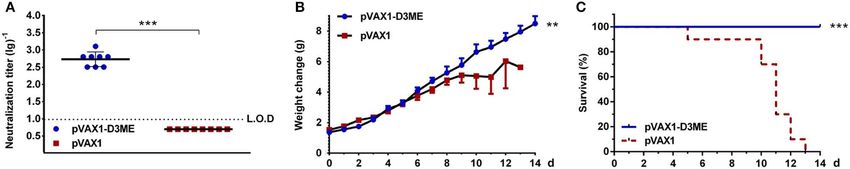
Feng et al.                                                                                                                          A DNA Vaccine Against DENV3

 FIGURE 5 | Long-term NAb response and active protective immunity against DENV3 challenge at the 12th month after the immunization (n = 6). (A) Endpoint titers of
 DENV3-specific NAbs in sera were detected by PRNT50 and recorded as GMT ± SD. The L.O.D. depicted as a dotted line represents the limit of detection of the
 assay. (B–D) Mice were challenged with DENV3 and monitored daily for 27 days. (B) Pathological symptoms recorded as the mean clinical sign scores. (C)
 Percentage of body weight from day 0. (D) Survival rate shown as the percentage of survivors. **p < 0.01; ***p < 0.001.

Long-Term DENV3-Specific NAb Response                                               body weight, and survival rates are shown in Figure 6. Three
and Active Protection Against DENV3                                                 weeks after the last immunization, after challenge with DENV1
                                                                                    and DENV4, 100% survival rates with relatively mild symptoms
Challenge
                                                                                    (Figures 6B,J, ∗∗∗ p < 0.001) as well as limited body weight
Moreover, at the 12th month after the immunization, the
                                                                                    loss (Figure 6C, ∗∗ p < 0.01 and Figure 6K, ∗∗∗ p < 0.001) were
DENV3-specific NAb in pVAX1-D3ME-immunized sera were
                                                                                    observed in the pVAX1-D3ME-immunized mice, whereas the
maintained at 1:50.4 (Figure 5A), suggesting a long-term
                                                                                    survival rates were 28.6% (2/7, Figure 6D, ∗∗ p < 0.01) against
humoral immune response against DENV3. When the mice were
                                                                                    DENV1 and 14.3% (1/7, Figure 6L, ∗∗ p < 0.01) against DENV4
challenged with a lethal dose of DENV3, all of the pVAX1-D3ME-
                                                                                    in the control mice, which were accompanied with obvious illness
immunized mice (6/6) survived (∗∗ p < 0.01), with imperceptible
                                                                                    signs and body weight loss.
symptoms (∗∗∗ p < 0.001), and body weight loss (∗∗∗ p < 0.001),
                                                                                        Meanwhile, we observed that, when the mice were infected
as compared with the control mice (Figures 5B–D). The
                                                                                    with DENV2, the pVAX1-D3ME-immunized group showed
results suggested that three doses of pVAX1-D3ME triggered
Feng et al. A DNA Vaccine Against DENV3 FIGURE 6 | Short-term cross-reactive NAb response and protection against DENV1 (A–D), DENV2 (E–H), and DENV4 (I–L) at the ninth week after the immunization (n = 7). (A,E,I) Endpoint titers of cross-reactive NAbs in sera were detected by PRNT50 and recorded as GMT ± SD. The L.O.D. depicted as a dotted line represents the limit of detection of the assay. (B–D,F–H,J–L). Mice were challenged with DENVs and monitored daily for 27 days. (B,F,J) Pathological symptoms recorded as the mean clinical sign scores. (C,G,K) Percentage of body weight from day 0. (D,H,L) Survival rate shown as the percentage of survivors. Results are representative of three independent experiments. *p < 0.05; **p < 0.01; ***p < 0.001. FIGURE 7 | Long-term cross-reactive NAb response and protection against DENV1 (A–D), DENV2 (E–H), and DENV4 (I–L) at the 12th month after the immunization. For DENV1, n = 7 in the pVAX1-D3ME group, n = 5 in the pVAX1 group. For DENV2, n = 6 in the pVAX1-D3ME group, n = 5 in the pVAX1 group. For DENV4, n = 7 in the pVAX1-D3ME group, n = 5 in the pVAX1 group. (A,E,I) Endpoint titers of cross-reactive NAbs in sera were detected by PRNT50 and recorded as GMT ± SD. The L.O.D. depicted as a dotted line represents the limit of detection of the assay. (B–D,F–H,J–L) Mice were challenged with DENVs and monitored daily for 27 days. (B,F,J) Pathological symptoms recorded as the mean clinical sign scores. (C,G,K) Percentage of body weight from day 0. (D,H,L) Survival rate shown as the percentage of survivors. *p < 0.05; **p < 0.01; ***p < 0.001. 1:26.92, and 1:32.81 (figure not shown) against DENV1, At the 12th month, the cross-reactive NAb titers decreased DENV2, and DENV4, which still showed cross-reactive to 1:24.38, 1:20, and 1:9.06 (Figures 7A,E,I). After challenged neutralizing capability compared with those of the corresponding with DENV1 and DENV4, the mice immunized with pVAX1- controls (∗∗∗ p< 0.001). D3ME showed 85.7% (6/7, Figure 7D, ∗∗ p < 0.01) and 100% (7/7, Frontiers in Cellular and Infection Microbiology | www.frontiersin.org 8 March 2020 | Volume 10 | Article 87
Feng et al.                                                                                                           A DNA Vaccine Against DENV3

Figure 7L, ∗∗ p < 0.01) survival rates, respectively. Meanwhile,             lethal challenge with DENV1, 2, 3, and 4 were 100, 90, 100, and
the symptoms were relatively mild (Figures 7B,J, ∗∗∗ p < 0.001)              100%, respectively, and the protective efficacy against all four
and the levels of the body weight loss were less than those of               serotypes was stable.
the controls (Figure 7C, ∗∗ p < 0.01; Figure 7K, ∗∗∗ p < 0.001).                 The effective protective immunity evoked by recombinant
After challenge with DENV2, when compared with that of                       plasmids expressing the prM and E proteins of DENVs
the control, although the survival rate of the pVAX1-D3ME-                   should first be attributed to the target molecules used in the
immunized mice was 33.33% (2/6) without statistic difference                 vaccine. It has been demonstrated that prM and E proteins
(Figure 7H, p = 0.0564), differences in symptom scores and body              are involved in inducing effective NAbs to DENV (Lin et al.,
weight loss (Figures 7F,G, ∗∗∗ p < 0.001) indicated the partial              2015). Additionally, T cell epitopes eliciting cellular immune
cross-reactive protection.                                                   responses may also be important. Although the vast majority of
   The aforementioned results demonstrated that different                    T cell epitopes have been identified on non-structural proteins,
degrees of the cross-reactive protection evoked by three doses               especially NS3 (Mathew et al., 2014; Elong Ngono et al., 2016),
of pVAX1-D3ME could last up to 12 months from the                            they have also been found on structural proteins, including
initial immunization.                                                        prM and E (Duan et al., 2015; Hussain et al., 2015; Wen
                                                                             et al., 2017). Theoretically, DNA vaccine-encoded proteins
                                                                             often display a native conformation with post-translational
DISCUSSION                                                                   modifications, including glycosylation, proteolytic processing,
                                                                             and lipid conjugations, which are essential for eliciting immune
Dengue disease is the most common mosquito-borne viral                       responses to conformational epitopes. Moreover, it has been
disease in the world. Severe dengue is a leading cause of death              demonstrated that electroporation delivery could facilitate DNA
among children in Southeast Asian and Latin American countries               vaccination to generate a significantly persistent antibody
(Kittigul et al., 2007). In China, the region affected by dengue             response and antigen-specific T cell response (Chen et al., 2016;
has expanded, and the incidence has increased steadily since 2012            Zheng et al., 2017; Sheng et al., 2019), which would contribute to
(Lai et al., 2015). In 2014, Guangdong Province in China suffered            the effective protection induced by pVAX1-D3ME.
the most serious dengue outbreak in its history, with more than                  Although the amino acid sequence of the E protein defines
60,000 cases (Zhu et al., 2019). The development of virus-specific           each DENV serotype, the amino acid residues of this protein
prevention is an urgent public health priority to control the                are well-conserved, with high similarities (90%–96%) among the
increased global incidence and to reduce the disease burden. At              four different serotypes (Cedillo-Barron et al., 2014). Moreover,
present, only Dengvaxia, Sanofi’s controversial dengue vaccine,              it has been reported that anti-prM antibodies exhibit distinctive
has been developed for use in humans. Recently, Dengvaxia was                cross-reactivity to the four DENV serotypes (Chan et al., 2012).
approved for the prevention of secondary DENV infection in                   In this study, pVAX1-D3ME-elicited antibodies showed cross-
individuals 9 through 16 years of age who have had laboratory-               neutralizing and durable reactivity toward heterologous DENVs.
confirmed dengue disease and who live in endemic regions                     More importantly, most of the pVAX1-D3ME-vaccinated mice
(FDA, 2019). However, the recommendation for extra safety                    survived the challenge with heterologous serotypes, without an
precautions for this vaccine by the WHO (Sridhar et al., 2018)               increase in viral infection. Speculatively, both of the cross-
points to the limitation of its application. Another live-attenuated         reactive NAbs and CD8+ T cells might contribute to the cross-
tetravalent dengue vaccine, TAK-003, from Takeda in Japan was                protection against heterotypic DENV infection (Zellweger et al.,
reported to have positive top-line results from a very large Phase           2015). Our results are consistent with a previous study from
III trial (Biswal et al., 2019; Sharma et al., 2019; Tong, 2019).            Reich et al., who reported that infection or immunization
Nevertheless, developing efficacious vaccines against dengue with            with one DENV serotype conferred effective cross-protection to
balanced immune responses, especially tetravalent protection, is             heterologous serotypes for an average duration of approximately
of great necessity.                                                          two years (Reich et al., 2013). Thus, the characteristics of cross-
    As a novel and rapidly developing approach, DNA vaccines                 reactive immunity to DENVs and its impact on dengue epidemics
offer a number of potential advantages including cost-                       need to be further investigated.
effectiveness, simplicity of manufacturing, high safety, and                     Several vaccines specifically targeting DENV3 have been
long-term expression of immunogens (Khan, 2013). More                        developed and evaluated in murine models. Most of them
importantly, antigens encoded by DNA vaccines can be                         expressed E protein as the specific immunogen. Chiang et al.
processed through either the MHC class I or II pathway,                      designed a subunit vaccine expressing recombinant DENV3
which are capable of stimulating robust cellular and humoral                 E protein domain III in a lipidated form (LD3ED III) and
immune responses. Moreover, DNA vaccines have been reported                  confirmed that LD3ED III induced broad profiles of humoral
to trigger balanced immune responses and provide effective                   and cellular immune responses even without formulation with
protection against DENVs (Beaumier et al., 2013). Previously,                exogenous adjuvants (Chiang et al., 2016). Versiani et al. used
we confirmed the effective protection induced by recombinant                 a diimide-activated amidation process to bind recombinant
plasmids expressing the prM and E proteins of DENV1 (Zheng                   DENV3 E proteins and developed multiwalled carbon nanotubes.
et al., 2017), DENV2 (Chen et al., 2016), or DENV4 (Sheng et al.,            The generation of both cell-mediated and NAb responses against
2019). With the same dosage and immunization strategy, two or                DENV3 increased substantially (Versiani et al., 2017). Hurtado-
three weeks after the third vaccination, the survival rates against          Melgoza et al. reported that the DNA vaccine candidate targeting

Frontiers in Cellular and Infection Microbiology | www.frontiersin.org   9                                        March 2020 | Volume 10 | Article 87
Feng et al. A DNA Vaccine Against DENV3
DENV3 NS3 triggered a favorable response with the activation be considered alternative immunogens in a dengue tetravalent
of T lymphocytes; however, immunization with this DNA vaccine formulation.
vaccine presented no detectable antibody titers against DENV3
(Hurtado-Melgoza et al., 2016). DATA AVAILABILITY STATEMENT
Regarding the limitations of this study, the possibility of
increasing immunogenicity with a lower dose of DNA than The raw data supporting the conclusions of this article will be
50 µg of each immunogen and a different regimen should made available by the authors, without undue reservation, to any
be considered. In our previous work, based on a consensus qualified researcher.
sequence of the ectodomain of E protein (cE80) of the four DENV
serotypes (Wang et al., 2019), heterologous (DNA prime-protein ETHICS STATEMENT
boost) regimens elicited greater systemic immune response and
more effective tetravalent protection than did homologous DNA This animal study was reviewed and approved by Institutional
immunization. As the optimal immunization regimen, DNA Animal Care and Use Committee of Chinese Capital
vaccination followed by protein boosting has been applied to a Medical University.
variety of infectious agents to improve the immunogenicity and
protective efficacy of vaccines (Menon et al., 2017; Wang et al., AUTHOR CONTRIBUTIONS
2017; Cai et al., 2018). Further investigation should be performed
to assess whether it is an appropriate strategy for our dengue JA and HC conceived and designed the experiments. KF, XZ,
DNA vaccine candidate. RW, DF, and ZS performed the experiments. KF, NG, HC, and
Taken together, this study demonstrated that three doses of JA analyzed the data. HC and KF prepared the manuscript draft.
50 µg of pVAX1-D3ME were sufficient to induce robust NAbs HC, JA, and HZ revised the manuscript. All authors read and
and strong antigen-specific T cell responses and provided long- approved the final manuscript.
term protective immunity against DENV3 infection. Moreover,
pVAX1-D3ME-elicited NAbs and protective immunity were not FUNDING
only type specific but also cross-reactive against heterotypes,
showing great promise for the further evaluation of a This research was funded by National Natural Science
dengue tetravalent DNA vaccine candidate. In a future Foundation of China, Grant Nos. 81772172, 81671971,
study, immunogenic motifs in the DENV proteins that are U1602223, and 81372935 and the Beijing Municipal Commission
conserved or highly homologous among all serotypes should of Education, Grant No. KZ201810025035.
REFERENCES Chiang, C. Y., Liu, S. J., Hsieh, C. H., Chen, M. Y., Tsai, J. P., Liu, H.
H., et al. (2016). Recombinant lipidated dengue-3 envelope protein domain
Beaumier, C. M., Gillespie, P. M., Hotez, P. J., and Bottazzi, M. E. (2013). New III stimulates broad immune responses in mice. Vaccine 34, 1054–1061.
vaccines for neglected parasitic diseases and dengue. Transl. Res. 162, 144–155. doi: 10.1016/j.vaccine.2016.01.009
doi: 10.1016/j.trsl.2013.03.006 Duan, Z., Guo, J., Huang, X., Liu, H., Chen, X., Jiang, M., et al. (2015).
Bhatt, S., Gething, P. W., Brady, O. J., Messina, J. P., Farlow, A. W., Moyes, C. Identification of cytotoxic T lymphocyte epitopes in dengue virus serotype 1.
L., et al. (2013). The global distribution and burden of dengue. Nature 496, J. Med. Virol. 87, 1077–1089. doi: 10.1002/jmv.24167
504–507. doi: 10.1038/nature12060 Eckels, K. H., and Putnak, R. (2003). Formalin-inactivated whole virus and
Biswal, S., Reynales, H., Saez-Llorens, X., Lopez, P., Borja-Tabora, C., Kosalaraksa, recombinant subunit flavivirus vaccines. Adv. Virus Res. 61, 395–418.
P., et al. (2019). Efficacy of a tetravalent dengue vaccine in healthy children and doi: 10.1016/S0065-3527(03)61010-9
adolescents. N. Engl. J. Med. 381, 2009–2019. doi: 10.1056/NEJMoa1903869 Elong Ngono, A., Chen, H. W., Tang, W. W., Joo, Y., King, K., Weiskopf, D.,
Bustos-Arriaga, J., Gromowski, G. D., Tsetsarkin, K. A., Firestone, C. Y., et al. (2016). Protective role of cross-reactive CD8 T cells against dengue virus
Castro-Jimenez, T., Pletnev, A. G., et al. (2018). Decreased accumulation of infection. EBioMedicine 13, 284–293. doi: 10.1016/j.ebiom.2016.10.006
subgenomic RNA in human cells infected with vaccine candidate DEN4Delta30 FDA (2019). First FDA-Approved Vaccine for the Prevention of Dengue Disease in
increases viral susceptibility to type I interferon. Vaccine 36, 3460–3467. Endemic Regions (U.S. FOOD & DRUG).
doi: 10.1016/j.vaccine.2018.04.087 Ferguson, N. M., Rodriguez-Barraquer, I., Dorigatti, I., Mier, Y. T.-R. L., Laydon,
Cai, D., Song, Q., Duan, C., Wang, S., Wang, J., and Zhu, Y. (2018). Enhanced D. J., and Cummings, D. A. (2016). Benefits and risks of the sanofi-pasteur
immune responses to E2 protein and DNA formulated with ISA 61 VG dengue vaccine: modeling optimal deployment. Science 353, 1033–1036.
administered as a DNA prime-protein boost regimen against bovine viral doi: 10.1126/science.aaf9590
diarrhea virus. Vaccine 36, 5591–5599. doi: 10.1016/j.vaccine.2018.07.054 Guirakhoo, F., Arroyo, J., Pugachev, K. V., Miller, C., Zhang, Z. X., Weltzin, R., et al.
Cedillo-Barron, L., Garcia-Cordero, J., Bustos-Arriaga, J., Leon-Juarez, M., and (2001). Construction, safety, and immunogenicity in nonhuman primates of a
Gutierrez-Castaneda, B. (2014). Antibody response to dengue virus. Microbes chimeric yellow fever-dengue virus tetravalent vaccine. J. Virol. 75, 7290–7304.
Infect. 16, 711–720. doi: 10.1016/j.micinf.2014.07.011 doi: 10.1128/JVI.75.16.7290-7304.2001
Chan, A. H., Tan, H. C., Chow, A. Y., Lim, A. P., Lok, S. M., Moreland, N. J., Henein, S., Swanstrom, J., Byers, A. M., Moser, J. M., Shaik, S. F., Bonaparte, M.,
et al. (2012). A human PrM antibody that recognizes a novel cryptic epitope on et al. (2017). Dissecting antibodies induced by a chimeric yellow fever-dengue,
dengue E glycoprotein. PLoS ONE 7:e33451. doi: 10.1371/journal.pone.0033451 live-attenuated, tetravalent dengue vaccine (CYD-TDV) in naive and dengue-
Chen, H., Zheng, X., Wang, R., Gao, N., Sheng, Z., Fan, D., et al. (2016). exposed individuals. J. Infect. Dis. 215, 351–358. doi: 10.1093/infdis/jiw576
Immunization with electroporation enhances the protective effect of a DNA Huang, X. Y., Ma, H. X., Wang, H. F., Du, Y. H., Su, J., Li, X. L., et al. (2014).
vaccine candidate expressing prME antigen against dengue virus serotype 2 Outbreak of dengue Fever in central China, 2013. Biomed. Environ. Sci. 27,
infection. Clin. Immunol. 171, 41–49. doi: 10.1016/j.clim.2016.08.021 894–897. doi: 10.3967/bes2014.125
Frontiers in Cellular and Infection Microbiology | www.frontiersin.org 10 March 2020 | Volume 10 | Article 87Feng et al. A DNA Vaccine Against DENV3 Hurtado-Melgoza, M. L., Ramos-Ligonio, A., Alvarez-Rodriguez, L. M., Meza- Prompetchara, E., Ketloy, C., Thomas, S. J., and Ruxrungtham, K. (2019). Menchaca, T., and Lopez-Monteon, A. (2016). Differential humoral and Dengue vaccine: global development update. Asian Pac J Allergy Immunol. cellular immunity induced by vaccination using plasmid DNA and protein doi: 10.12932/AP-100518-0309. [Epub ahead of print]. recombinant expressing the NS3 protein of dengue virus type 3. J. Biomed. Sci. Putnak, R., Porter, K., and Schmaljohn, C. (2003). DNA vaccines for 23:85. doi: 10.1186/s12929-016-0302-z flaviviruses. Adv. Virus Res. 61, 445–468. doi: 10.1016/S0065-3527(03) Hussain, M., Idrees, M., and Afzal, S. (2015). Development of global 61012-2 consensus of dengue virus envelope glycoprotein for epitopes Rai, D., Pham, N. L., Harty, J. T., and Badovinac, V. P. (2009). Tracking the total based vaccine design. Curr. Comput. Aided Drug Des. 11, 84–97. CD8 T cell response to infection reveals substantial discordance in magnitude doi: 10.2174/1573409911666150529130134 and kinetics between inbred and outbred hosts. J. Immunol. 183, 7672–7681. Kaech, S. M., Wherry, E. J., and Ahmed, R. (2002). Effector and memory T-cell doi: 10.4049/jimmunol.0902874 differentiation: implications for vaccine development. Nat. Rev. Immunol. 2, Reich, N. G., Shrestha, S., King, A. A., Rohani, P., Lessler, J., Kalayanarooj, 251–262. doi: 10.1038/nri778 S., et al. (2013). Interactions between serotypes of dengue highlight Khan, K. H. (2013). DNA vaccines: roles against diseases. Germs 3, 26–35. epidemiological impact of cross-immunity. J. R. Soc. Interface 10:20130414. doi: 10.11599/germs.2013.1034 doi: 10.1098/rsif.2013.0414 Kittigul, L., Pitakarnjanakul, P., Sujirarat, D., and Siripanichgon, K. (2007). Sharma, M., Glasner, D. R., Watkins, H., Puerta-Guardo, H., Kassa, Y., Egan, M. A., The differences of clinical manifestations and laboratory findings in et al. (2019). Magnitude and functionality of the NS1-specific antibody response children and adults with dengue virus infection. J. Clin. Virol. 39, 76–81. elicited by a live-attenuated tetravalent dengue vaccine candidate. J. Infect. Dis. doi: 10.1016/j.jcv.2007.04.006 221, 867–877. doi: 10.1093/infdis/jiz081 Kraemer, M. U. G., Reiner, R. C. Jr., Brady, O. J., Messina, J. P., Gilbert, Sheng, Z., Chen, H., Feng, K., Gao, N., Wang, R., Wang, P., et al. M., Pigott, D. M., et al. (2019). Past and future spread of the arbovirus (2019). Electroporation-mediated immunization of a candidate DNA vaccine vectors Aedes aegypti and Aedes albopictus. Nat. Microbiol. 4, 854–863. expressing dengue virus serotype 4 prM-E antigen confers long-term doi: 10.1038/s41564-019-0376-y protection in mice. Virol. Sin. 34, 88–96. doi: 10.1007/s12250-019-0 Kularatne, S. A. (2015). Dengue fever. BMJ 351:h4661. doi: 10.1136/bmj.h4661 0090-8 Lai, S., Huang, Z., Zhou, H., Anders, K. L., Perkins, T. A., Yin, W., et al. (2015). Shirin, T., Muraduzzaman, A. K. M., Alam, A. N., Sultana, S., Siddiqua, M., The changing epidemiology of dengue in China, 1990-2014: a descriptive Khan, M. H., et al. (2019). Largest dengue outbreak of the decade with high analysis of 25 years of nationwide surveillance data. BMC Med. 13:100. fatality may be due to reemergence of DEN-3 serotype in Dhaka, Bangladesh, doi: 10.1186/s12916-015-0336-1 necessitating immediate public health attention. New Microbes New Infect Lin, H. E., Tsai, W. Y., Liu, I. J., Li, P. C., Liao, M. Y., Tsai, J. J., et al. (2012). Analysis 29:100511. doi: 10.1016/j.nmni.2019.01.007 of epitopes on dengue virus envelope protein recognized by monoclonal Soo, K. M., Khalid, B., Ching, S. M., and Chee, H. Y. (2016). Meta- antibodies and polyclonal human sera by a high throughput assay. PLoS Negl. analysis of dengue severity during infection by different dengue virus Trop. Dis. 6:e1447. doi: 10.1371/journal.pntd.0001447 serotypes in primary and secondary infections. PLoS ONE 11:e0154760. Lin, Y., Wen, K., Guo, Y., Qiu, L., Pan, Y., Yu, L., et al. (2015). Mapping of doi: 10.1371/journal.pone.0154760 the B cell neutralizing epitopes on ED III of envelope protein from dengue Sridhar, S., Luedtke, A., Langevin, E., Zhu, M., Bonaparte, M., Machabert, virus. Bing Du Xue Bao 31, 665–673. doi: 10.13242/j.cnki.bingduxuebao.0 T., et al. (2018). Effect of dengue serostatus on dengue vaccine safety 02828 and efficacy. N. Engl. J. Med. 379, 327–340. doi: 10.1056/NEJMoa18 Manoff, S. B., Sausser, M., Falk Russell, A., Martin, J., Radley, D., Hyatt, D., 00820 et al. (2019). Immunogenicity and safety of an investigational tetravalent Tan, K. K., Zulkifle, N. I., Sulaiman, S., Pang, S. P., NorAmdan, N., MatRahim, recombinant subunit vaccine for dengue: results of a Phase I randomized N., et al. (2018). Emergence of the Asian lineage dengue virus type 3 clinical trial in flavivirus-naive adults. Hum. Vaccin. Immunother. 15, genotype III in Malaysia. BMC Evol. Biol. 18:58. doi: 10.1186/s12862-018- 2195–2204. doi: 10.1080/21645515.2018.1546523 1175-4 Mathew, A., Townsley, E., and Ennis, F. A. (2014). Elucidating the role of T Tong, A. (2019). Takeda claims PhIII success in dengue vaccine months cells in protection against and pathogenesis of dengue virus infections. Future after Dengvaxia implosion for rival Sanofi (ENDPOINTSNEWS). Available Microbiol. 9, 411–425. doi: 10.2217/fmb.13.171 online at: https://endpts.com/takeda-claims-phiii-success-in-dengue-vaccine- Mellado-Sanchez, G., Garcia-Cordero, J., Luria-Perez, R., Lazaro-Olan, L., Santos- months-after-dengvaxia-implosion-for-rival-sanofi/ Argumedo, L., Gutierrez-Castaneda, B., et al. (2005). DNA priming E and NS1 Versiani, A. F., Astigarraga, R. G., Rocha, E. S., Barboza, A. P., Kroon, constructs–homologous proteins boosting immunization strategy to improve E. G., Rachid, M. A., et al. (2017). Multi-walled carbon nanotubes immune response against dengue in mice. Viral Immunol. 18, 709–721. functionalized with recombinant Dengue virus 3 envelope proteins induce doi: 10.1089/vim.2005.18.709 significant and specific immune responses in mice. J. Nanobiotechnology 15:26. Menon, V., Ayala, V. I., Rangaswamy, S. P., Kalisz, I., Whitney, S., Galmin, L., doi: 10.1186/s12951-017-0259-4 et al. (2017). DNA prime/protein boost vaccination elicits robust humoral Wang, R., Zheng, X., Sun, J., Feng, K., Gao, N., Fan, D., et al. (2019). response in rhesus macaques using oligomeric simian immunodeficiency virus Vaccination with a single consensus envelope protein ectodomain sequence envelope and Advax delta inulin adjuvant. J. Gen. Virol. 98, 2143–2155. administered in a heterologous regimen induces tetravalent immune responses doi: 10.1099/jgv.0.000863 and protection against dengue viruses in mice. Front. Microbiol. 10:1113. Oliveira, E. R. A., de Alencastro, R. B., and Horta, B. A. C. (2017). New doi: 10.3389/fmicb.2019.01113 insights into flavivirus biology: the influence of pH over interactions Wang, S., Chou, T. H., Hackett, A., Efros, V., Wang, Y., Han, D., between prM and E proteins. J. Comput. Aided Mol. Des. 31, 1009–1019. et al. (2017). Screening of primary gp120 immunogens to formulate doi: 10.1007/s10822-017-0076-8 the next generation polyvalent DNA prime-protein boost HIV-1 vaccines. Osorio, J. E., Velez, I. D., Thomson, C., Lopez, L., Jimenez, A., Haller, A. A., et al. Hum. Vaccin. Immunother. 13, 2996–3009. doi: 10.1080/21645515.2017.13 (2014). Safety and immunogenicity of a recombinant live attenuated tetravalent 80137 dengue vaccine (DENVax) in flavivirus-naive healthy adults in Colombia: a Wen, J., Elong Ngono, A., Regla-Nava, J. A., Kim, K., Gorman, M. J., Diamond, randomised, placebo-controlled, phase 1 study. Lancet Infect. Dis. 14, 830–838. M. S., et al. (2017). Dengue virus-reactive CD8(+) T cells mediate cross- doi: 10.1016/S1473-3099(14)70811-4 protection against subsequent Zika virus challenge. Nat. Commun. 8:1459. Parveen, N., Islam, A., Tazeen, A., Hisamuddin, M., Abdullah, M., Naqvi, I. doi: 10.1038/s41467-017-01669-z H., et al. (2019). Circulation of single serotype of Dengue Virus (DENV- Whitehead, S. S. (2016). Development of TV003/TV005, a single dose, highly 3) in New Delhi, India during 2016: a change in the epidemiological immunogenic live attenuated dengue vaccine; what makes this vaccine different trend. J. Infect. Public Health 12, 49–56. doi: 10.1016/j.jiph.2018. from the Sanofi-Pasteur CYD vaccine? Expert Rev. Vaccines 15, 509–517. 08.008 doi: 10.1586/14760584.2016.1115727 Frontiers in Cellular and Infection Microbiology | www.frontiersin.org 11 March 2020 | Volume 10 | Article 87
Feng et al.                                                                                                                                     A DNA Vaccine Against DENV3

Zellweger, R. M., Tang, W. W., Eddy, W. E., King, K., Sanchez, M. C.,                      Conflict of Interest: The authors declare that the research was conducted in the
   and Shresta, S. (2015). CD8+ T cells can mediate short-term protection                  absence of any commercial or financial relationships that could be construed as a
   against heterotypic dengue virus reinfection in mice. J. Virol. 89, 6494–6505.          potential conflict of interest.
   doi: 10.1128/JVI.00036-15
Zheng, X., Chen, H., Wang, R., Fan, D., Feng, K., Gao, N., et al. (2017). Effective        Copyright © 2020 Feng, Zheng, Wang, Gao, Fan, Sheng, Zhou, Chen and An. This
   protection induced by a monovalent DNA vaccine against dengue virus (DV)                is an open-access article distributed under the terms of the Creative Commons
   serotype 1 and a bivalent DNA vaccine against DV1 and DV2 in mice. Front.               Attribution License (CC BY). The use, distribution or reproduction in other forums
   Cell. Infect. Microbiol. 7:175. doi: 10.3389/fcimb.2017.00175                           is permitted, provided the original author(s) and the copyright owner(s) are credited
Zhu, G., Xiao, J., Liu, T., Zhang, B., Hao, Y., and Ma, W. (2019). Spatiotemporal          and that the original publication in this journal is cited, in accordance with accepted
   analysis of the dengue outbreak in Guangdong Province, China. BMC Infect.               academic practice. No use, distribution or reproduction is permitted which does not
   Dis. 19:493. doi: 10.1186/s12879-019-4015-2                                             comply with these terms.

Frontiers in Cellular and Infection Microbiology | www.frontiersin.org                12                                                    March 2020 | Volume 10 | Article 87
You can also read


