Potential Application of the CRISPR/Cas9 System against Herpesvirus Infections
←
→
Page content transcription
If your browser does not render page correctly, please read the page content below
viruses
Review
Potential Application of the CRISPR/Cas9 System
against Herpesvirus Infections
Yuan-Chuan Chen 1,2,3         ID
                                   , Jingxue Sheng 2 , Phong Trang 2 and Fenyong Liu 1,2, *
 1    College of Life Sciences and Technology, Jinan University, Guangzhou 510632, China;
      yuchuan1022@gmail.com
 2    Program in Comparative Biochemistry, University of California, Berkeley, CA 94720, USA;
      jxsheng@berkeley.edu (J.S.); phong.trang@berkeley.edu (P.T.)
 3    National Applied Research Laboratories, Taipei 10636, Taiwan
 *    Correspondence: liu_fy@berkeley.edu; Tel.: +1-510-643-2436

 Received: 25 March 2018; Accepted: 19 May 2018; Published: 29 May 2018

 Abstract: The CRISPR/Cas9 system has been applied in the genome editing and disruption of latent
 infections for herpesviruses such as the herpes simplex virus, Epstein–Barr virus, cytomegalovirus,
 and Kaposi’s sarcoma-associated herpesvirus. CRISPR/Cas9-directed mutagenesis can introduce
 similar types of mutations to the viral genome as can bacterial artificial chromosome recombination
 engineering, which maintains and reconstitutes the viral genome successfully. The cleavage mediated
 by CRISPR/Cas9 enables the manipulation of disease-associated viral strains with unprecedented
 efficiency and precision. Additionally, current therapies for herpesvirus productive and latent
 infections are limited in efficacy and cannot eradicate viruses. CRISPR/Cas9 is potentially adapted
 for antiviral treatment by specifically targeting viral genomes during latent infections. This review,
 which focuses on recently published progress, suggests that the CRISPR/Cas9 system is not only
 a useful tool for basic virology research, but also a promising strategy for the control and prevention
 of herpesvirus latent infections.

 Keywords: CRISPR; clinical application; herpesvirus; latent infection; genome editing; HSV
 (Herpes Simplex Virus); EBV (Epstein–Barr Virus); CMV (cytomegalovirus); KSHV (Kaposi’s
 Sarcoma-Associated Herpesvirus)

1. Introduction
     Diseases caused by viruses are difficult to treat due to the high viral mutation rate and latent
infections. Particularly, it is almost impossible to eradicate latent viruses in the human host.
In this review, we focus on the therapy of herpesviruses such as herpes simplex virus 1 (HSV-1),
herpes simplex virus 2 (HSV-2), Epstein–Barr virus (EBV), cytomegalovirus (CMV), and Kaposi’s
sarcoma-associated herpesvirus (KSHV) [1]. The clustered regularly interspaced short palindromic
repeats (CRISPR)/Cas9 nuclease system [2,3], a genome editing technology, represents a novel and
exciting possibility for the treatment of herpesvirus infections in recent studies [4]. Here, we briefly
describe the characteristics of herpesviruses and the CRISPR/Cas9 system.

1.1. Properties of Herpesviruses
      Herpesviridae, also known as Herpesviruses, is a large family of DNA viruses that cause human
diseases and remain an important global health problem [1]. Herpesviruses can cause lytic or latent
infections, in which the viruses either enter a lytic pathway to lyse their host cells or a latent pathway
to stay silent within the host cells [5–8]. The productive form of herpesviruses is a spherical enveloped
virion with a diameter of 120 to 260 nm (usually about 150 nm). It consists of a large double-stranded,

Viruses 2018, 10, 291; doi:10.3390/v10060291                                      www.mdpi.com/journal/viruses
Viruses 2018, 10, 291 2 of 12
linear DNA genome encoding 100–200 genes wrapped in an icosahedral capsid, surrounded by
amorphous protein called the tegument, with an outer lipid bilayer membrane envelope containing
different glycoproteins. Herpesviruses replicate in their host’s nucleus, with a sequential transcription
and translation of immediate–early (IE), early (E), and late (L) genes, with the earlier genes regulating
the transcription of later genes. HSV-1, HSV-2, EBV, human CMV (HCMV), and KSHV are distinct
viruses in this family known to cause diseases in humans [5–8] (Table 1).
Table 1. Major human herpesviruses (HHV-1, -2, -4, -5, and -8).
Type (Synonym) Subfamily Primary Target Cells Latency Cells Pathophysiology
Oral or genital herpes
HHV-1
α (alpha) Mucoepithelial cells Sensory neurons (predominantly orofacial), cold
(HSV-1)
sores, keratitis, etc.
HHV-2 Oral or genital herpes
α (alpha) Mucoepithelial cells Sensory neurons
(HSV-2) (predominantly genital), etc.
Infectious mononucleosis, Burkitt’s
lymphoma, nasopharyngeal
HHV-4
γ (gamma) B cells, Epithelial cells B cells, Epithelial cells carcinoma, Hodgkin’s disease,
(EBV)
post-transplant lymphomas, CNS
lymphoma in AIDS patients, etc.
Infectious mononucleosis-like
Monocytes,
HHV-5 Peripheral monocytes, syndrome, retinitis, pneumonitis,
β (beta) Lymphocytes,
(HCMV) CD34+ progenitor cells gastrointestinal diseases, mental
Epithelial cells
retardation, vascular disorders, etc.
Kaposi’s sarcoma, primary effusion
HHV-8 Lymphocytes and B cells, lymphoma, some types of
γ
(KSHV) other cells Mononucleocytes multicentric Castleman’s
disease, etc.
HSV-1: herpes simplex virus 1; HSV-2: herpes simplex virus 2; EBV: Epstein–Barr virus; HCMV: human
cytomegalovirus; KSHV: Kaposi’s sarcoma-associated herpesvirus; CNS: central nervous system; AIDS: acquired
immunodeficiency syndrome.
1.2. Life Cycle of Herpesviruses
The life cycle of herpesviruses can be classified into lytic and latent infection stages. Infection is
initiated when an infectious virion attaches to a host cell with specific receptors on the cell surface.
Following binding of the viral envelope glycoproteins to cell membrane receptors, the virion penetrates
the host cell through receptor-mediated endocytosis or membrane fusion. The viral capsid breaks down
to release the viral DNA genome. The invading viral DNA takes over the host cell and manipulates its
enzymes to produce new viral DNA genomes and proteins. The new viral DNA genome and proteins
assemble to form new virions. Finally, new virions break free to search for new host cells. During
symptomatic infections, mRNAs are expressed from viral lytic genes in infected cells. However, during
latent infections, a small number of viral genes may transcribe latency-associated transcripts (LAT) in
some host cells. In this fashion, viruses can persist in host cells indefinitely. Though primary infection
may be accompanied by limited clinical illness, long-term latency has no signs and symptoms.
1.3. Latency and Reactivation of Herpesviruses
Virus latency is the ability of a virus to lie dormant within its hosts, denoted as the lysogenic part
of the viral life cycle [1]. A latent infection is a type of persistent viral infection, where the virus is still
present in the host and causes no overt symptoms but can be transmitted to others. When viruses are
stimulated by stress or when their host immune system is suppressed, the dormant virus can reactivate
to start producing large amounts of viral progeny to cause symptoms and illness. There are two kinds
of viral latency: proviral latency and episomal latency. For proviral latency, a provirus integrates into
the host genome. Human immunodeficiency virus (HIV) is a great example of establishing proviral
latency. HIV enters the host genome as a provirus, and its proviral DNA replicates synchronously
with the host. Episomal latency does not require the integration of host and viral genome and refers
to the use of genetic episomes during latency. The herpesvirus family utilizes episomal latency toViruses 2018, 10, 291 3 of 12
maintain its viral genome. Herpesvirus viral DNAs can reside in the cytoplasm or nucleus as linear
or lariat structures. Compared with a proviral genome, episomal viral DNAs are more vulnerable to
cellular sensors or degradation by the host’s enzymes. Both proviral and episomal latency may require
maintenance for continued infections and fidelity of viral genes. Latency is generally maintained by
viral genes expressed primarily during latency, in which the LAT genes’ products can keep the viral
genome from being destroyed. Reactivation of latent viruses has been implicated in many diseases.
When herpesviruses are reactivated, the transcription of viral genes switches from LAT genes to lytic
genes to enhance viral DNA replication and virion production. Clinically, the reactivation to lytic
infections is often accompanied by nonspecific symptoms such as low-grade fever, headache, sore
throat, malaise, and rash [1]. In addition, this reactivation may lead to death in some serious cases.
1.4. The Clustered Regularly Interspaced Short Palindromic Repeats (CRISPR)/Cas9 Nuclease System
The CRISPR/Cas system originated from a prokaryotic immune system that confers resistance
to foreign genetic elements such as plasmids and viruses (bacteriophages) and provides an acquired
immunity for the host [2,3]. CRISPRs are loci that contain multiple, short, direct repeats of DNA
sequences. Each repeat contains a series of base pairs followed by about 30 base pairs known as
“spacer DNA”. The spacers are short segments of DNA from a virus and serve as a ‘memory’ of past
exposures [2,3]. When encountering this specific phage again, the host can recognize the foreign DNAs
by complementation with the stored short spacer sequence in CRISPR RNA (crRNA). Hybridization
between crRNA and the complimentary foreign sequence initiates destruction of the invading
DNA/RNA by Cas nucleases. Back in 2012, researchers adopted CRISPR/Cas9 in the mammalian
system. Since then, CRISPR application in mammalian systems has been a tremendous success. Cas9
is an element of the CRISPR/Cas system in Streptococcus pyogenes. As part of Streptococcus pyogenes’
adaptive immunity process, foreign DNA is acquired as a spacer in the CRISPR locus. crRNA is
matured by pre-crRNA annealing with transactivating RNA (tracrRNA) followed by RNase III cleavage.
The mature crRNA and tracrRNA form a complex with Cas9 protein to cleave the target DNA [9].
For easier application in gene editing, scientists developed plasmids that could express crRNA and
tracrRNA together as single-guide RNA (sgRNA). sgRNA along with Cas9 protein can edit any
spot in the genome as long as the target sequence is followed by an NGG protospacer adjacent
motif (PAM) sequence. Cas9 is a DNA endonuclease whose structure is bilobed, composed of target
recognition domain and nuclease lobes. The nuclease lobe contains nucleases RuvC, HNH, and
a carboxyl-terminal domain for the PAM recognition [10] (Figure 1). When sgRNA and Cas9 are
expressed in the mammalian system, Cas9 protein can bind to the host genome PAM sequence through
3D diffusion [11,12]. The GG dinucleotide is recognized through major-groove interactions with
conserved arginine residues from the C-terminal domain of the Cas9 protein. Interactions of the
minor groove of the PAM duplex and the phosphodiester group at the +1 position in the target DNA
strand lead to local strand separation immediately upstream of the PAM [13]. If the target DNA
matches the sgRNA sequence, the Cas9 HNH endonuclease domain cleaves the complementary strand,
and the Cas9 RuvC cleaves the noncomplementary strand domain at 3 nucleotides (nt) upstream of
the PAM [14]. Studies have shown that nonspecific target binding events are transient and short-lived
compared with the specific target binding event. Once Cas9/sgRNA binds to its specific target,
the Cas9 protein goes through conformational changes to accommodate target DNA cleavage [11].
The double-stranded break (DSB) created by Cas9 can be repaired by either Non-Homologous End
Joining (NHEJ) or Homologous Repair. Homologous repair can happen when donor homologous
DNA is present. Otherwise, NHEJ will repair the DSB. NHEJ will introduce a small insertion/deletion
(indel) in the targeted DNA and will create in-frame amino acid deletions, insertions, or frameshift
mutations. This leads to premature stop codons within the open reading frame (ORF).Viruses 2018, 10, 291 4 of 12
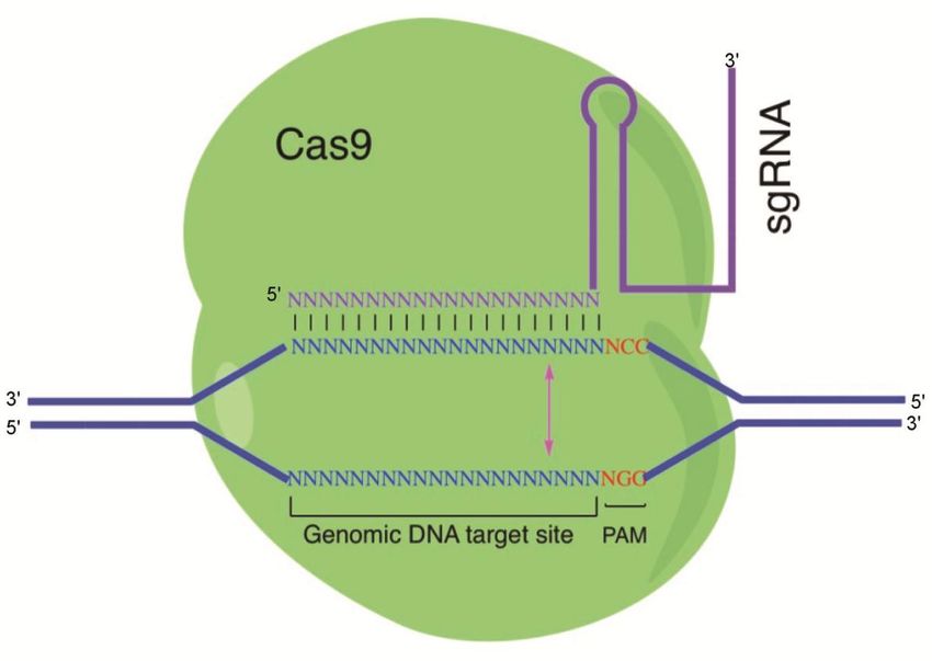
Viruses 2018, 10, x FOR PEER REVIEW 4 of 11
Figure
Figure 1.1. Illustration
Illustration of
of the
the CRISPR/Cas9 system: The
CRISPR/Cas9 system: The Cas9
Cas9 protein
protein interacts
interacts with
with the
the single-guide
single-guide
RNA
RNA (sgRNA)
(sgRNA) to to direct
direct endonuclease
endonuclease activity
activity proximal
proximal toto the
the protospacer
protospacer adjacent
adjacent motif (PAM)
motif (PAM)
sequence.
sequence. Custom-designed
Custom-designed sgRNAssgRNAs recognize
recognize their
their target
target sequence
sequence and
and allow
allow Cas9
Cas9 endonuclease
endonuclease
to
to cleave
cleave the
the sense
sense strand
strand 33 base
base pairs
pairs (bp)
(bp) and
and antisense
antisense strand
strand 33 bp
bp upstream
upstream ofof the
the PAM
PAM sequence
sequence
(NGG).
(NGG). Binding of sgRNAs to the target sites induces Cas9 endonuclease to create a double-strand
Binding of sgRNAs to the target sites induces Cas9 endonuclease to create a double-strand
break
break (blunt
(blunt end)
end) on
on the
the genomic
genomic target.
target.
2. CRISPR/Cas9 as a Tool
Tool for
for Studying
Studying Herpesvirus
Herpesvirus Host
Host Interaction
Interaction
The CRISPR/Cas9
CRISPR/Cas9 system system has
has been
been used
used inin prokaryotic
prokaryotic and eukaryotic cells for genome genome editing
editing
such asas silencing,
silencing, enhancing,
enhancing, or or modification
modification of of specific
specific genes
genes [2,3].
[2,3]. By constructing
constructing plasmids
plasmids
containing Cas9 genes and a specifically designed sgRNA, the organism’s genome can be cleaved at
most locations with
with the the only
onlylimitation
limitationbeing
beingthe theavailability
availabilityofof
thethe
PAMPAM binding
binding sequence,
sequence, NGG,
NGG, in
in
thethe targeting
targeting site.site. CRISPR/Cas9
CRISPR/Cas9 was shown
was first first shown to work
to work as a genome
as a genome engineering
engineering or editing
or editing tool in
tool
human in human cell culture,
cell culture, animals,animals, and [2].
and bacteria bacteria [2]. genome
Efficient Efficientmodifications
genome modifications have been
have been performed
performed in species such as baker’s yeasts (Saccharomycetes cerevisiae)
in species such as baker’s yeasts (Saccharomycetes cerevisiae) [15], zebrafish [16], [15], zebrafish [16], nematodes
(Caenorhabditis elegans) [17], plants [18], and
(Caenorhabditis and mice
mice [19].
[19]. Additionally,
Additionally, thethe CRISPR/Cas9
CRISPR/Cas9 system has has
been successfully
successfully applied
appliedin inbasic
basicresearch
researchfor forherpesviruses
herpesviruses bybyengineering,
engineering, targeting, activating,
targeting, or
activating,
repressing
or repressingspecific
specificgenes of interest
genes [4,20].
of interest These
[4,20]. findings
These andand
findings their implications
their implications may
maybe be
discussed
discussedin
the broadest
in the context
broadest contextpossible. Future
possible. research
Future researchdirections may
directions mayalsoalso
be highlighted.
be highlighted.
2.1.
2.1. HSV
HSV
The human interferon-inducible
The human interferon-induciblefactorfactor1616(IFI16)
(IFI16) is an
is an antiviral
antiviral factor
factor that that
bindsbinds nuclear
nuclear viral
viral DNA and promotes antiviral responses [21]. It is a nuclear protein that is
DNA and promotes antiviral responses [21]. It is a nuclear protein that is involved in the regulation involved in the
regulation of transcription and induction of interferon-β (IFN-β), and in the activation
of transcription and induction of interferon-β (IFN-β), and in the activation of inflammation. To of inflammation.
To investigate
investigate thethe
rolerole IFI16inincontrolling
ofofIFI16 controllingHSVHSV infection,
infection, CRISPR/Cas9
CRISPR/Cas9technology
technology waswas used
used to
to
generate knockouts of IFI16 [21,22]. These studies showed that IFI16 represses
generate knockouts of IFI16 [21,22]. These studies showed that IFI16 represses HSV-1 gene HSV-1 gene expression
to reduce virus
expression titers by
to reduce acting
virus as by
titers a restriction
acting as factor for HSV-1
a restriction andfor
factor preventing
HSV-1 and thepreventing
associationthe
of
association of important transcriptional activators with wild-type HSV-1 promoters. It restricts
wild-type HSV-1 replication and may play a direct or indirect role in histone modification [21,22].Viruses 2018, 10, 291 5 of 12
important transcriptional activators with wild-type HSV-1 promoters. It restricts wild-type HSV-1
replication and may play a direct or indirect role in histone modification [21,22].
UL7, an HSV-1 tegument protein, is highly conserved in viral infections and proliferation, though
its mechanism of action is still poorly understood. The replication rate of the HSV-1 UL7 mutant
(UL7-MU strain) was 10-fold lower than that of the wild-type (WT) strain [23]. The pathologic
effect of the UL7-MU strain was attenuated in infected mice compared with the wild-type HSV-1
strain. In latency, the expression of viral LAT in the central nervous system and trigeminal nerve was
lower in UL7-MU-infected mice than that in mice infected with wild-type HSV-1. By modulating the
transcription of the immediate early gene α4, UL7 may be involved in transcriptional regulation
through interacting with the transcript complex structure of the viral genome during HSV-1
infection [23]. In the study showing that the mutated tegument protein UL7 attenuated the virulence
of HSV-1, the UL7-MU strain was constructed using CRISPR/Cas9 technology [23].
2.2. EBV
EBV is etiologically responsible for Burkitt’s lymphoma and several human cancers, in which viral
genomes are maintained as multicopy episomes [6]. EBV infection in gastric epithelial cells triggers
malignant transformation by inducing resistance to oncogene-induced cell death. It is extremely
difficult to clone viral DNA because EBV-positive cancer cells are incompetent for progeny virus
production in cell culture. By CRISPR/Cas9-mediated strand break of the viral genome, bacterial
artificial chromosome (BAC) clones of EBV episomes were obtained [24]. EBV strains maintained in
two gastric cancer cell lines (SNU719 and YCCEL1) were cloned, and their complete genome sequences
were determined [24]. EBV variant strains may also be relevant to EBV-associated diseases, and the
determination of their viral genome sequences will facilitate the identification of any disease-specific
EBV strains. CRISPR/Cas9-mediated cleavage of EBV episomal DNA was found to enable the cloning
of disease-associated viral strains with unprecedented efficiency and precision [24]. As two gastric
cancer cell-derived EBV strains were cloned, the infection of epithelial cells with reconstituted viruses
revealed the mechanism of EBV-mediated epithelial carcinogenesis. The relationship between viral
genome variation and EBV-associated diseases should be established by these experiments [24].
2.3. CMV
Human CMV (HCMV) only propagates inside human cells; thus, it has developed methods
to protect itself against host stress responses and take over cellular processes to complete its life
cycle [7]. The mammalian target of rapamycin complex 1 (mTORC1) controls cell growth and anabolic
metabolism, and functions as a critical host factor activated by HCMV during successful infection [25].
Since mTORC1 is crucial for virus replication, HCMV maintains high mTORC1 activity. mTORC1
inhibitors suppressed HCMV replication in vitro and reduced the incidence of HCMV reactivation
in transplant recipients [25]. The multifunctional HCMV protein pUL38 can activate mTORC1 by
binding and antagonizing tuberous sclerosis complex protein 2 (TSC2) and plays a major role in
blocking endoplasmic reticulum stress-induced cell death during HCMV infection [25]. Therefore,
pUL38 inhibitors are a potential antiviral factor for HCMV infection and a mutant host cell line is
needed. In the study showing that pUL38 can activate mTORC1 both through TSC2-dependent
and -independent manners, the TSC2 knockout U373-MG strain was created using CRISPR/Cas9
technology [25].
The CMV genome is complex and its adaptations to cell culture have complicated the study of
infection in vivo [7]. Recombination engineering of CMV BACs enabled the study of mutant CMVs.
CRISPR/Cas9-based mutagenesis has been used to construct mutants of guinea pig CMV (GPCMV),
which can be used to infect guinea pigs, an animal model, to study CMV-associated pathogenesis
and diseases [26]. An alternative strategy for the mutagenesis of guinea pig CMV that utilizes
CRISPR/Cas9-mediated genome editing can introduce the same type of target mutations to the viral
genome as BAC-based methods. This method is highly efficient in introducing targeted insertions orViruses 2018, 10, 291 6 of 12
deletions to nonessential viral genes. Viral mutants can be recovered after limited viral replications,
minimizing the risk of spontaneous mutations. CRISPR/Cas9 avoided selection that might occur by
BAC recombination engineering and facilitated genetic manipulation of low-passage or clinical CMV
isolates [26].
2.4. KSHV
KSHV, a human oncogenic virus, adapts specific mechanisms to manipulate its host cellular
microenvironment and hijacks the host signaling pathways to maintain latent infections [5].
The pathogenesis of KSHV is closely related to its modulation of cellular signaling pathways, including
the extracellular signal-regulated kinase (ERK) pathway and the mitogen-activated protein kinase
(MAPK) pathway [20]. KSHV protein ORF45 activates the cellular kinase RSK (p90 ribosomal S6
kinase, a major functional mediator of ERK/MAPK signaling), and this activation is vital for KSHV
gene expression and virion production. Also, ORF45 is shown to contribute to the sustained activation
of both ERK and RSK during KSHV lytic replication [20]. The difference in protein phosphorylation is
significant upon induction of KSHV lytic replication in that the activation of RSK by ORF45 causes
differential phosphorylation of cellular and viral substrates [20]. A phosphoproteomic analysis of
KSHV-infected cells was performed to characterize the specific substrates of ORF45-activated RSK [27].
RSK was knocked out by CRISPR/Cas9; additionally, several cellular substrates were identified by
screening and the consequent effects on the regulation of gene expression and virion production were
characterized [27]. In the study to implicate ORF45-mediated activation of RSK in the regulation of viral
and host gene expression during KSHV lytic replication, the ORF45/RSK-dependent phosphorylation
was validated by CRISPR/Cas9-mediated knockout of RSK in KSHV-infected cells [27].
3. Treatment of Herpesvirus-Associated Diseases Based on CRISPR/Cas9
The advances in genome editing using CRISPR/Cas9 promote virological studies and may provide
a cure for persistent herpesvirus infections by directly targeting these viruses within infected cells.
Herpesvirus infection is currently treated using nucleoside analogs such as Ganciclovir, Valganciclovir,
and Foscarnet [1], but these drugs are designed to block viral polymerase activity [7]. As the
primary course of treatment is using nucleotide analogues to block viral polymerase replication,
another difficulty in herpesvirus infection treatment is the failure to control reactivation of the viruses.
The treatment is only effective when the virus genome is actively replicating; hence, the remaining
silenced viral genome can be reactivated at any time. With that being said, it is crucial to develop other
treatments against herpesvirus infections. We need to develop a treatment strategy that could disrupt
the viral genome. To conclude, a gene therapy that can edit and eliminate the herpesvirus genome is
the only solution for curing herpesvirus latent infections. Here, we review recent applications of the
CRISPR/Cas9 system and discuss its therapeutic potential to treat the productive and latent infections
of herpesviruses [20,28–30].
Roehm et al. adapted the CRISPR system to treating HSV-1 infection [31]. Roehm and colleagues
designed sgRNAs targeting ICP0, a crucial viral-encoded protein that can regulate viral gene expression
and replication. Although sgRNA targeting ICP0, an immediate early gene that promotes transcription
from the viral gene, exhibited a significant decrease in viral production, a mixture of sgRNAs targeting
ICP0, ICP4, and ICP27 eliminated HSV viral infection. These promising results suggest CRISPR as
a possible solution for treating HSV infection.
In 2016, Diemen et al. conducted a study that used CRISPR/Cas9 technology to suppress
herpesvirus virus replication in both latent and lytic infection models [4]. This study showed CRISPR’s
ability to suppress three prototypical members of the herpesvirus family: EBV, HSV, and HCMV.
For EBV, the authors designed sgRNAs for both the latent infection model and the lytic infection
model. For the latent infection model, sgRNAs were designed to target EBV viral miRNAs miR-BART5,
miR-BART6, and miR-BART16. The EBV latently infected gastric carcinoma cells were transduced to
express CRISPR/Cas9 sgRNAs. A luciferase assay followed by sequencing showed that the targetedViruses 2018, 10, 291 7 of 12
miRNAs were downregulated and edited. This experiment illustrates the application of CRISPR
technology in a herpesvirus latent infection model. For the lytic infection model, the authors designed
sgRNAs targeting the viral EBV nuclear antigen 1 (EBNA1) and several areas within the EBV origin
of replication (OriP). EBV-GFP infected cells had an almost complete loss of GFP after a combination
of sgRNAs treatment. A similar approach was tested in both HCMV and HSV. Both lytic infection
models showed almost complete suppression of viral infections. Additionally, CRISPR gene editing
could abrogate replication of HSV-1 reactivated from quiescence.
Other groups also proved the effectiveness of CRISPR/Cas9 in treating latent herpesvirus
infections. Wang and Quake investigated the application of CRISPR/Cas9 in treating latent virus
infection in EBV [32]. They designed several sgRNAs that targeted the EBV genome location
responsible for the viral structure, transformation, and latency. Patient-derived cells with latent
Epstein–Barr virus infection were treated with the designed CRISPR/Cas9. The results showed the
elimination of EBV genome in a quarter of the cells, and half of the cells showed a decrease in viral
load after CRISPR/Cas9 treatment. Yuen et al. designed sgRNAs targeting EBV genome region
EBNA1, OriP, and W repeats [33]. Their study showed a reduction of 50% in viral genome copy
number. Although the suppression of the EBV viral genome did not affect EBV latently infected cells,
the remaining infected cells were sensitized to chemotherapy.
While it is difficult to find good animal models for human herpesvirus infections, there are several
studies providing proof of concept evidence of CRISPR/Cas9 technology treating viral infections
in the animal model. Lin et al. conducted a study using CRISPR gene editing technology to treat
HBV infection both in vitro and in vivo [34]. They first tested the efficacy of sgRNAs targeting HBV
viral genomes in vitro. Then, the top two sgRNAs that had highest targeting efficiency were selected
for application in an in vivo study. They used the well-established HBV hydrodynamics-mouse
model of HBV infection. HBV expression vector and the CRISPR/Cas9 dual expression vectors were
coinjected into the tail veins of C57BL/6 mice by hydrodynamic transfection. Serum HBsAg levels
were significantly reduced in mice receiving HBV-specific sgRNAs targeting the HBV viral genome.
Sequencing results showed 27.8% of the clones contained indel in the targeted region. In another
study, Yin et al. demonstrated the CRISPR/Cas9 system in excising the HIV-1 provirus in different
animal models representing the different levels of clinical relevance [35]. They intravenously injected
quadruplex sgRNAs/saCas9 AAV-DJ/8 into mice. The results showed that CRISPR/Cas9 treatment
excised HIV-1 proviral DNA and significantly reduced viral RNA expression in several organs/tissues
of mice. Additionally, successful proviral excision was detected by PCR genotyping in different organs.
Both studies reviewed above showed the ability of delivering a CRISPR/Cas9 system effectively and
safely to the targeted cells and organs.
Taken together, this progress in adapting CRISPR/Cas9 in suppressing herpes viral replication,
and the advances in delivering the CRISPR system in animal models provide hope for finding a cure
for herpesvirus infections.
4. Challenges of CRISPR/Cas9 Delivery
Delivery tools for transfection or gene transfer are agents to facilitate nucleic acids entering
target cells. Their main function is to increase the transfection efficiency of DNA (including genes,
plasmid DNA, and DNA fragments) and RNA (including mRNA, miRNA, and siRNA) in vitro or
in vivo. The most common strategies for CRISPR/Cas9 delivery are lipoids [36,37], viruses [36,37],
nanoparticles [36,37], bacteria [38], gene guns [39], electroporation [40], and nanostraws [41] (Table 2).Viruses 2018, 10, 291 8 of 12
Table 2. Delivery tools for CRISPR/Cas9.
Delivery Tools Example Characteristic
The lipid subunits which form liposomes
entrap the transfection materials, allowing
themselves to overcome the electrostatic
Lipoid Lipofectamine, Liposome
repulsion of the cell membrane to let DNA or
RNA cross into the cytoplasm to access the
nuclei or organelles.
Lentivirus, Adenovirus, A specific virus is engineered to deliver DNA
Virus Adeno-associated virus (AAV), or RNA to target cells and used as a vector for
Baculovirus gene transfer.
Nanoparticles (1–100 nanometers in size),
Mesoporous silica nanoparticles
consist of a variety of compounds and
Nanoparticle (MSNs), Dendrimers, Carbon
materials, can be complexed with DNA or
Nanotubes, Cationic polymers
RNA for gene delivery.
An attenuated strain of Salmonella which is
invasive but nonpathogenic shows DNA
Bacterium Salmonella
transfer activity with little cytotoxicity and
pathogenicity in hosts.
The device, a biolistic particle delivery
system, is used for delivering exogenous
Gene gun PDS-1000/He Particle Delivery System
DNA to cells; the payload is an elemental
particle of a heavy metal coated with DNA.
An electric field is applied to cells to increase
Electroporation Electroporator the cell permeability, allowing DNA to be
introduced into the cell.
The device is used for creating a direct
Nanostraw Navan physical conduit to cells for DNA delivery,
mimicking the gap junction between cells.
CRISPR/Cas components must be transported to the target cells to exert a therapeutic effect.
The delivery will be critical to the success of therapeutic genome editing applications. Therefore,
the delivery tool is essential for CRISPR/Cas9 delivery to target cells and it is very crucial to
select a suitable delivery tool with high specificity, efficiency, and safety. However, the options
of CRISPR/Cas9 delivery tools present challenges due to the following issues.
1. The delivery tool is not specific enough: Some delivery tools are not very specific and may deliver
nucleic acids to nontarget cells. It is important to reduce the risk of nonspecific delivery, but the
evaluation of their benefits and risks is complex.
2. The delivery tool is not very efficient: Not all delivery tools are efficient; some of them are low in
efficiency and require multiple rounds of transfections. Additionally, it is hard to improve and
evaluate their efficiency, especially in animals and clinics.
3. The delivery tool is deficient in biosafety: Some delivery tools are toxic, biohazardous, or even
destructive to normal cells or recipient hosts. Some delivery tools such as lipoids, viruses, bacteria,
and nanoparticles may induce vector-associated immune responses in hosts, and immune barriers
must be overcome [36]. Thus, verifying their safety in preliminary testing is needed.
In several recent studies, encouraging progress has been made to possibly overcome the
challenges of delivering CRISPR/Cas9 in vivo (Table 3). Adeno-associated viruses (AAV) with low
immunogenicity enter the cells by endocytosis upon binding to the specific integrin and receptor
and integrate at a specific site called AAVS1 in the host genome. This site-specific integration
avoids unpredictable insertion mutation and other harmful consequences [42]. A novel approach
integrates large single-strand transgene cassettes into the genomes, increasing the knock-in efficiency
by combining CRISPR/Cas9 with AAV by 13.6–19.5-fold compared with conventional AAV-mediated
methods [43]. A single administration of lipid nanoparticles (LNP) that are biodegradable and well
tolerated can deliver CRISPR/Cas9 components to achieve high-efficiency in vivo genome editing withViruses 2018, 10, 291 9 of 12
a concomitant reduction of the mouse transthyretin (TTR) gene serum protein [44]. The packaging
of the newly discovered smaller Cas9 and its guide RNA into one AAV delivery vehicle allows for
efficient in vivo genome editing. The combination of small Cas9 orthologues, tissue-specific promoters,
specific AAV serotypes, and different routes of delivery has improved the efficiency and precision for
in vivo application and overcome the immunogenicity and toxicity of the delivery tools [45].
Table 3. Possible strategies for overcoming the challenges for CRISPR/Cas9 delivery.
Challenge Strategy
Specificity Discovery of a specific virus such as adeno-associated viruses (AAV).
Efficiency Application of a combination system such as AAV-CRISPR.
Combination with several factors such as smaller Cas9 orthologues,
tissue-specific minimal promoters, AAV serotypes, and different
Biosafety routes of administration;
Development of novel and safe delivery tools such as lipid
nanoparticles (LNP), AAV, and baculoviruses.
5. Conclusions
CRISPR/Cas9, a naturally occurring component of a bacterial immune system, uses a novel
nuclease system to protect bacteria from bacteriophage infections and has been harnessed for a variety
of genome editing applications. By delivering the Cas9 protein and appropriate guide RNAs into a cell,
the organism’s genome can be cut at most locations with the availability of the PAM binding sequence.
It can also be modified to make programmable transcription factors that allow scientists to target
and activate or repress specific genes. Several studies have illustrated the utility of CRISPR/Cas9
technology for the study of herpesviruses including HSV, EBV, CMV, and KSHV. Specific genes
and factors/proteins have proved to be essential for herpesvirus proliferation and latency through
CRISPR/Cas9-mediated engineering in host or viral genomes. These promising results in basic research
contribute to an increased understanding of herpesviruses, the establishment of specific cell lines or
potential animal models, as well as the development of new drugs for viral infections.
The pharmaceutical studies have made great achievements in drug discovery, and therapeutic
options have been expanded to include small molecules (e.g., siRNAs), antiviral agents, protease
inhibitors, and preventive vaccines for diseases caused by viral infections [1]. However, effective
treatments for persistent, recurrent, and highly prevalent herpesviruses are still unavailable.
This represents a significant unmet medical need for herpesvirus infections such as HSV, EBV, HCMV,
and KSHV. Current antiviral therapy is not able to eradicate latent viruses in humans efficaciously,
though the standard drugs are effective for the treatment of lytic infection and alleviation of symptoms
in patients. The advance of CRISPR/Cas9 technology presents a novel and promising strategy to
treat latent herpesvirus infections. Previous work has shown that CRISPR/Cas9 can be adapted for
antiviral treatment for latency ex vivo and/or in vivo. Furthermore, there is encouraging evidence that
the ever-important delivery challenge can be overcome with the use of AAV delivery vectors and lipid
nanoparticles. CRISPR/Cas9 may have the potential to be an effective therapy against latent viral
infections in humans, though more preclinical studies will be needed to demonstrate its effectiveness
in vivo.
Author Contributions: Y.-C.C., J.S., and F.L. wrote the manuscript, P.T. contributed to revise the manuscript.
Acknowledgments: We are grateful to Marco Paliza-Carre, and Ting Wang for critical comments and editorial
assistance. This research has been supported by grants from Guangdong Innovative and Entepreneurial Research
Team Program (No. 2014ZT05S136), National Mega Project on Major Drug Development (2009ZX09103-678,
2012ZX09102301-004, 2012ZX09103301-20, 2013ZX09102-031, 2014ZX09509001-001), Antiviral Cooperative
Innovation Center of Traditional Chinese Medicine at Shandong Province (XTCX2014B01-09), Project for
Construction of Guangzhou Key Laboratory of Virology (No. 201705030003), National Small Business InnovationViruses 2018, 10, 291 10 of 12
and Research (SBIR) Program of China, the Technology R & D Program of Jiangsu Province, China (BG2007035
and BG2008662), and NIH (RO1-AI041927, RO1-AI091536, RO1-DE023935, and RO1-DE025462)
Conflicts of Interest: The authors declare no conflict of interest.
References
1. Pellett, P.E.; Roizman, B. Herpesviridae. In Fields Virology; Lippincott-William & Wilkins: Philadelphia, PA,
USA, 2013; pp. 1802–1822.
2. Charpentier, E.; Doudna, J.A. Biotechnology: Rewriting a genome. Nature 2013, 495, 50–51. [CrossRef]
[PubMed]
3. Koonin, E.V.; Makarova, K.S.; Zhang, F. Diversity, classification and evolution of CRISPR-Cas systems.
Curr. Opin. Microbiol. 2017, 37, 67–78. [CrossRef] [PubMed]
4. Van Diemen, F.R.; Kruse, E.M.; Hooykaas, M.J.; Bruggeling, C.E.; Schurch, A.C.; van Ham, P.M.; Imhof, S.M.;
Nijhuis, M.; Wiertz, E.J.; Lebbink, R.J. CRISPR/Cas9-mediated genome editing of herpesviruses limits
productive and latent infections. PLoS Pathog. 2016, 12, e1005701. [CrossRef] [PubMed]
5. Damania, B.; Cesarman, E. Kaposi’s sarcoma-associated herpesvirus. In Fields Virology; Lippincott-William &
Wilkins: Philadelphia, PA, USA, 2013; pp. 2080–2128.
6. Longnecker, R.; Kieff, E.; Cohen, J.I. Epstein-Barr Virus/Replication and Epstein-Barr Virus. In Fields Virology;
Lippincott-William & Wilkins: Philadelphia, PA, USA, 2013; pp. 1898–1959.
7. Mocarski, E.S.; Shenk, T.; Griffiths, P.D.; Pass, R.F. Cytomegaloviruses. In Fields Virology; Lippincott-William
& Wilkins: Philadelphia, PA, USA, 2013; pp. 1960–2014.
8. Roizman, B.; Knipe, D.M.; Whitley, R.J. Herpes Simplex Viruses. In Fields Virology; Lippincott-William &
Wilkins: Philadelphia, PA, USA, 2013; pp. 1823–1897.
9. Jiang, F.; Doudna, J.A. CRISPR-Cas9 structures and mechanisms. Annu. Rev. Biophys. 2017, 46, 505–529.
[CrossRef] [PubMed]
10. Horvath, P.; Barrangou, R. CRISPR/Cas, the immune system of bacteria and archaea. Science 2010, 327,
167–170. [CrossRef] [PubMed]
11. Shibata, M.; Nishimasu, H.; Kodera, N.; Hirano, S.; Ando, T.; Uchihashi, T.; Nureki, O. Real-space and
real-time dynamics of CRISPR-Cas9 visualized by high-speed atomic force microscopy. Nat. Commun. 2017,
8, 1430. [CrossRef] [PubMed]
12. Knight, S.C.; Xie, L.; Deng, W.; Guglielmi, B.; Witkowsky, L.B.; Bosanac, L.; Zhang, E.T.; El Beheiry, M.;
Masson, J.B.; Dahan, M.; et al. Dynamics of CRISPR-Cas9 genome interrogation in living cells. Science 2015,
350, 823–826. [CrossRef] [PubMed]
13. Anders, C.; Niewoehner, O.; Duerst, A.; Jinek, M. Structural basis of PAM-dependent target DNA recognition
by the Cas9 endonuclease. Nature 2014, 513, 569–773. [CrossRef] [PubMed]
14. Jinek, M.; Chylinski, K.; Fonfara, I.; Hauer, M.; Doudna, J.A.; Charpentier, E. A programmable
dual-RNA-guided DNA endonuclease in adaptive bacterial immunity. Science 2012, 337, 816–821. [CrossRef]
[PubMed]
15. DiCarlo, J.E.; Norville, J.E.; Mali, P.; Rios, X.; Aach, J.; Church, G.M. Genome engineering in Saccharomyces
cerevisiae using CRISPR-Cas systems. Nucleic Acids Res. 2013, 41, 4336–4343. [CrossRef] [PubMed]
16. Hwang, W.Y.; Fu, Y.; Reyon, D.; Maeder, M.L.; Tsai, S.Q.; Sander, J.D.; Peterson, R.T.; Yeh, J.R.; Joung, J.K.
Efficient genome editing in zebrafish using a CRISPR-Cas system. Nat. Biotechnol. 2013, 31, 227–229.
[CrossRef] [PubMed]
17. Friedland, A.E.; Tzur, Y.B.; Esvelt, K.M.; Colaiacovo, M.P.; Church, G.M.; Calarco, J.A. Heritable genome
editing in C. elegans via a CRISPR-Cas9 system. Nat. Methods 2013, 10, 741–743. [CrossRef] [PubMed]
18. Jiang, W.; Zhou, H.; Bi, H.; Fromm, M.; Yang, B.; Weeks, D.P. Demonstration of CRISPR/Cas9/sgRNA-mediated
targeted gene modification in Arabidopsis, tobacco, sorghum and rice. Nucleic Acids Res. 2013, 41, e188.
[CrossRef] [PubMed]
19. Wang, H.; Yang, H.; Shivalila, C.S.; Dawlaty, M.M.; Cheng, A.W.; Zhang, F.; Jaenisch, R. One-step generation
of mice carrying mutations in multiple genes by CRISPR/Cas-mediated genome engineering. Cell 2013, 153,
910–918. [CrossRef] [PubMed]
20. Van Diemen, F.R.; Lebbink, R.J. CRISPR/Cas9, a powerful tool to target human herpesviruses. Cell. Microbiol.
2017, 19. [CrossRef] [PubMed]Viruses 2018, 10, 291 11 of 12
21. Diner, B.A.; Lum, K.K.; Toettcher, J.E.; Cristea, I.M. Viral DNA sensors IFI16 and cyclic GMP-AMP synthase
possess distinct functions in regulating viral gene expression, immune defenses, and apoptotic responses
during herpesvirus infection. MBio 2016, 7, e01553-16. [CrossRef] [PubMed]
22. Johnson, K.E.; Bottero, V.; Flaherty, S.; Dutta, S.; Singh, V.V.; Chandran, B. IFI16 restricts HSV-1 replication by
accumulating on the hsv-1 genome, repressing HSV-1 gene expression, and directly or indirectly modulating
histone modifications. PLoS Pathog. 2014, 10, e1004503. [CrossRef] [PubMed]
23. Xu, X.; Fan, S.; Zhou, J.; Zhang, Y.; Che, Y.; Cai, H.; Wang, L.; Guo, L.; Liu, L.; Li, Q. The mutated tegument
protein UL7 attenuates the virulence of herpes simplex virus 1 by reducing the modulation of alpha-4 gene
transcription. Virol. J. 2016, 13, 152. [CrossRef] [PubMed]
24. Kanda, T.; Furuse, Y.; Oshitani, H.; Kiyono, T. Highly efficient CRISPR/Cas9-mediated cloning and functional
characterization of gastric cancer-derived Epstein-Barr virus strains. J. Virol. 2016, 90, 4383–4393. [CrossRef]
[PubMed]
25. Bai, Y.; Xuan, B.; Liu, H.; Zhong, J.; Yu, D.; Qian, Z. Tuberous Sclerosis Complex Protein 2-Independent
Activation of mTORC1 by Human Cytomegalovirus pUL38. J. Virol. 2015, 89, 7625–7635. [CrossRef]
[PubMed]
26. Bierle, C.J.; Anderholm, K.M.; Wang, J.B.; McVoy, M.A.; Schleiss, M.R. Targeted mutagenesis of guinea
pig cytomegalovirus using CRISPR/Cas9-mediated gene editing. J. Virol. 2016, 90, 6989–6998. [CrossRef]
[PubMed]
27. Avey, D.; Tepper, S.; Li, W.; Turpin, Z.; Zhu, F. Phosphoproteomic Analysis of KSHV-Infected Cells Reveals
Roles of ORF45-Activated RSK during Lytic Replication. PLoS Pathog. 2015, 11, e1004993. [CrossRef]
[PubMed]
28. Liang, X.; Sun, L.; Yu, T.; Pan, Y.; Wang, D.; Hu, X.; Fu, Z.; He, Q.; Cao, G. A CRISPR/Cas9 and Cre/Lox
system-based express vaccine development strategy against re-emerging Pseudorabies virus. Sci. Rep. 2016,
6, 19176. [CrossRef] [PubMed]
29. Moos, W.H.; Pinkert, C.A.; Irwin, M.H.; Faller, D.V.; Kodukula, K.; Glavas, I.P.; Steliou, K. Epigenetic
treatment of persistent viral Infections. Drug Dev. Res. 2017, 78, 24–36. [CrossRef] [PubMed]
30. Pennington, M.R.; Van de Walle, G.R. Electric cell-substrateiImpedance sensing to monitor viral growth
and study cellular responses to infection with alphaherpesviruses in real time. mSphere 2017, 2. [CrossRef]
[PubMed]
31. Roehm, P.C.; Shekarabi, M.; Wollebo, H.S.; Bellizzi, A.; He, L.; Salkind, J.; Khalili, K. Inhibition of HSV-1
replication by gene editing strategy. Sci. Rep. 2016, 6, 23146. [CrossRef] [PubMed]
32. Wang, J.; Quake, S.R. RNA-guided endonuclease provides a therapeutic strategy to cure latent herpesviridae
infection. Proc. Natl. Acad. Sci. USA 2014, 111, 13157–13162. [CrossRef] [PubMed]
33. Yuen, K.S.; Wang, Z.M.; Wong, N.M.; Zhang, Z.Q.; Cheng, T.F.; Lui, W.Y.; Chan, C.P.; Jin, D.Y. Suppression of
Epstein-Barr virus DNA load in latently infected nasopharyngeal carcinoma cells by CRISPR/Cas9. Virus
Res. 2018, 244, 296–303. [CrossRef] [PubMed]
34. Lin, S.R.; Yang, H.C.; Kuo, Y.T.; Liu, C.J.; Yang, T.Y.; Sung, K.C.; Lin, Y.Y.; Wang, H.Y.; Wang, C.C.;
Shen, Y.C.; et al. The CRISPR/Cas9 system facilitates clearance of the intrahepatic HBV templates in vivo.
Mol. Ther. Nucleic Acids 2014, 19, e186. [CrossRef] [PubMed]
35. Yin, C.; Zhang, T.; Qu, X.; Zhang, Y.; Putatunda, R.; Xiao, X.; Li, F.; Xiao, W.; Zhao, H.; Dai, S.; et al. In vivo
excision of HIV-1 provirus by saCas9 and multiplex single-guide RNAs in animal models. Mol. Ther. 2017,
25, 1168–1186. [CrossRef] [PubMed]
36. Das, S.K.; Menezes, M.E.; Bhatia, S.; Wang, X.Y.; Emdad, L.; Sarkar, D.; Fisher, P.B. Gene therapies for cancer:
Strategies, challenges and successes. J. Cell. Physiol. 2015, 230, 259–271. [CrossRef] [PubMed]
37. Goncalves, G.A.R.; Paiva, R.M.A. Gene therapy: Advances, challenges and perspectives. Einstein (Sao Paulo)
2017, 15, 369–375. [CrossRef] [PubMed]
38. Bai, Y.; Gong, H.; Li, H.; Vu, G.P.; Lu, S.; Liu, F. Oral delivery of RNase P ribozymes by Salmonella inhibits
viral infection in mice. Proc. Natl. Acad. Sci. USA 2011, 108, 3222–3227. [CrossRef] [PubMed]
39. Gan, W.B.; Grutzendler, J.; Wong, W.T.; Wong, R.O.; Lichtman, J.W. Multicolor “DiOlistic” labeling of the
nervous system using lipophilic dye combinations. Neuron 2000, 27, 219–225. [CrossRef]
40. Guo, W.; Guo, Y.; Tang, S.; Qu, H.; Zhao, H. Dendritic cell-Ewing’s sarcoma cell hybrids enhance antitumor
immunity. Clin. Orthop. Relat. Res. 2008, 466, 2176–2183. [CrossRef] [PubMed]Viruses 2018, 10, 291 12 of 12
41. Stewart, M.P.; Sharei, A.; Ding, X.; Sahay, G.; Langer, R.; Jensen, K.F. In vitro and ex vivo strategies for
intracellular delivery. Nature 2016, 538, 183–192. [CrossRef] [PubMed]
42. Oggu, G.S.; Sasikumar, S.; Reddy, N.; Ella, K.K.R.; Rao, C.M.; Bokara, K.K. Gene delivery approaches for
mesenchymal stem cell therapy: Strategies to increase efficiency and specificity. Stem. Cell Rev. 2017, 13,
725–740. [CrossRef] [PubMed]
43. Xiao, Q.; Min, T.; Ma, S.; Hu, L.; Chen, H.; Lu, D. Intracellular generation of single-strand template increases
the knock-in efficiency by combining CRISPR/Cas9 with AAV. Mol. Genet. Genom. 2018; in press.
44. Finn, J.D.; Smith, A.R.; Patel, M.C.; Shaw, L.; Youniss, M.R.; van Heteren, J.; Dirstine, T.; Ciullo, C.;
Lescarbeau, R.; Seitzer, J.; et al. A single administration of CRISPR/Cas9 lipid nanoparticles achieves
robust and persistent in vivo genome editing. Cell Rep. 2018, 22, 2227–2235. [CrossRef] [PubMed]
45. Lau, C.H.; Suh, Y. In vivo genome editing in animals using AAV-CRISPR system: Applications to
translational research of human disease. F1000Res 2017, 6, 2153. [CrossRef] [PubMed]
© 2018 by the authors. Licensee MDPI, Basel, Switzerland. This article is an open access
article distributed under the terms and conditions of the Creative Commons Attribution
(CC BY) license (http://creativecommons.org/licenses/by/4.0/).You can also read


