Identification and characterization of satellite DNAs in two toed sloths of the genus Choloepus (Megalonychidae, Xenarthra) - Nature
←
→
Page content transcription
If your browser does not render page correctly, please read the page content below
www.nature.com/scientificreports
OPEN Identification and characterization
of satellite DNAs in two‑toed
sloths of the genus Choloepus
(Megalonychidae, Xenarthra)
Radarane Santos Sena1, Pedro Heringer1, Mirela Pelizaro Valeri1, Valéria Socorro Pereira2,
Gustavo C. S. Kuhn1 & Marta Svartman1*
Choloepus, the only extant genus of the Megalonychidae family, is composed of two living species of
two-toed sloths: Choloepus didactylus and C. hoffmanni. In this work, we identified and characterized
the main satellite DNAs (satDNAs) in the sequenced genomes of these two species. SATCHO1, the
most abundant satDNA in both species, is composed of 117 bp tandem repeat sequences. The second
most abundant satDNA, SATCHO2, is composed of ~ 2292 bp tandem repeats. Fluorescence in situ
hybridization in C. hoffmanni revealed that both satDNAs are located in the centromeric regions of
all chromosomes, except the X. In fact, these satDNAs present some centromeric characteristics in
their sequences, such as dyad symmetries predicted to form secondary structures. PCR experiments
indicated the presence of SATCHO1 sequences in two other Xenarthra species: the tree-toed sloth
Bradypus variegatus and the anteater Myrmecophaga tridactyla. Nevertheless, SATCHO1 is present
as large tandem arrays only in Choloepus species, thus likely representing a satDNA exclusively in this
genus. Our results reveal interesting features of the satDNA landscape in Choloepus species with the
potential to aid future phylogenetic studies in Xenarthra and mammalian genomes in general.
A significant part of eukaryotic genomes, ~ 30% in some plants to more than 50% in some insects and mammals,
is composed of tandemly organized highly repetitive sequences, known as satellite DNAs (satDNAs) (reviewed
in Ref.1). In general, satDNAs differ from other tandemly repetitive sequences by their organization, which
consists of long arrays that can extend up to megabases in length. SatDNAs are major components of the con-
stitutive heterochromatin present in fundamental chromosome structures, such as centromeres and telomeres
(reviewed in Refs.1,2).
They also have been shown to be important components of chromosome organization, pairing, and segre-
gation. For instance, their transcripts have been reported to participate in centromeric activity and genomic
regulation3–5. Some satDNAs also have protein binding motifs such as the CENP-B motif which, together with
the CENP-B protein, is known to be involved in kinetochore structuring by helping the assembly of the CENP-A
protein in m ammals6–8. Both the CENP-B protein and the CENP-B box motif are largely conserved in mamma-
lian centromeres, but despite this broad conservation, the role of the CENP-B proteins is still poorly understood
(reviewed in Ref.8).
Moreover, around 50% of some studied satDNAs have short inverted repeat (short dyad symmetry) sequences
within their monomers, which have been reported as essential to chromatin structure and/or function4,7,9–11.
Short dyad symmetry sequences have been identified in satellite DNA-free centromeres and in centromeric
satDNAs which lack CENP-B boxes7. Those dyad symmetries are predicted to adopt non-B-form DNA structures
such as cruciform, hairpins, triplexes, and single-stranded DNA, which are commonly identified in functional
centromeres4,7.
It is important to note that functional centromere sequences (those associated with CENP-A) are restricted
to relatively short segments of DNA nested within megabase arrays of pericentromeric satDNAs, each of them
having different epigenetic c ompositions1,11. Although pericentromeric satDNAs are involved in centromere
maintenance and stability, the factors determining their boundaries and intrinsic differences with functional
centromeric sequences are not fully k nown1.
1
Laboratório de Citogenômica Evolutiva, Departamento de Genética, Ecologia e Evolução, Instituto de Ciências
Biológicas, Universidade Federal de Minas Gerais, Belo Horizonte, MG, Brazil. 2Fundação de Parques Municipais e
Zoobotânica, Belo Horizonte, MG, Brazil. *email: svartmanm@ufmg.br
Scientific Reports | (2020) 10:19202 | https://doi.org/10.1038/s41598-020-76199-8 1
Vol.:(0123456789)
www.nature.com/scientificreports/
C. didactylus C. hoffmanni
Satellite DNA SATCHO1 SATCHO2 SATCHO1 SATCHO2
Satellite confidence High Low High Low
Satellite probability 0.986 0.0425 0.992 0.0426
Consensus size 117 bp 2292 bp 117 bp 2292 bp
Genome proportion 13% 0.62% 2.6% 0.23%
AT content 58.97% 54.97% 58.97% 54.97%
Table 1. Putative satDNAs identified by RepeatExplorer2 in C. didactylus and C. hoffmanni.
SatDNAs are important components in the evolution of eukaryotic genomes. They can evolve three times
faster than intergenic regions, which often results in significant differences between sequences, even among
closely related species (reviewed in Ref.1). This rapid evolution is thought to be a consequence of mechanisms
such as unequal crossing-over, gene conversion and replication s lippage12, which are all related with the process
known as molecular drive, described by Dover13. Because new mutations are constantly spread by molecular
drive, intraspecific satDNA arrays are often composed of very similar tandemly repeated sequences that have
the potential to be used as species-specific markers.
The study of repetitive DNAs has been significantly advanced with the introduction of next-generation
sequencing technologies and high-throughput in silico analyses of genomes (reviewed in Ref.1). One of the tools
used in these studies is RepeatExplorer, a pipeline that identifies repetitive DNA sequences de novo in genomes,
using the raw reads without the need of a reference library of known repetitive sequences14. This pipeline per-
forms graph-based clustering analyses, identifying read similarities by comparing pairwise reads all-to-all, before
grouping them into clusters.
Xenarthra is a basal eutherian group which originated and diversified entirely in South America15,16. With 31
recognized extant species, this superorder is divided into two orders: Cingulata, represented by armadillos; and
Pilosa, composed by anteaters (Vermilingua) and sloths (Folivora)17. Despite its importance as a basal placental
group, Xenarthra has been poorly studied in comparison with other mammals, mostly because of their strict
geographic distribution and collection difficulty because of their natural behavior. Hence, more information
about their ecology and genetics is essential to a better characterization of the g roup16.
Studies on the repetitive DNA fraction of Xenarthra genomes have been mostly restricted to the identification
of retrotransposon families. For instance, LINE (Long Interspersed Element) and SINE (Small Interspersed Ele-
ment) families have been described in six species: the sloths Choloepus hoffmanni and Bradypus tridactylus18,19,
the anteaters Tamandua tetradactyla and Myrmecophaga tridactyla18,20, and the armadillos Dasypus novemcinctus
and Euphractus sexcinctus18,21. Currently, the only Xenarthra species with an identified satDNA sequence is the
armadillo D. novemcinctus22, which has a satDNA with ~ 173 bp monomers. Mapping by fluorescence in situ
hybridization (FISH) revealed that this satDNA was present in the centromeres of all chromosomes in this
species22.
Two-toed sloths are the only extant representatives of the Megalonychidae family, composed by the single
living genus Choloepus23, with two species: C. didactylus and C. hoffmanni. Both species inhabit the tropical
forests of South and Central America with a small overlap area of occurrence in the Amazon forest in Peru,
southwestern Amazonas state and Acre state in Brazil. These two species can be differentiated mainly by mor-
phological characters, such as pelage color24, osteological features25, the mitochondrial COI and Cyt-b genes, and
the nuclear gene Enamelin26,27. Cytogenetic analyses of Choloepus have been mostly based on simple karyotypic
descriptions without banding p atterns26,28–34. These studies revealed a complex and confusing karyotypic scenario
with significant variation in diploid numbers in C. didactylus (2n = 52–67) and less variation in C. hoffmanni
(2n = 49–53), with translocations between the Y chromosome and different autosomes, occurrence of X0 females,
and unpaired chromosomes described as B chromosomes.
In this work we identified and characterized the most abundant satDNA sequences from the C. didactylus
and C. hoffmanni genomes using in silico methods. In addition, we mapped these sequences in the chromosomes
of C. hoffmanni. This is the first study to identify, characterize and map satDNAs in sloths, revealing interesting
aspects of the centromeric and repetitive fraction of their genomes.
Results
In silico identification and analysis of satDNAs. The RepeatExplorer2 analysis identified two abun-
dant putative satDNAs in the C. didactylus and C. hoffmanni genomes, which we named SATCHO1 and SAT-
CHO2 (Supplementary data 1 and 2) (Table 1). The analysis indicated differences in the proportion of satDNAs
in the two species: the satDNA content represents > 13% of the C. didactylus genome, whereas this value is
approximately 3% in C. hoffmanni.
SATCHO1, the most abundant satDNA sequence in both species, has ~ 117 bp monomers, low levels of
inter-repeat nucleotide variability (~ 3% on average) and AT content of ~ 59%. This satDNA represents 13% of
the C. didactylus and 2.6% of the C. hoffmanni genomes. SATCHO2 is the second most abundant satDNA and
has ~ 2292 bp monomers, inter-repeat nucleotide variability of ~ 24% on average and AT content of ~ 55%. It
corresponds to 0.62% and 0.23% of the C. didactylus and C. hoffmanni genomes, respectively. Although SAT-
CHO1 and SATCHO2 sequences are abundant in both genomes, we did not identify similar sequences in any
Scientific Reports | (2020) 10:19202 | https://doi.org/10.1038/s41598-020-76199-8 2
Vol:.(1234567890)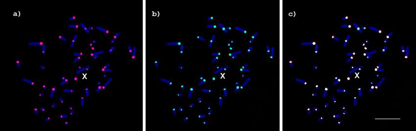
www.nature.com/scientificreports/
Figure 1. Identification of conserved (dark grey) and variable (light grey) satDNA segments of C. didactylus
and C. hoffmanni by sliding window analysis. Sliding window of 10 bp for (a) SATCHO1 and (b) SATCHO2.
Nucleotide diversity (Pi) is indicated by the red line, average nucleotide diversity is indicated by the solid line,
and average diversity ± 2 SD is indicated by the dotted line.
other species on Repbase or in searches against all sequences from the non-redundant nucleotide collection in
Genbank (accessed in 03/01/2020).
The analysis of nucleotide variability along both satDNAs revealed the presence of conserved regions within
their monomers (Fig. 1), even though satDNAs are expected to evolve neutrally, revealing regions under potential
selective constraints.
Phylogenetic and NMDS analyses. In order to infer the interspecific similarity between copies of SAT-
CHO1 and SATCHO2 in C. didactylus and C. hoffmanni, we constructed a Neighbor-Joining tree using a sample
of copies from each satDNA. The resulting tree showed that satDNA copies from both species are very similar
and did not segregate into species-specific branches for SATCHO1 and SATCHO2 sequences (Fig. 2a,c).
We also estimated the pairwise distance values of the same set of sequences to generate NMDS ordinations
for their Euclidean distances. The results also did not reveal any clear topological segregation between copies
from each species (Fig. 2b,d). Nevertheless, each satDNA appeared to evolve under distinct evolutionary rates,
as evidenced by their heterogeneous distribution across the NMDS ordinations.
Overall, both analyses indicate that the satDNA sequences from C. didactylus and C. hoffmanni have not
diverged enough to segregate into species-specific clusters.
Chromosome mapping of SATCHO1 and SATCHO2. The C. hoffmanni individual we studied pre-
sented a karyotype with a diploid number 2n = 51. GTG-banding allowed the identification of all chromosome
pairs and of an odd chromosome, which we identified as a B chromosome (Fig. 3a). The CBG-banding revealed
the presence of constitutive heterochromatin in the centromeric regions of all chromosomes, except the X
(Fig. 3b).
Our specimen has the same karyotype described earlier by Svartman et al.34 for C. hoffmanni (2n = 50), from
which it differs by the presence of the extra odd chromosome and by an inversion in pair 3 (metacentric in our
specimen and acrocentric in the one previously described).
SATCHO1 and SATCHO2 were both FISH mapped in the centromeric regions of all C. hoffmanni chro-
mosomes, except the X (Fig. 4), coinciding with the constitutive heterochromatin revealed after CBG-banding
(Fig. 3b). This finding suggests that both satDNAs could play a functional role in the centromeres of C. hoffmanni.
Centromeric features of SATCHO1 and SATCHO2. Because SATCHO1 and SATCHO2 were located
in the centromeric regions of C. hoffmanni chromosomes, we searched for putative CENP-B box-like motifs
within these satDNA sequences. These motifs are typical of mammalian centromeric sequences and are thought
to associate with kinetochore p roteins6,35,36. We found that SATCHO1 has a motif with 5 of the 9 conserved
nucleotides present in the evolutionary conserved domain (ECD) box (TTCGNNNNANNCGGG)22,37, hav-
ing 73% of overall similarity with its canonical structure and sharing 59% sequence similarity with the human
CENP-B box (Fig. 5). Interestingly, this putative CENP-B box-like motif from SATCHO1 overlaps with the con-
served region identified by DnaSP analysis on its distal portion (Fig. 1a). In the SATCHO2 sequence, we identi-
fied two segments separated by ~ 140 bp which together form a putative CENP-B box-like motif (Fig. 5). These
segments however constitute a broken motif and are thus unlikely to compose a functional sequence.
We also found some small palindromic sequences with 4–5 bp on both satDNAs (Fig. 6). As we have men-
tioned, these dyad symmetries have the potential to form secondary DNA structures which are commonly found
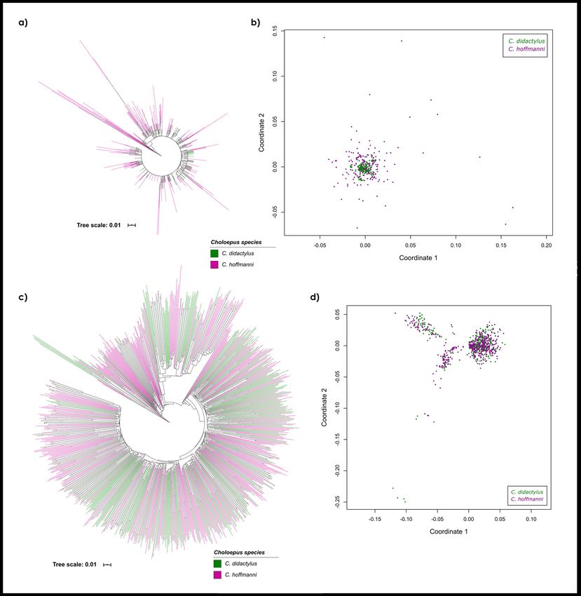
on functional centromeric sequences. Indeed, the analysis of nucleic acid folding prediction showed that several
Scientific Reports | (2020) 10:19202 | https://doi.org/10.1038/s41598-020-76199-8 3
Vol.:(0123456789)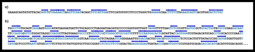
www.nature.com/scientificreports/

                                            Figure 2.  Comparative phylogenetic analyses of (a) SATCHO1 and (c) SATCHO2 sequences between C.
                                            didactylus and C. hoffmanni inferred by the Neighbor-Joining method with 1000 bootstraps. Minimum
                                            bootstrap support is 50%. Non-metric Multidimensional Scaling (NMDS) of evolutionary divergence among
                                            (b) SATCHO1 and (d) SATCHO2 sequences between C. didactylus and C. hoffmanni. The ordinations in (b)
                                            and (d) represent Euclidian distances for four dimensions. Each color represents sequences from one Choloepus
                                            species: C. didactylus (green) and C. hoffmanni (magenta).

                                            segments within SATCHO1 and SATCHO2 have the potential to form stable DNA secondary structures (Fig. 7).
                                            These results indicate that both satDNAs contain structural hallmarks of functional centromeric sequences.

                                            SATCHO1 and SATCHO2 in other Xenarthra. In order to verify if the satDNAs identified in Choloepus
                                            are also present outside the genus, we conducted Blastn searches against assembled Xenarthra genomes. For
                                            SATCHO1 we got hits in multiple contigs of the assembled genomes of B. variegatus, M. tridactyla and T. tetra-
                                            dactyla. However, the maximum number of tandemly repeated copies retrieved in a single contig was 42 on B.
                                            variegatus, and 3 on M. tridactyla and T. tetradactyla. In contrast, searches on both Choloepus species returned
                                            hundreds of contigs including considerable results, with some of them having up to 295 tandemly repeated cop-
                                            ies of SATCHO1.

          Scientific Reports |   (2020) 10:19202 |              https://doi.org/10.1038/s41598-020-76199-8                                                4

Vol:.(1234567890)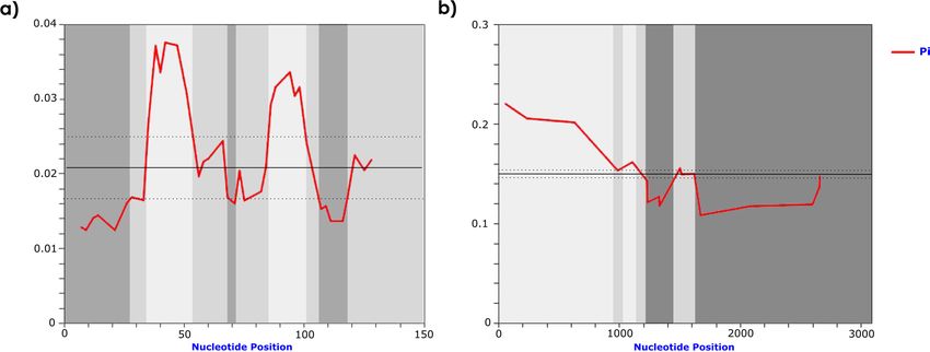
www.nature.com/scientificreports/

                                  Figure 3.  Karyotype of Choloepus hoffmanni (2n = 51) after: (a) GTG-banding and (b) CBG-banding.
                                  Bar = 10 µm.

                                  Figure 4.  Metaphases of Choloepus hoffmanni after FISH using (a) SATCHO1 (red) and (b) SATCHO2 (green)
                                  as probes. (c) Merged signals from (a) and (b). Chromosomes were counterstained with DAPI. Note the signals
                                  in the centromeric regions of all chromosomes, except the X. Bar = 10 µm.

                                  Figure 5.  CENP-B motifs identified in sequences from SATCHO1 and SATCHO2 aligned with Homo sapiens
                                  (HSA) and the evolutionary conserved domain (ECD). In red: conserved bases compared with HSA. In yellow:
                                  conserved bases compared with ECD. The CENP-B motif found in SATCHO2 is composed of two different
                                  fragments separated by ~ 140 bp, as indicated by the green symbol.

                                      Blastn searches on different assembled Xenarthra genomes using SATCHO2 as a query returned hits in mul-
                                  tiple contigs only in B. variegatus. However, we only found up to three tandemly repeated copies in this species.
                                  In the genus Choloepus however, Blastn searches retrieved hundreds of contigs with hits, and up to 60 tandemly
                                  arranged copies in a single contig. Interestingly, although SATCHO1 and SATCHO2 have a centromeric localiza-
                                  tion, we did not find contigs including both satDNA sequences in none of our Blast searches.

Scientific Reports |   (2020) 10:19202 |              https://doi.org/10.1038/s41598-020-76199-8                                                 5

                                                                                                                                            Vol.:(0123456789)
www.nature.com/scientificreports/
Figure 6. (a) SATCHO1 and (b) SATCHO2 sequences with dyad palindrome sequences. Each palindrome pair
is represented by the blue color and the direction arrow above them.
We also performed PCR experiments using the SATCHO1 and SATCHO2 primers in the genomic DNAs
of the three-toed sloth B. variegatus and the giant anteater M. tridactyla. SATCHO1 homologous sequences
were amplified from both species (Supplementary Fig. 1), which was confirmed by cloning and sequencing. The
two sequenced copies from B. variegatus showed an average of ~ 2% nucleotide divergence from the Choloepus
SATCHO1 consensus sequence, whereas the two copies of M. tridactyla presented an average of ~ 1% nucleotide
divergence. FISH with the SATCHO1 probe in M. tridactyla chromosomes did not produce any signal (data not
shown). The SATCHO2 sequence did not amplify by PCR with the genomic DNAs of neither B. variegatus nor
M. tridactyla (Supplementary Fig. 2).
These results suggest that, although SATCHO1 and SATCHO2 are present outside the genus Choloepus, these
sequences are not distributed as abundant long arrays of tandem repeats in other Xenarthra genera, in which
they should not be classified as satDNAs.
Discussion
In this work we identified two novel centromeric satDNAs in the genomes of C. didactylus and C. hoffmanni,
which could potentially have a centromeric function. Although both species have the same satDNAs, the results
from RepeatExplorer revealed some marked differences in the genome proportion of these sequences in each
species (Table 1). It is important to note that both species have approximately the same genome size (~ 3.3 Gb)
as indicated by their sequencing projects (C. didactylus accession: GCA_004027855.1, C. hoffmanni accession:
GCA_000164785.2). Despite the possibility that these observed differences reflect a real interspecific variation,
it is also likely that they constitute artifacts derived from distinct values of genome coverage and/or sequencing
platforms used for each species (Illumina HiSeq 2000 for C. hoffmanni, and Illumina HiSeq 2500 for C. didacty-
lus). Although it is currently not possible to rule out any of these possibilities, the high sequence similarity and
comparable number of Blastn results in both satDNAs between species indicate that a real large difference in
abundance is unlikely. Indeed, a recent study demonstrated that different sequencing platforms, or even differ-
ent versions of the same platform, have their own biases in representing the true proportion of highly abundant
repeats38.
Our phylogenetic and NMDS results revealed that both satDNAs do not segregate into different branches in a
species-specific manner. That result was unexpected, considering that satDNAs usually evolve rapidly through the
process of molecular drive, which also tends to produce a high degree of intra-species sequence homogeneity13.
Hence, this high level of sequence identity could be explained by one or more of the following hypotheses: (i)
C. didactylus and C. hoffmanni share a very recent common ancestor; (ii) they display a slow rate of molecular
evolution; (iii) they went through a recent process of hybridization; (iv) or that these satDNAs sequences have
been conserved by selective pressures. Regarding the first possibility, previous molecular data from different
studies showed considerable variation in the estimated divergence between C. didactylus and C. hoffmanni. For
instance, using the mitochondrial gene Cyt-b, the split of the two Choloepus species was estimated at ~ 18.7 Mya
with Bayesian inference and ~ 5.8 Mya with Median Joining N etwork27. Gibb et al.39 estimated the split varying
from 3.5 to 16.7 Mya, based on mitogenomic shotgun data with Bayesian and maximum likelihood phylogenetic
inferences. Hence, these estimates of divergence times argue against the hypothesis of a very recent common
ancestor of C. didactylus and C. hoffmanni. In relation to the second hypothesis, Choloepus species have been
shown to display a relatively slow rate of molecular evolution when compared to other Xenarthra groups39,
although the reason for that is not fully understood. However, even considering that a slower rate of molecular
evolution could partially explain the high sequence identity found between these satDNAs, it does not seem
likely that sequences evolving neutrally would keep this level of conservation after several million years. As to
the third possibility, it is worth mentioning that the two Choloepus species inhabit some overlapping areas of
the Amazon forest and there is no precise information about the collecting areas of most specimens s tudied27.
Hence, the chance of interspecific hybridization cannot be ruled out. Finally, the hypothesis that SATCHO1
and SATCHO2 could have been conserved by selective pressures is currently more difficult to evaluate beyond
the evidence we provided for a putative centromeric function, as its likelihood also depends on the exclusion of
the first three possibilities. Nevertheless, considering all the evidence provided here and elsewhere, we suggest
that the sequence conservation of these satDNAs between C. didactylus and C. hoffmanni likely derive from a
combination of selective pressures and a slow rate of molecular evolution.
More importantly, our results revealed that both satDNAS are located in the centromeric regions of all C.
hoffmanni chromosomes, except the X (Fig. 4a). It has been suggested that the most abundant tandem repeat
Scientific Reports | (2020) 10:19202 | https://doi.org/10.1038/s41598-020-76199-8 6
Vol:.(1234567890)www.nature.com/scientificreports/
Figure 7. The optimal secondary structure of (a) SATCHO1 and (b) SATCHO2 predicted by RNAfold.
Scientific Reports | (2020) 10:19202 | https://doi.org/10.1038/s41598-020-76199-8 7
Vol.:(0123456789)www.nature.com/scientificreports/
in a given genome likely corresponds to its centromeric sequence40, a feature that was observed for SATCHO1
in C. hoffmanni, and presumably also in C. didactylus. Although C. hoffmanni had the two satDNAs mapped to
centromeric regions, the resolution of our results does not enable us to determine how they are distributed along
the centromeric heterochromatin and if this distribution varies among chromosomes. As we have mentioned, it
is also not possible to determine if one or both satDNAs are part of the functional centromere. Further analyses
using long sequencing reads, chip-seq with CENP-A antibodies and immuno-fiber FISH experiments would be
important to address these issues.
In addition, we found conserved regions in SATCHO1 and SATCHO2, which include motifs sharing similari-
ties with CENP-B box-like sequences (Fig. 5). Although the CENP-B box-like motif of SATCHO2 is disrupted by
an intruding sequence, and thus is probably non-functional, its presence indicates that this large satDNA might
have been previously involved in centromeric activity during the evolution of Choloepus. It is also possible that
SATCHO2 currently has a secondary centromeric function, unrelated with the activity carried out by satDNAs
containing CENP-B box-like sequences. In any case, the conservation of such regions in these satDNAs suggests
that they could be under some sort of selective constraint. The fact that SATCHO1 and SATCHO2 also have an
enrichment of symmetric sequences capable of forming non-B DNA forms and secondary structures argues for
their putative centromeric function, as these nucleotide arrangements are thought to interact with centromere
components7,11.
Taken together, our data suggest a putative functional role for these satDNAs, which would explain their
centromeric localization in C. hoffmanni and remarkable conservation in both Choloepus species. Similar results
were reported in rodents of the genus Peromyscus, in which the centromeric satDNA PMsat was found in the
centromeres of seven s pecies41. Similarly to our results, PMsat monomers presented small sequence variation and
shared similarities with the human CENP-B box-like motif. Based on these observations, the authors suggested
that PMsat may have played some biological role which led to its maintenance in Peromyscus41.
Another interesting finding of our study is that SATCHO2 is composed by ~ 2292 bp monomers, an uncom-
monly large size for a satDNA sequence. Most satDNAs identified in plants and animals showed monomer lengths
around 150–180 bp and 300–360 bp, r espectively42,43. There is a limited number of species in which satDNAs with
monomers ranging from 1 kb to ~ 2 kb have been reported. That is the case of some whales44, South American
monkeys45, banana46, non-domestic Bovidae47, and the field bean48. SatDNA monomers larger than 2 kb have
been identified in b ovines49 and in the ant Monomorium subopacum50. To our knowledge, the only examples of
monomers significantly larger than SATCHO2 were reported in Bovidae: the satDNA 1.709 (SATIV) with ~ 3.8 kb
and the satDNA 1.711b with ~ 2.6 kb49,51.
Finally, several studies have demonstrated that satDNAs, especially those found in centromeres, are asso-
ciated with Robertsonian translocations, the main chromosome rearrangements related to Bovidae genome
evolution5,52–55. It would be interesting to investigate if there is also a link between satDNAs and chromosome
rearrangements in Xenarthra, as the number of available genomes of this group will certainly increase in the
near future.
Materials and methods
Identification and analysis of satDNA sequences. In order to identify the most abundant satDNA
sequences in the genomes of Choloepus species we performed a graph-based clustering analysis of sequence
reads using the pipeline R epeatExplorer214. The analysis was performed in a set of 357,044 random sampled
reads (~ 1.19% genome coverage) from the C. didactylus genome (accession: SRX4501348) and 789,160 random
sampled reads (~ 2.6% genome coverage) from the C. hoffmanni genome (accession: SRX282195). Identified
satDNA consensus sequences were used as queries in searches conducted on R epbase56 and GenBank (https://
www.ncbi.nlm.nih.gov/genbank/) in order to detect similarities with previously described sequences. To analyze
the satDNA copies directly in the species genomes, we retrieved a sample of each satDNA sequences from the C.
didactylus (accession: PVKG000000000.1) and C. hoffmanni (accession: ABVD00000000.2) assembled genomes
available on GenBank using Blastn searches with default parameters57. The software DnaSP 6.12.0358 was used to
identify DNA polymorphisms and nucleotide diversity along the satDNA sequences, by applying a window size
of 10 bp (SATCHO1 and SATCHO2) and a step size of 2 bp for SATCHO1 and 3 bp for SATCHO2. Windows
that exhibited standard deviation (S.D.) values ≥ 2, from the average variability, were considered highly variable,
while those with values ≤ 2 S.D. were considered conserved.
We searched putative CENP-B box-like motifs (CTTCGTTGGAAACGGGA)36 on the SATCHO1 and SAT-
CHO2 monomer sequences using the alignment algorithm MUSCLE59 on MEGA760. We also searched for dyad
symmetries in the satDNA sequences using the EMBOSS palindrome s oftware61 with a minimum palindrome
length of 4 bp and maximum gap between elements of 20 bp. We used the RNAfold web server (https://rna.
tbi.univie.ac.at/)62 to search for optimal secondary structure with minimum free energy on the SATCHO1 and
SATCHO2 sequences.
Pairwise evolutionary distances within each satDNA sequence from C. didactylus and C. hoffmanni were
estimated using MEGA760. The values were used to obtain non-metric multidimensional scaling (NMDS) ordi-
nations with the R package V egan63, representing Euclidian distances in four dimensions. We used Rstudio
64
v1.1.442 to conduct the NMDS analysis and plotting of the results. We constructed a phylogeny of the sequences
using the Neighbor-Joining method with 1000 replicates on M EGA760. The phylogenetic tree was edited using
65
iTOL4.4.1 (https://itol.embl.de/) .
Biological samples. Chromosome preparations and genomic DNAs were obtained from cultured fibro-
blasts of C. hoffmanni and M. tridactyla male individuals. Tissue and blood samples from both specimens were
obtained from Fundação de Parques Municipais e Zoobotânica de Belo Horizonte/MG, Brazil, under a license
Scientific Reports | (2020) 10:19202 | https://doi.org/10.1038/s41598-020-76199-8 8
Vol:.(1234567890)www.nature.com/scientificreports/
from IBAMA (Instituto Brasileiro do Meio Ambiente e dos Recursos Naturais Renováveis) conceded to M.
Svartman (Process Sisbio 28422-5). The C. hoffmanni individual came from an unknown location in Rondônia
estate, Brazil, and the M. tridactyla individual was apprehended by IBAMA in Esmeraldas, Minas Gerais, Brazil,
but its origin is unknown. We also used the genomic DNA from a male B. variegatus captured in Teófilo Otoni,
Minas Gerais, Brazil.
Cell cultures and chromosome spreads were obtained according to Stanyon and Galleni66 and genomic DNAs
were obtained with the Wizard Genomic Purification kit (Promega).
Molecular analysis. The identified satDNAs were amplified by polymerase chain reaction (PCR) from
C. hoffmanni genomic DNA with the following primers designed from the consensus sequences generated
on RepeatExplorer: SATCHO1-F (AGTTGTTTTTCAGCCCAGGG) and SATCHO1-R (CACGTGGGACTC
TGCGAAAG); SATCHO2-F (TCTCACCCGGATCTGAACCT) and SATCHO2-R (GGATACGGGGGTTTG
AAGCA). The thermocycling conditions were as follows: 95 °C-5 min, 30 cycles: 95 °C-1 min; 53.4 °C-1 min;
72 °C-1 min; final elongation: 72 °C-10 min. The PCR products were extracted from a 1% agarose gel, purified
with Wizard SV Gel and PCR Clean-up System kit (Promega), and cloned into a plasmid vector pGEM-T-Easy
cloning kit (Promega). The recombinant plasmids were sequenced with the ABI 3730 platform (Applied Biosys-
tems). The sequences obtained have GenBank accession numbers: MT505303–MT505310.
Banding patterns and fluorescence in situ hybridization (FISH). The GTG- and CBG-banding
of C. hoffmanni chromosomes were performed according to Seabright67 and Sumner68, respectively. FISH
was performed using the cloned satDNA sequences as probes after they were labeled by nick-translation
with digoxigenin-11-dUTP (DIG-Nick Translation mix, Roche Applied Science) for SATCHO1 and biotin-
16-dUTP (Biotin-Nick Translation mix, Roche Applied Science) for SATCHO2. The probes (~ 150 ng in 50%
formamide/2xSSC) were denatured for 10 min at 98 °C. Chromosomes were dehydrated in ethanol series (70%,
90%, 100%) and denatured in 70% formamide/2xSSC for 2 min at 75 °C. The hybridization was performed over-
night at 37 °C. Post-hybridization washes comprised two 5 min incubations in 2xSSC at 45 °C. Immunodetec-
tion was performed with anti-digoxigenin conjugated with rhodamine (Roche Applied Science) for SATCHO1
and avidin conjugated with FITC (Roche Applied Science) for SATCHO2. Chromosomes were counterstained
with DAPI 1:500 in Slowfade (Invitrogen). The analysis was performed under a Zeiss Axioimager 2 epifluo-
rescence microscope adapted with a CCD camera and image acquisition was performed with the AxioVision
(Zeiss) software (Carl Zeiss MicroImaging, Jena, Germany).
Verification of SATCHO1 and SATCHO2 in other Xenarthra species. To verify the presence of
the identified satDNAs in other Xenarthra species, we conducted Blastn searches on all assembled Xenarthra
genomes except Choloepus, using SATCHO1 and SATCHO2 consensus sequences as queries. We also performed
PCRs with genomic DNAs from B. variegatus and M. tridactyla using the same set of primers and conditions
applied to amplify SATCHO1 and SATCHO2 in C. hoffmanni. The genomic DNA of C. hoffmanni was used as a
positive control. PCR products from B. variegatus and M. tridactyla were cloned, sequenced (accession numbers:
MT505305–MT505308), and used as probes for FISH under the same conditions described above.
Data availability
The datasets generated during and/or analyzed in the current study are available in the GenBank repository (https
://www.ncbi.nlm.nih.gov/genbank/) under accession numbers: MT505303–MT505310.
Received: 23 July 2020; Accepted: 19 October 2020
References
1. Garrido-Ramos, M. A. Satellite DNA: An evolving topic. Genes (Basel). 8, 230 (2017).
2. Lower, S. S., McGurk, M. P., Clark, A. G. & Barbash, D. A. Satellite DNA evolution: Old ideas, new approaches. Curr. Opin. Genet.
Dev. 49, 70–78 (2018).
3. Kuhn, G. C. S. Satellite DNA transcripts have diverse biological roles in Drosophila. Heredity 115, 1–2. https://doi.org/10.1038/
hdy.2015.12 (2015).
4. Talbert, P. B. & Henikoff, S. Transcribing centromeres: Noncoding RNAs and kinetochore assembly. Trends Genet. 34, 587–599
(2018).
5. Escudeiro, A. et al. Bovine satellite DNAs—A history of the evolution of complexity and its impact in the Bovidae family. Eur.
Zool. J. 86, 20–37 (2019).
6. Masumoto, H., Nakano, M. & Ohzeki, J. I. The role of CENP-B and α-satellite DNA: De novo assembly and epigenetic maintenance
of human centromeres. Chromosome Res. 12, 543–556 (2004).
7. Kasinathan, S. & Henikoff, S. Non-B-form DNA is enriched at centromeres. Mol. Biol. Evol. 35, 949–962 (2018).
8. Gamba, R. & Fachinetti, D. From evolution to function: Two sides of the same CENP-B coin?. Exp. Cell Res. https://doi.
org/10.1016/j.yexcr.2020.111959 (2020).
9. Martínez-Balbás, A. et al. Satellite DNAs contain sequences that induce curvature. Biochemistry 29, 2342–2348 (1990).
10. Fitzgerald, D. J., Drydens, G. L., Bronsonl, E. C. & Anderson, J. N. Conserved patterns of bending in satellite and nucleosome
positioning DNA. J. Biol. Chem. 269, 21303–21314 (1994).
11. Plohl, M., Meštrović, N. & Mravinac, B. Satellite DNA evolution. Genome Dyn. 7, 126–152 (2012).
12. Ruiz-Ruano, F. J., López-León, M. D., Cabrero, J. & Camacho, J. P. M. High-throughput analysis of the satellitome illuminates
satellite DNA evolution. Sci. Rep. 6, 1–14 (2016).
13. Dover, G. Molecular drive: A cohesive mode of species evolution. Nature 299, 111–117 (1982).
14. Novák, P., Neumann, P., Pech, J., Steinhaisl, J. & MacAs, J. RepeatExplorer: A Galaxy-based web server for genome-wide charac-
terization of eukaryotic repetitive elements from next-generation sequence reads. Bioinformatics 29, 792–793 (2013).
Scientific Reports | (2020) 10:19202 | https://doi.org/10.1038/s41598-020-76199-8 9
Vol.:(0123456789)www.nature.com/scientificreports/
15. Delsuc, F., Vizcaíno, S. F. & Douzery, E. J. P. Influence of tertiary paleoenvironmental changes on the diversification of South
American mammals: A relaxed molecular clock study within xenarthrans. BMC Evol. Biol. 4, 1–13 (2004).
16. Superina, M. & Loughry, W. J. Why do xenarthrans matter?. J. Mammal. 96, 617–621 (2015).
17. Gardner, A. L. Mammals of South America, Volume 1- Marsupials, Xenarthrans, Shrews, and Bats. (University of Chicago Press,
Chicago, 2008).
18. Waters, P. D., Dobigny, G., Pardini, A. T. & Robinson, T. J. LINE-1 distribution in Afrotheria and Xenarthra: Implications for
understanding the evolution of LINE-1 in eutherian genomes. Chromosoma 113, 137–144 (2004).
19. Bao, W. & Jurka, J. Origin and evolution of LINE-1 derived “half-L1” retrotransposons (HAL1). Gene 465, 9–16 (2010).
20. Nishihara, H., Kuno, S., Nikaido, M. & Okada, N. MyrSINEs: A novel SINE family in the anteater genomes. Gene 400, 98–103
(2007).
21. Churakov, G., Smit, A. F. A., Brosius, J. & Schmitz, J. A novel abundant family of retroposed elements (DAS-SINEs) in the nine-
banded armadillo (Dasypus novemcinctus). Mol. Biol. Evol. 22, 886–893 (2005).
22. Alkan, C. et al. Genome-wide characterization of centromeric satellites from multiple mammalian genomes. Genome Res. 21,
137–145 (2011).
23. Engelmann, G. F. The phylogeny of the Xenarthra. In The Evolution and Ecology of Armadillos, Sloths and Vermilinguas (ed. Mont-
gomery, G.G.) 51–64 (Smithsonian Institution, Washington, DC, 1985).
24. Hayssen, V. Choloepus hoffmanni (Pilosa: Megalonychidae). Mamm. Species 43, 37–55 (2011).
25. Wetzel, R. & Avila-Pires, F. Identification and distribution of the recent sloths of Brazil (Edentata). Rev. Bras. Biol. 40, 831–836
(1980).
26. Steiner, C. C., Houck, M. L. & Ryder, O. A. Species identification and chromosome variation of captive two-toed sloths. Zoo Biol.
30, 623–635 (2011).
27. Ruiz-García, M., Chacón, D., Plese, T., Schuler, I. & Shostell, J. M. Mitogenomics phylogenetic relationships of the current sloth’s
genera and species (Bradypodidae and Megalonychidae). Mitochondrial DNA Part A DNA Mapp. Seq. Anal. 29, 281–299 (2018).
28. Corin-Frederick, J. Les formules gonosomiques dites aberrantes chez les mammifères euthériens. Chromosoma 27, 268–287 (1969).
29. Jorge, W., Meritt, D. J. & Bernirschke, K. Chromosome studies in Edentata. Cytobios 18, 157–172 (1978).
30. Jorge, W. Estudo cromossômico de algumas espécies da ordem Edentata (UNESP, São Paulo, 1981).
31. Jorge, W., Orsi-Souza, A. T. & Best, R. The somatic chromosomes of Xenarthra. In The Evolution and Ecology of Armadillos, Sloths,
and Vermilinguas (ed. Montgomery, G. G.) 121–129 (Smithsonian Institution, Washington, DC, 1985).
32. Sonta, S. I., Hayata, I., Sasaki, M. & Kondo, N. Karyotype and sex determining in the two-toed sloth, Choloepus didactylus. Chro-
mosome Inf. Serv. 28, 15–17 (1980).
33. Dobigny, G. et al. Low rate of genomic repatterning in Xenarthra inferred from chromosome painting data. Chromosome Res. 13,
651–663 (2005).
34. Svartman, M., Stone, G. & Stanyon, R. The ancestral Eutherian karyotype is present in Xenarthra. PLoS Genet. 2, 1006–1011 (2006).
35. Earnshaw, W. C. & Rothfield, N. Identification of a family of human centromere proteins using autoimmune sera from patients
with scleroderma. Chromosoma 91, 313–321 (1985).
36. Muro, Y. et al. Centromere protein B assembles human centromeric α-satellite DNA at the 17-bp sequence, CENP-B box. J. Cell
Biol. 116, 585–596 (1992).
37. Stitou, S., Díaz De La Guardia, R., Jiménez, R. & Burgos, M. Isolation of a species-specific satellite DNA with a novel CENP-B-like
box from the North African rodent Lemniscomys barbarus. Exp. Cell Res. 250, 381–386 (1999).
38. Flynn, J. M., Long, M., Wing, R. A. & Clark, A. G. Evolutionary dynamics of abundant 7 bp satellites in the genome of Drosophila
virilis. Mol. Biol. Evol. 37, 1362–1375 (2020).
39. Gibb, G. C. et al. Shotgun mitogenomics provides a reference phylogenetic framework and timescale for living xenarthrans. Mol.
Biol. Evol. 33, 621–642 (2016).
40. Melters, D. P. et al. Comparative analysis of tandem repeats from hundreds of species reveals unique insights into centromere
evolution. Genome Biol. 14, 1–20 (2013).
41. Smalec, B. M., Heider, T. N., Flynn, B. L. & O’Neill, R. J. A centromere satellite concomitant with extensive karyotypic diversity
across the Peromyscus genus defies predictions of molecular drive. Chromosome Res. 27, 237–252 (2019).
42. Schmidt, T. & Heslop-Harrison, J. S. Genomes, genes and junk: The large-scale organization of plant chromosomes. Trends Plant
Sci. 3, 195–199 (1998).
43. Henikoff, S., Ahmad, K. & Malik, H. S. The centromere paradox: Stable inheritance with rapidly evolving DNA. Science 293,
1098–1102 (2001).
44. Árnason, Ú., Grétarsdóttir, S. & Widegren, B. Mysticete (baleen whale) relationships based upon the sequence of the common
cetacean DNA satellite. Mol. Biol. Evol. 9, 1018–1028 (1992).
45. Fanning, T. G., Seuánez, H. N. & Forman, L. Satellite DNA sequences in the New World primate Cebus apella (Platyrrhini, Pri-
mates). Chromosoma 102, 306–311 (1993).
46. Hřibová, E. et al. Repetitive part of the banana (Musa acuminata) genome investigated by low-depth 454 sequencing. BMC Plant
Biol. 10, 1–10 (2010).
47. Kopecna, O. et al. Tribe-specific satellite DNA in non-domestic Bovidae. Chromosome Res. 22, 277–291 (2014).
48. Ávila Robledillo, L. et al. Satellite DNA in Vicia faba is characterized by remarkable diversity in its sequence composition, associa-
tion with centromeres, and replication timing. Sci. Rep. 8, 1–11 (2018).
49. Skowronski, J., Plucienniczak, A. & Bednarek, A. Bovine l.709 satellite recombination hotspots and dispersed repeated sequences.
J. Mol. Biol. 177, 399–416 (1984).
50. Lorite, P., Carrillo, J. A., Aguilar, J. A. & Palomeque, T. Isolation and characterization of two families of satellite DNA with repetitive
units of 135 bp and 2.5 kb in the ant Monomorium subopacum (Hymenoptera, Formicidae). Cytogenet. Genome Res. 105, 83–92
(2004).
51. Taparowsky, E. J. & Gerbi, S. A. Structure of 1.711 lb gm/cm3 bovine satellite DNA: Evolutionary relationship to satellite I. Nucleic
Acids Res. 10, 5503–5515 (1982).
52. Jobse, C. et al. Evolution and recombination of bovine DNA repeats. J. Mol. Evol. 41, 277–283 (1995).
53. Modi, W. S., Gallagher, D. S. & Womack, J. E. Evolutionary histories of highly repeated DNA families among the Artiodactyla
(Mammalia). J. Mol. Evol. 42, 337–349 (1996).
54. Adega, F., Guedes-Pinto, H. & Chaves, R. Satellite DNA in the karyotype evolution of domestic animals—Clinical considerations.
Cytogenet. Genome Res. 126, 12–20 (2009).
55. Escudeiro, A., Adega, F., Robinson, T. J., Heslop-Harrison, J. S. & Chaves, R. Conservation, divergence, and functions of centromeric
satellite DNA families in the bovidae. Genome Biol. Evol. 11, 1152–1165 (2019).
56. Jurka, J. et al. Repbase update, a database of eukaryotic repetitive elements. Cytogenet. Genome Res. 110, 462–467 (2005).
57. Altschul, S. F., Gish, W., Miller, W., Myers, E. W. & Lipman, D. J. Basic local alignment search tool. J. Mol. Biol. 215, 403–410 (1990).
58. Rozas, J. et al. DnaSP v6: DNA sequence polymorphism analysis of large data sets. Mol. Biol. Evol. 34, 3299–3302 (2017).
59. Edgar, R. C. MUSCLE: Multiple sequence alignment with high accuracy and high throughput. Nucleic Acids Res. 32, 1792–1797
(2004).
60. Kumar, S., Stecher, G. & Tamura, K. MEGA7: Molecular evolutionary genetics analysis Version 7.0 for bigger datasets. Mol. Biol.
Evol. 33, 1870–1874 (2016).
Scientific Reports | (2020) 10:19202 | https://doi.org/10.1038/s41598-020-76199-8 10
Vol:.(1234567890)www.nature.com/scientificreports/
61. Rice, P., Longden, L. & Bleasby, A. EMBOSS: The European molecular biology open software suite. Trends Genet. 16, 276–277
(2000).
62. Gruber, A. R., Lorenz, R., Bernhart, S. H., Neuböck, R. & Hofacker, I. L. The Vienna RNA websuite. Nucleic Acids Res. 36, 70–74
(2008).
63. Dixon, P. VEGAN, a package of R functions for community ecology. J. Veg. Sci. 14, 927–930 (2003).
64. RStudio Team. RStudio: Integrated Development for R. [Computer sotware]. RStudio, Inc. https://www.rstudio.com/ (2020).
65. Letunic, I. & Bork, P. Interactive Tree Of Life (iTOL) v4: Recent updates and new developments. Nucleic Acids Res. 47, W256–W259
(2019).
66. Stanyon, R. & Galleni, L. A rapid fibroblast culture technique for high resolution karyotypes. Bolletino di Zool. 58, 81–83 (1991).
67. Seabright, M. A rapid banding technique for human chromosomes. Lancet 2, 971–972 (1971).
68. Sumner, A. T. A simple technique for demonstrating centromeric heterochromatin. Exp. Cell Res. 75, 304–306 (1972).
Acknowledgements
R.S.S., P.H. and M.P.V. received doctoral fellowships from Coordenação de Aperfeiçoamento de Pessoal de Nível
Superior (CAPES). G.C.S.K. and M.S. are recipients of productivity fellowships from Conselho Nacional de
Desenvolvimento Científico e Tecnológico (CNPq—processes 308386/2018-3 and 310433/2018-5, respectively).
Author contributions
R.S.S., P.H. and M.P.V. conducted the bioinformatics and data analyses, designed the study and drafted the
manuscript; R.S.S. and M.P.V. carried out the cytogenetic and molecular analyses; R.S.S. prepared all figures;
V.S.P. provide the tissue and blood samples; G.C.S.K. and M.S. conceived and coordinated the study, and helped
drafting the manuscript.
Competing interests
The authors declare no competing interests.
Additional information
Supplementary information is available for this paper at https://doi.org/10.1038/s41598-020-76199-8.
Correspondence and requests for materials should be addressed to M.S.
Reprints and permissions information is available at www.nature.com/reprints.
Publisher’s note Springer Nature remains neutral with regard to jurisdictional claims in published maps and
institutional affiliations.
Open Access This article is licensed under a Creative Commons Attribution 4.0 International
License, which permits use, sharing, adaptation, distribution and reproduction in any medium or
format, as long as you give appropriate credit to the original author(s) and the source, provide a link to the
Creative Commons licence, and indicate if changes were made. The images or other third party material in this
article are included in the article’s Creative Commons licence, unless indicated otherwise in a credit line to the
material. If material is not included in the article’s Creative Commons licence and your intended use is not
permitted by statutory regulation or exceeds the permitted use, you will need to obtain permission directly from
the copyright holder. To view a copy of this licence, visit http://creativecommons.org/licenses/by/4.0/.
© The Author(s) 2020
Scientific Reports | (2020) 10:19202 | https://doi.org/10.1038/s41598-020-76199-8 11
Vol.:(0123456789)You can also read