Downstream Targets of Homeobox Gene HLX Show Altered Expression in Human Idiopathic Fetal Growth Restriction
←
→
Page content transcription
If your browser does not render page correctly, please read the page content below
The American Journal of Pathology, Vol. 176, No. 1, January 2010
                                                                Copyright © American Society for Investigative Pathology
                                                                DOI: 10.2353/ajpath.2010.090187

Metabolic, Endocrine and Genitourinary Pathobiology

Downstream Targets of Homeobox Gene HLX Show
Altered Expression in Human Idiopathic Fetal Growth
Restriction

Gayathri Rajaraman, Padma Murthi,                               Fetal growth restriction (FGR), also known as intrauterine
Niroshani Pathirage, Shaun P. Brennecke,                        growth retardation, is a significant pregnancy disorder
and Bill Kalionis                                               associated with major perinatal complications including
                                                                stillbirth, prematurity, fetal compromise, infant morbidity
From the Department of Obstetrics and Gynaecology, University
                                                                and mortality. Moreover, later in life, FGR-affected indi-
of Melbourne and Pregnancy Research Centre, The Royal
                                                                viduals also have an increased risk of chronic disorders
Women’s Hospital, Parkville, Australia
                                                                such as ischemic heart disease, maturity onset diabetes,
                                                                and psychiatric disorders.1,2 A common definition of FGR
                                                                is a birth weight at or below the 10th percentile for ges-
                                                                tational age and gender, failure of the fetus to grow to its
Fetal growth restriction (FGR) , a clinically significant       genetically determined potential size, and the likely pres-
pregnancy disorder , is poorly understood at the mo-            ence of an underlying pathological process that inhibits
lecular level. This study investigates idiopathic FGR           the expression of the normal intrinsic growth potential.
associated with placental insufficiency. Previously,            Obvious causes of FGR are fetal (eg, genetic defects),
we showed that the homeobox gene HLX is expressed               placental (eg, infarcts), and maternal factors (eg, maturity
in placental trophoblast cells and that HLX expres-             onset diabetes, certain autoantibody disorders, alcohol
sion is significantly decreased in human idiopathic             abuse, and smoking), but these factors account for only
FGR. Here , we used the novel approach of identifying           30% of FGR cases. The remaining 70% of FGR cases
downstream targets of HLX in cell culture to detect             with no obvious causes are classified as “idiopathic” and
potentially important genes involved in idiopathic              are commonly attributed to uteroplacental insufficiency.
FGR. Downstream targets were revealed by decreas-                  Placental insufficiency is associated with abnormal tro-
ing HLX expression in cultured trophoblast cells with           phoblast function in FGR. Villous outgrowth, which is
HLX-specific small interfering RNAs to model human              determined by trophoblast proliferation, is reduced in the
idiopathic FGR and comparing these levels with con-             FGR-affected placenta together with increased apoptosis
trols using a real-time PCR-based gene profiling sys-           of these cells.3 Defective trophoblast function also results
tem. Changes in candidate HLX target mRNA levels                in growth restriction of the fetus due to the reduction in
were verified in an independent trophoblast cell line,          transfer of nutrients and growth factors to the fetus.4
and candidate target gene expression was assessed in            Another significant defect in FGR is uteroplacental isch-
human idiopathic FGR-affected placentae (n ⴝ 25)                emia due to failure of the specialized extravillous tropho-
compared with gestation-matched controls (n ⴝ 25).              blast cells to proliferate, invade, and subsequently trans-
The downstream targets RB1 and MYC , cell cycle
regulatory genes , showed significantly increased
mRNA levels in FGR-affected tissues compared with               Supported by National Health and Medical Research Council project
                                                                grant 509140. The authors would also like to thank Lynne Quayle Chari-
gestation-matched controls , whereas CCNB1 , ELK1 ,
                                                                table Trust Fund (Equity Trustees) and the Marian E.H. Flack Trust for
JUN , and CDKN1 showed significantly decreased
                                                                funding support for this project and also the University of Melbourne for
mRNA levels (n ⴝ 25 , P < 0.001 , t-test). The changes          the award of an Early Career Researcher Grant (to P.M.) G.R was
for RB1 and CDKN1C were verified by Western blot                awarded the Felix Meyer Postgraduate Scholarship from the University of
analysis in FGR-affected placentae compared with                Melbourne and the Royal Women’s Hospital Postgraduate Scholarship.
gestation-matched controls (n ⴝ 6). We conclude                   Accepted for publication September 18, 2009.
that cell cycle regulatory genes RB1 , MYC , CCNB1 ,              Address reprint requests to Gayathri Rajaraman, Ph.D., Department
ELK1 , JUN , and CDKN1C , which control important               of Obstetrics and Gynaecology, University of Melbourne, RWH Cam-
trophoblast cell functions , are targets of HLX. (Am            pus, 20 Flemington Rd., Parkville, Victoria 3052, Australia. E-mail:
J Pathol 2010, 176:278 –287; DOI: 10.2353/ajpath.2010.090187)   graja@unimelb.edu.au.

278
HLX Targets in Fetal Growth Restriction 279
AJP January 2010, Vol. 176, No. 1
form and remodel the maternal spiral arteries in the types in early placental development.28 Furthermore, we
placental bed.5 Villous structural defects, vascular abnor- postulated that reduced levels of HLX are required for
malities, and reduced branching of the villous structure cytotrophoblast differentiation and that dysregulation of
are also seen in FGR.6 – 8 These defects also prevent HLX expression contributed to the aberrant cytotropho-
adequate nutrient transfer to the fetus.9 blast proliferation and differentiation associated with pla-
Mouse knockout studies of transcription factors pro- cental pathologies.28 Recently, we also provided evi-
vide genetic proof of the critical role of transcription fac- dence of HLX regulation by growth factors and cytokines
tors in regulating placental development.10 –14 Some and established that HLX is an important regulator for
knockout gene phenotypes show the hallmarks of major signal transduction-mediated proliferation of human tro-
human placental disorders13 and thus may play an im- phoblast cells.29 By using four independent small inter-
portant role in the pathogenesis of human idiopathic fering RNA (siRNA) oligonucleotides, we have reported a
FGR. significant 60 to 85% decrease in HLX mRNA and 73 to
The homeobox gene family of transcription factors is 87% decrease in protein expression by HLX siRNA trans-
extensively characterized in the human placenta and fection compared with the mock-transfected control tro-
embryo.15,16 These genes are known to play important phoblast cells.29 Finally, we showed that HLX mRNA and
roles in implantation, placentation, and embryonic devel- protein expression is significantly decreased in human
opment in the mouse.11–13,17–20 Only a few studies of idiopathic FGR,30 suggesting that HLX expression may
human homeobox genes have provided evidence that be of pathological significance.
homeobox genes are important in human placental de- The focus of this study is to identify downstream target
velopment and particularly in trophoblast development genes of HLX in cultured trophoblast and to identify the
and differentiation.10,16,21,22 Targeted disruption of the molecular pathways that are affected in human idiopathic
Hlx homeobox gene in the mouse has shown that Hlx also FGR.
plays a fundamental role in visceral organogenesis.23 Hlx Signal transduction pathways that regulate trophoblast
mutant mice resulted in developing gut and liver diver- functions include the phosphatidylinositol 3-kinase-Akt,
ticulum defects. In addition, Hlx mutation also showed a protein kinase A, protein kinase C, p38/c-Jun N-terminal
defect in proliferation and resulted in embryonic death kinase, Jak-Stat, and mitogen-activated protein kinase
due to liver failure.23 (MAPK) pathways.31–33 Of these, the two pathways that
Our interest is in the homeobox gene HLX, the human are frequently used by trophoblast cells are the Jak-Stat
homologue of mouse Hlx homeobox gene, which is highly and MAPK pathways.33 The human MAPK signaling
expressed in hematopoietic progenitors and shows de- pathway PCR array was used in this study.
creased expression levels in activated lymphocytes.24 We hypothesize that placental HLX downstream tar-
HLX inactivation impairs CD34⫹ bone marrow cell prolif- get gene expression will be significantly altered in
eration in response to stimulation by cytokines while in- human idiopathic FGR pregnancies compared with un-
ducing differentiation of these cells. Moreover, HLX inac- complicated pregnancies. In this study, an in vitro cell
tivation reduces the levels of c-myc, c-fos, cyclin B, and culture model using HLX siRNA, in trophoblast cells,
p34cdc2 mRNA expression,24 which are cell cycle regu- was used. PCR array comparisons were made between
lator genes implicated in trophoblast cell function.25 HLX siRNA-treated cells and control cells transfected
Therefore, c-myc, c-fos, cyclin B, and p34cdc2 were the with a siRNA that does not target human genes. Candi-
first identified targets of HLX. date HLX target gene expression levels were then as-
Slavotinek et al26 have described HLX mutations in sessed by real-time PCR, using validated probes, in hu-
human congenital diaphragmatic hernia patients. In this man idiopathic FGR-affected placentae compared with
study, the HLX gene was sequenced in 119 congenital gestation age-matched control placentae.
diaphragmatic hernia patients, because HLX is in the
deleted interval at ch1q41-1q42 for human congenital
diaphragmatic hernia, and four amino acid substitution
mutations (p.A235V, p.S12F, p.S18L, and p.D173Y) re-
Materials and Methods
sulted in congenital diaphragmatic hernia phenotype.26 Patient Details and Tissue Sampling
Slavotinek et al26 concluded that HLX mutations are eti-
ologically important in human diaphragm formation by Human placentae from pregnancies complicated by id-
interaction with other genetic or environmental factors. iopathic FGR (n ⫽ 25) and gestation-matched control
Furthermore, Suttner et al27 have determined that HLX pregnancies (n ⫽ 25) were obtained with informed pa-
gene variants influence the development of childhood tient consent and with approval from the Research and
asthma. Their study identified 19 polymorphisms in the Ethics committees of The Royal Women’s Hospital. These
HLX gene, and two tagging single nucleotide polymor- samples have been characterized and used in our pre-
phisms representing seven polymorphisms were associ- vious gene expression studies.30,34 Ultrasound was used
ated with childhood asthma in a study population of 3099 for the prospective identification of growth-restricted fe-
German children. Suttner et al27 concluded that genetic tuses. The clinical characteristics of FGR-affected preg-
alterations in HLX contributed to the pathogenesis of nancies and the gestation age-matched controls used in
childhood asthma. this study are as previously published by Murthi et al.30,34
Previously, we provided evidence that HLX is ex- As described,30,34 the inclusion criteria for this study
pressed primarily in the proliferating cytotrophoblast cell were a birth weight less than the 10th percentile for280 Rajaraman et al
AJP January 2010, Vol. 176, No. 1
gestation age using Australian growth charts35 and any were grown in Ham’s F-10 nutrient mixture/RPMI 1640
two of the following criteria diagnosed on antenatal ultra- medium supplemented with 10% fetal bovine serum for
sound: abnormal umbilical artery Doppler flow velocim- 48 hours (2 ⫻ 105 cells/well in 6-well plates) and then
etry, oligohydramnios as determined by amniotic fluid transfected with two individual HLX siRNAs [HLX-si1 se-
index ⬍7 on antenatal ultrasound performed before de- quence: r(UGAAUUUCUUGGGUUCGAG)d(TT); HLX-si2
livery, or asymmetric growth of the fetus as quantified sequence: r(AAACCUUUUCUCCAGG CCU)d(TT)], us-
from the head circumference-to-abdominal circumfer- ing RNAiFect transfection reagent (Qiagen, Victoria, Aus-
ence ratio (⬎1.2). Placental tissue subjected to maternal tralia). The mock-transfected control wells received all
smoking, chemical dependency, multiple pregnancies, reagents, except the siRNA oligonucleotide. Cells were
pre-eclampsia, placental abruption, prolonged rupture of returned to incubation at 37°C in 5% CO2 for 72 hours.
the membranes, fetal congenital anomalies, or suspicion Negative control siRNAs consisted of a pool of enzyme-
of intrauterine viral infection were excluded from this generated siRNA oligonucleotides of 15 to 19 bp in
study. Pre-eclampsia cases with associated FGR were length, which were not specific for any known human gene
also excluded from this study. The last menstrual period (catalog number 0652-13-7000, Superarray Bioscience,
dates were used to calculate the gestation age for both Frederick, MD) and showed no sequence similarity to HLX.
FGR and control patients and further confirmed by early
pregnancy ultrasound. Control patients were selected to
match FGR cases according to gestation. All control RNA Extraction
patients presented with either spontaneous labor or re-
Total RNA from excised placental tissue was isolated as
quired elective delivery by induction of labor/cesarean
described previously by Murthi et al.30,34 Briefly, 300 mg
section. Preterm control patients presented in spontane-
of placental tissue was homogenized, and total RNA was
ous idiopathic preterm labor or underwent elective deliv-
isolated by acid guanidium isothiocyanate-phenol-chlo-
ery for conditions not associated with placental dysfunc-
roform extraction and lithium chloride precipitation ac-
tion (eg, elective cesarean section due to maternal breast
cording to the method of Chomzynski and Sacchi38 and
cancer). Particularly, control patients did not have clinical
as modified by Puissant and Houdebine.39 Total cellular
evidence of FGR, pre-eclampsia, placental abruption,
RNA from cultured trophoblast cells was isolated using
prolonged rupture of the membranes, or ascending in-
the QIAShredder and an RNeasy Microkit (Qiagen) ac-
fection. All control patients gave birth to normally formed
cording to the manufacturer’s instructions. First-strand
babies with birth weights appropriate for gestation. All
synthesis for real-time PCR cDNA preparation was per-
control placentae obtained were grossly normal with no
formed on 2 g of total RNA from placental tissue and
observed abnormalities such as infarcts.
cultured cells, using Superscript III ribonuclease H-re-
Placental tissues were excised from randomly selected
verse transcriptase (Invitrogen, Victoria, Australia). First-
areas of central placental cotyledons, after dissecting
strand synthesis of cDNA for RT2-profiler PCR array was
away any attached decidua. Samples were processed
performed using the RT2 First Strand kit (catalog number
within 10 minutes of delivery of the placenta. Samples of
c-03; SuperArray Bioscience). Briefly, 10 l of reverse tran-
fresh placental tissues were divided into small pieces and
scriptase mixture (containing reverse transcriptase buffer,
thoroughly washed in 0.9% PBS to minimize blood contam-
primer, external control mix and reverse transcriptase en-
ination and then snap frozen and stored at ⫺80°C for RNA
zyme mix) was added to 10 l of genomic DNA elimination
analysis.
mixture (containing 1 g of RNA, gDNA elimination buffer,
and RNase-free water) and incubated at 42°C for 15 min-
Cell Culture utes. RNA degradation and reverse transcriptase inactiva-
tion were performed by heating at 95°C for 5 minutes.
Human trophoblast cell lines are a well established model
for examining trophoblast function in vitro. In this study,
two well-characterized, human extravillous trophoblast PCR Array
cell lines SGHPL-4 and HTR-8/SVneo were used, be-
The downstream target genes of HLX were determined
cause these cell lines retain many features of normal
using the real-time PCR based “RT2 profiler PCR array”
extravillous trophoblast cells, including human placental
for gene profiling (SuperArray Bioscience Corporation,
lactogen, human chorionic gonadotropin, and HLA-
catalog no. PHAS-061). The human MAPK signaling
Class1.36,37 The cell lines used in this study are long
pathway PCR array was used in this study. This commer-
term, which senesce after ⬃30 passages. For this work,
cially available PCR array encompassed the gene ex-
SGHPL-4 cells (passages 5 to 12) were cultured in sup-
pression profiles of 84 genes related to the MAPK signal-
plemented Ham’s F-10 medium, and the HTR-8/Svneo
ing pathway, which is an important signaling pathway in
cells (passages 16 to 19) were cultured in supplemented
the regulation of human trophoblast function.33 Members
RPMI 1640 medium, as described previously.29
of the MAP kinase kinase kinase (MKKK), MAP kinase
kinase (MKK), and MAPK families are represented on this
Inactivation of HLX by siRNA array as well as transcription factors and genes whose
expression is induced by MAPK signaling. In addition,
Inactivation of HLX by siRNA was performed as previ- the array also included genes related to scaffolding and
ously described.29 Briefly, cultured trophoblast cells anchoring. Furthermore, the PCR array chosen in thisHLX Targets in Fetal Growth Restriction 281
AJP January 2010, Vol. 176, No. 1
study contained several cell cycle regulatory genes, in- Real-Time PCR
cluding CCNB1, MYC, CDKN1C, and JUN, which have
been previously identified to be the targets of HLX in Verification of candidate HLX target gene mRNA ex-
hematopoietic progenitor cells.24 pression (identified from PCR array) in mock control
Procedures were performed according to the manu- and HLX siRNA-transfected SGHPL-4 and HTR-8/
facturer’s protocol. Briefly, diluted cDNA prepared using Svneo cells was performed on an ABI Prism 7700
the RT2 First-Strand kit (as described above) was added (PerkinElmer-Applied Biosystems) using prevalidated
to a master mix containing the fluorescent SYBR green/ TaqMan Gene Expression Assays (RB1 Assay ID
ROX dye. Aliquots from this mix were added to a 96-well Hs00153108_m1, EGR1 Assay ID Hs00152928_m1,
plate that contained 84 predispensed gene-specific MYC Assay ID Hs00153408_m1, JUN Assay ID
primer sets. The plates (one for each HLX-siRNA and Hs00277190_s1, Cyclin B1 Assay ID Hs00259126_m1,
controls) were then placed in an ABI Prism 7700 Se- ELK1 Assay ID Hs00428286_g1, and CDKN1C Assay ID
quence Detector (PerkinElmer-Applied Biosystems, Vic- Hs00175938_m1; Applied Biosystems). Gene expression
toria, Australia) for quantitation of gene expression under quantitation was performed as the second step in a two-
the following cycling parameters: 95°C for 10 minutes, step RT-PCR protocol according to the manufacturer’s in-
followed by 40 cycles of denaturation at 95°C for 15 structions. A total of 20 l of PCR mix containing TaqMan
seconds and primer extension at 60°C for 1 minute. Each Universal PCR master mix, TaqMan Gene Expression As-
plate contained a panel of five housekeeping gene primers say mix and 3.5 ng of cDNA was used. Amplification was
for normalization of the PCR array data, as well as estima- performed for 40 cycles, including denaturation at 95°C for
tion of the linear dynamic range of the assay. The house- 15 seconds and annealing/extension at 60°C for 60 sec-
keeping gene panel consisted of 2-micrglobulin (B2M),
onds. Gene expression for the housekeeping gene GAPDH
hypoxanthine phosphoribosyltansferase 1 (HPRT1), ribo-
was co-amplified with HLX in the same reaction well. Murthi
somal protein L13a (RPL13A), glyceraldehyde-3-phosphate
et al34 from our laboratory previously showed that GAPDH is
dehydrogenase (GAPDH), and -actin (ACTB). Further-
a suitable endogenous reference gene for relative gene
more, for each reaction, both negative control (no reverse
expression studies in placental tissues from human
transcriptase enzyme added) and template control (no
idiopathic FGR, and its expression is not altered in FGR
cDNA added) were included as controls. Raw data were
acquired and processed with ABI Sequence Detector placentae compared with control placentae. The
System software version 1.0 (PerkinElmer-Applied Bio- GAPDH primers (5⬘-GCACCACCAACTGCTTAGCA-3⬘ and
systems) to calculate the threshold cycle (Ct) value. Rel- 5⬘-GTCTTCTGGGTGGCAGTGATG-3⬘) and TaqMan probe
ative gene expression values (fold change) were subse- (5⬘-VIC-TCGTGGAAGGACTCATGACCACAGTCC-TAMRA-
quently determined according to the manufacturer’s 3⬘) were designed using Primer Express 1.5 software
instructions. Briefly, the fold change for each gene from the (Applied Biosystems). The cycling conditions for GAPDH
mock control array and the HLX-siRNA-treated array was were as per the HLX target genes. Relative quantitation of
calculated as 2⫺⌬⌬Ct, normalized to the average Ct value HLX target gene expression normalized to GAPDH was
of the five housekeeping genes. Fold changes ⬎1 were calculated according to the 2⫺⌬⌬CT method of Livak and
fold increase, and for fold changes ⬍1, the negative Schmittgen,40 using the mock control as a calibrator (ABI
inverse of the result was reported as a fold decrease. Prism 7700 Sequence detection system, User Bulletin
Candidate gene prioritization was based on the highest number 2, 2001). These candidate HLX target genes
positive and lowest negative fold change differences. were then further tested for expression in FGR-affected
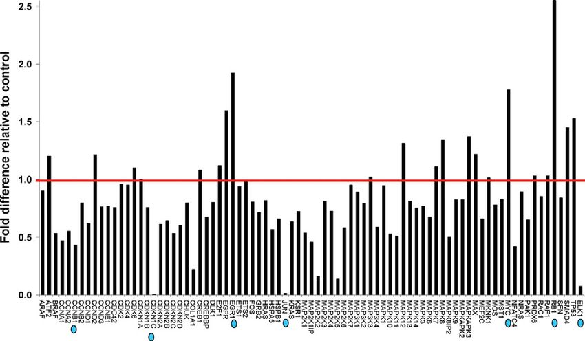
Figure 1. Identification of HLX downstream tar-
get genes. RNA was extracted from SGHPL-4
cells transfected with HLX siRNA, transcribed
into first strand cDNA, and the RT2 profiler PCR
array was performed for gene profiling. Follow-
ing initial denaturation at 95°C for 10 minutes,
the 84 predispensed genes and a panel of house-
keeping genes were amplified for 40 cycles in-
cluding denaturation at 95°C for 15 seconds and
primer extension at 60°C for 1 minute. Relative
gene expression values (fold change above or
below threshold value of 1) were subsequently
calculated for the HLX siRNA-treated plate, rela-
tive to the mock control plate, normalized to the
housekeeping gene panel. The red line shows
the threshold value at 1. The prioritized candi-
date HLX target genes are indicated by blue dots.282 Rajaraman et al
AJP January 2010, Vol. 176, No. 1
human placental samples compared with gestation age- Results
matched controls, as per the protocol described above.
Identification of HLX Downstream Targets
Western Immunoblotting The SGHPL-4 human trophoblast cell line was trans-
Samples were electrophoresed on a 10% PAGE/0.1% SDS fected with two independent siRNA oligonucleotides de-
gel in running buffer (250 mmol/L Tris and 1.92 mmol/L
glycine) and transferred electrophoretically to a nitrocellu-
lose membrane at 100 V for 30 minutes in transfer buffer
(255 mmol/L Tris, 1.92 mmol/L glycine, and 0.1% SDS). The
filter was blocked with 10% (w/v) nonfat milk powder/TBS for
1 hour. Incubation with the RB1 (0.5 g/ml), CDKN1C (1
g/ml) (Sapphire Biosciences, New South Wales, Australia)
or GAPDH (0.5 g/ml) (Imgenex, San Diego, CA) antibodies
was performed overnight at 4°C. The filter was then washed
in Tris-buffered saline and incubated with biotinylated anti-
rabbit antibody (1.5 g/ml) (Amersham Life Sciences, New
South Wales, Australia) in 5% (w/v) nonfat milk powder/TBS
for at least 1 hour at room temperature. Streptavidin-HRP
(1.5 g/ml) in 2% nonfat milk powder/Tris-buffered saline
buffer was added and incubation was performed at room
temperature for 1 hour. Tyramide signal amplification was
then performed, according to the manufacturer’s instruc-
tions (PerkinElmer), for CDKN1C blots. RB1 and GAPDH
antibodies did not require tyramide signal amplification.
Detection of protein bands was performed using ECL-Lu-
milyte autoradiography kit according to the manufacturer’s
instructions (PerkinElmer).
Data Analysis
All parameters for the gestation age-matched, FGR-af-
fected pregnancies and controls were described as
mean ⫾ SEM. Differences between the clinical character-
istics of the FGR-affected pregnancies and the control pa-
tients were investigated using either 2 test or Student’s
t-test where appropriate. The difference in mRNA expres-
sion of the HLX target genes between siRNA-treated and
control-cultured trophoblast cells and between FGR-af-
fected and control pregnancies was assessed by t-test. A
value of P ⬍ 0.001 was considered significant.
Figure 3. Validation of target genes that showed increased expression fol-
lowing siRNA-mediated HLX silencing. cDNA (3.5 ng) from SGHPL-4 and
HTR-8/SVneo cells transfected with HLX-siRNA and the mock control were
amplified for 40 cycles including denaturation at 95°C for 15 seconds and
annealing/extension at 60°C for 60 seconds (using prevalidated Taqman
gene expression assays from Applied Biosystems). Gene expression quanti-
tation for the housekeeping gene GAPDH was coamplified with each HLX
Figure 2. Prioritized HLX downstream target genes. HLX downstream target target gene. Relative quantification of RB1, MYC. and EGR1 (A, B, and C,
genes, as identified in Figure 1, were prioritized on the basis of most respectively) expression in SGHPL-4 and HTR-8 cells normalized to GAPDH
increased and most decreased gene expression with HLX siRNA transfection, was calculated according to the 2⫺⌬⌬CT method of Livak and Schmittgen,40
compared with the control. Fold changes greater than 1 were reported as fold using the mock control as a calibrator. Significance at *P ⬍ 0.001 (n ⫽ 3,
increase, and for fold changes ⬍1, the negative inverse of the result was t-test). The black and gray bars represent the SGHPL-4 and HTR-8/SVneo cell
reported as a fold decrease. lines, respectively.HLX Targets in Fetal Growth Restriction 283
AJP January 2010, Vol. 176, No. 1
signed to target different regions of the HLX cDNA as reduction, with negative fold changes of 38.7, 25.29,
described in Materials and Methods. HLX-si3 and HLX-si4 24.98, and 15.55 respectively.
were chosen for this study, because they resulted in a
maximal decrease of HLX expression in both SGHPL-4
and HTR-8/SVneo cell lines from our previous experi- Validation of Prioritized HLX Target Genes in
ments.29 The HLX gene was inactivated at the mRNA and Independent Trophoblast Cell Lines
protein levels, in cultured trophoblast cells as described
The prioritised HLX downstream target genes identified
previously.29 PCR array for identification of potential HLX
from PCR-array were further validated by individual real-
downstream targets was performed using the human
time PCR using independent gene-specific PCR primers
MAPK pathway specific array. Gene expression (fold
(validated Taqman Assays), in both trophoblast cell lines
change) was calculated relative to five different house-
SGHPL-4 and HTR-8/SVneo, using the two siRNA oligo-
keeping genes included in the array. The relative mRNA
nucleotides HLX-si3 and HLX-si4. The independent vali-
expression of the 84 predispensed genes following
dation results were consistent with the PCR-array. RB1,
siRNA-mediated reduction of HLX expression is shown in
MYC, and EGR-1 mRNA levels were significantly in-
Figure 1. Of the 84 genes in the array, genes that showed
creased (n ⫽ 3, P ⬍ 0.001, t-test) (Figure 3), whereas
significantly altered mRNA levels with HLX inactivation
CDKN1C, ELK1, CCNB1, and JUN mRNA levels were
using two individual HLX siRNAs were prioritized as can-
significantly decreased (n ⫽ 3, P ⬍ 0.001, t-test) with HLX
didate downstream targets of HLX. As shown in Figure 2,
inactivation using HLX-si3 and HLX-si4, in both the cell
retinoblastoma-1 (RB1), myelocytomatosis viral onco-
lines tested (Figure 4).
gene homolog (MYC), and early growth response-1
(EGR-1) mRNA levels were highly increased with positive
fold changes of 35.41, 20.57, and 13.98, respectively. On mRNA Expression of HLX Target Genes in
the other hand, cyclin-dependent kinase inhibitor-1C Human Idiopathic FGR
(CDKN1C), ELK1 (a member of the ETS oncogene fam-
ily), cyclin B1 (CCNB1), and JUN oncogene mRNA levels Expression of the HLX downstream target genes identi-
were substantially decreased as a result of HLX gene fied in cultured trophoblast cells were tested in FGR-
Figure 4. Validation of target genes that showed decreased expression following siRNA-mediated HLX silencing. Relative quantification of CDKN1C, ELK1,
CCNB1, and JUN (A–D) expression in SGHPL-4 and HTR-8/SVneo cells normalized to GAPDH was calculated according to the 2⫺⌬⌬CT method of Livak and
Schmittgen,40 using the mock control as a calibrator. Significance at *P ⬍ 0.001 (n ⫽ 3, t-test). The black and gray bars represent the SGHPL-4 and HTR-8/SVneo
cell lines, respectively.284 Rajaraman et al
AJP January 2010, Vol. 176, No. 1
25, P ⬍ 0.001, t-test) (Figure 6). However, the mRNA level
of EGR-1 was unchanged in FGR-affected placentae
compared with controls (4.8 ⫾ 0.77 control versus 4.55 ⫾
0.93 FGR, n ⫽ 25, P ⫽ 0.83, t-test).
Protein Expression of RB1 and CDKN1C in
Human Idiopathic FGR
The expression of RB1 and CDKN1C, which showed the
highest increase and decrease in mRNA levels, respec-
tively, in FGR-affected placental tissues, was further as-
sessed at the protein level. As shown in Figure 7, RB1
protein expression was significantly increased in FGR-
affected placentae (n ⫽ 6, P ⬍ 0.001, t-test) (Figure 7A),
whereas CDKN1C protein expression was significantly
decreased in FGR-affected placentae compared with
gestation-matched controls, relative to the housekeeping
gene GAPDH (n ⫽ 6, P ⬍ 0.001, t-test), which was
consistently expressed in all samples used (Figure 7B).
Discussion
Homeobox genes control transcription by binding to the
regulatory elements in the promoter regions of their target
genes. These downstream targets of homeobox genes
Figure 5. Increased HLX target gene expression in FGR-affected placentae. often perform specialized roles in controlling various cell
Real-time PCR for relative quantitation of RB1 (A) and MYC (B) normalized
to the housekeeping gene GAPDH were performed in all gestation-matched functions. Therefore, homeobox target genes are referred
controls (n ⫽ 25) and FGR-affected placentae (n ⫽ 25). Data were analyzed to as genes that act downstream of the homeobox, which
according to the 2⫺⌬⌬CT method of Livak and Schmittgen.40 Statistical com- may be involved, directly or indirectly, in the mediation of
parisons were performed using t-test. Significance at *P ⬍ 0.001.
homeobox gene function.
In this study, a well-defined group of human pregnan-
affected placentae compared with gestation age- cies with severe idiopathic FGR, which is frequently as-
matched control placentae. As shown in Figure 5, RB1 sociated with uteroplacental insufficiency, were used.
and MYC mRNA levels were significantly increased (n ⫽ These samples have been used in gene expression stud-
25, P ⬍ 0.001, t-test) whereas CCNB1, ELK1, JUN, and ies from this laboratory.30,34 The pregnancies compli-
CDKN1C mRNA levels were significantly decreased (n ⫽ cated by FGR are at risk due to poor placental function41
Figure 6. Decreased HLX target gene expres-
sion in FGR-affected placentae. Relative quanti-
tation of CDKN1C, ELK1, CCNB1, and JUN (A–D)
normalized to the housekeeping gene GAPDH
were performed in all controls (n ⫽ 25) and
FGR-affected placentae (n ⫽ 25). Real-time PCR
data were analyzed according to the 2⫺⌬⌬CT
method of Livak and Schmittgen.40 Statistical
comparisons were performed using t-test. Signifi-
cance at *P ⬍ 0.001.HLX Targets in Fetal Growth Restriction 285
AJP January 2010, Vol. 176, No. 1
A key pathway in trophoblast function is the MAPK
pathway.33 This study has shown that reduced HLX ex-
pression in cultured trophoblast cells significantly alters
genes in the MAPK pathway. Therefore, HLX may play an
important role in directly signaling trophoblast function
via the MAPK pathway, which consists of several tumor
suppressor genes and cell cycle regulators.
In this study, we have identified cell cycle regulatory
genes as downstream targets of the homeobox gene HLX
in cultured trophoblast cells, namely RB1, MYC, EGR1,
CDKN1C, ELK1, CCNB1, and JUN. RB1 and MYC mRNA
expression was increased with HLX inactivation, whereas
EGR1, CDKN1C, ELK1, CCNB1, and JUN mRNA expres-
sion was decreased compared with mock-transfected
control cells. These findings are not only consistent in two
independent trophoblast cell lines, SGHPL-4 and HTR-8/
SVneo, but also reflected in FGR-affected human placen-
tal tissue that is associated with abnormal trophoblast
function. RB1 and MYC mRNA expression was signifi-
cantly increased in idiopathic FGR placentae, whereas
CDKN1C, ELK1, CCNB1, and JUN expression was signif-
icantly decreased. Although a significant increase in
Figure 7. HLX target protein expression in FGR-affected placentae. Follow-
ing protein extraction of human idiopathic FGR-affected placentae (n ⫽ 6)
EGR-1 mRNA expression in HLX-inactivated cultured tro-
and control placentae (n ⫽ 6), protein assays were performed. Protein phoblast cells was observed, EGR-1 expression was not
samples (25 g) were then electrophoresed and transferred to a nitrocellu- significantly altered in idiopathic FGR placental tissue
lose membrane. Immunoblotting was performed using commercially avail-
able RB1, CDKN1C, and GAPDH rabbit polyclonal antibodies. Composite compared with control placentae. This difference in
images of autoradiographic detection of RB1 (100 kDa), CDKN1C (57 kDa), EGR-1 expression between cultured trophoblast cells
and GAPDH (40 kDa) are shown in A. Lanes 1– 6 are control placentae, and human placental tissue suggests that multiple cell
whereas lanes 7–12 are FGR-affected placental samples. Lanes 13 and 14 are
the primary antibody omitted and rabbit serum negative controls, respec- types present in the whole placenta may regulate EGR-1
tively. B: The densitometric quantitation of RB1 and CDKN1C in A, as the expression.
percentage densitometric values relative to GAPDH. Significance at *P ⬍
0.001, n ⫽ 6, t-test. Where indicated by white dividing lines, lanes 1 and 2 are
Various studies have shown that RB1, MYC, EGR1,
from different areas of the same blot, whereas lanes 13 and 14 are from CDKN1C, ELK1, CCNB1, and JUN are expressed in hu-
independent blots. man extravillous trophoblast cells and are implicated in
and are characterized by asymmetric growth of the fetus,
altered umbilical artery diastolic velocities, and reduced
liquor volume.6,42 Typically, the placentae are smaller
than controls and have a variety of morphological and
functional defects. Hence, in this study, the expression
levels of HLX target genes were determined in placental
samples that meet the clinical selection criteria for FGR.30
Previous studies have shown that reduced trophoblast
proliferation41 and increased apoptosis43 occur in the
villous core of FGR-affected placentae, whereas de-
creased trophoblast invasion44 and migration45 are seen
in the FGR placental bed. We have previously shown that
siRNA-mediated HLX inactivation significantly decreases
trophoblast proliferation in the SGHPL-4 and HTR8-SV/
neo cell lines by up to 80%, compared with mock-trans-
fected cells, and that HLX is a mediator of the cytokine
colony-stimulating factor-1-dependent trophoblast prolif-
eration.29 We have also shown that trophoblast migration
significantly decreases by 70% with HLX-siRNA transfec-
tion and that HLX is a mediator of hepatocyte growth
factor-dependent trophoblast migration, but siRNA-medi-
ated HLX inactivation does not affect trophoblast invasion
(our unpublished data). Therefore, a functional correla-
tion between HLX and trophoblast function is evident and
the placental samples used in this study reflect the func-
Figure 8. Schematic diagram: Summary of HLX downstream target gene
tional activity of HLX in the human placenta (ie, tropho- expression changes that alter trophoblast cell functions associated with
blast proliferation and migration). human idiopathic FGR.286 Rajaraman et al
AJP January 2010, Vol. 176, No. 1
the regulation of cell proliferation and migration.46 –52 This study. Therefore, this study has demonstrated that the HLX
is consistent with our finding of HLX expression in the homeobox gene targets cell cycle regulatory genes in two
human placenta in actively proliferating and migrating independent cell types, and these targets are significantly
trophoblast cells.28 Results from this study suggest that altered in human idiopathic FGR placentae compared with
RB1, MYC, EGR1, CDKN1C, ELK1, CCNB1, and JUN are gestation age-matched controls.
direct or indirect targets of homeobox gene HLX and that In conclusion, this is the first study to identify downstream
HLX-mediated target gene expression in trophoblast targets of a homeobox gene in the human placenta. This
cells may cause the reduction in trophoblast proliferation study shows that candidate downstream target genes of the
and in migration associated with idiopathic FGR. The homeobox gene HLX are significantly altered in human
gene expression changes of HLX downstream targets in idiopathic FGR-affected placentae, compared with gesta-
trophoblast cells, and their interrelationship with human tion-matched control placentae from uncomplicated
idiopathic FGR is summarized in Figure 8. pregnancies. Most importantly, the findings of this study
Mouse knockout studies for HLX target genes identi- demonstrate that in vitro models for siRNA-mediated
fied in this study provide evidence that HLX target gene knockdown of HLX expression in cultured trophoblast
mutations directly result in an FGR-like phenotype due to cells show consistent changes to those observed in hu-
placental defects. Targeted disruption of c-myc gene man idiopathic FGR where HLX levels are reduced.
(homolog of MYC) in the mouse model system results in These results suggest that reduced levels of HLX seen in
severe placental defects and embryonic death due to FGR cause direct or indirect effects on target genes that
placental insufficiency.53 have been shown to be altered in FGR. Therefore, re-
The product of the RB1 gene is a nuclear phosphopro- duced HLX levels directly or indirectly cause gene ex-
tein that may act as an inhibitor of cell proliferation.54 Wu pression changes in targets that have deleterious effects
et al55 have demonstrated that reduction of RB1 gene on trophoblast function.
expression in the mouse model system results in ex-
cessive proliferation of trophoblast cells and a severe
disruption of the normal labyrinth architecture in the Acknowledgments
placenta. This is accompanied by a decrease in vascu-
larization and a reduction in placental transport function We thank the Clinical Research Midwife Ms. Susan Nisbet
and ultimately embryonic death.55 In our results, RB1 for the collection of placental tissues and Dr. Joanne Said
showed the highest increase in mRNA levels in FGR- for the characterization of clinical samples used in this
affected placentae compared with control placentae, and study.
this was shown to be reflected in increased protein levels.
Therefore, increased RB1 expression levels in FGR
may reduce trophoblast proliferation and result in a References
fewer number of trophoblast cells available to migrate
and invade into the maternal decidua. This reduction in 1. Rosso IM, Cannon TD, Huttunen T, Huttunen MO, Lonnqvist J,
Gasperoni TL: Obstetric risk factors for early-onset schizophrenia in a
trophoblast proliferation may lead to the shallow, inad-
Finnish birth cohort. Am J Psychiatry 2000, 157:801– 807
equate remodeling of the maternal spiral arteries associ- 2. Gale CR, Martyn CN: Birth weight and later risk of depression in a
ated with FGR. national birth cohort. Br J Psychiatry 2004, 184:28 –33
Studies have shown that targeted disruption of 3. Axt R, Kordina AC, Meyberg R, Reitnauer K, Mink D, Schmidt W:
CDKN1C in the mouse model system results in severe pla- Immunohistochemical evaluation of apoptosis in placentae from nor-
mal and intrauterine growth-restricted pregnancies. Clin Exp Obstet
cental defects.56 CDKN1C knockout mice have displayed Gynecol 1999, 26:195–198
an array of pre-eclampsia-like symptoms, including placen- 4. Regnault TR, Marconi A, Smith C, Glazier J, Novak D, Sibley C,
tal abnormalities, hypertension, proteinuria, and premature Jansson T: Placental amino acid transport systems and fetal growth
labor.56 CDKN1C is a maternally imprinted gene that is restriction—a workshop report. Placenta 2006, 26(Suppl A): S76 –S80
5. Kaufmann P, Black S, Huppertz B: Endovascular trophoblast
important in the regulation of embryonic implantation and
invasion: implications for the pathogenesis of intrauterine growth re-
development, placental growth, and the pathogenesis of tardation and pre-eclampsia. Biol Reprod 2003, 69:1–7
proliferative trophoblastic diseases.48 This is consistent 6. Kingdom J, Huppertz B, Seaward G, Kaufmann P: Development of
with our data, suggesting that HLX-mediated reduction the placental villous tree and its consequences for fetal growth. Eur J
of CDKN1C expression may reduce trophoblast prolifer- Obstet Gynecol Reprod Biol 2000, 92:35– 43
7. Chaddha V, Viero S, Huppertz B, Kingdom J: Developmental biology
ation. Because CDKN1C was the most decreased gene of the placenta and the origins of placental insufficiency. Semin Fetal
in FGR out of the prioritized HLX target genes, its expression Neonatal Med 2004, 9:357–369
was also confirmed at the protein level. These results show 8. Arroyo JA, Winn VD: Vasculogenesis and angiogenesis in the IUGR
that CDKN1C protein expression is also significantly de- placenta. Semin Perinatol 2008, 32:172–177
9. Ahmed A, Perkins J: Angiogenesis and intrauterine growth restriction.
creased in idiopathic human FGR placentae compared with
Baillieres Best Pract Res Clin Obstet Gynaecol 2000, 14:981–998
controls. Therefore, the reduced levels of HLX may directly 10. Knoefler M, Kalionis B, Huelseweh B, Bilban M, Morrish DW: Novel
or indirectly cause the reduction in CDKN1C mRNA and genes and transcription factors in placental development—a work-
protein expression seen in idiopathic FGR. shop report. Placenta 2000, 21(Suppl A): S71–S73
Most importantly, the four HLX downstream target genes 11. Hemberger M, Cross JC: Genes governing placental development.
Trends Endocrinol Metab 2001, 12:162–168
CCNB1, MYC, CDKN1C, and JUN, previously identified as 12. Rossant J, Cross JC: Placental development: lessons from mouse
HLX target genes in hematopoietic progenitor cells,24 were mutants. Nat Rev Genet 2001, 2:538 –548
confirmed as HLX targets in cultured trophoblast cells in this 13. Sapin V, Blanchon L, Serre AF, Lemery D, Dastugue B, Ward SJ: UseHLX Targets in Fetal Growth Restriction 287
AJP January 2010, Vol. 176, No. 1
of transgenic mice model for understanding the placentation: towards 35. Gauran RL, Wein P, Sheedy M, Walstab J, Beischer NA: Update of
clinical applications in human obstetrical pathologies? Transgenic growth percentiles for infants born in an Australian population. Aust
Res 2001, 10:377–398 NZ J Obstet Gynaecol 1994, 34:39 –50
14. Cross JC, Baczyk D, Dobric N, Hemberger M, Hughes M, Simmons 36. Choy MY, Manyonda IT: The phagocytic activity of human first trimes-
DG, Yamamoto H, Kingdom JC: Genes, development and evolution ter extravillous trophoblast cells. Hum Reprod 1998, 13:2941–2949
of the placenta. Placenta 2003, 24:123–130 37. Shiverick KT, King A, Frank H, Cartwright JE, Schneider H: Cell
15. Quinn LM, Johnson BV, Nicholl J, Sutherland GR, Kalionis B: Isolation culture models of human trophoblast II: trophoblast cell lines a work-
and identification of homeobox genes from the human placenta in- shop report. Placenta 2001, 22(Suppl A):S104 –S106
cluding a novel member of the Distal-less family, DLX4. Gene 1997, 38. Chomzynski P, Sacchi N: Single-step method of RNA isolation by acid
187:55– 61 guanidinium thiocyanate-phenol-chloroform extraction. Anal Biochem
16. Murthi P, So M, Gude NM, Doherty VL, Brennecke SP, Kalionis B: 1987, 162:156 –159
Homeobox genes are differentially expressed in macrovascular hu- 39. Puissant C, Houdebine LM: An improvement of the single-step
man umbilical vein endothelial cells and microvascular placental method of RNA isolation by acid guanidinium thiocyanate-phenol-
endothelial cells. Placenta 2007, 28:219 –223 chloroform extraction. Biotechniques 1990, 8:148 –149
17. Maas R, Bei M: The genetic control of early tooth development. Crit 40. Livak KJ, Schmittgen TD: Analysis of relative gene expression data
Rev Oral Biol Med 1997, 8:4 –39 using real-time quantitative PCR and the 2⫺⌬⌬C(T) method. Methods
18. Kraus P, Lufkin T: Mammalian Dlx homeobox gene control of cranio- 2001, 25:402– 408
facial and inner ear morphogenesis. J Cell Biochem 1999, Suppl 41. Chen CP, Bajoria R, Aplin JD: Decreased vascularization and cell
32–33:133–140 proliferation in placentas of intrauterine growth-restricted fetuses with
19. Rinkenberger JL, Cross JC, Werb Z: Molecular genetics of implanta- abnormal umbilical artery flow velocity waveforms. Am J Obstet Gy-
tion in the mouse. Dev Genet 1997, 21:6 –20 necol 2002, 187:764 –769
20. Simmons DG, Cross JC: Determinants of trophoblast lineage and cell 42. Jackson MR, Walsh AJ, Morrow RJ, Mullen JB, Lye SJ, Ritchie JW:
subtype specification in the mouse placenta. Dev Biol 2005, 284:12–24 Reduced placental villous tree elaboration in small-for-gestation age
21. Quinn LM, Latham SE, Kalionis B: The homeobox genes MSX2 and pregnancies: relationship with umbilical artery doppler waveforms.
MOX2 are candidates for regulating epithelial-mesenchymal cell inter- J Obstet Gynecol 1995, 172(2 Pt 1):518 –525
actions in the human placenta. Placenta 2000, 21(Suppl A):S50 –S54 43. Huppertz B, Kadyrov M, Kingdom JC: Apoptosis and its role in the
22. Murthi P, Hiden U, Rajaraman G, Liu H, Borg AJ, Coombes FJ, trophoblast. Am J Obstet Gynecol 2006, 195:29 –39
Desoye G, Brennecke SP, Kalionis B: Novel homeobox genes are 44. Kaufmann P, Black S, Huppertz B: Endovascular trophoblast
differentially expressed in placental microvascular endothelial cells invasion: implications for the pathogenesis of intrauterine growth
compared with macrovascular cells. Placenta 2008, 29:624 – 630
retardation and preeclampsia. Biol Reprod 2003, (69) 1:1–7
23. Hentsch B, Lyons I, Li R, Hartley L, Lints TJ, Adams JM, Harvey RP: Hlx
45. Burrows TD, King A, Loke YW: Trophoblast migration during human
homeobox gene is essential for an inductive tissue interaction that drives
placental implantation. Hum Reprod Update 1996, 2:307–321
expansion of embryonic liver and gut. Genes Dev 1996, 10:70 –79
46. Quenby S, Brazeau C, Drakeley A, Lewis-Jones DI, Vince G: Onco-
24. Kehrl JH, Deguchi Y: Potential roles for two human homeodomain
gene and tumour suppressor gene products during trophoblast dif-
containing proteins in the proliferation and differentiation of human
ferentiation in the first trimester. Mol Hum Reprod 1998, 4:477– 481
hematopoietic progenitors. Leuk Lymphoma 1993, 10:173–176
47. Roncalli M, Bulfamante G, Viale G, Springall DR, Alfano R, Comi A,
25. Morrish DW, Dakour J, Li H: Life and death in the placenta: new
Maggioni M, Polak JM, Coggi G: c-Myc and tumour suppressor gene
peptides and genes regulating human syncytiotrophoblast and ex-
product expression in developing and term human trophoblast. Pla-
travillous cytotrophoblast lineage formation and renewal. Curr Protein
centa 1994, 15:399 – 409
Pept Sci 2001, 2:245–259
48. Chilosi M, Piazzola E, Lestani M, Benedetti A, Guasparri I, Granchelli
26. Slavotinek AM, Moshrefi A, Lopez Jiminez N, Chao R, Mendell A,
G, Aldovini D, Leonardi E, Pizzolo G, Doglioni C, Menestrina F,
Shaw GM, Pennacchio LA, Bates MD: Sequence variants in the HLX
Mariuzzi GM: Differential expression of p57kip2, a maternally im-
gene at chromosome 1q41-1q42 in patients with diaphragmatic her-
printed cdk inhibitor, in normal human placenta and gestational tro-
nia. Clin Genet 2009, 75:429 – 439
phoblastic disease. Lab Invest 1998, 78:269 –276
27. Suttner K, Ruoss I, Rosenstiel P, Depner M, Pinto LA, Schedel M,
49. Kauma S, Hayes N, Weatherford S: The differential expression of
Adamski J, Illig T, Schreiber S, Von Mutius E, Kabesch M: HLX1 gene
variants influence the development of childhood asthma. J Allergy hepatocyte growth factor and met in human placenta. J Clin Endo-
Clin Immunol 2009;123:82– 88 crinol Metab 1997, 82:949 –954
28. Rajaraman G, Murthi P, Quinn L, Brennecke SP, Kalionis B: Homeodo- 50. Korgun ET, Celik-Oxenci C, Acar N, Cayli S, Desoye G, Demir R:
main protein HLX is expressed primarily in cytotrophoblast cell types in Location of cell cycle regulators cyclin B1, cyclin A, PCNA, Ki67 and
the early human placenta. Reprod Fertil Dev 2008, 20:357–367 cell cycle inhibitors p21, p27 and p57 in human first trimester pla-
29. Rajaraman G, Murthi P, Leo B, Brennecke SP, Kalionis B: Homeobox centa and deciduas Histochem Cell Biol 2006, 125:615– 624
gene HLX1 is a regulator of colony stimulating factor-1 dependent 51. Bamberger AM, Bamberger CM, Aupers S, Milde-Langosch K, Loning T,
cell proliferation. Placenta 2007, 28:991–998 Makrigiannakis A: Expression pattern of the activating protein-1 family
30. Murthi P, Doherty V, Said J, Donath S, Brennecke SP, Kalionis B: of transcription factors in the human placenta. Mol Hum Reprod 2004,
Homeobox gene HLX1 expression is decreased in idiopathic human 10:223–228
fetal growth restriction. Am J Pathol 2006, 168:511–518 52. Takai N, Ueda T, Narahara H, Miyakawa I: Expression of c-Ets1 protein
31. LaMarca HL, Dash PR, Vishnuthevan K, Harvey E, Sullivan DE, Morris in normal human placenta. Gynecol Obstet Invest 2006, 61:15–20
CA, Whitley GS: Epidermal growth factor-stimulated extravillous cytotro- 53. Dubois NC, Adolphe C, Ehninger A, Wang RA, Robertson EJ, Trumpp
phoblast motility is mediated by the activation of PI3-K, Akt and both p38 A: Placental rescue reveals a sole requirement for c-Myc in embry-
and p42/44 mitogen activiated protein kinases. Hum Reprod 2008, onic erythroblast survival and hematopoietic stem cell function. De-
23:1733–1741 velopment 2008, 135:2455–2465
32. Knerr I, Schubert SW, Wich C, Amann K, Aigner T, Vogler T, Jung R, 54. Jeanblanc M, Mousli M, Hopfner R, Bathami K, Martinet N, Abbady
Dotsch J, Rascher W, Hashemolhosseini S: Stimulation of GCMa and AQ, Siffert JC, Mathieu E, Muller CD, Bronner C: The retinoblastoma
syncytin via cAMP mediated PKA signaling in human trophoblastic gene and its product are targeted by ICBP90: a key mechanism in the
cells under normoxic and hypoxic conditions. FEBS Lett 2005, G1/S transition during the cell cycle. Oncogene 2005, 24:7337–7345
579:3991–3998 55. Wu L, de Bruin A, Saavedra HI, Starovic M, Trimboli A, Yang Y,
33. Fitzgerald JS, Busch S, Wengenmayer T, Foerster K, de la Motte T, Opavska J, Wilson P, Thompson JC, Ostrowski MC, Rosol TJ, Woollett
Poehlmann TG, Markert UR: Signal transduction in trophoblast inva- LA, Weinstein M, Cross JC, Robinson ML, Leone G: Extra-embryonic
sion. Chem Immunol Allergy 2005, 88:181–199 function of Rb is essential for embryonic development and viability.
34. Murthi P, Fitzpatrick E, Borg AJ, Donath S, Brennecke SP, Kalionis B: Nature 2003, 421:942–947
GAPDH, 18S rRNA and YWHAZ are suitable endogenous reference 56. Knox KS, Baker JC: Genome-wide expression profiling of placentas
genes for relative gene expression studies in placental tissues from in the p57Kip2 model of pre-eclampsia. Mol Hum Reprod 2007,
human idiopathic fetal growth restriction. Placenta 2008, 29:798 – 801 13:251–263You can also read