Review Article HCG variants, the growth factors which drive human malignancies
←
→
Page content transcription
If your browser does not render page correctly, please read the page content below
Am J Cancer Res 2012;2(1):22-35
www.ajcr.us /ISSN:2156-6976/ajcr0000084

Review Article
HCG variants, the growth factors which drive
human malignancies
Laurence A Cole

USA hCG Reference Service, University of New Mexico, Albuquerque NM 87104, USA

Received August 25, 2011; accepted October 6, 2011; Epub November 20, 2011; Published January 1, 2012

Abstract: The term human chorionic gonadotropin (hCG) refers to a group of 5 molecules, each sharing the common
amino acid sequence but each differing in meric structure and carbohydrate side chain structure. The 5 molecules
are each produced by separate cells and each having separate biological functions. hCG and sulfated hCG are hor-
mones produced by placental syncytiotrophoblast cells and pituitary gonadotrope cells. Hyperglycosylated hCG is an
autocrine produced by placental cytotrophoblast cells. Hyperglycosylated hCG drives malignancy in placental cancers,
and in testicular and ovarian germ cell malignancies. hCGβ and hyperglycosylated hCGβ are autocrines produce by
most advanced malignancies. These molecules, particularly the malignancy promoters are presented in this review
on hCG and cancer. hCGβ and hyperglycosylated hCGβ are critical to the growth and invasion, or malignancy of most
advanced cancers. In many ways, while hCG may appear like a nothing, a hormone associated with pregnancy, it is
not, and may be at the center of cancer research.

Keywords: Human chorionic gonadotropin (hCG), variants, groth factors, human malignancies, cancer

Introduction                                                  perglycosylated hCG, hCGβ and hyperglycosy-
                                                              lated hCGβ all can seemingly antagonize a TGFβ
Human chorionic gonadotropin (hCG) is a glyco-                receptor [5, 6]. As described later in this review,
protein hormone comprising an α-subunit and β                 all these molecules are autocrine cancer pro-
-subunit (Figure 1). hCG is considered the most               moters that seemingly act by antagonizing a
acidic and most glycosylated glycoprotein (Table              TGFβ receptor on cancer cells.
1). The sugars form a key part of hCG’s struc-
ture. The structure of the sugars on hCG are                  Hyperglycosylated hCG is a second major form
shown in Figure 2. Figure 1 shows the 3 dimen-                of hCG that seemingly functions as a TGFβ an-
sional structure as predicted from X-ray crystal-             tagonist [6]. As such the amino acid sequence
lography [2].                                                 generates two independent dimeric molecules,
                                                              hCG and hyperglycosylated hCG. While hCG
Interestingly, the β-subunit (hCGβ) has common                functions as a hormone acting on the joint hCG/
evolutionary sequences with transforming                      luteinizing hormone (LH) receptor, hyperglycosy-
growth factor β (TGFβ) [3, 4]. Examination of the             lated hCG functions as an autocrine as an ap-
crystal structure of hCG [2] shows the presence               parent TGFβ antagonist and is produced by cyto-
on hCGβ of a cystine knot structure also com-                 trophoblast cells [6, 7]. hCG and hyperglycosy-
mon to TGFβ and other cytokines. This site of                 lated hCG act together to control implantation of
this cystine knot structure is shown in Figure 1.             pregnancy and placental growth and function
It comprises 4 overlapping β-subunit peptides,                during pregnancy. Hyperglycosylated hCG is an
β30-45, β80-100, β1-15 and β50-65 linked by                   over-glycosylated variant of hCG. As shown in
3 disulfide bridges, β34-88, β9-57 and β38-90.                Table 1 and Figure 2, hyperglycosylated hCG
While the hormone hCG does not apparently                     has double size O-linked oligosaccharides and
expose these sequences and structures com-                    extra-large N-linked oligosaccharides. Consider-
mon to TGFβ, hCG variants can. As found, hy-                  ing the size of these oligosaccharides, they ac-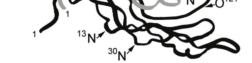
HCG and cancer
count for 39% of the molecular weight (Table 1).
Hyperglycosylated hCG is the principal molecule
produced in the first 3 weeks of pregnancy. At
this time is controls implantation of pregnancy,
and cytotrophoblast cell growth and invasion
during the first trimester of pregnancy [8-14]. It
is our understanding that antagonization of the
cytotrophoblast cell TGFβ receptor leads to a
cancer-like process, blockage of apoptosis, and
secretion of invasive enzymes, metallopro-
teinases and collagenases, leading to growth
and proteolytic invasion [15-25].
Hyperglycosylated hCG function for the length of
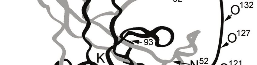
Figure 1. Crystal structure of deglycosylated regular pregnancy promoting root cytotrophoblast cell
hCG, as shown by Lathorn et al [2]. The unfolded β- growth. The combination of hCG and hyperglyco-
subunit C-terminal peptide is added (missing in crys- sylated hCG promote villous placental tissue
tal structure). It is inferred that this structure is not growth, hyperglycosylated hCG promoting cyto-
folded since the sequence comprises primarily a poly- trophoblast growth and hCG promoting the fu-
mer of proline and serine residues. The symbols N
and O indicate the sites of attachment of N-linked
sion of cytotrophoblast cells to syncytiotro-
and O-linked oligosaccharides. Residue β48 and β93 phoblast cells. hCG also promoted umbilical
are indicated as the site of cleavage of the β-subunit. artery angiogenesis and formation of the umbili-
The symbol K indicates the site of the cystine knot cal circulation. All these system come together
structure. The α-subunit is shown in grey, and β- in formation of hemochorial placentation [26-
subunit is shown in black. 34].
Table 1. Properties of 5 independent variants of hCG. Amino acid content, molecular weight and sugar
contents determined from published structures as determined by Elliott et al. for hCG and hyperglycosy-
lated hCG [90], Birken et al. for sulfated pituitary hCG [80] and Valmu et al. for hyperglycosylated hCGβ
[91]. The molecular weight of common hCG dimer amino acid backbone is that as determined by Mor-
gan et al. [122]. Molecular weight of N- and O-linked sugar side chains is added to these values. Isoelec-
tric points are those published by Sutton et al. [92], and metabolic clearance rates are those established
[1, 80].
Parameter hCG Sulfated hCG Hyperglycosy- hCGβ Hyperglycosylated
lated hCG hCGβ
Type of molecule Hormone Hormone Autocrine Autocrine Autocrine
Total Molecular 37,180 36,150 42,800 23,300 27,600
weight
Amino acids α- 92 92 92 -
subunit
Amino acids β- 145 145 145 145
subunit
Peptide molecular 26,200 26,200 26,200 16,000 16,000
weight
O-linked sugar units 4 4 4 4 4
N-linked sugar units 4 4 4 2 2
Molecular weight 10,980 9,950 16,600 7,300 11,600
sugars
Percentage sugars 30% 28% 39% 31% 42%
Isoelectric point (pI), 3.5 Not known 3.2 Not known 3.5
principal peak
Metabolic clearance 36 h 20 h Not known 0.72 h Not Known
rate
23 Am J Cancer Res 2012;2(1):22-35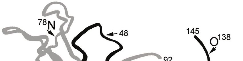
HCG and cancer

Figure 2. The carbohydrate structure of hCG, hCGβ, hyperglycosylated hCG, hyperglycosylated hCGβ and sulfated hCG
[80,90,91].

hCG and hyperglycosylated hCG evolved with                   humans [35, 36]. This was needed to permit the
humans [35, 36]. During their evolution came                 development of the human brain and humans
super-CG (chorionic gonadotropin) and super-                 [35, 36]. The human genome harbors genes to
hyperglycosylated CG two extremely potent                    express super-hyperglycosylated CG and its de-
growth factors that permitted hemochorial pla-               rivatives, super-CGβ and super-hyperglycosy-
centation to extend its efficiency multiple-fold in          lated CGβ. These are expressed in human can-

24                                                                          Am J Cancer Res 2012;2(1):22-35
HCG and cancer

Table 2. Parallelisms between placental implantation and invasion characteristics in primates, presence
and sugar structure on chorionic gonadotropin (CG) or LH, and relative brain masses. Table summarizes
published data [37-40,49,55].
Species               Implantation      Depth of Invasion   Sugar structures,    Brain mass         First appearance
                      characteristics                       acidity or pI        (% of body weight)
Humans                Hemochorial       1/3rd myometrium CG, 8 oligosaccha- 2.4%                      0.1 million year ago
                                                         rides, pI 3.5
Advanced simian       Hemochorial       1/10th myometrium CG, 6 oligosaccha- 0.74%                    20 million year ago
primates                                                  rides, pI 4.9
Early simian pri-     Hemochorial       through decidua     CG, 5 oligosaccha- 0.17%                  37 million year ago
mates                                                       rides, pI 6.3
Prosimian primate Epitheliochorial no-invasion              No CG produced, LH 0.07%                  55 million years ago
                                                            produced,
                                                            3 oligosaccharides,
                                                            pI 8.4

  Table 3. Use of serum free β-subunit (hCGβ plus hyperglycosylated hCGβ) as a tumor marker for detec-
  tion of malignancies. Averages are determined by combining total positive cases from multiple reports
  (89-79,100-113).
  Malignancy                                       Number of Cases         Sensitivity (>3 fmol/ml)
  Ovarian cancer                                   150                     38%
  Cervical cancer                                  60                      37%
  Endometrial cancer                               55                      33%
  Vulvar                                           50                      38%
  Bladder cancer                                   170                     35%
  Lung cancer                                      243                     18%
  Colorectal cancer                                436                     17%

  TOTAL                                            1164                    Mean 30% detection

cers, and just as they permitted super-biology in                simian primates such as platyrrhine or the new
human evolution, so will they permit unfortu-                    world monkey, CG and hyperglycosylated CG
nately, super-biology in driving human cancers.                  first evolved, and along with these molecules
This is the topic of this review.                                came primitive hemochorial placentation [35,
                                                                 36]. Hemochorial placentation, or fetal circula-
Since this super-CG, super-hyperglycosylated CG                  tion filtration by syncytiotrophoblast cell sur-
driven process evolved to drive human evolution                  rounded by maternal blood, is much more effi-
[35, 36]. It is important to understand human                    cient.
evolution first, before we consider human can-
cer, or a human evolution process gone hay-                      In 1980 Fiddes and Goodman [37], examined
wire. The earliest primates, prosimian primates                  the DNA sequence for the β-subunits of CG and
such as lemurs, had small brains, 0.07% of                       LH in humans and primates, and showed that
body weight (Table 2). This is because                           the evolution of CG from LH occurred by a single
prosimian primates used inefficient non-                         deletion mutation in LH β-subunit DNA and read
invasive epitheliochorial placentation. With the                 -through into the 3’-untranslated region in early
evolution of the next level of primates, early                   simian primates. In 2002 Maston and Ruvolo

25                                                                               Am J Cancer Res 2012;2(1):22-35
HCG and cancer
Table 4. Use of urine β-subunit core fragment as a tumor marker for detection of malignancies. Data
from multiple reports (89-79,100-113).
Malignancy Number of cases Sensitivity (>3 fmol/ml)
Ovarian cancer 207 66%
Cervical cancer 410 48%
Endometrial cancer 157 47%
Pancreatic cancer 29 55%
Bladder cancer 102 48%
Lung cancer 122 24%
TOTAL 1027 Mean 48% detection
[38], investigated the DNA sequences of the β- human CG has 4 O-linked and 4 N-linked oligo-
subunit of CG in 14 primates and showed that saccharides all terminating in sialic acid resi-
the genes to make CG and its variants were not dues. These acidify hCG resulting in a molecule
present in prosimians or primitive primates with a mean isoelectric point (pI) of 3.5, and a
(example: Lemur), but evolved by the indicated circulating half-life of 36 hours or 2160 minutes
deletion mutation with the early simian primates [1]. At the other extreme, is LH (pI 9.0 [43]), the
(platyrrhine or new world monkey). The first or molecule that CG evolved from, has just 3 N-
early simian primates CG and hyperglycosylated linked oligosaccharides. The metabolic clear-
CG molecules had just 3 N-linked and 2 O-liked ance half-life of LH Is just 25 minutes [44], or
oligosaccharides (Table 2). These evolved with 86 fold shorter that human CG. Human CG cir-
the species about 37 million year ago (Table 2). culates for approximately 86 times longer than
With the evolution of advanced simian primates LH, raising the circulation concentration propor-
about 20 million ago (examples: orangutan and tionately. A regression equation linking the num-
chimpanzee), with further point mutations a ber of oligosaccharides and the metabolic clear-
form of CG and hyperglycosylated CG evolved ance rates them was formed. If clearance rate
that had 3 N-linked and 3 O-linked oligosaccha- (minutes) half-life is CR and number of oligosac-
rides (Table 2). With the evolution of humans, charides is #O then CR = (2.4#O x 1.9). Using
approximately 0.1 millon year ago, and with this equation it was calculated that that the
further point mutations came the evolution of clearance rate half-life of early simian primate
human CG and hyperglycosylated CG having 4 N CG was approximately 2.5 hours and the clear-
-linked and 4 O-linked oligosaccharides. This ance rate half-life of advanced simian primate
increasing numbers of oligosaccharides and CG was approximately 6 hours.
acidic sugars, 3 N-linked 2 O-linked, to 3 N-
linked 3 O-linked and 4 N-linked 4 O-linked led The size of the brain in mammals is directly re-
to the evolution of a CG with an extreme acidity. lated to the combination of body mass and the
Acidity ranged from pI 6.3 in early simians, to pI metabolic support of the developing progeny
4.8 in advanced simians and on to super-acidic [45]. The larger brain size, seen in advanced
pI 3.5 molecules in humans [35, 36, 38-40]. primates and humans, correlates with dispro-
portionately large energy demands by the devel-
The metabolic clearance rate or circulating lev- oping fetuses [45-51]. Numerous studies sup-
els of CG were very much changed with acidity port the concept that advanced primates, and
and evolution. As CG evolved with additional to a greater extent humans, had to develop
oligosaccharides containing sialic acid, it very more efficient or super efficient placentation
much lengthened metabolic clearance rate of mechanisms to support the increasing nutri-
molecules and their effective bio-potency [38 tional demands of their embryonic brain (Table
41, 42]. As an example, at one extreme, regular 2) [39, 40, 45-55].
26 Am J Cancer Res 2012;2(1):22-35HCG and cancer
The prosimian primate had an average size and super-hyperglycosylated CG or hCG and
mammalian brain, 0.07% of body mass (Table hyperglycosylated hCG were created, and this
2). In this species, epitheliochorial placentation cancer story starts. Two potent growth promot-
was sufficient. Hemochorial placentation ers, normally reserved for evolution, and for
started with the evolution of CG in early simian pregnancy [35, 36]. Human cancers use hyper-
primate. It was only with the appearance of CG glycosylated hCG and its free subunit variants to
and hyperglycosylated CG in early simian pri- drive the most efficient possible malignancy. It
mates, that the signals to implant placentas is at this point that this review starts.
inside the uterus [8, 9, 12-14], and the signals
to generate villous placenta [36, 36], to pro- HCG, one name shared by five independent
mote angiogenesis of uterine vasculature [29- molecules
32] and development of the umbilical cord [33,
34] that hemochorial placentation happened Research in the last 10 years has shown that
[35]. Hemochorial placentation was primitive in the molecule generally called human chorionic
early simian primates, implanting only through gonadotropin (hCG) is not one independent
the depth of the decidua, leading to a larger molecule, but rather is 5 separate molecules
brain 0.17% of body mass (Table 2). It was with with independent functions. The five separate
evolution and the development of more-acidic forms of hCG all share a common amino acid
more-potent CG and hyperglycosylated CG that backbone, thus have a common name. They
hemochorial placentation went deeper to 1/10th vary greatly, however, in carbohydrate side
myometrial depth in advance simian primates chain structure and meric structure (Table 1).
(Table 2). This supported the development of a
much larger brain, 0.74% of body mass (Table hCG is a hormone made by placental syncyti-
2). otrophoblast cells [7]. hCG comprises a 92
amino acid α-subunit and a 145 amino acid β-
With the evolution of humans and the multiple subunit. The β-subunit of hCG, while structurally
mutations needed to produce their super-CG similar to the β-subunit of LH, differentiates hCG
with 2 additional oligosaccharides, hemochorial from other glycoprotein hormones. hCG, like LH,
placentation went to the extreme. CG jumped in is a hormone, and binds a common hCG/LH
acidity from metabolic clearance rate half-life of hormone receptor.
360 to 2160 minutes. With this hemochorial
placentation went deeper to 1/3rd the thickness For the first 3 weeks of pregnancy, hCG pro-
of the myometrium (Table 2). Hemochorial pla- motes production of progesterone by ovarian
centation reached the efficiency needed to sup- corpus luteal cells [56-58]. Multiple research
port a human brain, 2.4% of body mass. groups have shown that hCG also functions dur-
ing pregnancy to promote angiogenesis in the
Nutrition transfer and placentation were taken uterine vasculature [29-32]. This insures maxi-
to the extreme in the humans. Human CG has a mal blood supply to the invading placenta, an
circulating half-time of 2160 minutes. This important function during pregnancy. While hy-
leads to invasion to one third the thickness of perglycosylated hCG may promote cytotro-
the myometrium and to the super-efficient pla- phoblast cell growth during pregnancy [6, 9-14],
centation that is needed to support the nutri- hCG promotes the fusion of cells and their dif-
tional transfer necessary for a brain of 2.4% ferentiation to syncytiotrophoblast cells [28]. It
body mass or 3 fold greater than that of ad- is the combination of these two processes that
vanced simians [46, 48, 49, 55]. Considering leads to villous trophoblast tissue formation and
the relationship between regular CG, hyperglyco- hemochorial placentation in pregnancy [35, 36].
sylated CG and hemochorial placentation, and Multiple groups show that hCG promotes an anti
between advancing acidity of CG and advancing -macrophage inhibitory factor or a macrophage
invasion and angiogenesis, it would not be un- migration inhibitory factor that prevents destruc-
reasonable to claim that the evolution of CG in tion of the foreign feto-placental by the mother’s
early simians started primates on the evolution tissue during pregnancy [59, 60]. Other groups
path to advanced brains, or is at the root of hu- have shown that hCG also controls uterine
man evolution [35, 36]. growth during pregnancy [61, 62], and yet other
groups have shown that hCG also relaxes myo-
It is with these evolution stories that super-CG metrial contractions during pregnancy [63, 64].
27 Am J Cancer Res 2012;2(1):22-35HCG and cancer
It has been shown that hCG also control umbili- all other malignancies use a similar TGFβ an-
cal cord growth and circulation and develop- tagonism pathway when they can become ad-
ment during pregnancy [33, 34]. New research vanced and can reach a state of differentiation
is finding receptors in fetal organs and a further whereby they express an hCGβ gene [69-79].
role for hCG in fetal growth during pregnancy These cancer cells seemingly lack the ability to
[65, 66]. combine hCG subunits and just secrete hCGβ or
hyperglycosylated hCGβ. Both of these mole-
The structure of the N-linked and O-linked oligo- cules can antagonize the TGFβ receptor and
saccharide side chains attached to the hormone promote malignancy [5, 6]. As now demon-
hCG are shown in Figure 2. The three dimen- strated, all advanced cancers are directly pro-
sional structure of hCG dimer was shown by moted to grow, invade and metastasize by an
Lapthorn and colleagues (Figure 1) [2]. As autocrine hCGβ or hyperglycosylated hCGβ [69-
shown, the β-subunit wraps itself around the α- 79]. Actions include inhibition of apoptosis in
subunit (Figure 1). Hyperglycosylation of hCG cancer cells and promotion of invasion prote-
subunits leads to incomplete folding, this leads ases by cancer cells [41-48]. As demonstrated,
to exposure of sequences otherwise hidden on recently, hyperglycosylated hCG, hCGβ and hy-
hCG. These are the evolutionary TGFβ struc- perglycosylated hCGβ are inter-changeable pro-
tures. Hyperglycosylated hCG is an autocrine, moters, that all can promote choriocarcinoma or
and not a hormone like hCG, it seemingly binds other advanced malignancies [6].
and antagonizes TGFβ receptors on the cytotro-
phoblast cells that make hyperglycosylated hCG A fifth or final variant of hCG is made by pituitary
[6, 8-26]. This is part of the process of preg- gonadotrope cells during the menstrual cycle
nancy implantation. Hyperglycosylated hCG pro- [80-83]. This is the sulfated variant of hCG with
motes blockage of apoptosis in these cells, and sulfated oligosaccharides as shown in Table 1
production of collagenases and metallopro- and Figure 2 [80]. Research by Odell and Griffin
teinases needed for invasion in the implantation [81, 82] using an ultrasensitive hCG assay
process [8-26]. Hyperglycosylated hCG also pro- shows that sulfated hCG is produced during the
motes cytotrophoblast cells or placental growth length of the menstrual cycle, following the se-
during the length of pregnancy [11-14]. cretion pattern of LH. hCG and LH bind a com-
mon receptor. Research in Cole’s laboratory
Hyperglycosylated hCG drives invasion as oc- shows that sulfated hCG production in 277
curs in the fastest growing human malignancy, menstrual cycles at the time of the LH peak
choriocarcinoma. Classically, a women may averages 1.54 ± 0.90 mIU/ml [83]. It appears
have a normal pregnancy, and deliver with just that sulfated hCG matches LH function in pro-
a few cytotrophoblast cell remaining at the im- moting androstenedione production by theca
plantation site. Transformation may occur in cells, progesterone production by corpus luteal
one of these remaining cells. Just 6 to 10 weeks cells and in enhancing ovulation.
later, the new mother may show at an emer-
gency room with difficulty breathing and sei- Choriocarcinoma and germ cell malignancies
zures, due to choriocarcinoma spreading to her
lungs, and in her brain. This is choriocarcinoma, Choriocarcinoma is a gestational trophoblastic
a malignancy totally driven by hyperglycosylated disease, residing at the interface of obstetrics
hCG and seemingly by the TGFβ antagonism and oncology. Transformation in choriocarci-
process normally reserved for pregnancy im- noma cases seemingly involves blockage of
plantation [6, 9, 11, 27, 67, 68]. cytotrophoblast cells from fusing to form syncyti-
otrophoblast cells [9, 11, 27, 67, 68]. Cytotro-
Choriocarcinoma is not the only malignancy that phoblast cells are the site of hyperglycosylated
produces hyperglycosylated hCG, and uses hy- hCG production, the driving force behind chorio-
perglycosylated hCG to drive its malignancy. carcinoma [9, 11].
Testicular and ovarian germ cell malignancies
take on a cytotrophoblast histology and are The big question is what is the best tumor
driven by hyperglycosylated hCG [9, 27]. These marker? Only one set of tumor markers fit this
are the only malignancies that misuse this evo- criterion, total hCG and hyperglycosylated hCG
lution growth factor to drive their malignancy, [67, 84-87]. Both of these tumor markers are
hyperglycosylated hCG. As we now understand, 100% sensitive for choriocarcinoma. This is be-
28 Am J Cancer Res 2012;2(1):22-35HCG and cancer
cause choriocarcinoma cannot exist without hyperglycosylated hCG act though similar
hyperglycosylated hCG, as measured as hyper- mechanisms, TGFβ receptor antagonism, to
glycosylated hCG or total hCG immunoassays. control apoptosis, to control cell growth, and
No other tumor marker can make this claim. As promote collagenases and metalloproteinases
demonstrated, when choriocarcinoma cells are promoting invasion [5, 6, 15-26].
grown in a nude mouse, they grow very rapidly.
When an antibody is given to bind hyperglycosy- The story with choriocarcinoma and germ cell
lated hCG, all growth completely stops [9, 11]. malignancies does not stop here. Choriocarci-
Similarly, when nude mice are administered noma is an important part of cancer history. It
choriocarcinoma cells in which the hCG subunit has always been at the root of major discover-
genes are blocked with anti-sense cDNA, all ies. It was at the root of discovery of chemother-
growth ceases [88, 89]. It is concluded that apy as a cure for cancer. As was known, chorio-
choriocarcinoma cannot exist without hypergly- carcinoma is an extremely fast growing malig-
cosylated hCG. nancy. As Dr. Roy Hertz reasoned, why doesn’t
an inhibitor of cell division or DNA synthesis
The USA hCG Reference Service uses the B152 block choriocarcinoma cancer growth. As rea-
antibody hyperglycosylated hCG assay. This test soned, methotrexate blocks the synthesis of the
detects hyperglycosylated hCG and its free β- critical DNA nucleotide thymidine. Why doesn’t
subunit, hyperglycosylated hCGβ [90]. In the methotrexate block choriocarcinoma growth? As
USA hCG Reference Service experience this tu- shown by Dr. Hertz in the nineteen fifties, meth-
mor marker detects 100% of choriocarcinoma, otrexate makes an effective treatment of chorio-
persistent hydatidiform mole, testicular germ carcinoma [93-95]. This discovery led to mod-
cell malignancy and ovarian germ cell malig- ern chemotherapy treatment for cancer.
nancy cases. This test is diagnostic, it demon-
strates malignant vs. quiescent or benign dis- Now here, we start again with choriocarcinoma
ease (HCG and cancer
actually hyperglycosylated hCGβ, a variant of lated hCGβ antagonize this receptor [5, 6, 69,
hCGβ similar to the β-subunit of hyperglycosy- 70]. As reported, hCGβ and hyperglycosylated
lated hCG. Why some cancers produce primarily hCGβ promote the production of collagenases
hyperglycosylated hCGβ versus hCGβ is not and metalloproteinases, invasion proteases
known. produced by cancer cells [69], leading to metas-
tases.
The literature shows that all advanced malig-
nancies secrete hCGβ or hyperglycosylated As shown recently [6], cancers other than chori-
hCGβ [98, 99], yet only a small proportion of ocarcinoma and germ cell malignancies pro-
malignancy cases, about 30%, have hCGβ or duce hCGβ and hyperglycosylated hCG. Hyper-
hyperglycosylated hCGβ in blood (Table 3), or glycosylated hCG, hCGβ and hyperglycosylated
their degradation product, β-core fragment pre- hCGβ are all interchangeable. Just as hCGβ can
sent in urine of 48% of cancer cases (Table 4). do hyperglycosylated hCG’s job with choriocarci-
This is because hCGβ and hyperglycosylated noma, so can hyperglycosylated hCG do hCGβ’s
hCGβ are rapidly cleaved by the enzyme leuko- job with other malignancies [6]. It appears that
cyte elastase, produced my macrophages and they all are interchangeable markers, all seem-
leukocytes upon upon secretion. This enzyme ingly acting on a TGFβ receptor to antagonize it.
first nicks or cleaves the molecules at β47-48
upon secretion, and then cleaves this mole- In recent years, hCGβ vaccines are being evalu-
cule’s C-terminal, or major acidic component by ated for patients with advanced cancers [116-
cleavage at β92-93 (Figure 1) [71, 115]. The 121]. Initial clinical trials are extremely promis-
resulting degradion products are rapidly cleared ing, showing a 2-fold extention of cancer sur-
from the circulation by the liver and kidney, with vival [118-121]. The vaccine studies confirms
circulating half lives of a few minutes verses 36 the key role that hCGβ/hyperglycosylated hCGβ
hour like hCG [71, 115]. This makes detection has in cancer metastasis and its action in all
of the hCGβ or hyperglycosylated hCGβ in can- cancer cases.
cer cases very difficult, yielding a detection rate
in blood of just 30%. It is my understanding that choriocarcinoma,
persistent hydatidiform mole and germ cell ma-
An accumulation of studies (Tables 3 and 4) lignancies are promoted by hyperglycosylated
shows that most malignancies produce this hCG in all stages. These are eutopic malignan-
molecule [98, 99]. Urine β-core fragment is a cies or malignancies driven by hyperglycosy-
useful tumor marker in gynecologic oncology, lated hCG. Hyperglycosylated hCG is seemingly
detecting 47% of endometrial, 48% of cervical the single cancer promoter, since cancer is
and 66% of ovarian malignancies. Urine β-core brought to a complete halt in nude mice when
fragment can be used as a simple three hCG supply is blocked by antibody or DNA fac-
monthly screening test in women with familial tors [11, 88, 89]. Other malignancies produce
ovarian cancer. Urine β-core fragment can be hCGβ and hyperglycosylated hCGβ. This is only
used as a wide spectrum cancer screening test. produced in advanced disease [5, 69-79, 98-
Yes, it detects 48% of all cancers, but a person 114]. It seems that the other or ectopic malig-
positive in a β-core fragment assay can only nancies have to be advanced to differentiate
then be screened with MRIs of the head and tissues and to express ectopic hCGβ. From the
pelvis and chest CT to determine the site of ma- time that hCGβ is ectopically expressed on-
lignancy. wards hCGβ may be the principal driver of the
malignancies. Based on the vaccine studies, it
Examination of the crystal structure of hCG [2], appears, as suggested [98, 99], that all malig-
shows that the β-subunit has common evolu- nancies may be controlled in advanced stages
tionary sequences with TGFβ [3, 4], and a by hCGβ and/or hyperglycosylated hCGβ. It ap-
cystine knot structure unique to hCG, TGFβ, pears that once advanced malignancies start to
platelet-derived growth factor and nerve growth express hCGβ and/or hyperglycosylated hCGβ
factor. The site of this cystine knot structure is that the malignancy may then be controlled by
shown in Figure 1. As demonstrated [5], hCGβ the TGFβ antagonism choriocarcinoma-like
antagonizes a TGFβ receptor site inhibiting route by a molecule like hCGβ/
apoptosis in the cancer cells, indicating that hyperglycosylated hCGβ. It appears that hCGβ/
hCGβ, hyperglycosylated hCG and hyperglycosy- hyperglycosylated hCGβ should be the target of
30 Am J Cancer Res 2012;2(1):22-35HCG and cancer
much cancer research, it is the future, the mole- anthelix motif in the TGF-β superfamily by
cules that seemingly drive advanced malignan- molecular 3D-Rapid Prototyping. Materialwis-
cies. senschaft und Werkstofftechnik 2003; 34:
1113-1119.
[4] Lehnert SA, Akhurst RA. Embryonic expression
The future pattern of TGF beta type-1 RNA suggests both
paracrine and autocrine mechanisms of ac-
In conclusion, it appears that hyperglycosylated tion. Developm 1988; 104: 263-273.
hCG, hCGβ and hyperglycosylated hCGβ are an [5] Butler SA, Ikram MS, Mathieu S, Iles RK. The
inter-related set of molecules [6]. That seem- increase in bladder carcinoma cell population
ingly drive cancer through a TGFβ antagonism induced by the free beta subunit of hCG is a
pathway [5, 6, 23, 25]. Choriocarcinoma and result of an anti-apoptosis effect and not cell
germ cell malignances are all seemingly driven proliferation. Brit J Cancer 2000; 82: 1553-
in early and advanced stages by this highly inva- 1556.
[6] Cole LA, Butler SA. Hyperglycosylated hCG,
sive pathway. In contrast, most other cancers hCGβ and Hyperglycosylated hCGβ: Inter-
are driven by alternative pathways until they changeable Cancer Promoters. Molec Cell
become advance and express the hCGβ gene. Endocrinol 2011; in press.
They seeming adopt this viscous TGFβ antago- [7] Kovalevskaya G, Genbacev O, Fisher SJ, Ca-
nism pathway. This may be the key cancer cere E, O'Connor JF. Trophoblast origin of hCG
physiology pathway. isoforms: cytotrophoblasts are the primary
source of choriocarcinoma-like hCG. Mol Cellul
This review presents research on cancers taking Endocrinol 2002; 194: 147-155.
this pathway and promises for the future. Anti- [8] Sasaki Y, Ladner DG, Cole LA. Hyperglycosyla-
ted hCG the source of pregnancy failures.
bodies to hyperglycosylated hCG may seemingly Fertil Steril 2008; 89: 1871-86.
cure choriocarcinoma and germ cell malignan- [9] Cole LA, Khanlian SA, Riley JM, Butler SA. Hy-
cies in the future, and vaccines to hCGβ and perglycosylated hCG (hCG-H) in Gestational
administered antibodies may significantly ex- Implantation, and in Choriocarcinoma and
tend the lives of all advanced cancer patients. Testicular Germ Cell Malignancy Tumorigene-
Vaccines may not work in some advanced stage sis. J Reprod Med 2006; 51: 919-929.
cases, in patients with compromised immune [10] Cole LA. Biological function of hyperglycosy-
systems. This is where administered antibodies lated hCG, in: Cole LA (ed), HCG (hCG). El-
may be most warranted. sevier, Burlington MA 2010; pp: 49-65.
[11] Cole LA, Dai D, Butler SA, Leslie KK, Kohorn
EI. Gestational trophoblastic diseases: 1.
In evolution, the molecule hyperglycosylated Pathophysiology of hyperglycosylated hCG-
hCG was recruited to drive human evolution as regulated neoplasia. Gynecol Oncol 2006;
an extreme growth factor. A growth factor that 102: 144-149.
drove placental implantation deeper and growth [12] Guibourdenche J, Handschuh K, Tsatsaris V,
to extremes. Unfortunately cancers take advan- Gerbaud MC, Legul F, Muller D, Evain-Brion, D,
tage of the availability of the extreme growth Fournier T. Hyperglycosylated hCG is a marker
factors. It appears that the hCGβ/ of early human trophoblast invasion. J Clin
hyperglycosylated hCGβ TGFβ pathway may be Endocrinol Metab 2010; 95: E240-4.
[13] Handschuh K, Guibourdenche J, Tsatsaris V,
the central pathway to treatment of all ad- Guesnon M, Laurendeau I, Evain Brion D,
vanced cancers. Fournier T. HCG expression in human tropho-
blasts from early placenta, comparative study
Address correspondence to: Dr. Laurence A Cole, USA between villous and extravillous trophoblastic
hCG Reference Service, University of New Mexico, cells. Placenta 2007; 28: 175-84.
Albuquerque, NM 87131, USA Tel: 505-263-9635; E- [14] Handschuh K, Guibourdenche J, Tsatsari V,
mail: larry@hcglab.com Guesnon M, Laurendeau I, Evain Brion D,
Fournier T. HCG produced by the invasive tro-
References phoblast but not the villous trophoblast pro-
motes cell invasion and is down-regulated by
[1] Wehmann RE, Nisula BC. Metabolic and renal peroxisome proliferator-activated receptor-a.
clearance rates of purified hCG. J Clin Invest Endocrinol 2007; 148: 5011-19.
1981; 68: 184-94. [15] Schuster N, Krieglstein K. Mechanisms of TGF-
[2] Lapthorn AJ, Harris DC, Littlejohn A, Lustbader β-mediated apoptosis. Cell Tissue Res 2002;
JW, Canfield RE, Machin KJ. Crystal structure 307: 1-14.
of hCG. Nature 1994; 369: 455-461. [16] Kamijo T, Rajabi MR, Mizunuma H, Ibuki Y.
[3] Laub M, Jennissen HP. Identification of the Biochemical evidence for autocrine/paracrine
31 Am J Cancer Res 2012;2(1):22-35HCG and cancer
regulation of apoptosis in cultured uterine [29] Toth P, Li X, Rao CV, Lincoln SR, Sanfillipino
epithelial cells during mouse embryo implan- JS, Spinnato JA, Yussman MA. Expression of
tation in vitro. Molec Human Reprod 1998; 4: functional hCG/human luteinizing hormone
990-8. receptor gene in human uterine arteries. J Clin
[17] Pampferf S. Apoptosis in rodent peri- Endocrinol Metab 1994; 79: 307-315.
implantation embryos: differential susceptibil- [30] Zygmunt M, Herr F, Keller Schoenwetter S,
ity of inner cell mass and trophectoderm cell Kunzi Rapp K, Munstedt K, Rao CV, Lang U,
lineages-A review. Placenta 2000; 21: S3-S10. Preissner KT. Characterization of hCG as a
[18] Shooner C, Caron PC, Fréchette Frigon G, novel angiogenic factor. J Clin Endocrinol Me-
Leblanc V, Déry MC, Asselin E. TGF-beta ex- tab 2002; 87: 290-296.
pression during rat pregnancy and activity on [31] Lei ZM, Reshef E, Rao CV. The expression of
decidual cell survival. Reprod Biol Endocrinol hCG/ luteinizing hormone receptors in human
2005; 3: 20. endometrial and myometrial blood vessels. J
[19] Liu YX, Gao F, Wei P, Chen XL, Gao HJ, Zou RZ, Clin Endocrinol Metab 1992; 75: 651-659.
Siao LJ, Xu FH, Feng Q, Liu K, Hu ZY. Involve- [32] Zygmunt M, Herr F, Keller Schoenwetter S,
ment of molecules related to angiogenesis, Kunzi Rapp K, Munstedt K, Rao CV, Lang U,
proteolysis and apoptosis in implantation in Preissner KT. Characterization of hCG as a
rhesus monkey and mouse. Contraception novel angiogenic factor. J Clin Endocrinol Me-
2005; 71: 249-62. tab 2002; 87: 290-5296.
[20] Knittel T, Mehde M, Kobold D, Saile B, Dinter [33] Rao CV, Li X, Toth P, Lei ZM, Cook VD. Novel
C, Ramadori G. Expression patterns of matrix expression of functional hCG/luteinizing hor-
metalloproteinases and their inhibitors in par- mone receptor in human umbilical cords. J
enchymal and non-parenchymal cells of rat Clin Endocrinol Metab 1993; 77: 1706-1714.
liver regulation by TNF-alpha and TGF-beta1. J [34] Wasowicz G, Derecka K, Stepien A, Pelliniemi
Hepatol 1999; 30: 48-60. L, Doboszynska T, Gawronska B, Ziecik AJ.
[21] Murphy G, Reynolds JJ, Whitham SE, Docherty Evidence for the presence of luteinizing hor-
AJ, Angel P, Heath JK. Transforming growth mone-chorionic gonadotrophin receptors in
factor beta modulates the expression of colla- the pig umbilical cord. J Reprod Fertil 1999;
genase and metalloproteinase inhibitor. Euro 117: 1-9.
Molec Biol Org J 1987; 6: 1899-1904. [35] Cole LA. hCG and hyperglycosylated hCG in the
[22] Qureshi HY, Sylvester J, El Mabrouk M, Zafa- establishment and evolution of hemochorial
rullah M. TGF-beta-induced expression of tis- placentation. J Reprod Immunol 2009; 82:
sue inhibitor of metalloproteinases-3 gene in 111-117.
chondrocytes is mediated by extracellular [36] Cole LA, Khanlian SA, Kohorn EI. Evolution of
signal-regulated kinase pathway and Sp1 tran- the Human Brain, Chorionic Gonadotropin and
scription factor. J Cell Physiol 2005; 203: 345- Hemochorial Implantation of the Placenta:
52. Insights into Origins of Pregnancy Failures,
[23] Stetler Stevenson WG, Brown PD, Onisto M, Preeclampsia and Choriocarcinoma. J Reprod
Levy AT, Liotta LA. Tissue inhibitor of metallo- Med 2008; 53: 449-557.
proteinases-2 (TIMP-2) mRNA expression in [37] Fiddes JC, Goodman HM. The cDNA for the β-
tumor cell lines and human tumor tissues. J subunit of hCG suggests evolution of a gene
Biol Chem 1990; 265: 13933-13938. by readthrough into the 3’-untranslated re-
[24] Staun Ram E, Shaleu E. Human trophoblast gion. Nature 1980; 286: 684-687.
function during the implantation process. Re- [38] Maston GA, Ruvolo M. Chorionic gonadotropin
prod Biol Endocrinol 2005; 3: 56-68. has a recent origin within primates and an
[25] Fisher SJ, Tian yi C, Li Z, Hartman L, Grahl K, evolutionary history of selection. Mol Biol Evol
Zhang GY, Tarpey J, Damsky CH. Adhesive and 2002; 19: 320-334.
degradative properties of human placental [39] Bambra CS. Purification and properties of
cytotrophoblast cell in vitro. J Cell Biol 1989; baboon chorionic gonadotropin. J Reprod Fer-
109: 891-902. til 1987; 19: 421-430.
[26] Khoo NK, Bechberger JF, Shepherd T, Bond [40] Crawford RJ, Tegear GW, Niall HD. The nucleo-
SL, McCrae KR, Hamilton GS, Lala PK. SV40 tide sequence of baboon chorionic gonadotro-
Tag transformation of the normal invasive pin β-subunit genes have diverged from the
trophoblast results in a premalignant pheno- human. Gene 1986; 46: 161-169.
type I Mechanisms responsible for hyperinva- [41] Cassels JW, Mann K, Blithe DL, Nisula RC,
siveness and resistance to anti-invasive action Wehmann RE. Reduced metabolic clearance
of TGFβ. Intl J Cancer 1998; 77: 429-39. of acidic variants of human choriogonadotro-
[27] Cole LA. Hyperglycosylated hCG. Placenta pin from patients with testicular cancer. Can-
2007; 28: 977-986. cer 1989; 64: 2313-2318.
[28] Shi QJ, Lei ZM, Rao CV, Lin J. Novel role of [42] Rosa C, Amr S, Birken S, Wehmann R, Nisula
hCG in differentiation of human cytotro- B. Effect of desialylation of hCG on its meta-
phoblasts. Endocrinol 1993; 132: 387-395. bolic clearance rate in humans, J Clin Endocri-
32 Am J Cancer Res 2012;2(1):22-35HCG and cancer
nol Metab 1984; 59: 1215-1219. Univ ZU 1919; 23: 63-70.
[43] Keel BA, Grotjan HE. Characterization of rat [59] Akoum A, Metz CN, Morin M. Marked increase
pituitary luteinizing hormone charge microhet- in macrophage migration inhibitory factor syn-
erogeneity in male and female rats using chro- thesis and secretion in human endometrial
matofocussing: effect of castration. Endocrinol cells in response to hCG hormone. J Clin Endo-
1985; 117: 354-360. crinol Metab 2005; 90: 2904-2910.
[44] Valdhuis JD, Fraioli F, Rogol AD, Dufau ML. [60] Wan H, Marjan A, Cheung VW, Leenen PJM,
Metabolic clearance of biologically active lu- Khan NA, Benner R, Kiekens RCM. Chorionic
teinizing hormone in men. J Clin Invest 1986; gonadotropin can enhance innate immunity by
77: 1122-1128. stimulating macrophage function. J Leukocyte
[45] Martin RD. Relative brain size and basal meta- Biol 2007; 82: 926-933.
bolic rate in terrestrial vertebrates. Nature [61] Reshef E, Lei ZM, Rao CV, Pridham DD, Che-
1981; 293: 57-60. gini N, Luborsky JL. The presence of gonad-
[46] Gibbons A. Solving the brain’s energy crisis. otropin receptors in nonpregnant human
Science 1998; 280: 1345-1347. uterus, human placenta, fetal membranes,
[47] Robillard PY, Hulsey TC, Chaouat G. Pree- and decidua. J Clin Endocrinol Metab 1990;
clampsia and human reproduction. An essay 70: 421-430.
of a long term reflection. J Reprod Immunol [62] Zuo J, Lei ZM, Rao CV. Human myometrial
2003; 59: 93-100. chorionic gonadotropin/luteinizing hormone
[48] Martin RD. Scaling of the mammalian barrier: receptors in preterm and term deliveries. J
the maternal energy hypothesis. News Physiol Clin Endocrinol Metab 1994; 79: 907-911.
Sci 1996; 4: 149-154. [63] Eta E, Ambrus G, Rao V. Direct regulation of
[49] Cunnane SC, Herbige LS, Crawford MA. The human myometrial contractions by hCG. J Clin
importance of energy and nutrient supply in Endocrinol Metab 1994; 79: 1582-1586.
human brain evolution. Nutr Health 1993; 9: [64] Doheny HC, Houlihan DD, Ravikumar N, Smith
19-35. TJ, Morrison JJ. Human chorionic gonadotro-
[50] Martin RD. Human reproduction: a compara- phin relaxation of human pregnant myo-
tive background for medical hypotheses. J metrium and activation of the BKCa channel. J
Reprod Immunol 2003; 59: 111-135. Clin Endocrinol Metab 2003; 88: 4310-4315.
[51] Pjenenborg R. The placental bed. Hypertens [65] Goldsmith PC, McGregor WG, Raymoure WJ,
Pregn 1996; 15: 7-23. Kuhn RW, Jaffe RB. Cellular localization of
[52] Kliman HJ. Uteroplacental blood flow. The chorionic gonadotropin in human fetal kidney
story of decidualization, menstruation, and and liver. J Clin Endocrinol Metab 1983; 57:
trophoblast invasion. Am J Path 2000; 157: 54-61.
1759-1768. [66] Rao CV, Lei ZM. The past, present and future
[53] Robillard PY, Chaline J, Chaouat G, Hulsey TC. of nongonadal hCG/LH actions in reproductive
Preeclampsia/eclampsia and the evolution of biology and medicine. Mol Cell Endocrinol
the human brain. Current Anthropol 2003; 44: 2007; 269: 2-8.
130-135. [67] Cole LA, Butler SA, Khanlian SA, Giddings A,
[54] Jauniaux E, Poston L, Burton GJ. Placental- Muller CY, Seckl MJ, Kohorn EI. Gestational
related diseases of pregnancy: involvement of trophoblastic diseases: 2. Hyperglycosylated
oxidative stress and implications in human hCG as a Reliable Marker of Active Neoplasia.
evolution. Human Reprod Updt 2006; 12: 747 Gynecol Oncol 2006; 102: 150-158.
-755. [68] Cole LA, Muller Y. hCG in the Management of
[55] Lockett WP. Comparative development and Quiescent and Chemorefractory Gestational
evolution of the placenta in primates. Contrib Trophoblastic Diseases. Gynecol Oncol 2010;
Primatol 1974; 3: 42-234. 116: 3-9.
[56] Schmitt EJ, Barros CM, Fields PA M, Fields MJ, [69] Butler SA, Iles RK. Biological function of the
Diaz T, Kluge JM, Thatcher WW. A cellular and free β-subunit: expression and treatment tar-
endocrine characterization of the original and get in cancer, in: Cole LA (ed), HCG (hCG).
induced corpus luteum after administration of Elsevier, Burlington MA 2010; pp: 153-172.
a gonadotropin-releasing hormone agonist or [70] Iles RK. Ectopic hCGβ expression by epithelial
hCG on day five of the estrous cycle. J Anim cancer: Malignant behavior metastasis and
Sci 1996; 74: 1915-1929. inhibition of tumor cell apoptosis. Molec Cellul
[57] Richardson MC, Masson GM. Progesterone Endocrinol 2007; 260: 264-270.
production by dispersed cells from human [71] Cole LA, Birken S, Sutphen S, Hussa RO, Pat-
corpus luteum: stimulation by gonadotrophins tillo RA. Absence of the COOH-terminal peptide
and prostaglandin F2α; lack of response to on ectopic hCG β-subunit (HCG-β). Endocrinol
adrenaline and isoprenaline. J Endocrinol 1982; 110: 2198-2200.
1980; 87: 247-254. [72] Butler SA, Iles RK. Ectopic human chorionic
[58] Hirose T. Experimentalle histologische studie gonadotrophin β secretion by epithelial tumors
zur genese corpus luteum. Mitt Med Fakultd and human chorionic gonadotrophin β-
33 Am J Cancer Res 2012;2(1):22-35HCG and cancer
induced apoptosis in Karposi’s sarcoma, is 159.
there a connection? Clin Cancer Res 2003; 9: [89] Hamade AL, Nakabayashi K, Sato A, Kiyoshi K,
4666-4673. Takamatsu Y, Laoag-Fernandez JB, Ohara N,
[73] Carter WB, Sekharem M, Coppola D. HCG in- Maruo T. Transfection of antisense chorionic
duces apoptosis in breast cancer. Breast Can- gonadotropin β gene into choriocarcinoma
cer Res Treatm 2006; 100: S243-S244. cells suppresses the cell proliferation and
[74] Bellet D, Lazar V, Bleche I, Paradis V, Giovan- induces apoptosis. J Clin Endocrinol Metab
grandi Y, Paterliru P. Malignant transformation 2005; 90: 4873-4879.
of nontrophoblastic cells in association with [90] Elliott MM, Kardana A, Lustbader JW, Cole LA.
the expression of chorionic gonadotropin β Carbohydrate and Peptide structure of the α-
genes normally transcribed in trophoblastic and β-subunits of hCG from normal and aber-
cells. Cancer Res 1997; 57: 516-523. rant pregnancy and choriocarcinoma. Endo-
[75] Cosgrove DE, Campain JA, Cox GS. Chorionic crine 1997; 7: 15-32.
gonadotropin synthesis by human tumor cell [91] Valmu L, Alfthan H, Hotakainen K, Birken S,
lines: Examination of subunit accumulation Stenman UH. Site-specific glycan analysis of
steady-state levels of mRNA and gene struc- hCG {beta}-subunit from malignancies and
ture. Biochem Biophys Acta 1989; 1007: 44- pregnancy by liquid chromatography - electros-
54. pray mass spectrometry. Glycobiology 2006;
[76] Marcillac I, Cottu P, Theodore C, Lacombe MJ, 16: 1207-1218.
Bellet D, Droz JP. Free hCG beta subunit as [92] Sutton JM. Charge variants in serum and urine
tumour marker in urothelial cancer. Lancet hCG. Clin Chem Acta 2004; 341: 199-203.
1993; 341: 1354-1355. [93] Hertz R, Bergenstal DM, Lipsett MB, Price EB,
[77] Cole LA. β-core fragment (β-core UGP or UGF). Hilbish TF. Chemotherapy of choriocarcinoma
Tumor Marker Update 1994; 6: 69-75. and related trophoblastic tumors in women. J
[78] Iles RK. Human chorionic gonadotrophin and Am Med Assoc 1958; 18: 845-854.
its fragments as markers of prognosis in blad- [94] Li MC, Hartz R, Bergenstal DM. Therapy of
der cancer. Tumor Marker Update 1995; 7: choriocarcinoma and related trophoblastic
161-166. tumors with folic acid and purine antagonists.
[79] Gillott DJ, lles RK, Chard T. The effects of β- New Engl J Med 1958; 10: 66-74.
human chorionic gonadotrophin on the in vitro [95] Hertz R, Li MC, Spencer DB. Effect of meth-
growth of bladder cancer cell lines. Br J Can- otrexate therapy upon choriocarcinoma and
cer 1996; 73: 323-326. chorioadenoma. Proc Soc Exp Biol Med 1956;
[80] Birken S, Maydelman Y, Gawinowicz MA, 93: 361-366.
Pound A, Liu Y, Hartree AS. Isolation and char- [96] Cole LA, Hussa RO. Use of glycosidase di-
acterization of human pituitary chorionic go- gested hCG β-subunit to explain the partial
nadotropin. Endocrinol 1996; 137: 1402-11. binding of glycoprotein hormones to Con A.
[81] Odell WD, Griffin J. Pulsatile secretion of hCG Endocrinol 1981; 109: 2276-2279.
in normal adults. N Engl J Med 1987; 317: [97] Cole LA, Hussa RO, Rao CV. Discordant syn-
1688-91. thesis and secretion of hCG and subunits by
[82] Odell WD, Griffin J. Pulsatile secretion of chori- cervical cancer cells. Cancer Res 1981; 41:
onic gonadotropin during the normal men- 1615-1619.
strual cycle. J Clin Endocrinol Metab 1989; [98] Acevedo HF, Hartstock RJ. Metastatic pheno-
69: 528-32. type correlates with high expression of mem-
[83] Cole LA, Ladner DG, Cole LA, Gutierrez JM. brane-associated complete β-hCG in vivo.
Production of hCG during the menstrual cycle. Cancer 1996; 78: 2388-2399.
J Reprod Med 2009; 54: 245-250. [99] Regelson W. Have we found the "definitive
[84] Hancock BW, Newlands ES, Berkowitz RS, cancer biomarker"? The diagnostic and thera-
Cole LA. Chapman and Hall. London UK 1997; peutic implications of hCG-beta statement as
pp: 1-502. a key to malignancy. Cancer 1995; 76: 1299-
[85] Cole LA, Laidler L, Muller C. USA hCG Refer- 1301.
ence Service, 10 year report. Clin Biochem [100] Iles RK. Human chorionic gonadotrophin and
2010; 43: 1013-1022. its fragments as markers of prognosis in blad-
[86] Cole LA, Muller Y. hCG in the Management of der cancer. Tumor Marker Update 1995; 7:
Quiescent and Chemorefractory Gestational 161-166.
Trophoblastic Diseases. Gynecol Oncol 2010; [101] Schwartz PE, Chambers JT, Taylor KJ, Cole LA,
116: 3-9. Makuch R. Urinary gonadotropin fragments.
[87] Cole LA. Human Chorionic Gonadotropin and Anticancer Res 1993; 13: 1722-1725.
Associated Molecules. Expert Rev Molec Diag [102] Muller C, Cole LA. The Quagmire of hCG and
2009; 9: 51-73. hCG Testing in Gynecologic Oncology. Gynecol
[88] Lei ZM, Taylor DD, Gercel Taylor C, Rao CV. Oncol 2009; 112: 663-672.
HCG promotes tumorigenesis of choriocarci- [103] Rosen SW, Calvert I, Weintraub BD, Tseng JS,
noma JAR cells. Troph Res 1999; 13: 147- Rabson AS. Stimulation of N6O2'-dibutyryl
34 Am J Cancer Res 2012;2(1):22-35HCG and cancer
cyclic adenosine 3':5'-monophosphate of ec- [115] Cole LA, Kardana A, Andrade Gordon P, Gawi-
topic production of the free beta subunit of nowicz MA, Morris JC, Bergert, ER, O'Connor J,
chorionic gonadotropin by a human brain tu- Birken S. The Heterogeneity of hCG: III. The
mor cell line. Cancer Res 1980; 40: 4325- occurrence, biological and immunological
4328. activities of nicked hCG. Endocrinology 1991;
[104] Cook AM, Huddart RA, Jay G, Norman A, Dear- 129: 1559-1567.
naley DP, Horwich A. The utility of tumour [116] Delves PJ, Iles RK, Roitt IM, Lund T. Designing
markers in assessing the response to chemo- a new generation of anti-hCG vaccines for
therapy in advanced bladder cancer. British cancer therapy. Molec Cellular Endocrinol
Journal of Cancer 2000; 82: 1952-1957. 2007; 260: 276-281.
[105] Cole LA, Tanaka A, Kim GS, Park SY, Koh MW, [117] Iversen PL, Mourich DV, Moulton HM. Mono-
Schwartz PE, Chambers JT, Nam JH. Beta-Core clonal antibodies to two epitopes of β-hCG for
Fragment (beta-Core/UGF/UGP), a Tumor the treatment of cancer. Cur Opin Molec
Marker: A 7-Year Report. Gynecol Oncol 1996; Therap 2003; 5: 156-160.
60: 264-270. [118] Moulton HM, Yoshihara PH, Mason DH,
[106] Acevedo HF, Tong JY, Hartsock RJ. HCG-beta Iversen PL, Triozzi PL. Active specific immuno-
subunit gene statement in cultured human therapy with β-hCG peptide vaccine in patients
fetal and cancer cells of different types and with metastatic colorectal cancer: Antibody
origins. Cancer 1995; 76: 1467-75. response is associated with improved survival.
[107] Nishimura R, Baba S, Hasegawa K, Kinugasa Clin Cancer Res 2002; 8: 2044-2051.
M, Okamura M, Kimura A, Ohtsu F, Takeuchi [119] Morse MA, Chapman R, Powderly J, Blackwell
K. Characterization of immunoreactive hCG K, Keler T, Green J, Riggs R, He LZ, Rama-
beta-subunit in cultured fluids of the cell lines krishna V, Vitale L, Zhao B, Butler SA, Hobeika
derived from gynecologic malignant tumors. A, Osada T, Davis T, Clay T, Lyerly HK. Phase I
Nippon Sanka Fujinka Gakkai Zasshi 1990; Study Utilizing a Novel Antigen-Presenting Cell-
42: 1471-1476. Targeted Vaccine with Toll-like Receptor
[108] Bepler G, Jaques G, Oie HK, Gazdar AF. HCG Stimulation to Induce Immunity to Self-
and related glycoprotein hormones in lung antigens in Cancer Patients. Clin Cancer Res
cancer cell lines. Cancer Lett 1991; 58: 145- 2011; 17: 4844-4853.
150. [120] He LZ, Ramakrishna V, Connolly JE, Wang XT,
[109] Ozturk M, Bellet D, Isselbacher KJ, Wands J. Smith P, Jones CL, Valkova Valchanova M,
Ectopic beta-hCG production by a human Arunakumari A, Treml JF, Goldstein J, Wallace
hepatoma cell line (FOCUS): isolation and PK, Keler T, Endres MJ. A Novel Human Can-
immunochemical characterization. Endocrinol cer Vaccine Elicits Cellular Responses to the
1987; 120: 559-566. Tumor-Associated Antigen, HCG β. Clin Cancer
[110] Udagawa Y, Nozawa S, Chin K, Sakayori M, Res 2004; 10: 1920-1924.
Mikami M, Ohta K, Tsukazaki K. Biological [121] Triozzi PL, Stevens VC. HCG as a target for
properties of two newly established cell lines cancer vaccines. Oncol Rep 1999; 6: 7-17.
(SKG- 3a,3b) from a human uterine cervical [122] Morgan FJ, Birken S, Canfield RE. The Amino
epidermoid carcinoma. Nippon Sanka Fujinka Acid Sequence of HCG. J Biol Chem 1975;
Gakkai Zasshi 1984; 36: 237-246. 250: 5247-5258.
[111] Cole LA, Schwartz PE, Wong Y. Urinary gonad-
otropin fragments (UGF) in cancers of the
female reproductive system: I Sensitivity and
specificity comparison with other markers.
Gynecol Oncol 1988; 31: 82-90.
[112] Szturmowicz M, Slodkowska J, Zych J, Rudzin-
ski P, Sakowicz A, Rowinska Zakrzewska E.
Frequency and Clinical Significance of β-
Subunit HCG Expression in Non-Small Cell
Lung Cancer Patients. Tumor Biol 1999; 20:
99-104.
[113] Kinugasa M, Nishimura R, Koizumi T, Morisue
K, Higashida T, Natazuka T, Nakagawa T, Is-
obe T, Baba S, Hasegawa K. Combination
assay of urinary beta-core fragment of hCG
with serum tumor markers in gynecologic can-
cers. Jpn J Cancer Res 1995; 86: 783-789.
[114] Ruddon RW, Norton SE. Use of biological
markers in the diagnosis of cancers of un-
known primary tumor. Semin Oncol 1993; 20:
251-260.
35 Am J Cancer Res 2012;2(1):22-35You can also read


