Structure-function relationships of SMC protein complexes for DNA loop extrusion
←
→
Page content transcription
If your browser does not render page correctly, please read the page content below
pISSN 2288-6982 l eISSN 2288-7105
https://doi.org/10.34184/kssb.2021.9.1.1
Biodesign
MINI REVIEW P 1-13
Structure-function relationships of SMC
protein complexes for DNA loop extrusion
Hansol Lee1, Haemin Noh1 and Je-Kyung Ryu2,*
1
Department of Biological Sciences, KAIST, Daejeon 34141, Republic of Korea
2
Department of Bionanoscience, Kavli Institute for Nanoscience, Delft University of Technology, 2628 CJ Delft, The Netherlands
*Correspondence: workrjk@gmail.com
Structural Maintenance of Chromosome (SMC) complexes are vital for chromosome organization. They extrude DNA loops
to compact 2 meters of DNA into a micrometer-sized chromosome structure. The DNA loop extrusion process is believed
to be a universal mechanism of SMC complexes for spatiotemporal chromosome organization conserved in almost all
species from prokaryotes to eukaryotes. However, the molecular mechanism of DNA loop extrusion by SMC complexes
is under debate; various tentative mechanistic models have been suggested, but there is no clear consensus. Here, we
review the structural studies of various SMC complexes from prokaryotes to eukaryotes to understand the structure-
function relationships of SMC complexes involved in DNA loop extrusion. We introduce controversial observations of the
conformations of SMC complexes based on previous reports and discuss various proposed mechanisms of DNA loop
extrusion suggested by experimental observations of the conformations of diverse SMC complexes.
INTRODUCTION fluorescence experiments. In particular, recent in vitro single-
The key proteins for chromosome organization, named Structural molecule fluorescence (Terakawa et al., 2017; Ganji et al., 2018;
Maintenance of Chromosomes (SMC) complexes – including Davidson et al., 2019; Kim et al., 2019; Golfier et al., 2020;
condensin, cohesin, and the SMC5/6 complex – are essential Kong et al., 2020) and Atomic Force Microscopy (AFM) (Ryu et
and conserved through all kingdoms of life from prokaryotes to al., 2020a) studies directly observed that SMC complexes are
eukaryotes (Nasmyth and Haering, 2005; Aragon et al., 2013; mechanochemical motor proteins that extrude DNA into large
Jeppsson et al., 2014; Hirano, 2016; Uhlmann, 2016; Rowley and loops using ATP hydrolysis. These studies showed very high
Corces, 2018; van Ruiten and Rowland, 2018). SMC complexes speeds of DNA loop extrusion (600 bp/s) despite low ATPase
are vital for the spatiotemporal organization of chromosomes, activity (~2 ATP/s) (Ganji et al., 2018). This indicates that SMC
playing key roles in sister chromatid cohesion, chromosome complexes generate large step sizes (~50 nm) for DNA loop
resolution, chromosome segregation, DNA replication, and extrusion, suggesting they fall within an entirely different class of
DNA repair (Nasmyth and Haering, 2005; Haering and Gruber, motor proteins from other DNA-related motor enzymes such as
2016). Moreover, many mutations in the SMC complexes are translocases, polymerases, and helicases, which generate small
related to human diseases such as Cornelia de Lange syndrome steps (~1 bp/ATP) (Seidel et al., 2004; Pease et al., 2005; Graham
(CdLS) and many cancers (Romero-Pérez et al., 2019; Waldman, et al., 2010; Sirinakis et al., 2011; Liu et al., 2014; Hsieh et al.,
2020). Therefore, elucidating the molecular mechanisms of SMC 2015). Although uncovering special characteristics like the large
complexes is crucial to understanding the most fundamental step size is crucial to understanding chromosomal structure,
building blocks of chromosome organization and determinants of the molecular mechanism of DNA loop extrusion driven by SMC
chromosome-related diseases. complexes is still largely under debate due to many controversial
SMC complexes are believed to be key universal mechano observations.
chemical motor proteins that extrude DNA loops processively This review focuses on recent structural studies that suggest
using ATP hydrolysis, contributing to macroscale chromosome mechanisms for DNA loop extrusion by SMC complexes. We
organization. Because this “DNA loop extrusion hypothesis” introduce conserved SMC architectures and the emerging
can explain the fundamental principles of how the chromosome evidence for DNA loop extrusion. In addition, we introduce
is precisely organized, a recent article in Nature described the several controversial studies on the structures of different SMC
phenomenon as “a complete revolution in DNA enzymology” complexes and discuss contradictory working models.
and “kind of the Higgs boson” of the chromosomal biology field
(Dolgin, 2017). Moreover, emerging evidence has supported CONSERVED ARCHITECTURES OF SMC COMPLEXES
the DNA loop extrusion hypothesis using chromosome capture The protein complexes classified as the SMC protein family
methods (such as High-throughput sequencing experiment are diverse: cohesin, condensin, and SMC5/6 complexes in
(Hi-C)), 3D polymer simulations, and in vitro single-molecule eukaryotes; Smc-ScpAB, MukBEF, and MskBEF in prokaryotes
www.bdjn.org Bio Design l Vol.9 l No.1 l Mar 30, 2021 1
Mechanism of SMC-driven DNA loop extrusion

(Figure 1A and Table 1). The key feature of this family is a unique            its middle, which is called a “hinge” (Haering et al., 2002). Each
tripartite ring structure, which is formed by the connection                   SMC polypeptide is folded at its central hinge domain, forming
between the ends of two SMC subunits and one kleisin subunit.                  an anti-parallel coiled-coil (Jeppsson et al., 2014) (Figure 1B).
Each SMC complex includes peripheral subunits classified as                    The N-terminal Walker A motif and the C-terminal Walker B
HAWK or KITE proteins, which interact with the kleisin subunit                 motif of an SMC subunit are combined and form an ATPase
(Figure 1A and Table 1) (Palecek and Gruber, 2015; Wells et al.,               “head” domain which is classified as an ATP-binding cassette
2017).                                                                         (ABC) ATPase (Lőwe et al., 2001). Two SMC subunits form a
   SMC subunits are the main components of the SMC                             homodimer in prokaryotes and a heterodimer in eukaryotes
complexes, of which amino acid sequences and architectures are                 through the interaction interfaces in their hinge domain (Figure
conserved through species (Bürmann et al., 2017; Haering et al.,               1B, C) (Haering et al., 2002; Haering and Gruber, 2016). A pair
2002). Every SMC subunit is a long polypeptide which consists                  of head domains also forms a transient dimer by sandwiching
of 1,000-1,500 amino acids and contains a globular domain at                   two ATP molecules between their Walker A/B motifs (Figure

FIGURE 1 I Structural features of SMC complexes. (A) Overall architectures of representative SMC family proteins. Eukaryotic cohesin, condensin,
Smc5/6, prokaryotic Smc-ScpAB and MukBEF are displayed (left to right). Names of each subunit are listed in Table 1. Various colorations are used
to identify each subunit. Nomenclatures of the subunits of eukaryotic proteins follow the names of homologous proteins of S. cerevisiae species. The
names of HAWK and KITE subunits of each SMC complex are depicted in the bottom and listed in Table 1 with red and blue colors, respectively. (B)
Schematic of the folding of an SMC dimer molecule and the binding sites for its kleisin partner. κ-SMC (cyan) designates the SMC subunit binding to
the C-terminal winged-helix domain of kleisin (green) while ν-SMC (blue) designates the SMC subunit binding to the N-terminal helical bundle of kleisin.
The binding interfaces of kleisin are denoted by dashed green lines. (C) Representative crystal structures of SMC hinge domains. Homodimer structure
of the hinge domain from P. furiosus Smc (PDB ID: 4RSJ, left) and heterodimer structure of the hinge domain from S. cerevisiae Smc2/4 (4RSI, right). (D)
Representative crystal structures of SMC head domains. ATP-engaged Smc head dimer from P. furiosus (1XEX, left), Smc1 head binding the C-terminal
WH domain of Scc1 from S. cerevisiae (1W1W, middle), and Smc3 head with proximal coiled-coil binding the N-terminal helices of Scc1 from S.
cerevisiae (4UX3). Colorization follows that of Figure 1A. (E) Experimentally determined structures of HAWK proteins. Pds5 (5F0O), Scc3 (4UVK), Scc2
(5T8V), Ycs4 (6QJ3), and Ycg1 (5OQR) (left to right). Nomenclatures of the subunits of eukaryotic proteins follow the names of homologous proteins
from S. cerevisiae . (F) Structural alignment of KITE proteins. B. Subtilis ScpB (4I98) and H. Sapiens Nse1/3 (5WY5) were superimposed based on their
secondary structures.

2      Bio Design l Vol.9 l No.1 l Mar 30, 2021                                                                                         www.bdjn.org
Hansol Lee, Haemin Noh and Je-Kyung Ryu
TABLE 1 I Subunit composition of diverse and conserved SMC complexes
Protein
Cohesin Condensin SMC5/6 Prokaryotic SMC complex
name
H. sapiens Smc-ScpAB MukBEF MksBEF
Species S. cerevisiae H. sapiens S. cerevisiae S. cerevisiae H. sapiens Distributed
Most
condensin I condensin II Enterobacteria among
Prokaryotes
bacteria
κ-SMC Smc1 SMC1 Smc4 SMC4 SMC4 Smc5 SMC5 Smc MukB MksB
ν-SMC Smc3 SMC3 Smc2 SMC2 SMC2 Smc6 SMC3 Smc MukB MksB
SCC, NSE4A,
Kleisin Scc1 Brn1 CAP-H CAP-H2 Nse4 ScpA MukF MksF
RAD21L NSE4B
PDS5A,
HAWKA Pds5 Ycs4 CAP-D2 CAP-D3 Nse1 NSE1 - - -
PDS5B
SA1,
HAWKB Scc3 Ycg1 CAP-G CAP-G2 Nse3 NSE3 - - -
SA2
KITEA - - - - - Nse5 NSE5
ScpB MukE MksE
KITEB - - - - - Nse6 NSE6
Components of eukaryotic cohesin, condensin, SMC5/6, and prokaryotic condensin are listed from left to right, respectively. For eukaryotic SMC complexes, subunit
names of S. cerevisiae and H. sapiens were listed as representative species. HAWK and KITE subunits are highlighted with red and blue colors.
1D) (Lammens et al., 2004). Each SMC subunit interacts with (Wells et al., 2017). HAWK proteins consist of several HEAT
the kleisin subunit through the two binding interfaces of kleisin: (Huntingtin/EF3/PP2A/Tor1) repeat domains, and the HEAT
the proximal coiled-coils of its head (called the “neck”) and the repeat domain contains partial alpha-solenoids which consist of
bottom part of its head (“cap”). The SMC subunit that binds the the repeats of two alpha-helices. In addition, every eukaryotic
neck of the kleisin subunit is called “ν-SMC “ (ν from “neck”) SMC complex adopts distinct HAWK components: Scc3, Pds5,
while the other SMC subunit binding the cap of the kleisin and Scc2 homologs for cohesin; Ycg1 and Ycs4 homologs for
subunit is called “κ-SMC “ (κ from “cap”) (Figure 1B, D and Table condensin; and Nse5/6 for SMC5/6 complex in S. cerevisiae
1). (Table 1). Although the HAWK proteins show a low amino-acid
Each SMC complex interacts with only one kleisin subunit: sequence similarity and differ in three-dimensional structures,
an Scc1 homolog, a Brn1 homolog, Nse4, ScpA, MukF, and their structures show a common hook-shaped feature, especially
MksF are kleisin subunits for eukaryotic cohesin, eukaryotic when they form a subcomplex with kleisin (Figure 1E) (Kschonsak
condensin, SMC5/6 complex, Smc-ScpAB, MukBEF, and et al., 2017; Kong et al., 2020; Lee et al., 2020; Shi et al., 2020).
MksBEF, respectively. The kleisin subunits differ in their amino In the case of cohesin, while NimblScc2, the human homolog of
acid sequences, but share similar structural features in their Scc2, is directly involved in the DNA loop extrusion process by
N- and C-termini: an N-terminal helical bundle binds to the stimulating the ATPase activity of the cohesin complex, Pds5
neck of ν-SMC, and a C-terminal winged-helix (WH) domain is not necessary for DNA loop extrusion (Davidson et al., 2019).
binds to the cap of κ-SMC (Figure 1B, D). A crystal structure of However, Pds5, the unloading factor of cohesin, competes
MukF revealed that the N-terminal domain provides the binding against a cohesin loader, NimblScc2, for the same binding site
interface for a kleisin homodimer (Woo et al., 2009) (Figure on the kleisin protein of the cohesin complex (Murayama and
1A, right-most panel). This binding characteristic of MukF and Uhlmann, 2015) (Figure 1A, left-most panel). In the case of
subsequent reports suggest that two MukBEF complexes act condensin, Ycg1 seems to anchor condensin onto DNA using
as a functional unit (Fennell-Fezzie et al., 2005; Badrinarayanan a safety-belt mechanism with Brn1 (Kschonsak et al., 2017). In
et al., 2012; Sherratt, 2020). The middle region of the kleisin contrast, KITE proteins consist of tandem WH domains; two WH
subunits seems largely unstructured based on their predicted domains are connected by a flexible linker to form a homo- or
secondary structures and experimentally determined partial heterodimer in prokaryotic SMC complexes and the eukaryotic
structures (Woo et al., 2009; Bürmann et al., 2013; Kamada et SMC5/6 complex (Woo et al., 2009; Bürmann et al., 2013;
al., 2013). Kamada et al., 2013; Palecek and Gruber, 2015). Moreover,
The peripheral subunits interacting with the kleisin subunits alignment of WH domains based on secondary structure shows
can be separated into two superfamilies: HAWK (HEAT proteins high structural similarity (Figure 1F). Although the structural
Associated With Kleisin) with eukaryotic cohesin, condensin, stabilization of the kleisin-KITE subcomplex is certainly important
and SMC5/6; and KITE (Kleisin Interacting Tandem winged to control aspects of the SMC complex such as ATPase activity
helical Elements of SMC complexes) with eukaryotic SMC5/6 (Kamada et al., 2013), the functional role of the KITE proteins is
and prokaryotic SMC complexes (Figure 1A, bottom; Table 1) still unknown.
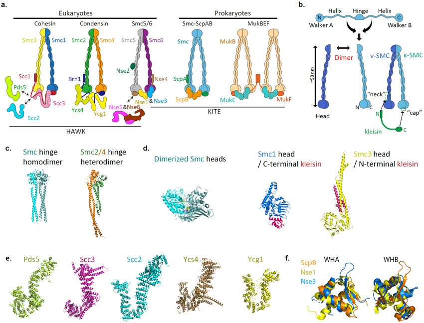
www.bdjn.org Bio Design l Vol.9 l No.1 l Mar 30, 2021 3Mechanism of SMC-driven DNA loop extrusion DNA LOOP EXTRUSION: FUNDAMENTAL PRINCIPLES OF are sequenced and displayed in a matrix of contact frequencies SMC-MEDIATED CHROMOSOME ORGANIZATION as a heat map. In a Hi-C map, it was shown that the cohesin The loop extrusion hypothesis proposed by Arthur Riggs was complex extrudes DNA loops to fold chromatin into Topologically that an unspecified enzyme reels DNA in to induce 50 – 100 Associating Domains (TADs), 100 kb – 1 Mb regions of high kbp DNA loops for chromosome organization (Riggs, 1990). self-interaction frequency present in interphase in vertebrate Over two decades ago, cohesin was suggested to be a new cells (Dixon et al., 2012; Sofueva et al., 2013; Rao et al., 2014; class of mechanochemical motor that forms chromosome loops Gassler et al., 2017; Schwarzer et al., 2017; Wutz et al., 2017). (Guacci et al., 1993; Strunnikov et al., 1993); condensin was also Figure 2A shows a typical Hi-C map of TADs, organized by suggested to extrude DNA in a processive manner to induce cohesin complex. Each TAD is separated by distinct boundary loops (Hirano and Mitchison, 1994; Kimura et al., 1999; Nasmyth, regions which are normally defined by the orientation of CCCTC- 2001). Furthermore, emerging evidence supports the long- binding factor (CTCF)-binding sites (Dixon et al., 2012; Sofueva standing hypothesis that every SMC protein uses a DNA loop et al., 2013; de Wit et al., 2015; Guo et al., 2015; Holzmann et extrusion mechanism for chromosome organization universally al., 2019; Busslinger et al., 2017; Wutz et al., 2017), and stable from prokaryotes to eukaryotes, although the exact function of corner peaks at some TADs are observed at convergent CTCF each SMC protein complex seems to differ. sites, suggesting the cohesin complex is an active DNA loop Recent progress using chromosome conformation capture extruder that translocates along chromatin until it encounters methods such as Hi-C and micro-C has provided evidence of a CTCF site in a proper orientation (Sanborn et al., 2015; DNA loop extrusion by SMC complexes (Sofueva et al., 2013; Fudenberg et al., 2016). This process is strongly supported by Rao et al., 2014; Gibcus et al., 2018; Costantino et al., 2020; Hi-C control experiments. Firstly, a cohesin depletion experiment Krietenstein et al., 2020). To obtain contact probability maps of showed the disruption of both TADs and the peaks located at the chromosome within a nucleus, the chromosome is cross- CTCF convergent sites. In addition, the depletion strengthened linked and digested into short DNA pieces, and these pieces compartmentalization patterns (Rao et al., 2017). Secondly, FIGURE 2 I Experimental basis of the DNA loop extrusion hypothesis. (A) Cartoon representation of the interaction signals underlying a Hi-C map (top) (Dixon et al., 2012; Sexton et al., 2012; Lajoie et al., 2015; Rowley and Corces, 2018) and a schematic model of the structure of a TAD in a metazoan cell (bottom). Both dark and light magenta areas represent Hi-C signals (top). Peak ChIP-seq signals (black wedges) imply the binding of the boundary protein, CTCF, to DNA (middle). Smaller subTADs characterized by darker magenta are shown in a TAD (top), and CTCF loops shown as dots at the apexes of each TAD domain correspond to DNA loops (middle) formed by cohesin complexes between CTCF sites (bottom). The coloration of cohesin follows that of Figure 1A. The direction of each CTCF site is depicted by the orientations of the yellow arrowheads in the schematic model. (B) Cartoon representation of a Hi-C map of the B. subtilis strain BWX3352 contains a single ParS site close to 00 (top) (Wang et al., 2017). Maps display contact frequencies during the propagation of the juxtaposition of chromosome of B. subtilis . The extension of off-diagonal (dark magenta, top) is highly correlated with the localizations of Smc-ScpAB observed by a ChIP-seq experiment (red lines; middle), implying that loading and translocation of SMC proteins (cyan) form juxtaposed chromosome arms from a site adjacent to the replication origin of DNA to the opposite site (bottom). (C) Cartoon representation of the simulated TAD organization mediated by cohesin complex and CTCF (Fudenberg et al., 2016). Directionalities (green and red arrows) of boundary elements (CTCF proteins: yellow arrowheads) determine the direction of loop-extruding factors such as cohesin complex (cyan rings) during interphase loop extrusion (left). The Hi-C simulation results correspond to the topological features of DNA tethered by boundary elements (right). (D) Cartoon representation of an in vitro single-molecule DNA loop extrusion experiment (Ganji et al., 2018). Single DNA loop, extruded by a loop extruding factor (cyan ring), is visualized during during orthogonal fluid flow (blue arrow). 4 Bio Design l Vol.9 l No.1 l Mar 30, 2021 www.bdjn.org
Hansol Lee, Haemin Noh and Je-Kyung Ryu

depletion of Nipbl, a human cohesin loader, disrupted TADs            complex differs from that of the condensin complex (Davidson
and peaks at CTCF binding sites (Busslinger et al., 2017), while      et al., 2019; Kim et al., 2019). One possible explanation for
depletion of Wapl, the unloading factor of the cohesin complex,       the discrepancy in symmetricity is that, in the case of cohesin,
induced more TADs and longer loops and formed a vermicelli            CTCF might function as an anchor when bound to one of the
shape of the chromosome structure (Haarhuis et al., 2017).            HAWK subunits (Li et al., 2020). The in vitro experiments showed
Lastly, an ATP depletion experiment using oligomycin showed           that SMC complexes reel in DNA up to 1.5 kbp/s at low forces
a dramatic change of TAD distribution across the genome (Vian         despite low ATPase activity (~2 ATP/s), suggesting a large step
et al., 2018). These results strongly support that the cohesin        size (~50 nm) per hydrolyzed ATP molecule. How the interaction
complex is a motor protein that has ATPase activity, and that         between SMC complexes and DNA induces the large step is still
CTCF functions as a roadblock to stall the DNA loop extrusion         under debate, although numerous studies have been done to
process driven by the cohesin complex (Vian et al., 2018).            elucidate the DNA binding sites and conformational changes of
   Hi-C experiments investigating the prokaryotic SMC complex         SMC complexes.
also support a DNA loop extrusion mechanism. For example,
the Hi-C pattern of Smc-ScpAB showed close contacts between           INTERACTIONS BETWEEN DNA AND SMC COMPLEXES
the juxtaposed chromosome arms of the circular chromosome             To reel in DNA for loop extrusion, SMC complexes must have
in B. subtilis (Figure 2B) (Le et al., 2013; Marbouty et al., 2015;   at least two DNA binding sites. Recent studies report that SMC
Wang et al., 2015). The Hi-C results suggest that the Smc-            subunits bind DNA mainly through two interfaces: the hinge
ScpAB complexes were loaded at parS sites close to the                domain with its adjacent coiled-coil, and the ATP-bound head
replication origin (close to 00) via interaction with ParB (Le et     dimer. In addition, HAWK subunits are also known to provide
al., 2013; Wilhelm et al., 2015), and the ATPase activity of Smc-     additional interaction sites with DNA through their multi-
ScpAB seems to be used for translocation from parS sites to the       conformational HEAT-repeats.
replication terminus (Wang et al., 2018). After that, Smc-ScpAB         Various studies report DNA-binding properties of the dimerized
is likely unloaded by XerD at ter region (Karaboja et al., 2021).     hinge at the inner part of the ring. Since the inner region of the
The rapid processive translocation speed of Smc-ScpAB along           hinge contains a positively-charged patch, it is plausible that
the juxtaposition of the opposite chromosome arms (up to 800          DNA inside the ring can bind to the hinge domain (Griese et al.,
bp/s) strongly indicates the existence of the DNA loop extrusion      2010; Soh et al., 2015; Diebold-Durand et al., 2017). Indeed, gel-
process in bacteria.                                                  shift assays and fluorescence anisotropy experiments showed
   Meanwhile, in silico polymer simulations strongly support the      DNA binding affinity to the hinge domain of mouse condensin
DNA loop extrusion model suggested from Hi-C experiments. A           (Griese et al., 2010), yeast condensin (Piazza et al., 2014),
simulated Hi-C map successfully reproduced the TAD patterns           bovine cohesin (Chiu et al., 2004), and prokaryotic condensin
shown in Hi-C experiments by assuming that a cohesin complex          (Hirano and Hirano, 2006). Previous AFM studies also showed
translocates along DNA to extrude a cis DNA loop until it meets a     DNA binding by the hinge domain of E. coli MukB (Kumar et al.,
boundary element (Figure 2C) (Fudenberg et al., 2016). Also, this     2017), S. cerevisiae condensin (Ryu et al., 2020a), and S. pombe
simulation predicts that the exceedingly strong corner peaks of       condensin (Yoshimura et al., 2002), suggesting a conserved
TADs indicate the activity of cohesin in the region between two       DNA-binding capability of the hinge domain. DNA binding affinity
boundary elements – that is, CTCFs.                                   measurements of hinge domains with varied coiled-coil lengths
   Additionally, single-molecule double-tethered DNA assays           imply that the juxtaposed hinge-proximal coiled-coil should
have directly visualized the DNA loop extrusion process driven        be opened to provide space for DNA binding (Griese et al.,
by S. cerevisiae condensin, human cohesin and condensin I/            2010). The hinge and its adjacent coiled-coil seem to undergo
II, and Xenopus cohesin and condensin (Ganji et al., 2018;            a conformational change upon binding of DNA to the hinge and
Davidson et al., 2019; Kim et al., 2019; Golfier et al., 2020;        ATP to the head (Soh et al., 2015; Minnen et al., 2016; Ryu et al.,
Kong et al., 2020; Ryu et al., 2020a). To reconstitute the DNA        2020a). The ATPase activity of the head domain seems to control
loop extrusion process, SMC complexes and ATP are added               DNA binding to the distant hinge domain (Hirano and Hirano,
to an immobilized λDNA, and a flow perpendicular to the DNA           2006; Soh et al., 2015; Diebold-Durand et al., 2017). Recent
strand is applied (Figure 2D). A clear DNA loop structure can         HS-AFM studies showed that the hinge angle of the O-shaped
be observed, indicating direct visualization of loop extrusion        condensin changes dynamically with ATP hydrolysis, indicating
mediated by the SMC complex. This in vitro assay showed that          that the hinge domain is very flexible (Ryu et al., 2020a). This
DNA loop extrusion by a single yeast condensin complex is             is consistent with previous crystal structures of hinge domains
asymmetric because condensin anchors DNA using the safety-            that showed two different states of the fully associated and half-
belt mechanism of Ycg1-Brn1 and reels it in from only one side        dissociated conformations (Haering et al., 2002; Griese et al.,
(Kschonsak et al., 2017; Ganji et al., 2018). On the contrary, the    2010; Ku et al., 2010; Li et al., 2010; Niki, 2017).
human cohesin complex extrudes a DNA loop symmetrically,                SMC head monomers show low binding affinity to DNA;
suggesting that the anchoring mechanism of the cohesin                however, the binding affinity largely increases when they

www.bdjn.org                                                                               Bio Design l Vol.9 l No.1 l Mar 30, 2021    5
Mechanism of SMC-driven DNA loop extrusion
are dimerized upon ATP binding. A crystal structure of large step along DNA. Recently, quite enormous structural
a dimerized E. coli MukB head domain shows a positive studies on SMC proteins using various techniques such as
patch possibly capable of binding a DNA strand (Woo et X-ray crystallography, negative stain EM, rotary shadow EM,
al., 2009). A dimerized Rad50 head domain shows stable cryo-EM, and AFM have been performed and many different
DNA binding based on X-ray crystallography (Seifert et al., configurations, such as I, O, V, Y, B, P, and a folded state have
2016). Recent Cryo-EM structures of the cohesin complex been observed (Figure 3A) (Diebold-Durand et al., 2017; Eeftens
also showed that DNA binds the ATP-bound head dimer and Dekker, 2017; Bürmann et al., 2019; Ryu et al., 2020a).
(Collier et al., 2020; Higashi et al., 2020; Shi et al., 2020). However, it is still not clear which of the different conformations
In addition, HAWK subunits were observed to have DNA the SMC proteins use to extrude DNA loops (Table 2).
binding affinity. Based on the crystal structure of the Ycg1- We can categorize the controversial studies of the
Brn1 complex, Ycg1 contains a positive patch inside its groove, conformational changes of SMC complexes according to
providing a strong DNA binding site for condensin. The Ycg1- the shape transitions that they report: (i) I to O – from a rigid,
binding DNA is embraced by a kleisin loop, and this loop is extended I-shape (the apo state) to an O-shape (the ATP-bound
considered to be a safety belt during the sliding movement of state) (Figure 3B), (ii) I to folded state (Figure 3C), and (iii) O to
condensin complex along DNA (Kschonsak et al., 2017). The B (Figure 3D) – from the apo state to the ATP-bound state. The
partial positive patch in the inner groove responsible for DNA I-to-O shape transition is supported by the crystal structure of
binding is also conserved in Scc3 of the cohesin complex (Li bacterial SMC proteins and negative stain EM (Diebold-Durand
et al., 2018). Recent Cryo-EM structures also report that the et al., 2017; Kamada et al., 2017). The structure of the full length
parts of HEAT-repeats of the Scc2/4 cohesin loader complex of the SMC arms of Smc-ScpAB showed the closely juxtaposed
also participate in DNA binding (Collier et al., 2020; Higashi et SMC coiled-coils (I-shape) and ATPγS-bound dimerized heads
al., 2020; Shi et al., 2020). In contrast of HAWK subunits, the with open SMC arms (O-shape), suggesting the lever arm
DNA-binding ability of KITE subunits has rarely been reported. movement of the head domains induces the O-shape via ATP
ScpB was regarded as a DNA-binding protein because it binding. This dimerization process seems to tilt the proximal
contains a WH domain, which is one of the representative DNA- coiled-coils of the head domains and generate a large tension
binding motifs, but its DNA-binding affinity seems very low and on the coiled-coils to open the closed ring. On the other hand,
not necessary for the function of Smc-ScpAB complexes in the I-to-folded-state transition is supported by recent Cryo-EM
prokaryotes (Jeon et al., 2020; Kim et al., 2006). A DNA-binding studies of MukBEF, yeast cohesin, yeast condensin (Bürmann
capability of MukE has also not been reported. et al., 2019), and an early AFM study on S. pombe condensin
(Yoshimura et al., 2002). However, it is not clear how ATP
CONTROVERSIAL STRUCTURAL STUDIES OF SMC binding changes the structure of the SMC arms in these studies.
PROTEINS On the contrary, recent liquid-phase HS-AFM imaging on yeast
Comparison of ATP hydrolysis rates in bulk and the measure condensin complex shows a predominantly extended O-shape
ments of DNA loop extrusion speeds suggest that condensin to collapsed B-shape (Ryu et al., 2020a). Interestingly, real-time
reels in DNA with large step sizes on the scale of the SMC imaging using the EQ mutant, which blocks the ATP hydrolysis of
complex (~50 nm) in a single ATP cycle. This indicates that SMC proteins, showed the O-to-B shape transition, suggesting
an extensive conformational change is necessary to take a that the B-shape is induced by ATP binding to the head domains.
FIGURE 3 I Various configurations of SMC complexes. (A) Various shapes of SMC proteins according to alphabet letters. (B) Conformational change
of SMC from the I-shape to the ATP-bound O-shape. The binding of two ATP molecules (red dots) promotes head-head engagement and opening of
rigidly juxtaposed coiled-coil pairs. (C) Folding of the I-shape. Kinks in the middle of the coiled-coil (termed the elbow) seem to enable bending and
folding of rigid SMC proteins. (D) Transition of SMC states from O- to B-shape upon ATP binding. Some experimental results imply dynamic flexibility
or bending of SMC which enables its B-shaped state.
6 Bio Design l Vol.9 l No.1 l Mar 30, 2021 www.bdjn.orgHansol Lee, Haemin Noh and Je-Kyung Ryu
TABLE 2 I List of reported experiments implying the conformational changes of SMC complexes
Conformation Species Protein Method Note Reference
Four segments of the SMC arms were
used to reconstruct the full length of SMC arms.
I and O B. subtilis Smc coiled-coil X-ray crystallography (Diebold-Durand et al., 2017)
I shape : apo state
O shape : ATP-bound state
condensin
S. pombe Dry AFM apo state (Yoshimura et al., 2002)
holocomplex
X-ray crystallography, MukB coiled-coil elbow was used for
Folded state E. coli MukBEF holocomplex negative stained EM, crystallography
and Cryo-EM apo state (Bürmann et al., 2019)
S. cerevisiae cohesin holocomplex Negative staining EM apo state
S. cerevisiae Smc2/4-Brn1-Ycs4 Cryo-EM apo state and ATP-bound state (Lee et al., 2020)
condensin Dry AFM and liquid- O shape: apo state
S. cerevisiae (Ryu et al., 2020a)
holocomplex phase HS AFM B shape: ATP-bound state
cohesin holocomplex With DNA
H. sapiens Cryo-EM (Shi et al., 2020)
and Nipbl B shape: ATP-bound state
O and B
with DNA
S. cerevisiae Smc1/3-Scc1-Scc2 Cryo-EM (Collier et al., 2020)
B shape: ATP-bound state
cohesin holocomplex with DNA
S. pombe Cryo-EM (Higashi et al., 2020)
with Mis4-Ssl3 B shape: ATP-bound state
Structural studies which support the transition from I-shape (the apo state) to O-shape (the ATP state) or folded state (the apo state), and O-shape (the apo state) to
B-shape (the ATP-bound state).
This result is also supported by the recent Cryo-EM studies of which also contradicts the observation that the hinge can be an
human cohesin and yeast cohesin complexes showing that the anchor (Chiu et al., 2004; Griese et al., 2010; Piazza et al., 2014;
hinge domain is engaged with the heads and HAWK (Collier et Soh et al., 2015; Diebold-Durand et al., 2017). Furthermore, with
al., 2020; Higashi et al., 2020; Takaki et al., 2021). this model, it is difficult to explain how the hinge replacement
of Smc to a structurally distinct zinc-hook domain of Rad50 did
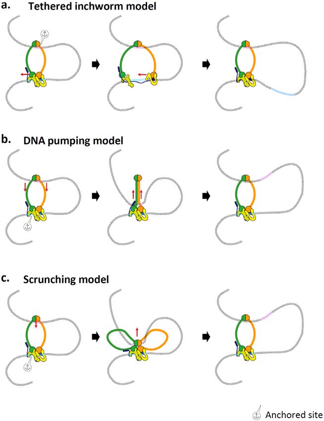
CONTROVERSIAL MECHANISMS FOR DNA LOOP EXTRU not eliminate the functional activity of the SMC complex during
SION fast cell division in B. subtilis (Bürmann et al., 2017). Moreover,
The discrepancy in the observed conformations of SMC this model seems to contradict the recent Cryo-EM and HS-AFM
complexes may suggest different mechanisms. In a tethered results that showed the collapsed state of the SMC complex
inchworm model (Nichols and Corces, 2018), the hinge domain where the hinge is in close proximity with heads and HAWK
of the SMC complex anchors DNA, and two head domains subunits (Bürmann et al., 2019; Higashi et al., 2020; Lee et al.,
– each head domain binds a HAWK or a KITE domain – are 2020; Ryu et al., 2020a; Shi et al., 2020).
tethered by a kleisin subunit (Figure 4A). The SMC protein moves On the other hand, the structural studies on the Smc-ScpAB
along DNA using head-head motions through the transition complex in prokaryotes suggested a ‘DNA pumping model’
from the V-shape to the O-shape. In the V-shape the two that involves a transition between opening and closing of the
heads are separated, and ATP binding induces the transition SMC arms (Figure 4B) (Diebold-Durand et al., 2017; Marko et
to the O-shape via dimerization of the two head domains. The al., 2019). Structural and biochemical studies revealed that
dimerization process of the heads can generate a DNA loop- Smc-ScpAB takes an I-shape in the absence of ATP, and the
extrusion step by pulling the DNA region bound to another binding of non-hydrolyzable ATP analogs induced the O-shape
head domain, while ATP hydrolysis releases the head domain by the dimerization of heads and the opening of the two SMC
to find a new target DNA region to repeat the cycle. In addition, arms from the head domains. Because full-length SMC arms
the kleisin subunit would be stretched and relaxed because are difficult to crystallize, crystal structures of the four different
the distance between two head domains is restricted by the segments of the SMC arms were obtained and co-aligned to
length of the kleisin subunit. These conformational changes reconstruct the structure of full-length SMC arms. In the DNA
of the kleisin subunit may also be involved in pulling the DNA pumping model (Diebold-Durand et al., 2017; Marko et al., 2019),
strand. However, this contradicts the previous observation that a zipping of the two SMC arms pushes pseudo-topologically
Ycg1-Brn1 of yeast condensin provides a DNA anchoring site, entrapped DNA from the hinge domain to the head domains,
because this model assumes that the hinge domain works as a while an ATP-induced dimerization of the head domains changes
DNA anchor. In addition, both eukaryotic and prokaryotic SMC the SMC protein conformation from an I-shape to an O-shape to
proteins have weak binding affinity to dsDNA in the hinge region, target new binding of DNA for subsequent ATP hydrolysis cycles.
www.bdjn.org Bio Design l Vol.9 l No.1 l Mar 30, 2021 7Mechanism of SMC-driven DNA loop extrusion
the hinge domain (Figure 4C). When the DNA binds to the head
and non-SMC subunits, the hinge releases it to subsequently
grab a new region of DNA to reel in (Hassler et al., 2019; Ryu
et al., 2020a; Terakawa et al., 2017). This model is supported
by recent Cryo-EM and AFM experiments that showed the
folded state or B-shape when the hinge domain contacts the
heads and HAWKs. Moreover, recent in vivo studies of cohesin
support that the hinge can be in close proximity with the head
domain. Furthermore, the force-dependent step sizes observed
by magnetic tweezers fits a simulation based on the scrunching
model (Ryu et al., 2020b; Takaki et al., 2021). A recent study
suggested a Brownian ratchet model in which the conformational
changes between the O- and B-shapes are induced by thermal
fluctuations due to the flexible SMC arms (Hwang and Karplus,
2019). Further reports indicate that transitions between the two
states are determined by the interaction between the hinge
domain and the globular domain via ATP binding (Higashi et al.,
2020; Ryu et al., 2020a; Shi et al., 2020). ATP binding induces
the dimerization of the head domains and engagement of the
hinge domainwith the globular head and non-SMC subunits
to form a B-shape while ATP hydrolysis or release induces the
dissociation of the hinge from the head and non-SMC subunits
to form an O-shape. For eukaryotic cohesin and condensin,
which have HAWK subunits, the scrunching model seems to be
the most relevant.
FIGURE 4 I Possible models of loop extrusion mechanism driven DISCUSSION
by active SMC complexes. (A) Tethered inchworm model. (B) DNA
pumping model. (C) Extended scrunching model. The coloration of the
In this review, we introduced the current understanding of the
SMC complex follows that of the condensin complex in Figure 1A. Black molecular mechanism of DNA loop extrusion, and we explored
anchor symbols denote the anchored positions of the hinge or head on the structure-function relationships of SMC proteins and DNA
DNA. Hinge-proximal (magenta) and head-proximal sites (cyan) of DNA
are marked to indicate the positional movement of DNA. Details are
loop extrusion. Different mechanisms for DNA loop extrusion
described in the main text. were discussed based on controversial structural results. Below,
we discuss the limitations of current studies, which might be able
to explain the controversial observations.
Moreover, this is strongly supported by electron paramagnetic
resonance (EPR-DEER) which measures the heterogeneous POSSIBLE REASONS FOR THE CONTROVERSIAL
distance between two different regions (1.5 ~ 10 nm) of a protein OBSERVATIONS
by measuring the coupling of electron spins (Nunez et al., 2021). The reason for the differences between previous structural
However, this seems to contradict recent eukaryotic SMC studies studies of SMC proteins may result from different organisms, the
with Cryo-EM and AFM imaging that showed folded states or protein purification process, and sample preparation methods.
B-shapes where the hinge region is close to the heads and First, SMC proteins from different organisms show different
HAWK subunits. Thus, the DNA pumping model seems to work shapes. For example, S. cerevisiae cohesin, human cohesin, and
for Smc-ScpAB, but not for eukaryotic condensin or cohesin, yeast condensin were found to adopt a folded conformation or
and future studies need to show whether the different results B-shape (Bürmann et al., 2019; Higashi et al., 2020; Ryu et al.,
were due to the differences of methods or intrinsic properties 2020a; Shi et al., 2020), but B. subtilis SMC proteins showed an
of Smc-ScpAB. Because Smc-ScpAB shows a different Hi-C I-shape in the apo state and an open shape in the ATP-bound
pattern from that of eukaryotic cohesin or condensin, the intrinsic state (Soh et al., 2015; Diebold-Durand et al., 2017; Bürmann et
behavior of SMC complexes may vary (Rao et al., 2017; Wang et al., 2019). Second, the purification process may induce certain
al., 2017). configurations because SMC complexes are macrocomplexes;
In a ‘scrunching model’, a HAWK subunit anchors DNA, and purifying intact functional complexes is quite challenging. Before
the hinge domain cyclically binds another region of the DNA performing structural studies, purified proteins should be assayed
and transfers it to the heads and non-SMC subunits using an for their ability to extrude DNA loops to ensure functional
extension and retraction of the flexible SMC arms connected to complexes are used. To date, purified yeast condensin, human
8 Bio Design l Vol.9 l No.1 l Mar 30, 2021 www.bdjn.orgHansol Lee, Haemin Noh and Je-Kyung Ryu
condensin I/II, and human cohesin have shown clear DNA loop To understand the reasons for these contradictory results,
extrusion activity (Ganji et al., 2018; Davidson et al., 2019; Kim systematic approaches are needed to compare the experiments
et al., 2019; Kong et al., 2020). Moreover, during purification, from various SMC proteins, methods, and conditions. Firstly, we
different post-translational modifications may also alter the need to understand whether every SMC complex can extrude
conformational state. For example, cohesin contains numerous a DNA loop and characterize biophysical properties the DNA
post-translational modification sites, such as for acetylation or loop extrusion by each SMC complex. For example, it is clear
phosphorylation, and some modifications were suggested to be that yeast condensin forms a single SMC complex to extrude
involved in the interaction between the two SMC arms, inducing a single loop, but with human cohesin it is not clear whether a
the juxtaposed structure (Kulemzina et al., 2016). Still, it is largely single or double complex is involved (Ganji et al., 2018; Davidson
unclear how the post-translational modifications are exactly et al., 2019; Kim et al., 2019). On the other hand, the MukBEF
involved in the structural transitions of SMC proteins. Third, complexes are dimerized via the N-terminal helical bundle of
different sample preparation methods can induce or bias the MukF, suggesting two MukBEF complexes can act as a single
observation of certain configurations. During sample preparation functional unit for the DNA loop extrusion process (Fennell-Fezzie
for structural studies, experimental factors such as surface et al., 2005; Woo et al., 2009; Badrinarayanan et al., 2012).
adhesion, metal staining, crystallization, and air exposure during However, DNA loop extrusion mediated by MukBEF has not yet
sample preparation can induce structural changes in proteins. been observed. In addition, whether the different SMC proteins
extrude DNA loops symmetrically or asymmetrically should be
LIMITATIONS OF THE METHODS FOR SMC PROTEIN also tested, as yeast condensin clearly showed asymmetric
STUDIES DNA loop extrusion while the process was symmetric for human
Recently, the development of state-of-the-art techniques such cohesin (Ganji et al., 2018; Davidson et al., 2019). Therefore,
as high-resolution Cryo-EM, and liquid-phase HS-AFM have there is a possibility that the conformational changes could vary
led to improved structural studies of SMC proteins and brought with each SMC protein. Secondly, each SMC complex needs to
greater insight into their mechanistic functions. However, there be tested using both liquid-phase HS-AFM and Cryo-EM in the
is still no perfect technique to clarify the structure-function presence of all subunits and DNA via controlled conditions of
relationships of SMC proteins. Crystallography has been largely ATP addition. These two techniques are complementary because
used to obtain atomic-resolution structures of subunits or parts HS-AFM provides fast and dynamic information while Cryo-
of the SMC arms. There remains no crystal structure of the full- EM can provide static atomic-resolution structural information.
length SMC arms, although four partial crystal structures of Thirdly, simultaneous observation of the conformation of SMC
truncated arms of bacterial Smc-ScpAB were determined to complexes during DNA loop extrusion would provide the most
display the structure of the full length of the SMC arms. The full- direct evidence to explain the mechanism. We expect that future
length SMC arms might be difficult to crystallize due to their studies will soon clearly show the molecular mechanism of the
flexibility, as observed by liquid-phase HS-AFM (Eeftens et al., DNA loop extrusion process, and answer specific questions such
2016; Ryu et al., 2020a). In the case of Cryo-EM, although the as the causes of one-sided or two-sided DNA loop extrusion and
recent revolution in resolution has enabled atomic-resolution the reason for its unidirectional motion.
structures of SMC proteins, single-particle analysis can bias the
structure of the flexible SMC arms because of an ever-luring CONCLUSION AND PERSPECTIVES
effect (Lyumkis, 2019). Moreover, as a macromolecule, the Summing up, we have discussed three different mechanistic
SMC complex can be easily exposed to an air-water interface models of DNA loop extrusion by SMC proteins. Although DNA
due to its large structure (~50 nm) because protein samples are loop extrusion seems to be a universal function of the entire SMC
trapped in a thin ice layer during sample preparation (thickness protein family, the mechanism has remained mysterious. Further
~50 nm). This air exposure can cause a preferred orientation studies of the conformational changes of SMC proteins will likely
(Noble et al., 2018) or denaturation (D’Imprima et al., 2019) of unravel their modes of action. Although various conformations
proteins. Moreover, with crystallography and Cryo-EM, it is not of SMC complexes were observed in precise experiments,
possible to obtain the dynamics of structural transitions, but the results can be affected by buffer composition, species of
liquid-phase HS-AFM imaging is a unique tool that can show the SMC protein, or detection methods. Therefore, systematic
the dynamics of the structural changes of a protein in aqueous studies to compare many different SMC proteins using different
solution without any staining or labeling. One limitation of this methods will be necessary to clearly describe the mechanism of
technique is that it cannot avoid non-specific binding of proteins DNA loop extrusion by SMC proteins.
to the surface. Furthermore, many structural studies have
complemented crosslinking experiments to support their results, ACKNOWLEDGEMENTS
but the possibility of crosslinking artifacts cannot be excluded. We thank Kevin Whitley at Newcastle Univ. and Hogyu David
Therefore, more studies on the architecture of SMC complexes Seo at Cleveland Clinic for help in editing the manuscript.
are necessary to form a clear picture of their mechanisms.
www.bdjn.org Bio Design l Vol.9 l No.1 l Mar 30, 2021 9Mechanism of SMC-driven DNA loop extrusion
Eeftens, J.M., Katan, A.J., Kschonsak, M., Hassler, M., de Wilde, L.,
Original Submission: Feb 25, 2021 Dief, E.M., Haering, C.H., and Dekker, C. (2016). Condensin Smc2-Smc4
Revised Version Received: Mar 17, 2021 dimers are flexible and dynamic. Cell Rep 14, 1813-1818.
Accepted: Mar 19, 2021
Fennell-Fezzie, R., Gradia, S.D., Akey, D., and Berger, J.M. (2005). The
MukF subunit of Escherichia coli condensin: architecture and functional
relationship to kleisins. EMBO J 24, 1921-1930.
REFERENCES Fudenberg, G., Imakaev, M., Lu, C., Goloborodko, A., Abdennur, N.,
Aragon, L., Martinez-Perez, E., and Merkenschlager, M. (2013). Condensin, and Mirny, L.A. (2016). Formation of chromosomal domains by loop
cohesin and the control of chromatin states. Curr Opin Genet Dev 23, extrusion. Cell Rep 15, 2038-2049.
204-211.
Ganji, M., Shaltiel, I.A., Bisht, S., Kim, E., Kalichava, A., Haering, C.H.,
Badrinarayanan, A., Reyes-Lamothe, R., Uphoff, S., Leake, M.C., and and Dekker, C. (2018). Real-time imaging of DNA loop extrusion by
Sherratt, D.J. (2012). In vivo architecture and action of bacterial structural condensin. Science 360, 102-105.
maintenance of chromosome proteins. Science 338, 528-531.
Gassler, J., Brandão, H.B., Imakaev, M., Flyamer, I.M., Ladstätter, S.,
Bürmann, F., Shin, H.C., Basquin, J., Soh, Y.M., Giménez-Oya, V., Kim, Bickmore, W.A., Peters, J.M., Mirny, L.A., and Tachibana, K. (2017).
Y.G., Oh, B.H., and Gruber, S. (2013). An asymmetric SMC-kleisin bridge A mechanism of cohesin-dependent loop extrusion organizes zygotic
in prokaryotic condensin. Nat Struct Mol Biol 20, 371-379. genome architecture. EMBO J 36, 3600-3618.
Bürmann, F., Basfeld, A., Vazquez Nunez, R., Diebold-Durand, M.L., Gibcus, J.H., Samejima, K., Goloborodko, A., Samejima, I., Naumova,
Wilhelm, L., and Gruber, S. (2017). Tuned SMC arms drive chromosomal N., Nuebler, J., Kanemaki, M.T., Xie, L., Paulson, J.R., Earnshaw, W.C.,
loading of prokaryotic condensin. Mol Cell 65, 861-872.e869. Mirny, L.A., and Dekker, J. (2018). A pathway for mitotic chromosome
Bürmann, F., Lee, B.G., Than, T., Sinn, L., O’Reilly, F.J., Yatskevich, formation. Science 359, eaao6135.
S., Rappsilber, J., Hu, B., Nasmyth, K., and Löwe, J. (2019). A folded Golfier, S., Quail, T., Kimura, H., and Brugués, J. (2020). Cohesin and
conformation of MukBEF and cohesin. Nat Struct Mol Biol 26, 227-236. condensin extrude DNA loops in a cell cycle-dependent manner. Elife 9,
Busslinger, G.A., Stocsits, R.R., van der Lelij, P., Axelsson, E., Tedeschi, e53885.
A., Galjart, N., and Peters, J.M. (2017). Cohesin is positioned in Graham, J.E., Sherratt, D.J., and Szczelkun, M.D. (2010). Sequence-
mammalian genomes by transcription, CTCF and Wapl. Nature 544, 503- specific assembly of FtsK hexamers establishes directional translocation
507. on DNA. Proc Natl Acad Sci U S A 107, 20263-20268.
Chiu, A., Revenkova, E., and Jessberger, R. (2004). DNA interaction and Griese, J.J., Witte, G., and Hopfner, K.P. (2010). Structure and DNA
dimerization of eukaryotic SMC hinge domains. J Biol Chem 279, 26233- binding activity of the mouse condensin hinge domain highlight common
26242. and diverse features of SMC proteins. Nucleic Acids Res 38, 3454-3465.
Collier, J.E., Lee, B.G., Roig, M.B., Yatskevich, S., Petela, N.J., Metson, Guacci, V., Yamamoto, A., Strunnikov, A., Kingsbury, J., Hogan, E., Meluh,
J., Voulgaris, M., Gonzalez Llamazares, A., Löwe, J., and Nasmyth, K.A. P., and Koshland, D. (1993). Structure and function of chromosomes in
(2020). Transport of DNA within cohesin involves clamping on top of mitosis of budding yeast. Cold Spring Harb Symp Quant Biol 58, 677-
engaged heads by Scc2 and entrapment within the ring by Scc3. Elife 9, 685.
e59560.
Guo, Y., Xu, Q., Canzio, D., Shou, J., Li, J., Gorkin, D.U., Jung, I., Wu, H.,
Costantino, L., Hsieh, T.S., Lamothe, R., Darzacq, X., and Koshland, D. Zhai, Y., Tang, Y., Lu, Y., Wu, Y., Jia, Z., Li, W., Zhang, M.Q., et al. (2015).
(2020). Cohesin residency determines chromatin loop patterns. Elife 9, CRISPR inversion of CTCF sites alters genome topology and enhancer/
e59889. promoter function. Cell 162, 900-910.
de Wit, E., Vos, E.S., Holwerda, S.J., Valdes-Quezada, C., Verstegen, Haarhuis, J.H.I., van der Weide, R.H., Blomen, V.A., Yáñez-Cuna, J.O.,
M.J., Teunissen, H., Splinter, E., Wijchers, P.J., Krijger, P.H., and de Laat, Amendola, M., van Ruiten, M.S., Krijger, P.H.L., Teunissen, H., Medema,
W. (2015). CTCF binding polarity determines chromatin looping. Mol Cell R.H., van Steensel, B., Brummelkamp, T.R., de Wit, E., and Rowland,
60, 676-684. B.D. (2017). The cohesin release factor WAPL restricts chromatin loop
D’Imprima, E., Floris, D., Joppe, M., Sánchez, R., Grininger, M., and extension. Cell 169, 693-707.e614.
Kühlbrandt, W. (2019). Protein denaturation at the air-water interface and Haering, C.H., and Gruber, S. (2016). SnapShot: SMC protein complexes
how to prevent it. Elife 8, e42747. Part I. Cell 164, 326-326.e1.
Davidson, I.F., Bauer, B., Goetz, D., Tang, W., Wutz, G., and Peters, J.M. Haering, C.H., Löwe, J., Hochwagen, A., and Nasmyth, K. (2002).
(2019). DNA loop extrusion by human cohesin. Science 366, 1338-1345. Molecular architecture of SMC proteins and the yeast cohesin
Diebold-Durand, M.L., Lee, H., Ruiz Avila, L.B., Noh, H., Shin, H.C., Im, complex. Mol Cell 9, 773-788.
H., Bock, F.P., Bürmann, F., Durand, A., Basfeld, A., Ham, S., Basquin, Hassler, M., Shaltiel, I.A., Kschonsak, M., Simon, B., Merkel, F., Thärichen,
J., Oh, B.H., and Gruber, S. (2017). Structure of full-length SMC and L., Bailey, H.J., Macošek, J., Bravo, S., Metz, J., Hennig, J., and Haering,
rearrangements required for chromosome organization. Mol Cell 67, 334- C.H. (2019). Structural basis of an asymmetric condensin ATPase
347.e335. cycle. Mol Cell 74, 1175-1188.e1179.
Dixon, J.R., Selvaraj, S., Yue, F., Kim, A., Li, Y., Shen, Y., Hu, M., Liu, Haering, C.H., and Gruber, S. (2016). SnapShot: SMC Protein Complexes
J.S., and Ren, B. (2012). Topological domains in mammalian genomes Part I. Cell 164, 326-326.e1
identified by analysis of chromatin interactions. Nature 485, 376-380.
Higashi, T.L., Eickhoff, P., Sousa, J.S., Locke, J., Nans, A., Flynn, H.R.,
Dolgin, E. (2017). DNA’s secret weapon against knots and tangles. Nature Snijders, A.P., Papageorgiou, G., O’Reilly, N., Chen, Z.A., O’Reilly, F.J.,
544, 284-286. Rappsilber, J., Costa, A., and Uhlmann, F. (2020). A structure-based
Eeftens, J., and Dekker, C. (2017). Catching DNA with hoops-biophysical mechanism for DNA entry into the cohesin ring. Mol Cell 79, 917-933.
approaches to clarify the mechanism of SMC proteins. Nat Struct Mol Biol e919.
24, 1012-1020. Hirano, T. (2016). Condensin-based chromosome organization from
10 Bio Design l Vol.9 l No.1 l Mar 30, 2021 www.bdjn.orgHansol Lee, Haemin Noh and Je-Kyung Ryu bacteria to vertebrates. Cell 164, 847-857. regulated by lysine acetylation and is required for cohesin association with Hirano, M., and Hirano, T. (2006). Opening closed arms: long-distance the DNA. Mol Cell 63, 1044-1054. activation of SMC ATPase by hinge-DNA interactions. Mol Cell 21, 175- Kumar, R., Grosbart, M., Nurse, P., Bahng, S., Wyman, C.L., and Marians, 186. K.J. (2017). The bacterial condensin MukB compacts DNA by sequestering Hirano, T., and Mitchison, T.J. (1994). A heterodimeric coiled-coil protein supercoils and stabilizing topologically isolated loops. J Biol Chem 292, required for mitotic chromosome condensation in vitro. Cell 79, 449-458. 16904–16920. Holzmann, J., Politi, A.Z., Nagasaka, K., Hantsche-Grininger, M., Walther, Lajoie, B.R., Dekker, J., and Kaplan, N. (2015). The Hitchhiker’s guide to N., Koch, B., Fuchs, J., Dürnberger, G., Tang, W., Ladurner, R., Stocsits, Hi-C analysis: practical guidelines. Methods 72, 65-75. R.R., Busslinger, G.A., Novák, B., Mechtler, K., Davidson, I.F., et al. (2019). Lammens, A., Schele, A., and Hopfner, K.P. (2004). Structural Absolute quantification of cohesin, CTCF and their regulators in human biochemistry of ATP-driven dimerization and DNA-stimulated activation of cells. Elife 8, e46269. SMC ATPases. Curr Biol 14, 1778-1782. Hsieh, T.H., Weiner, A., Lajoie, B., Dekker, J., Friedman, N., and Rando, Le, T.B., Imakaev, M.V., Mirny, L.A., and Laub, M.T. (2013). High-resolution O.J. (2015). Mapping nucleosome resolution chromosome folding in yeast mapping of the spatial organization of a bacterial chromosome. Science by Micro-C. Cell 162, 108-119. 342, 731-734. Hwang, W., and Karplus, M. (2019). Structural basis for power stroke vs. Lee, B.G., Merkel, F., Allegretti, M., Hassler, M., Cawood, C., Lecomte, L., Brownian ratchet mechanisms of motor proteins. Proc Natl Acad Sci U S A O’Reilly, F.J., Sinn, L.R., Gutierrez-Escribano, P., Kschonsak, M., Bravo, 116, 19777-19785. S., Nakane, T., Rappsilber, J., Aragon, L., Beck, M., et al. (2020). Cryo- Jeon, J.H., Lee, H.S., Shin, H.C., Kwak, M.J., Kim, Y.G., Gruber, S., and EM structures of holo condensin reveal a subunit flip-flop mechanism. Nat Oh, B.H. (2020). Evidence for binary Smc complexes lacking kite subunits Struct Mol Biol 27, 743-751. in archaea. IUCrJ 7(Pt 2), 193-206. Li, Y., Schoeffler, A.J., Berger, J.M., and Oakley, M.G. (2010). The Jeppsson, K., Kanno, T., Shirahige, K., and Sjögren, C. (2014). The crystal structure of the hinge domain of the Escherichia coli structural maintenance of chromosome structure: positioning and functioning of maintenance of chromosomes protein MukB. J Mol Biol 395, 11-19. SMC complexes. Nat Rev Mol Cell Biol 15, 601-614. Li, Y., Muir, K.W., Bowler, M.W., Metz, J., Haering, C.H., and Panne, Kamada, K., Miyata, M., and Hirano, T. (2013). Molecular basis of SMC D. (2018). Structural basis for Scc3-dependent cohesin recruitment to ATPase activation: role of internal structural changes of the regulatory chromatin. Elife 7, e38356. subcomplex ScpAB. Structure 21, 581-594. Li, Y., Haarhuis, J., Sedeño Cacciatore, Á., Oldenkamp, R., van Ruiten, Kamada, K., Su’etsugu, M., Takada, H., Miyata, M., and Hirano, T. (2017). M.S., Willems, L., Teunissen, H., Muir, K.W., de Wit, E., Rowland, B.D., Overall shapes of the SMC-ScpAB complex are determined by balance and Panne, D. (2020). The structural basis for cohesin-CTCF-anchored between constraint and relaxation of its structural parts. Structure 25, loops. Nature 578, 472-476. 603-616.e4. Liu, S., Chistol, G., Hetherington, C.L., Tafoya, S., Aathavan, K., Karaboja, X., Ren, Z., Brandão, H.B., Paul, P., Rudner, D.Z., and Wang, Schnitzbauer, J., Grimes, S., Jardine, P.J., and Bustamante, C. (2014). A X. (2021). XerD unloads bacterial SMC complexes at the replication viral packaging motor varies its DNA rotation and step size to preserve terminus. Mol Cell 81, 756-766.e8. subunit coordination as the capsid fills. Cell 157, 702-713. Kim, J.S., Shin, D.H., Pufan, R., Huang, C., Yokota, H., Kim, R., and Kim, Löwe, J., Cordell, S.C., and van den Ent, F. (2001). Crystal structure of the S.H. (2006). Crystal structure of ScpB from Chlorobium tepidum, a protein SMC head domain: an ABC ATPase with 900 residues antiparallel coiled- involved in chromosome partitioning. Proteins 62, 322-328. coil inserted. J Mol Biol 306, 25-35. Kim, Y., Shi, Z., Zhang, H., Finkelstein, I.J., and Yu, H. (2019). Human Lyumkis, D. (2019). Challenges and opportunities in cryo-EM single- cohesin compacts DNA by loop extrusion. Science 366, 1345-1349. particle analysis. J Biol Chem 294, 5181-5197. Kimura, K., Rybenkov, V.V., Crisona, N.J., Hirano, T., and Cozzarelli, N.R. Mäkelä, J., and Sherratt, D.J. (2020). Organization of the Escherichia coli (1999). 13S condensin actively reconfigures DNA by introducing global Chromosome by a MukBEF Axial Core. Mol Cell 78, 250-260.e5. positive writhe: implications for chromosome condensation. Cell 98, 239- Marbouty, M., Le Gall, A., Cattoni, D.I., Cournac, A., Koh, A., Fiche, 248. J.B., Mozziconacci, J., Murray, H., Koszul, R., and Nollmann, M. (2015). Kong, M., Cutts, E.E., Pan, D., Beuron, F., Kaliyappan, T., Xue, C., Condensin- and replication-mediated bacterial chromosome folding and Morris, E.P., Musacchio, A., Vannini, A., and Greene, E.C. (2020). Human origin condensation revealed by Hi-C and super-resolution Imaging. Mol condensin I and II drive extensive ATP-dependent compaction of Cell 59, 588-602. nucleosome-bound DNA. Mol Cell 79, 99-114.e9. Marko, J.F., De Los Rios, P., Barducci, A., and Gruber, S. (2019). DNA- Krietenstein, N., Abraham, S., Venev, S.V., Abdennur, N., Gibcus, J., segment-capture model for loop extrusion by structural maintenance of Hsieh, T.S., Parsi, K.M., Yang, L., Maehr, R., Mirny, L.A., Dekker, J., and chromosome (SMC) protein complexes. Nucleic Acids Res 47, 6956- Rando, O.J. (2020). Ultrastructural details of mammalian chromosome 6972. architecture. Mol Cell 78, 554-565.e557. Minnen, A., Bürmann, F., Wilhelm, L., Anchimiuk, A., Diebold-Durand, Kschonsak, M., Merkel, F., Bisht, S., Metz, J., Rybin, V., Hassler, M., and M.L., and Gruber, S. (2016). Control of Smc coiled coil architecture by the Haering, C.H. (2017). Structural basis for a safety-belt mechanism that ATPase heads facilitates targeting to chromosomal ParB/parS and release anchors condensin to chromosomes. Cell 171, 588-600.e24. onto flanking DNA. Cell Rep 14, 2003-2016. Ku, B., Lim, J.H., Shin, H.C., Shin, S.Y., and Oh, B.H. (2010). Crystal Murayama, Y., and Uhlmann, F. (2015). DNA entry into and exit out of the structure of the MukB hinge domain with coiled-coil stretches and its cohesin ring by an interlocking gate mechanism. Cell 163, 1628-1640. functional implications. Proteins 78, 1483-1490. Nasmyth, K. (2001). Disseminating the genome: joining, resolving, and Kulemzina, I., Ang, K., Zhao, X., Teh, J.T., Verma, V., Suranthran, S., separating sister chromatids during mitosis and meiosis. Annu Rev Genet Chavda, A.P., Huber, R.G., Eisenhaber, B., Eisenhaber, F., Yan, J., and 35, 673-745. Ivanov, D. (2016). A reversible association between Smc coiled coils is Nasmyth, K., and Haering, C.H. (2005). The structure and function of SMC www.bdjn.org Bio Design l Vol.9 l No.1 l Mar 30, 2021 11
You can also read