Dynein mediates the localization and activation of mTOR in normal and human cytomegalovirus-infected cells - Genes Dev
←
→
Page content transcription
If your browser does not render page correctly, please read the page content below
Downloaded from genesdev.cshlp.org on January 31, 2021 - Published by Cold Spring Harbor Laboratory Press

Dynein mediates the localization
and activation of mTOR in normal and
human cytomegalovirus-infected cells
Amy J. Clippinger and James C. Alwine1
Department of Cancer Biology, Abramson Family Cancer Research Institute, School of Medicine, University of Pennsylvania,
Philadelphia, Pennsylvania 19104, USA

Activation of stress signaling pathways normally leads to inhibition of the mammalian target of rapamycin
complex 1 (mTORC1); however, human cytomegalovirus (HCMV) infection maintains mTORC1 activity in the
presence of numerous types of stress. We previously demonstrated that HCMV infection maintains mTORC1
activity during amino acid deprivation through a Ras-related GTP-binding (Rag) protein-independent mechanism.
This depends on the colocalization of mTOR and its activator, Rheb (Ras homology enriched in brain)-GTP, to
a perinuclear position that corresponds to the viral cytoplasmic assembly compartment (AC). The data presented
here show that the HCMV-induced, amino acid depletion-resistant perinuclear localization and activation of
mTORC1 occurs as early as 8 h post-infection, prior to AC formation. We show that the molecular motor dynein is
required for perinuclear localization of mTORC1 in both uninfected and HCMV-infected cells. Association
between dynein and mTOR is shown by coimmunoprecipitation, and inhibition of dynein function using RNAi or
the small molecule inhibitor ciliobrevin A inhibits mTORC1 activity in both uninfected and HCMV-infected
cells. The data suggest that mTORC1 activation requires dynein-dependent transport to a position in the cell
where it can be activated. Thus, the HCMV commandeers a cellular dynein-dependent mTORC1 activation
mechanism to maintain stress-resistant mTORC1 activity during infection and to form the AC.
[Keywords: dynein; HCMV; assembly compartment; mTOR]
Supplemental material is available for this article.
Received May 11, 2012; revised version accepted July 27, 2012.

The mammalian target of rapamycin (mTOR) kinase is                             et al. 2005; Mamane et al. 2006; Reiling and Sabatini
found in two multiprotein complexes: mTOR complex                              2006). Maintaining mTOR activity and translation is
1 (mTORC1) and mTORC2. A major difference between                              vital to the success of an HCMV infection. However, a
the two complexes is their substrate specificity factors:                      viral infection induces stress response signaling that nor-
raptor is present in mTORC1, and rictor is present in                          mally inhibits mTORC1 and translation. Consequently,
mTORC2; consequently, the two complexes have differ-                           HCMV has evolved mechanisms to circumvent these
ent substrate specificities (Kim et al. 2002; Sarbassov                        inhibitory stress responses and maintain mTORC1 activity
et al. 2004). Human cytomegalovirus (HCMV), the largest                        (Kudchodkar et al. 2004, 2006; Walsh et al. 2005; Alwine
of the human herpesviruses, activates both complexes                           2008; Moorman et al. 2008; Clippinger et al. 2011a).
during infection (Kudchodkar et al. 2006). However, the                           Many cellular stress responses lead to the inhibition of
results of studies using shRNAs specific for raptor or                         mTORC1 through the activation of the guanosine tri-
rictor suggest that mTORC1 has a greater effect on viral                       phosphatase (GTPase)-activating protein (GAP) function
growth; specifically, raptor depletion is more deleterious                     of the tuberous sclerosis complex (TSC) (Fig. 1; for review,
to HCMV infection than rictor depletion (Moorman and                           see Alwine 2008; Buchkovich et al. 2008b). Activated
Shenk 2010; Clippinger et al. 2011a). The role of mTORC1                       TSC stimulates the intrinsic GTPase activity of Rheb
in translational control has been well-established; mTORC1-                    (Ras homology enriched in brain), converting it from
mediated phosphorylation of p70 S6 kinase (S6K) and the                        Rheb-GTP to Rheb-GDP. Rheb-GTP is an activator of
eukaryotic initiation factor 4E-binding protein 1 (4E-BP1)                     mTORC1, while Rheb-GDP is not. Thus, when the TSC
is necessary for the maintenance of translation (Sarbassov                     is inactivated, for example, through phosphorylation by
                                                                               Akt (protein kinase B), Rheb-GTP levels remain high,
1
                                                                               leading to activation of mTORC1 (Fig. 1; Sancak et al.
  Corresponding author
E-mail alwine@mail.med.upenn.edu                                               2007). Conversely, many cellular stresses activate the
Article is online at http://www.genesdev.org/cgi/doi/10.1101/gad.196147.112.   TSC, resulting in TSC-dependent inhibition of mTORC1.

       GENES & DEVELOPMENT 26:2015–2026 Ó 2012 by Cold Spring Harbor Laboratory Press ISSN 0890-9369/12; www.genesdev.org              2015
Downloaded from genesdev.cshlp.org on January 31, 2021 - Published by Cold Spring Harbor Laboratory Press

Clippinger and Alwine

                                                                      expected; however, it is resistant in HCMV-infected cells
                                                                      (Clippinger et al. 2011b). This suggested that HCMV
                                                                      infection bypasses normal Rag-dependent amino acid
                                                                      signaling. Indeed, we established that shRNA-mediated
                                                                      depletion of the Rag proteins had little effect on
                                                                      mTORC1 activity in infected cells but inhibited activity
                                                                      in mock-infected cells. However, Rheb was necessary, as
                                                                      its depletion inhibited mTOR activity in both mock- and
                                                                      HCMV-infected cells (Clippinger et al. 2011b). In mock-
                                                                      infected cells, mTOR activity in the presence of amino
                                                                      acids correlated with a perinuclear localization of mTOR
                                                                      with Rheb. This localization dispersed following the
                                                                      depletion of amino acids and correlated with the loss of
                                                                      mTOR activity (Clippinger et al. 2011b). In HCMV-
Figure 1. mTORC1 regulation. Activation of the PI3K (phos-            infected cells, mTOR was also found in a perinuclear
phatidylinositol 3-kinase)–Akt–TSC–mTORC1 pathway is crit-            localization with Rheb but did not disperse upon amino
ical for the phosphorylation of 4E-BP1 and S6K and the
                                                                      acid depletion, and mTOR activity was maintained
maintenance of translation. Numerous types of stresses inhibit
this pathway, halting translation. HCMV has multiple mecha-           (Clippinger et al. 2011b). At late times in HCMV in-
nisms to circumvent the effects of stress signaling to maintain       fection, the perinuclear localization of mTOR coincided
translation under stress conditions (see the text for details). The   with the cytoplasmic assembly compartment (AC), a peri-
mechanisms for amino acid control of mTORC1 are also shown.           nuclear, HCMV-specific structure that is needed for virion
                                                                      maturation and egress. Detailed studies of AC structure
                                                                      propose a three-dimensional model of a cylindrical AC
However, HCMV protects mTORC1 from TSC-depen-                         composed of organelle-specific vesicles (Golgi, trans-
dent inhibition (Kudchodkar et al. 2004, 2007; Moorman                Golgi network, and early endosomes), which form nested
et al. 2008). In part, this is accomplished through an in-            cylinders (Das et al. 2007; Das and Pellett 2011). The AC
teraction between the viral UL38 protein and the TSC,                 forms on a microtubule-organizing center (MTOC), and
which is believed to inactivate the TSC (Fig. 1; Moorman              microtubules radiate out from the AC (Sanchez et al.
et al. 2008). Additionally, the levels of Rheb increase in            2000). Our previous work and the work of others have
infected cells, which may also overcome the inhibitory                shown that formation of the AC on the MTOC is mediated
effects of TSC activation (Clippinger et al. 2011b). These            by the molecular motor dynein (Buchkovich et al. 2010;
findings suggest that any stress response that exerts its             Indran et al. 2010). Thus, we questioned whether the move-
effect on, or upstream of, the TSC will be circumvented,              ment of mTOR to its perinuclear location in the AC may
and Rheb-GTP levels and mTORC1 activity will remain                   be dynein-mediated and, correspondingly, whether dynein
high. However, some types of stress, including oxidative              is involved in the normal translocation and activation of
stress and amino acid deprivation, inhibit mTORC1                     mTORC1 in uninfected cells.
through a TSC-independent mechanism (Smith et al.                        In the present study, we show that HCMV infection
2005; Nobukuni et al. 2007). We showed recently that                  induces a perinuclear localization of mTOR by 8 h post-
mTORC1 activity is maintained in HCMV-infected cells                  infection (hpi), which is very early in infection, prior to
under both oxidative stress (Tilton et al. 2011) and amino            the formation of the AC. This localization is maintained
acid depletion (Clippinger et al. 2011b).                             during amino acid deprivation and corresponds with
   Amino acids signal to mTORC1 through the Ras-                      maintenance of mTORC1 activity. Uninfected, growing
related GTP-binding (Rag) proteins by stimulating GTP                 human fibroblasts (HFs) show a perinuclear localization
charging of Rag proteins, thereby promoting the associa-              of mTOR that is less compact than in infected cells but
tion of the Rag proteins with mTORC1 (Fig. 1; Kim et al.              disperses upon amino acid depletion, corresponding with
2008; Sancak et al. 2008, 2010). It has been proposed that            the loss of mTORC1 activity. We show that the molec-
the interaction of the Rag proteins with mTORC1 pro-                  ular motor dynein is required for the perinuclear locali-
motes mTORC1 translocation to a membrane-bound com-                   zation of mTOR in both uninfected and HCMV-infected
partment that contains Rheb-GTP (Kim et al. 2008; Sancak              cells; association between dynein and mTOR is shown by
et al. 2008). More recent work suggests that a protein                coimmunoprecipitation. Inhibition of dynein function
complex termed Ragulator interacts with the Rag pro-                  using RNAi or the small molecule inhibitor ciliobrevin
teins, resulting in the localization of the Rag dimer–                A (CA) inhibits mTORC1 activity in both uninfected and
mTORC1 complex to lysosomal membranes, which is                       HCMV-infected cells. The data show that in normal,
essential for mTORC1 activation (Sancak et al. 2010).                 uninfected cells, mTORC1 activation is dependent on
Constitutive targeting of mTORC1 to the lysosomal sur-                dynein-mediated transport. Thus, HCMV commandeers
face renders mTORC1 activity insensitive to amino acid                a cellular, dynein-dependent mTOR activation function
deprivation and, correspondingly, independent of Rag and              in order to (1) maintain mTOR in a location where it is
Ragulator but not Rheb-GTP (Sancak et al. 2010).                      constitutively active and resistant to stress inhibition and
   We showed recently that mTORC1 activity is sensitive               (2) form the AC, where mTOR may be further protected
to amino acid deprivation in mock-infected cells as                   from inhibitory stress signaling.

2016      GENES & DEVELOPMENT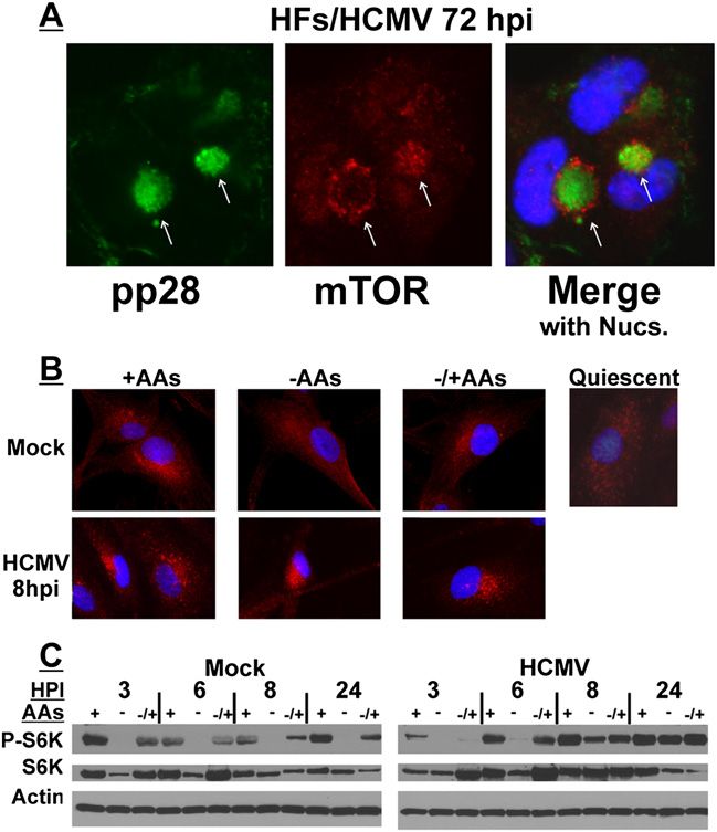
Downloaded from genesdev.cshlp.org on January 31, 2021 - Published by Cold Spring Harbor Laboratory Press

                                                                                                             Dynein activates mTOR

Results                                                              for 72 h. The AC was visualized using antibodies to the
                                                                     HCMV protein pp28 (Fig. 2A, green), an AC resident
The activity and perinuclear translocation of mTORC1                 protein. The two arrows in Figure 2A indicate two mor-
is resistant to amino acid deprivation by 8 h after                  phologies of mTOR localization (red) in the AC; in one
HCMV infection                                                       morphology (which is more predominantly seen), mTOR and
Our previously published results demonstrated that in                pp28 colocalize in the AC core. In the other morphology,
HCMV-infected cells, mTORC1 and Rheb-GTP localize                    mTOR is in an outer ring surrounding a core of pp28. We
to the cytoplasmic viral AC, where mTORC1 activity is                observed similar mixed morphologies for other AC com-
independent of Rag proteins and resistant to inhibition by           ponents, suggesting that localization of these compo-
amino acid deprivation (Clippinger et al. 2011b). The                nents is dynamic (Buchkovich et al. 2008a, 2009, 2010).
immunofluorescence micrograph in Figure 2A reiterates                   The AC does not begin to form until 24–36 hpi, as
these data using cells that have been HCMV-infected                  measured by the accumulation and perinuclear position-
                                                                     ing of viral protein markers of the AC, such as pp28 and
                                                                     glycoprotein B (gB) (Krzyzaniak et al. 2007; Buchkovich
                                                                     2010). Thus, we examined earlier times in infection to
                                                                     determine whether the perinuclear localization of mTOR
                                                                     and the resulting resistance to amino acid depletion pre-
                                                                     ceded or coincided with AC formation.
                                                                        In the top panel of Figure 2B, mTOR localization (red)
                                                                     was examined in subconfluent, actively growing, mock-
                                                                     infected HFs. In the presence of complete medium
                                                                     (containing amino acids, +AAs), mTOR is localized in a
                                                                     perinuclear position. A much more diffuse cytoplasmic
                                                                     localization of mTOR is observed after cells are main-
                                                                     tained in amino acid-free medium ( AAs) for 50 min; this
                                                                     is similar to the predominant localization of mTOR in
                                                                     confluent, contact-inhibited, quiescent HFs (Fig. 2B, quies-
                                                                     cent). In the subconfluent, actively growing HFs, the
                                                                     perinuclear localization of mTOR returns when cells
                                                                     are returned to amino acid-containing medium for 30 min
                                                                     following the 50 min of amino acid withdrawal (Fig. 2B, top
                                                                     panel, /+AAs). In the left panel of Figure 2C, phosphor-
                                                                     ylation of p70 S6K was examined as an indicator of
                                                                     mTORC1 activity in uninfected cells at 3, 6, 8, and 24 h
                                                                     after mock infection. At each time point, mTOR is active
                                                                     when cells are maintained in complete medium (+) but is
                                                                     inhibited after 50 min of amino acid depletion ( ). mTOR
                                                                     activity is regained when cells are returned to amino acid-
                                                                     containing medium for 30 min following the 50 min
Figure 2. The perinuclear localization and activity of mTORC1        depletion ( /+). These data are in agreement with pre-
are maintained following amino acid deprivation as early as 8 h      vious reports of mTORC1 activity under amino acid
after HCMV infection. (A) HCMV-infected HFs (72 hpi) were            depletion conditions (Sancak et al. 2008; Clippinger
stained for pp28 or mTOR followed by immunofluorescence              et al. 2011b). The bottom panel of Figure 2B shows mTOR
microscopy. This shows the characteristics of perinuclear viral      localization (red) in HCMV-infected HFs at 8 hpi. In
cytoplasmic ACs and the enlarged kidney-shaped nuclei around
                                                                     complete medium (+AAs), the perinuclear localization
them. (Green) pp28; (red) mTOR; (blue) nuclei; (arrows) ACs. (B)
Mock- or HCMV-infected HFs (8 hpi) were maintained in amino
                                                                     of mTOR becomes more compact and punctate. Remark-
acid-containing medium (+AAs), amino acid-starved for 50 min         ably, by 8 hpi, the tight perinuclear localization does not
prior to cell fixation ( AAs), or amino acid-starved for 50 min      disperse following 50 min of amino acid deprivation
followed by the readdition of amino acids for 30 min prior to cell   ( AAs). The results of Western analysis in the right panel
fixation ( /+AAs). Cells were then stained for mTOR followed         of Figure 2C show that this corresponds to the retention
by immunofluorescence microscopy. (Red) mTOR; (blue) nuclei.         of mTOR activity in the absence of amino acids at 8 h
(Top right panel) An example of mTOR staining (red) in               after HCMV infection. Thus, very early in HCMV in-
quiescent, mock-infected HFs is shown to contrast the more           fection, conditions are induced to maintain active mTOR
diffuse mTOR localization pattern of quiescent cells with its        in a perinuclear position even in the absence of amino
more concentrated localization in actively growing HFs (+AAs).
                                                                     acids. This corresponds with the virus-induced mainte-
(C) The phosphorylation of S6K was examined by Western
analysis at 3, 6, 8, and 24 h after mock or HCMV infection.
                                                                     nance of mTORC1 activity when amino acids are de-
HFs were maintained in amino acid-containing medium (+),             pleted. In addition, the very early perinuclear localization
amino acid-starved for 50 min prior to cell collection ( ), or       and activation of mTOR at 8 hpi precedes the formation
amino acid-starved for 50 min followed by the readdition of          of the AC, suggesting that mTOR perinuclear localiza-
amino acids for 30 min prior to cell collection ( /+).               tion may be a prerequisite for AC formation.

                                                                                                 GENES & DEVELOPMENT         2017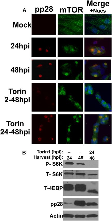
Downloaded from genesdev.cshlp.org on January 31, 2021 - Published by Cold Spring Harbor Laboratory Press

Clippinger and Alwine

  Recent reports suggest that mTORC1, complexed with
Rag heterodimers, is recruited to lysosomal membranes,
which is essential for mTORC1 activation (Sancak et al.
2010). Supplemental Figure S1A shows that the lyso-
somal marker LAMP2 colocalizes with perinuclear mTOR
in uninfected cells as well as with mTOR in the AC of
infected cells; previous studies have established the pres-
ence of lysosomal markers in the AC (Das and Pellett
2011). These data suggest that the association of active
mTOR with lysosomal membranes is maintained in
HCMV-infected cells. Additionally, Supplemental Figure
S1B shows that Rag proteins are also located perinuclearly.

Dynein function is needed for mTOR localization
in uninfected and HCMV-infected cells
Dynein is a multiprotein complex that functions as the
predominant minus end-directed microtubule motor in
eukaryotic cells. We and others showed previously that
AC formation requires dynein (Buchkovich et al. 2010;
Indran et al. 2010). In addition, the formation and main-
tenance of the characteristic enlarged, kidney-shaped
nucleus found wrapped around the AC in HCMV-infected
cells depends on dynein function (Buchkovich et al. 2010).
Dynein interacts with dynactin, an adaptor complex that
allows dynein to bind its cargo. A projecting arm of
dynactin, made up of a dimer of the p150Glued subunit,
binds directly to dynein (Karki and Holzbaur 1995).
Overexpression of p150Glued results in a wide range of
motility defects; however, constructs have been made
that express only the coiled-coil domain 1 (CC1; amino
acid residues 217–548) of p150Glued. Overexpression of
CC1 has far fewer adverse effects than overexpression
of p150Glued (Quintyne et al. 1999; Kwinter et al. 2008;
Maier et al. 2008). CC1 binds dynein directly, acting as a
competitive inhibitor for the interaction between dynein
and dynactin; thus, dynein cannot load its cargo (Quintyne
et al. 1999).
  Our previous studies used CC1 to inhibit dynein
function and show its critical role in AC formation
(Buchkovich et al. 2010). A similar approach was used
with CC1 to assess dynein’s involvement in mTOR
localization. HFs were infected for 24 h and then
electroporated with a plasmid expressing GFP-labeled
CC1. Forty-eight hours after transfection (72 hpi), the
cells were fixed, stained, and examined by immunofluo-              Figure 3. Dynein function is required for perinuclear localiza-
rescence microscopy. In Figure 3A, two HCMV-infected                tion of mTOR in mock- and HCMV-infected cells. (A) mTOR
cells are shown; the one on the left, indicated by the              (green) and gB (red) staining in HCMV-infected HFs (72 hpi); the
arrow, has been transfected and expresses GFP-CC1                   cell on the left (indicated by the arrow) is also expressing CC1
(white). Both cells have been infected, as indicated by             (white). (B) mTOR (red) staining in mock-infected U373-MG
                                                                    cells; three cells, indicated by arrows, are expressing CC1 (green).
the staining for HCMV gB (Fig. 3A, red), another viral
                                                                    (C) mTOR (green) staining in mock-infected HFs; the cell in-
marker of the AC. In the cell not expressing CC1, gB is             dicated by the arrow is expressing CC1 (white). (D) mTOR (green)
in the AC next to an enlarged, kidney-shaped nucleus.               staining in HCMV-infected U373-MG cells; the red cells were also
However, in the CC1-expressing cell, the nucleus is                 electroporated with siRNA that specifically targets dynein heavy
neither enlarged nor kidney-shaped, and gB has become               chain and siGLO, a fluorescently labeled, nonspecific siRNA that
dispersed throughout the cytoplasm; this is more easily             marks the transfected cells. The cell indicated by the arrow has
seen in the longer exposure of the gB staining (gB*).               taken up no siRNA.
These results agree with our previous observations that
dynein function is needed for AC formation and nuclear
remodeling (Buchkovich et al. 2010). The staining for

2018      GENES & DEVELOPMENT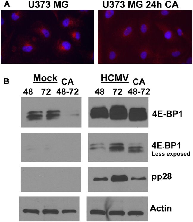
Downloaded from genesdev.cshlp.org on January 31, 2021 - Published by Cold Spring Harbor Laboratory Press

                                                                                                            Dynein activates mTOR

mTOR (Fig. 3A, green) shows that mTOR colocalizes to               while mTOR is much more diffuse in the siRNA trans-
the AC in the cell that is not expressing CC1; however,            fected cells. In the examination of numerous fields, we
mTOR is dispersed throughout the cytoplasm in the                  found that when siRNA transfection was noted, the AC
CC1-expressing cell. Examination of 30 CC1-expressing,             was either undetectable or very diffuse compared with
HCMV-infected cells found none with an intact AC. These            untransfected, infected cells. The results of this alterna-
results suggest that dynein function is required for the           tive approach for disrupting dynein function confirm the
localization of mTOR to the AC.                                    results of the CC1 experiments in Figure 3A and reiterate
   In our previous study of HCMV’s maintenance of                  that dynein is required for perinuclear localization of mTOR.
mTORC1 activity under amino acid depletion condi-                     The data in Figure 3, combined with our previous data,
tions, we used the glioblastoma cell line U373-MG                  suggest that dynein functions in the localization of
(Clippinger et al. 2011b). These studies showed that,              mTOR, contributing to its activation in uninfected human
in U373-MG cells, mTOR is active and predominately                 cells, and that HCMV commandeers this function to
perinuclear under normal, uninfected conditions. How-              (1) localize mTOR to a perinuclear position very early
ever, just like in HFs, the perinuclear localization of            in infection, where mTOR is active and resistant to
mTOR in uninfected U373-MG cells was lost upon de-                 stress inhibition, and (2) form the AC later in infection
pletion of amino acids and regained when amino acid-               (Buchkovich et al. 2010; Clippinger et al. 2011b).
containing medium was restored (Clippinger et al. 2011b).
We examined whether the perinuclear localization of
                                                                   Coimmunoprecipitation of dynein and mTOR
mTOR in uninfected U373-MG cells was dynein-dependent.
The GFP-CC1-expressing plasmid was electroporated                  The data above suggest that dynein is involved in
into uninfected U373-MG cells, and 48 h post-electro-              transporting mTOR to a perinuclear region where it
poration, the cells were fixed, stained, and examined by           can be activated. Thus, we asked whether an interaction
immunofluorescence microscopy. Figure 3B shows a field             between mTOR and dynein could be demonstrated by
of U373-MG cells; three of these cells (indicated by arrows)       immunoprecipitation. Extracts from mock-infected and
express GFP-CC1 (green)—two at high levels, and one at             HCMV-infected (72 hpi) HFs were immunoprecipitated
a much lower level. The relatively tight perinuclear local-        using anti-dynein intermediate chain, anti-mTOR, or a
ization of mTOR (Fig. 3B , red) is noted in all of the cells       nonspecific control IgG. The precipitates were analyzed
except the three expressing GFP-CC1, which show a dif-             by Western analysis probed for mTOR and dynein. The
fuse, cytoplasmic localization of mTOR. These results              results (Fig. 4) show that precipitation of dynein coim-
suggest that dynein is necessary for the perinuclear local-        munoprecipitated mTOR (Fig. 4A), and precipitation of
ization of mTOR observed in uninfected U373-MG cells.
   An additional control was performed to rule out
the possibility that the dynein-dependent localization of
mTOR seen in U373-MG cells was a phenomenon spe-
cific to a transformed cell line. We examined the effect of
CC1 inhibition on dynein function in normal, growing
HFs in complete medium. Figure 3C shows a field of
three, subconfluent, actively growing HFs, one of which
is expressing GFP-CC1 (white). In the CC1-expressing
cell, mTOR localization (Fig. 3C, green) is very diffuse
throughout the cytoplasm, while it has a more perinu-
clear localization in the cells not expressing GFP-CC1.
All of the CC1-expressing cells that we examined showed
diffuse mTOR staining. These results support the con-
clusion that dynein is required for perinuclear mTOR
localization in uninfected cells.
   To further verify the CC1 results in infected cells, we
tested siRNAs that specifically target the dynein heavy
chain. U373-MG cells were first electroporated with the
dynein siRNA and siGLO, a fluorescently labeled, non-
specific siRNA that marks the transfected cells. At 6 h
post-electroporation, the cells were infected with HCMV,
and at 72 h post-electroporation, the cells were fixed and
stained for mTOR (Fig. 3D, green). The left two panels of
                                                                   Figure 4. Coimmunoprecipitation of dynein and mTOR.
Figure 3D show the siRNA-containing cells, as indicated
                                                                   Mock- or HCMV-infected HF lysates (72 hpi) were immunopre-
by siGLO (red); two exposures are shown, and the lighter           cipitated with mouse anti-dynein intermediate chain (A), goat
one is used in the merge so that details of mTOR staining          anti-mTOR (B), or a nonspecific goat IgG control (C); a mouse
are not obscured. The longer exposure shows that one cell          IgG control gave the same result as the goat IgG control (not
contains no siRNA fluorescence (Fig. 3D, arrow), and only          shown). Western analysis was then performed for mTOR and
this cell has mTOR concentrated in the perinuclear AC,             dynein heavy chain. (M) Mock-infected; (V) HCMV-infected.

                                                                                                GENES & DEVELOPMENT         2019
Downloaded from genesdev.cshlp.org on January 31, 2021 - Published by Cold Spring Harbor Laboratory Press

Clippinger and Alwine

mTOR coimmunoprecipitated dynein (Fig. 4B). The spec-
ificity of the precipitations is indicated by the control
IgG, which precipitated neither mTOR nor dynein (Fig.
4C). The data in Figure 3, A and C, suggest that dynein
is involved in the localization of mTOR in mock- and
HCMV-infected HFs; the coimmunoprecipitation results
support this finding, showing that dynein and mTOR
coimmunoprecipitate similarly in mock- and HCMV-
infected HFs.
   It is important to note that, in comparison with the
input, the amount of dynein–mTOR complexes is rela-
tively low; the input represents ;5% of the lysate. This
may indicate that the complexes are unstable. Alterna-
tively, if the dynein–mTOR association occurs only
during transport, one would expect that only a small
amount of mTOR would be in active transport, and
therefore associated with dynein, at any given time. In
sum, the data show an interaction between dynein and
mTOR; this would be expected if dynein function is
needed to move and localize mTOR to a perinuclear
position in normal and infected HFs. Also of note in the
input lanes in Figure 4, A–C, is the observation that dynein
levels are not notably altered by HCMV infection.

mTOR activity is not needed for mTOR localization
to the AC in an established HCMV infection
We next examined whether the dynein-mediated AC
localization of mTOR required mTOR kinase activity.
To inhibit mTOR kinase, we used Torin1, a highly-
specific, small molecule inhibitor that binds the kinase
domain (Thoreen and Sabatini 2009). We and others
previously characterized Torin1 as an effective mTOR
inhibitor during HCMV infection (Moorman and Shenk
2010; Clippinger et al. 2011a). For these experiments,
we used confluent HFs; the mTOR staining of these
cells (Fig. 5A, Mock) provides a very good example of the
diffuse staining of mTOR seen in confluent, contact-
inhibited, quiescent, uninfected cells (also described in
Fig. 2B). Untreated HFs were HCMV-infected and fixed                Figure 5. mTOR activity is not necessary for its perinuclear
at 24 or 48 hpi for immunofluorescence staining. Other              localization in an established infection. (A) Immunofluores-
HCMV-infected cultures were treated with Torin1 from                cence analysis of mock- or HCMV-infected HFs at 24 or 48 hpi.
2 to 48 hpi or from 24 to 48 hpi and then fixed for                 Where applicable, HCMV-infected cells were treated with the
examination. Figure 5A shows that pp28 (red) and mTOR               mTOR kinase inhibitor Torin1 from 2 to 48 hpi (Torin 2-48hpi) or
(green) are localized to the perinuclear AC region by               from 24 to 48 hpi (Torin 24-48hpi). (Red) pp28; (green) mTOR;
24 hpi and become more concentrated in the AC by                    (blue) nuclei. (B) Western analysis of phosphor S6K (P-S6K), total
                                                                    S6K (T-S6K), and total 4E-BP1 (T-4E-BP1) shows the effectiveness
48 hpi. It is known that Torin1 significantly delays the
                                                                    of Torin1 inhibition of mTORC1. Note also the expected de-
course of HCMV infection if added from the start of the
                                                                    crease in pp28 detected in HCMV-infected cells treated with
infection (at 2 hpi) (Clippinger et al. 2011a). This can be         Torin1 from 2 to 48 and 24 to 48 hpi compared with untreated
seen in the sample treated with Torin1 from 2 to 48 hpi,            cells at 48 hpi.
where pp28 is not detected due to the delayed infection.
In addition, mTOR localization to the perinuclear re-
gion is more diffuse in the samples treated from 2 to               alternative approach of allowing the infection to establish
48 hpi than it is at 48 hpi in untreated cells; however,            for 24 h before adding Torin1. Figure 5A shows that the
mTOR localization is still more perinuclear than in con-            addition of Torin1 between 24 and 48 hpi had little effect
fluent, mock-infected cells. Thus, inhibition of mTOR               on the perinuclear AC localization of pp28 and mTOR.
activity at 2 hpi had only a partial effect on mTOR                 The Western analysis in Figure 5B shows that Torin1
localization by 48 hpi; this could be due to pleiotropic            inhibition of mTOR’s ability to phosphorylate S6K and
effects resulting from the delay of the lytic cycle caused          4E-BP was very effective during the 24–48 hpi treatment.
by Torin1 (Clippinger et al. 2011a). Therefore, we took the         Taken together, the data in Figure 5 suggest that once the

2020      GENES & DEVELOPMENT
Downloaded from genesdev.cshlp.org on January 31, 2021 - Published by Cold Spring Harbor Laboratory Press
Dynein activates mTOR
infection is established (by 24 hpi), mTOR activity is not
necessary for the maintenance of the AC.
Dynein depletion inhibits mTORC1 activity
We next asked whether the depletion of functional dynein
causes a loss of mTOR activity under conditions in which
it is usually active. HFs were infected with lentiviruses
expressing shRNAs specific for luciferase (a control) or the
dynein heavy chain, followed by mock or HCMV infection
at 24 h after shRNA addition. At 72 h after HCMV in-
fection, the cells were lysed and proteins were separated
by sodium dodecyl sulfate–polyacrylamide gel electro-
phoresis (SDS-PAGE) followed by Western analysis for
total 4E-BP1, phospho-4E-BP1, dynein, or actin. Figure 6
Figure 7. Inhibition of dynein function using CA causes loss of
shows that dynein knockdown resulted in a decrease in
mTORC1 activity. HFs were mock- or HCMV-infected for 48 or
mTORC1 activity, as demonstrated by loss of the more 72 h and then extracted, or treated with the dynein inhibitor CA
hyperphosphorylated forms of 4E-BP1 in both mock- and (60 mg/mL) between 48 and 72 h after mock or HCMV infection
HCMV-infected HFs. This decrease is seen despite an and then harvested. Western analysis was performed for total
incomplete depletion of dynein heavy chain by shRNAs; 4E-BP1 (T-4E-BP1), phospho-S6K (P-S6K), total p70 S6K (T-S6K),
it is likely that a more substantial loss of mTORC1 activity pp28, and actin.
could be observed if more complete dynein depletion could
be accomplished. Nevertheless, these results suggest that
48 and 72 h after HCMV infection or mock infection de-
dynein function is necessary to maintain mTORC1 activ-
creased the hyperphosphorylation of 4E-BP1 and the phos-
ity in both normal and HCMV-infected HFs.
phorylation of S6K in both mock- and HCMV-infected cells
in comparison with the levels in untreated cells 72 h after
Dynein inhibition by CA inhibits mTORC1 activity
HCMV infection or mock infection. In addition, the levels
and disturbs the formation of the AC
of the viral protein pp28 are lowered by the CA treatment;
Recent studies have shown that the small molecule in- this may be due to inhibition of translation by hypophos-
hibitor CA is a specific dynein inhibitor due to its ability to phorylated 4E-BP1. Figure 8 shows the immunofluores-
interfere with dynein’s ATPase activity and dynein-depen- cence examination of AC formation as indicated by pp28
dent microtubule gliding (Firestone et al. 2012). Thus, we (green) and mTOR (red) staining; we see a more compact
further analyzed the role of dynein in mTORC1 activation and intense AC at 72 hpi as compared with 48 hpi. The
using CA inhibition. For these experiments, we allowed panels in Figure 8 showing GFP indicate the infected cells,
the mock infection and the HCMV infection to establish since the virus carries an inserted GFP gene. This is shown
for 48 h and then added 60 mg/mL CA or a solvent control because the 48 and 72 hpi samples have a few uninfected
for 24 h and then extracted or fixed the cells at 72 hpi. cells that provide an internal, uninfected control; these
Figure 7 shows the activity of mTORC1 as measured by cells are indicated by the white arrows in Figure 8. In
S6K and 4E-BP1 phosphorylation. CA treatment between contrast to the AC formation at 48 and 72 hpi, dynein in-
hibition by CA between 48 and 72 hpi results in malfor-
mation of the AC, shown by a more diffuse staining of
mTOR and pp28.
As mentioned above, we used uninfected U373-MG
cells to study the perinuclear localization of mTOR
because mTOR is predominately perinuclear under nor-
mal growth conditions; this localization was lost upon
depletion of amino acids, correlating with the loss of
mTORC1 activity, and was regained when amino acid-
containing medium was restored, corresponding with
the return of mTORC1 activity (Clippinger et al. 2011b).
In Figure 3, we show that inhibition of dynein using
CC1 disrupted mTOR localization to the perinuclear
position in U373-MG cells. Figure 9A shows that in-
hibition of dynein by CA also disrupts the perinuclear
Figure 6. Dynein depletion inhibits mTORC1 activity. HFs
localization of mTOR in U373-MG cells. Figure 9B
were infected with lentiviral vectors expressing shRNA specific
for luciferase (Luc), a control, or dynein heavy chain (Dyn) for shows the effect of CA on mTORC1 activity by exam-
24 h followed by mock or HCMV infection. Seventy-two hours ining phosphorylation of 4E-BP1; in this experiment,
post HCMV infection, cells were lysed and proteins were sepa- samples at 48 and 72 h after HCMV infection or mock
rated by SDS-PAGE followed by Western analysis for total 4E-BP infection were compared with samples treated with CA
(T-4E-BP1), phospho-4E-BP (P-4-EBP1 37/46), dynein, and actin. for 24 h between 48 and 72 h after HCMV infection or
GENES & DEVELOPMENT 2021Downloaded from genesdev.cshlp.org on January 31, 2021 - Published by Cold Spring Harbor Laboratory Press
Clippinger and Alwine
Figure 8. Inhibition of dynein function using CA affects AC formation. HFs were mock- or HCMV-infected for 48 or 72 h and then
fixed, or treated with the dynein inhibitor CA (60 mg/mL) between 48 and 72 h and then fixed. Immunofluorescence analysis for pp28
(green) and mTOR (red) is shown; nuclei were stained with 4969-diamidino-2-phenylindole (DAPI) (blue). The HCMV used encodes GFP;
thus, infected cells are indicated in the GFP panels (green). This allows uninfected cells (arrows) to be seen as internal, uninfected
controls.
mock infection. Again, we observed that hyperphosphor- such as amino acid depletion, must also be dealt with
ylation of 4E-BP1 is lost in both cases. Additionally, the during infection. Our previous studies showed that
total level of 4E-BP1 is reduced, particularly in the mock- HCMV infection induces mTOR colocalization with its
infected cells, in agreement with the recent observation activator, Rheb-GTP, in a perinuclear region, where it
that severely hypophosphorylated 4E-BP1 is degraded by is maintained in an active state regardless of the presence
the KLHL25-CUL3 ubiquitin ligase (Yanagiya et al. 2012). of amino acids (Clippinger et al. 2011b). This contrasts
In sum, our data show that dynein function is needed with uninfected cells where the localization and activity
for mTORC1 activity, apparently to move mTORC1 to of mTORC1 are lost following amino acid depletion. At
locations in the cell where it will be active. HCMV has later times in infection, the perinuclear localization of
commandeered this function of dynein to insure the active mTOR coincides with the viral cytoplasmic AC,
activation of mTORC1 and the later formation of the AC. a perinuclear body that forms on an MTOC and is im-
portant in virion maturation. The nucleus and the AC are
well integrated; the nucleus is tightly pulled around the
Discussion
AC in a kidney shape. Both the formation of the AC and
During HCMV infection, mTORC1 activity is main- the reshaping of the nucleus around it require movement
tained even in the face of stress response signaling, which of the molecular motor dynein toward the MTOC at
would inhibit it in uninfected cells. Existing data show the center of the AC (Buchkovich et al. 2010; Indran et al.
that this is in part accomplished during the first 12 h of 2010). In the work presented here, we show that dynein
infection by transient activation of Akt, leading to the also mediates the movement of mTOR to the AC.
inactivation of the TSC (Fig. 1; Yu and Alwine 2002; Additionally, our data establish that dynein-mediated peri-
Kudchodkar et al. 2004). However, by 12 hpi, the viral nuclear localization of mTOR is induced very early
UL38 protein is produced and binds to the TSC, which in HCMV infection (by 8 hpi), well before the formation
is believed to inactivate the TSC, bypassing the need of the AC.
for Akt (Fig. 1; Moorman et al. 2008). Thus, any stress In uninfected cells, we show that mTOR localization to
signaling that normally inactivates mTORC1 through perinuclear sites also requires dynein function. Signifi-
activation of the TSC, such as hypoxia and energy dep- cantly, we found that mTORC1 activity, as measured by
rivation, is potentially blocked (Kudchodkar et al. 2004, 4E-BP1 and S6K phosphorylation, is dependent on dynein
2007). In addition, in HCMV-infected cells, the level of function. Thus, the movement and activity of mTOR
Rheb, the mTORC1 activator, is increased, potentially are dependent on a normal cellular function of dynein;
offering an additional means of bypassing the effects of HCMV commandeers this to ensure the constitutive activa-
the TSC (Clippinger et al. 2011b). However, stress responses tion of mTOR, the protection of mTOR from stress in-
that inhibit mTORC1 in a TSC-independent manner, hibition, and the formation of the AC. The use of molecular
2022 GENES & DEVELOPMENTDownloaded from genesdev.cshlp.org on January 31, 2021 - Published by Cold Spring Harbor Laboratory Press
Dynein activates mTOR
(Das and Pellett 2007, 2011; Das et al. 2007). Such func-
tional modification may affect the lysosomal component
of the AC, since there is no clear link between the AC and
autophagosome–lysosome fusion. In this regard, HCMV
has been shown to modulate autophagy differentially.
Very early in infection, HCMV stimulates autophagy
(McFarlane et al. 2011) by a mechanism that is indepen-
dent of de novo viral protein synthesis. However, at later
times of infection, coinciding with AC formation, HCMV
blocks autophagy (Chaumorcel et al. 2008) by a mecha-
nism that requires de novo viral protein expression. One
viral protein synthesized during this period is TRS1,
which has been shown to block autophagosome biogen-
esis (Chaumorcel et al. 2012). Interestingly, TRS1 has
been shown to be present in the AC (Buchkovich et al.
2009). Additionally, it has been shown that active Rheb
disrupts the interaction between dynein and misfolded
protein cargos and therefore blocks aggresome formation
by inhibiting dynein-dependent transportation of mis-
folded proteins (Zhoua et al. 2009). As mentioned above,
Rheb levels are greatly increased during HCMV infection
(Clippinger et al. 2011b); this may aid in uncoupling
dynein from misfolded protein cargos in order to direct
it to AC formation and block aggresome formation.
Recent studies have shown that targeting mTORC1 to
Figure 9. Inhibition of dynein function using CA affects the lysosomal surfaces maintains mTORC1 activity (Sancak
activity and perinuclear localization of mTOR in uninfected U373- et al. 2010). Additionally, the involvement of lysosomes
MG cells. (A) Monolayers of U373-MG cells were treated with CA in the activation of mTORC1 and in degrading autopha-
or the solvent control for 24 h and then stained for mTOR (red) and gic substrates has been associated with lysosomal posi-
the nuclei (blue). (B) Monolayers of U373-MG cells were mock- tioning (Korolchuk et al. 2010). In these HeLa cell studies,
infected or HCMV-infected for 48 and 72 h or treated with CA
activation of mTORC1 by nutrients (serum and amino
between 48 and 72 h after mock or HCMV infection. Cells were
extracted for Western analysis of 4E-BP1, pp28, and actin. A lighter
acids) correlated with its association with peripheral
exposure of the 4E-BP1 analysis is provided in order to see the lysosomes, whereas starvation (removal of serum and
details in the HCMV-infected cell samples. amino acids) caused perinuclear clustering of lyso-
somes and mTOR, which correlated with loss of mTOR
activity. In contrast, our present and previous experi-
motors for transport of virion components, nucleocap- ments (Clippinger et al. 2011b), conducted in U373-MG
sids, and virions in the assembly and egress process has cells and HFs, as well as studies done in HEK-293T cells
long been known (for review, see Dodding and Way 2011); (Sancak et al. 2008) show that the diffuse cytoplasmic
our studies extend these findings, showing that viral localization of mTOR correlates with inactivity (for
manipulation of molecular motors modulates fundamen- example, in the absence of amino acids) and that the
tal cellular signaling mechanisms. perinuclear localization correlates with activity. Addi-
The dynein-mediated formation of the perinuclear AC tionally, our present data show that the perinuclear
on the MTOC is reminiscent of perinuclear aggresome localization of mTOR in HCMV-infected cells corre-
formation, which provides a means to dispose of misfolded lates with mTORC1 activity. While these experiments
proteins that exceed the degradative capacity of ubiquitin– use different cells and conditions, the contrasting re-
proteasome and autophagy–lysosome systems. Previous sults suggest that the status of mTOR activity in either
studies have shown that decreased dynein function the perinuclear or diffuse cytoplasmic locations may be
impairs autophagic clearance of aggregate-prone pro- influenced by other cellular conditions or signaling. For
teins (Ravikumar et al. 2005). These studies suggest example, the use of serum is quite different between the
that loss of dynein function results in premature aggre- experiments; therefore, growth status or the availability
gate formation and impaired autophagosome–lysosome of serum growth factors may alter whether mTORC1 is
fusion. Thus, one interpretation of HCMV’s utilization of active or inactive in the perinuclear or diffuse cytoplasmic
dynein in AC formation would be to promote aggregate locations. Such control would allow localized activity of
formation and autophagosome–lysosome fusion. However, mTORC1, providing, for example, localized translation.
the AC has characteristics that are distinctly different from Localized translation mediated by localized changes in
aggresomes. A significant amount of evidence suggests mTORC1 activity has been demonstrated in axons (Lin
that the highly vesicular AC results from HCMV-induced and Holt 2008; Richter and Klann 2009)
remodeling of the membrane transport apparatus, result- Overall, our data suggest that HCMV commandeers a
ing in new functional profiles for the transport apparatus normal dynein function in mTOR localization and acti-
GENES & DEVELOPMENT 2023Downloaded from genesdev.cshlp.org on January 31, 2021 - Published by Cold Spring Harbor Laboratory Press
Clippinger and Alwine
vation in order to ensure the constitutive activation of Amino acid starvation
mTORC1. In this process, the virus bypasses the Rag HFs grown in six-well plates were mock- or HCMV-infected (MOI =
protein requirement in mTORC1 activation (Clippinger 3). At the indicated time points, cells were washed three times
et al. 2011b) and constitutively localizes mTOR in a with 13 phosphate-buffered saline (PBS) and incubated for 50 min
perinuclear position, where it is constitutively activated in RPMI 1640 medium without amino acids. Following the 50-min
and protected from inhibitory stress response signaling. incubation, cells were harvested or incubated for an additional
The data also suggest that HCMV’s takeover of dynein 30 min in complete, amino acid-containing medium prior to harvest.
function is prerequisite for the formation of the AC and
the reshaping of the nucleus around it. Most importantly, Viral infections and Western analysis
our data establish that dynein function is necessary for
HFs grown in six-well plates were serum-starved for 24 h
mTORC1 activity under normal cellular conditions.
followed by mock or HCMV infection (MOI = 3). At various
time points after infection, cells were collected and resuspended
Materials and methods in cold RIPA lysis buffer (1% NP-40, 1% sodium deoxycholate,
0.1% SDS, 0.15 M NaCl, 0.01 M sodium phosphate at pH 7.2,
Cell culture 2mM EDTA, 50 mM sodium fluoride, 200 mM sodium orthovan-
adate, 1 mM phenylmethylsulfonyl fluoride, 1.5 mg/mL aprotinin,
Life-extended foreskin HFs (Bresnahan et al. 2000) or the human 1 mg/mL leupeptin). Lysates were then centrifuged for 10 min
glioblastoma–astrocytoma cell line U373-MG were cultured at 4°C at maximum speed in a Beckman microcentrifuge. The
at 37°C in 5% CO2 in Delbecco’s modified Eagle’s medium supernatant was transferred to a fresh tube and stored at 20°C
(DMEM) supplemented with 10% fetal calf serum (FCS), 100 U/mL until ready for Western analysis. Loading dye (33) (187.5 mM
penicillin, 100 mg/mL streptomycin, and 2 mM Gluta-Max Tris-HCl at pH 6.8, 6% sodium dodecyl sulfate, 30% glycerol,
(Gibco, 35050). HFs were used between passages 2 and 10 after 0.3% bromophenol blue, 467 mM b-mercaptoethanol) was
thawing. For amino acid starvation experiments, cells were added to lysates, and the samples were boiled for 5 min. Pro-
incubated in RPMI 1640 medium without amino acids (US teins were separated by SDS-PAGE on an 8% or 12% gel and
Biological, R8999-04A) for 50 min. transferred to nitrocellulose membranes. The membranes were
blocked for 1 h in 5% nonfat dry milk in Tris-buffered saline with
0.1% Tween 20 (TBST) and then incubated in primary antibody
Reagents, plasmids, and lentiviral vectors overnight at 4°C with the appropriate antibody diluted in 2%
Torin1 was kindly provided by David Sabatini (Whitehead In- bovine serum albumin in TBST. Membranes were then incu-
stitute, Cambridge, MA) and was used at a concentration of 250 bated for 1 h with horseradish peroxidase-conjugated secondary
nM. The CC1-GFP expression plasmid, which expresses the CC1 antibodies (Thermo Scientific) diluted in 5% milk and visualized
(amino acids 217–548) of p150Glued, was a gift from Erika Holzbaur with ECL reagents (Roche).
(University of Pennsylvania, Philadelphia, PA) (Quintyne et al.
1999; Kwinter et al. 2008; Maier et al. 2008). The siRNA directed
against the dynein heavy chain (GenBank NM_001376; 59-GAGA Indirect immunofluorescence
GGAGGUUAUGUUUAAUU-39) and the siGLO transfection in- Collagen-coated coverslips (BD Biosciences, BD BioCoat, 354089)
dicator were purchased from Thermo Scientific Dharmacon. CA containing HFs or U373-MG cells were mock- or HCMV-infected
(Sigma Aldrich, HPI-4) was used at a concentration of 60 mM in (MOI = 3). At the indicated time point, cells were washed three
DMSO, which was used as the solvent control (Firestone et al. 2012). times with PBS and fixed for 20 min in 4% paraformaldehyde at
Primary antibodies used in this study were mTOR, total 4E- room temperature. Cells were permeabilized in PBS containing
BP1 and phospho-4E-BP1 (Thr37/46), and total S6K and phospho- 0.5% Triton X-100 and blocked in PBS containing 10% human
S6K (T389) (Cell Signaling); Rag A and Rag C (Cell Signaling); serum (blocking buffer) (Buchkovich et al. 2009). Primary and
LAMP2 (Abcam); pp28 (Santa Cruz Biotechnology); gB (East secondary antibodies (Alexa Fluor 594 and Alexa Fluor 647,
Coast Bio); and b-actin (Millipore). A dynein intermediate chain Invitrogen) were diluted in blocking buffer. Coverslips were washed
antibody (Millipore) or an mTOR antibody (Santa Cruz Biotech- three times in PBS, rinsed in H2O, and mounted on slides using
nology, N-19) was used for immunoprecipitation experiments. VectaShield mounting medium containing 4969-diamidino-2-phe-
Plasmids encoding lentiviruses that express shRNAs against nylindole (DAPI) (Vector Laboratories). Slides were examined using
dynein heavy chain (NM_001376; TRCN0000116326) were pur- a Nikon Eclipse E600 (403) microscope, and pictures were taken
chased from Open Biosystems. The lentivirus containing the using a Hamamatsu camera.
luciferase shRNA, used as a control, was constructed in this For the CC1 experiments, cells were infected for 24 h and then
laboratory (Yu et al. 2011). Lentiviruses were prepared in 293T electroporated with the CC1-GFP plasmid. Briefly, at 24 hpi,
cells as previously described (Yu and Alwine 2008). Where appli- cells grown on 6-cm plates were trypsinized and centrifuged to
cable, lentiviruses were incubated with cells for 7 h in the presence pellet the cells. The cell pellet was resuspended in 250 mL of
of 10 mg/mL polybrene. Cells were then maintained in serum- DMEM plus the CC1 plasmid and electroporated at 260 V/950 mF
containing medium for 24 h before infection. At 72 h after HCMV using a Bio-Rad Gene Pulser II. Cells were then replated on
infection, cells were collected and lysed for Western blot analysis. collagen-coated coverslips in six-well plates. Forty-eight hours
after transfection (72 hpi), the cells were fixed and stained for
immunofluorescence.
Virus preparation, titration, and infection
For the sidynein experiments, cells were first electroporated,
HCMV (Towne strain) stocks were prepared and purified as as described above, with siRNA directed against the dynein
previously described (Kudchodkar et al. 2004). Titers were heavy chain (GenBank NM_001376; 59-GAGAGGAGGUUAU
determined using the 50% tissue culture infective dose method GUUUAAUU-39; Dharmacon) and then infected with HCMV
(TCID50). All experiments were performed using a multiplicity 6 h post-electroporation. The sidynein was coelectroporated
of infection (MOI) of 3. with siGLO transfection indicator (Thermo Scientific Dharmacon)
2024 GENES & DEVELOPMENTDownloaded from genesdev.cshlp.org on January 31, 2021 - Published by Cold Spring Harbor Laboratory Press
Dynein activates mTOR
to monitor which cells took up the siRNA. At 72 h post- autophagy via its interaction with Beclin 1. J Virol 86: 2571–
electroporation, the cells were fixed and stained for mTOR. 2584.
Clippinger AJ, Maguire TG, Alwine JC. 2011a. The changing
role of mTOR kinase in the maintenance of protein synthesis
Immunoprecipitation
during human cytomegalovirus infection. J Virol 85: 3930–
Three 10-cm plates of HFs were mock-infected, and three 10-cm 3939.
plates were HCMV-infected. At 72 hpi, cells were washed once in Clippinger AJ, Maguire TG, Alwine JC. 2011b. Human cyto-
PBS and lysed in 300 mL of immunoprecipitation lysis buffer megalovirus infection maintains mTOR activity and its
(100 mM NaCl, 1 mM EGTA, 50 mM Trip at pH 8.0, 1% Igepal, perinuclear localization during amino acid deprivation. J Virol
PMSF, leupeptin, sodium orthovanadate, aprotinin). Following a 85: 9369–9376.
5-min incubation on ice, cells were centrifuged at maximum speed Das S, Pellett PE. 2007. Members of the HCMV US12 family
in a microfuge for 10 min at 4°C. Dynabead Protein G beads of predicted heptaspanning membrane proteins have unique
(Invitrogen) were used according to the manufacturer’s protocol. intracellular distributions, including association with the
Briefly, the dynabeads were conjugated to 10 mg of dynein inter- cytoplasmic virion assembly complex. Virology 361: 263–
mediate chain antibody (Millipore), mTOR/FRAP antibody (Santa 273.
Cruz Biotechnology), or a nonspecific control antibody (IgG; Santa Das S, Pellett PE. 2011. Spatial relationships between markers
Cruz Biotechnology). The antibody–bead complexes and cell lysate for secretory and endosomal machinery in human cytomega-
were incubated overnight at 4°C with rotation. The next day, the lovirus-infected cells versus those in uninfected cells. J Virol
complex was washed with immunoprecipitation wash buffer 85: 5864–5879.
(100 mM NaCl, 1 mM EGTA, 50 mM Tris at pH 8.0, PMSF, Das S, Vasanji A, Pellett PE. 2007. Three-dimensional structure
leupeptin, sodium orthovanadate, aprotinin), transferred to a fresh of the human cytomegalovirus cytoplasmic virion assembly
tube, and resuspended in 33 load dye for Western analysis. complex includes a reoriented secretory apparatus. J Virol 81:
11861–11869.
Dodding MP, Way M. 2011. Coupling viruses to dynein and
Acknowledgments
kinesin-1. EMBO J 30: 3527–3539.
We thank the members of the Alwine laboratory for support and Firestone AJ, Weinger JS, Maldonado M, Barlan K, Langston
helpful suggestions. This work was supported by NIH grants LD, O’Donnell M, Gelfand VI, Kapoor TM, Chen JK. 2012.
R01CA157846 and R01CA157679 awarded to J.C.A. from the Small-molecule inhibitors of the AAA+ ATPase motor cyto-
National Cancer Institute. plasmic dynein. Nature 484: 125–129.
Indran SV, Ballwstas ME, Britt WJ. 2010. Bicaudal D1-dependent
trafficking of HCMV tegument protein pp150 in virus
References
infected cells. J Virol 84: 3162–3177.
Alwine JC. 2008. Modulation of host cell stress responses by Karki S, Holzbaur EL. 1995. Affinity chromatography demon-
human cytomegalovirus. In Current topics in microbiology strates a direct binding between cytoplasmic dynein and the
and immunology, human cytomegalovirus (ed. TE Shenk dynactin complex. J Biol Chem 270: 28806–28811.
and MF Stinski), pp. 263–279. Springer, New York. Kim DH, Sarbassov DD, Ali SM, King JE, Latek RR, Erdjument-
Bresnahan WA, Hultman GE, Shenk T. 2000. Replication of wild Bromage H, Tempst P, Sabatini DM. 2002. mTOR interacts
type and mutant human cytomegalovirus in life-extended with raptor to form a nutrient-sensitive complex that signals
human diploid fibroblasts. J Virol 74: 10816–10818. to the cell growth machinery. Cell 110: 163–175.
Buchkovich NJ. 2010. ‘‘The role of the unfolded protein response Kim E, Goraksha-Hicks P, Li L, Neufeld T, Guan K-L. 2008.
regulator BiP in HCMV virion assembly and egress.’’ PhD Regulation of TORC1 by Rag GTPases in nutrient response.
thesis, University of Pennsylvania, Philadelphia, PA. Nature Cell Biology 10: 935–945.
Buchkovich NJ, Maguire TG, Paton AW, Paton JC, Alwine JC. Korolchuk VI, Saiki S, Lichtenberg M, Siddiqi FH, Roberts EA,
2008a. Human cytomegalovirus specifically controls the Imarisio S, Jahreiss L, Sarkar S, Futter M, Menzies FM, et al.
levels of the endoplasmic reticulum chaperone BiP/ 2010. Lysosomal positioning coordinates cellular nutrient
GRP78 which is required for virion assembly. J Virol 82: responses. Nat Cell Biol 13: 453–460.
31–39. Krzyzaniak M, Mach M, Britt WJ. 2007. The cytoplasmic tail of
Buchkovich NJ, Yu Y, Zampieri CA, Alwine JC. 2008b. The glycoprotein M (gpUL100) expresses trafficking signals re-
TORrid affairs of viruses: Effects of mammalian DNA quired for human cytomegalovirus assembly and replication.
viruses on the PI3K–Akt–mTOR signaling pathway. Nat J Virol 81: 10316–10328.
Rev Microbiol 6: 265–275. Kudchodkar S, Yu Y, Maguire T, Alwine JC. 2004. Human
Buchkovich NJ, Maguire TG, Paton AW, Paton JC, Alwine JC. cytomegalovirus infection induces rapamycin insensitive
2009. The endoplasmid reticulum chaperone BiP/GRP78 is phosphorylation of downstream effectors of mTOR kinase.
important in the structure and function of the HCMV J Virol 78: 11030–11039.
assembly compartment. J Virol 83: 11421–11428. Kudchodkar SB, Yu Y, Maguire TG, Alwine JC. 2006. Human
Buchkovich NJ, Maguire TG, Alwine JC. 2010. Role of the cytomegalovirus infection alters the substrate specificities
endoplasmic reticulum chaperone BiP, SUN domain pro- and rapamycin sensitivities of raptor- and rictor-containing
teins, and dynein in altering nuclear morphology during complexes. Proc Natl Acad Sci 103: 14182–14187.
human cytomegalovirus infection. J Virol 84: 7005–7017. Kudchodkar SB, Del Prete GQ, Maguire TG, Alwine JC. 2007.
Chaumorcel M, Souquere S, Pierron G, Codogno P, Esclatine A. AMPK-mediated inhibition of mTOR kinase is circum-
2008. Human cytomegalovirus controls a new autophagy- vented during immediate-early times of human cytomegalo-
dependent cellular antiviral defense mechanism. Autophagy virus infection. J Virol 81: 3649–3651.
4: 1–8. Kwinter DM, Lo K, Mafi P, Silverman MA. 2008. Dynactin
Chaumorcel M, Lussignol A, Mouna L, Cavignac Y, Fahie K, regulates bidirectional transport of dense-core vesicles in the
Cotte-Laffitte J, Geballe A, Brune W, Beau I, Codogno P, et al. axon and dendrites of cultured hippocampal neurons. Neu-
2012. The human cytomegalovirus protein TRS1 Inhibits roscience 162: 1001–1010.
GENES & DEVELOPMENT 2025Downloaded from genesdev.cshlp.org on January 31, 2021 - Published by Cold Spring Harbor Laboratory Press Clippinger and Alwine Lin AC, Holt CE. 2008. Function and regulation of local axonal Walsh D, Perez C, Notary J, Mohr I. 2005. Regulation of the translation. Curr Opin Neurobiol 18: 60–68. translation initiation factor eIF4F by multiple mechanisms Maier KC, Godfrey JE, Echeverri CJ, Cheong FK, Schroer TA. in human cytomegalovirus-infected cells. J Virol 79: 8057– 2008. Dynamitin mutagenesis reveals protein-protein inter- 8064. actions important for dynactin structure. Traffic 9: 481–491. Yanagiya A, Suyama E, Adachi H, Svitkin YV, Aza-Blanc P, Mamane Y, Petroulakis E, LeBacquer O, Sonenberg N. 2006. Imataka H, Mikami S, Martineau Y, Ronai ZA, Sonenberg N. mTOR, translation initiation and cancer. Oncogene 25: 2012. Translational homeostasis via the mRNA cap-binding 6416–6422. protein, eIF4E. Mol Cell 46: 847–858. McFarlane S, Aitken J, Sutherland JS, Nicholl MJ, Preston VG, Yu Y, Alwine JC. 2002. Human cytomegalovirus major imme- Preston CM. 2011. Early induction of autophagy in human diate-early proteins and simian virus 40 large T antigen can fibroblasts after infection with human cytomegalovirus or inhibit apoptosis through activation of the phosphatidylino- herpes simplex virus 1. J Virol 85: 4212–4221. sitide 39-OH kinase pathway and cellular kinase Akt. J Virol Moorman NJ, Shenk T. 2010. Rapamycin-resistant mTORC1 76: 3731–3738. kinase activity is required for herpesvirus replication. J Virol Yu Y, Alwine JC. 2008. Interaction between simian virus 40 84: 5260–5269. large T antigen and insulin receptor substrate 1 is dis- Moorman NJ, Cristea IM, Terhune SS, Rout MP, Chait BT, rupted by the K1 mutation, resulting in the loss of large Shenk T. 2008. Human cytomegalovirus protein UL38 in- T antigen-mediated phosphorylation of Akt. J Virol 82: hibits host cell stress responses by antagonizing the tuberous 4521–4526. sclerosis protein complex. Cell Host Microbe 3: 1–10. Yu Y, Maguire TG, Alwine JC. 2011. Human cytomegalovirus Nobukuni T, Kozma SC, Thomas G. 2007. hvps34, an ancient activates glucose transporter 4 expression to increase glucose player, enters a growing game: mTOR complex1/S6K1 sig- uptake during infection. J Virol 85: 1573–1580. naling. Curr Opin Cell Biol 19: 135–141. Zhoua X, Ikenouea T, Chena X, Lib L, Inokia K, Guana K-L. Quintyne NJ, Gill SR, Eckley DM, Crego CL, Compton DA, 2009. Rheb controls misfolded protein metabolism by inhib- Schroer TA. 1999. Dynactin is required for microtubule iting aggresome formation and autophagy. Procatl Acad Sci anchoring at centrosomes. J Cell Biol 147: 321–334. USA 102: 8923–8928. Ravikumar B, Acevedo-Arozena A, Imarisio S, Berger Z, Vacher C, O’Kane CJ, Brown SDM, Rubinsztein DC. 2005. Dynein mutations impair autophagic clearance of aggregate-prone proteins. Nat Genet 37: 771–776. Reiling JH, Sabatini DM. 2006. Stress and mTORture signaling. Oncogene 25: 6373–6383. Richter JD, Klann E. 2009. Making synaptic plasticity and memory last: Mechanisms of translational regulation. Genes Dev 23: 1–11. Sancak Y, Thoreen C, Peterson T, Lindquist R, Kang S, Spooner E, Carr S, Sabatini D. 2007. PRAS40 is an insulin-regulated inhibitor of the mTORC1 protein kinase. Mol Cell 25: 903–915. Sancak Y, Peterson T, Shaul Y, Lindquist R, Thoreen C, Bar-Peled L, Sabatini D. 2008. The Rag GTPases bind raptor and mediate amino acid signaling to mTORC1. Science 320: 1496–1501. Sancak Y, Bar-Peled L, Zoncu R, Markhard AL, Nada S, Sabatini DM. 2010. Ragulator–Rag complex targets mTORC1 to the lysosomal surface and is necessary for its activation by amino acids. Cell 141: 290–303. Sanchez V, Greis KD, Sztul E, Britt WJ. 2000. Accumulation of virion tegument and envelope proteins in a stable cytoplas- mic compartment during human cytomegalovirus replica- tion: Characterization of a potential site of virus assembly. J Virol 74: 975–986. Sarbassov DD, Ali SM, Kim DH, Guertin DA, Latek RR, Erdjument- Bromage H, Tempst P, Sabatini DM. 2004. Rictor, a novel binding partner of mTOR, defines a rapamycin-insensitive and raptor-independent pathway that regulates the cytoskel- eton. Curr Biol 14: 1296–1302. Sarbassov DD, Ali SM, Sabatini DM. 2005. Growing roles for the mTOR pathway. Curr Opin Cell Biol 17: 596–603. Smith EM, Finn SG, Tee AR, Browne GJ, Proud CG. 2005. The tuberous sclerosis protein TSC2 is not required for the regulation of the mammalian target of rapamycin by amino acids and certain cellular stresses. J Biol Chem 280: 18717– 18727. Thoreen CC, Sabatini DM. 2009. Rapamycin inhibits mTORC1, but not completely. Autophagy 5: 725–726. Tilton C, Clippinger AJ, Maguire T, Alwine JC. 2011. Human cytomegalovirus induces multiple means to combat reactive oxygen species. J Virol 85: 12585–12593. 2026 GENES & DEVELOPMENT
You can also read