RNA-Seq Analysis Illuminates the Early Stages of Plasmodium Liver Infection - mBio
←
→
Page content transcription
If your browser does not render page correctly, please read the page content below
RESEARCH ARTICLE
                                                                                                                                                   Host-Microbe Biology

RNA-Seq Analysis Illuminates the Early Stages of Plasmodium
Liver Infection
Maria Toro-Moreno,a Kayla Sylvester,b Tamanna Srivastava,a Dora Posfai,b Emily R. Derbyshirea,b

a
Department of Chemistry, Duke University, Durham, North Carolina, USA

                                                                                                                                                                                  Downloaded from http://mbio.asm.org/ on April 18, 2021 by guest
b   Department of Molecular Genetics and Microbiology, Duke University, Durham, North Carolina, USA

Maria Toro-Moreno and Kayla Sylvester contributed equally. Order was decided by contribution to drafting of the manuscript.

ABSTRACT The apicomplexan parasites Plasmodium spp. are the causative agents
of malaria, a disease that poses a significant global health burden. Plasmodium spp.
initiate infection of the human host by transforming and replicating within hepato-
cytes. This liver stage (LS) is poorly understood compared to other Plasmodium life
stages, which has hindered our ability to target these parasites for disease preven-
tion. We conducted an extensive transcriptome sequencing (RNA-Seq) analysis
throughout the Plasmodium berghei LS, covering as early as 2 h postinfection (hpi)
and extending to 48 hpi. Our data revealed that hundreds of genes are differentially
expressed at 2 hpi and that multiple genes shown to be important for later infection
are upregulated as early as 12 hpi. Using hierarchical clustering along with coexpres-
sion analysis, we identified clusters functionally enriched for important liver-stage
processes such as interactions with the host cell and redox homeostasis. Further-
more, some of these clusters were highly correlated to the expression of ApiAP2
transcription factors, while showing enrichment of mostly uncharacterized DNA
binding motifs. This finding indicates potential LS targets for these transcription fac-
tors, while also hinting at alternative uncharacterized DNA binding motifs and tran-
scription factors during this stage. Our work presents a window into the previously
undescribed transcriptome of Plasmodium upon host hepatocyte infection to enable
a comprehensive view of the parasite’s LS. These findings also provide a blueprint
for future studies that extend hypotheses concerning LS gene function in P. berghei
to human-infective Plasmodium parasites.
IMPORTANCE The LS of Plasmodium infection is an asymptomatic yet necessary
stage for producing blood-infective parasites, the causative agents of malaria. Block-
ing the liver stage of the life cycle can prevent clinical malaria, but relatively less is
known about the parasite’s biology at this stage. Using the rodent model P. berghei,
we investigated whole-transcriptome changes occurring as early as 2 hpi of hepato-
cytes. The transcriptional profiles of early time points (2, 4, 12, and 18 hpi) have not
                                                                                                                              Citation Toro-Moreno M, Sylvester K,
been accessible before due to the technical challenges associated with liver-stage in-                                        Srivastava T, Posfai D, Derbyshire ER. 2020. RNA-
fections. Our data now provide insights into these early parasite fluxes that may fa-                                          Seq analysis illuminates the early stages of
cilitate establishment of infection, transformation, and replication in the liver.                                            Plasmodium liver infection. mBio 11:e03234-19.
                                                                                                                              https://doi.org/10.1128/mBio.03234-19.
                                                                                                                              Editor John C. Boothroyd, Stanford University
KEYWORDS P. berghei, Plasmodium, RNA sequencing, liver stage, malaria,                                                        Copyright © 2020 Toro-Moreno et al. This is an
transcription                                                                                                                 open-access article distributed under the terms
                                                                                                                              of the Creative Commons Attribution 4.0
                                                                                                                              International license.

P   lasmodium spp., the causative agents of malaria, are eukaryotic parasites with a
    largely conserved and complex life cycle that begins in the mammalian host by
invasion of hepatocytes. In these host cells, a single parasite, termed a sporozoite, will
                                                                                                                              Address correspondence to Emily R.
                                                                                                                              Derbyshire, emily.derbyshire@duke.edu.
                                                                                                                              Received 9 December 2019
                                                                                                                              Accepted 12 December 2019
transform and then replicate asexually to form thousands of merozoites, or blood-                                             Published 4 February 2020
infective forms (1). After maturation and release from the liver, parasites replicate within
                                                                                                                                                             ®
January/February 2020 Volume 11 Issue 1 e03234-19                                                                                                                mbio.asm.org 1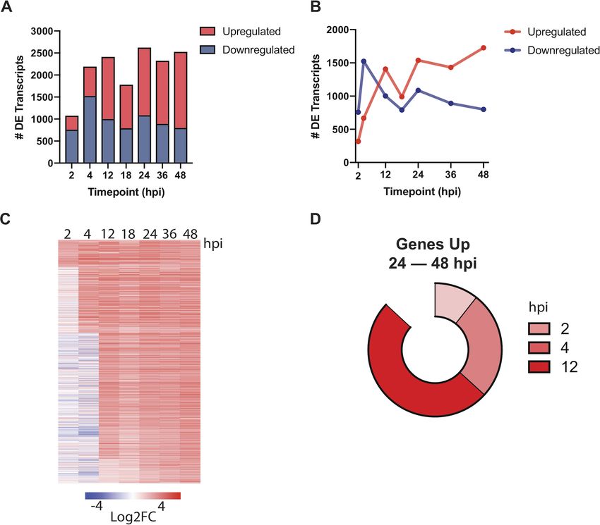
®
Toro-Moreno et al.

erythrocytes, causing the clinical manifestation of malaria. Some parasites differentiate
into sexual forms (gametocytes) that are ingested by an Anopheles mosquito during a
blood meal. In the mosquito, female and male gametocytes undergo sexual reproduc-
tion, and a series of developmental changes lead to a transformation into sporozoites.
Inoculation of these sporozoites in the host via a mosquito bite perpetuates the life
cycle (2). Despite the significant global burden of malaria (3), our molecular under-
standing of the Plasmodium life cycle is incomplete, hindering our ability to target these
parasites to prevent disease and reduce transmission. In particular, the changes that
enable sporozoites to transform and then develop within hepatocytes are largely
unknown.
    Transcriptomic studies have been instrumental in revealing gene expression varia-
tion that accompanies stage transitions and developmental processes in Plasmodium.

                                                                                                              Downloaded from http://mbio.asm.org/ on April 18, 2021 by guest
Subsequent analyses of these data have also identified transcription factors (TFs) that
are critical for controlling parasite progression at various stages [reviewed in reference
4]). However, only a few transcriptome analyses have been completed in the liver stage
(LS) relative to other parasite forms, likely owing to the technical challenges associated
with studying this stage. Still, these studies have provided important insight into
LS-specific biological processes (5), including hypnozoite markers (6, 7), and compara-
tive gene expression analysis with other stages (8), even at a single-cell resolution (9).
These studies examined gene expression upon the establishment of a LS-trophozoite
(24 h postinfection [hpi] and thereafter); however, the early stages of LS infection (0 to
24 hpi) for any Plasmodium species remain unresolved.
    Our current understanding of the early stages of LS development comes from
ultrastructural (10) and immunofluorescence (11) studies. Upon traversal and inva-
sion of hepatocytes, rod-shaped sporozoites expulse unnecessary organelles into
the parasitophorous vacuole (PV), which is accompanied by the formation of a
protrusion, a bulbous expansion, and a transformation into a spherical, replication-
competent trophozoite (10). Although this metamorphosis is obvious at the cellular
level, the molecular events underpinning this sequence of events remain obscure.
Previous studies have examined the gene expression of sporozoites grown axeni-
cally since sporozoites can complete this transformation extracellularly if activated
by bovine serum albumin, calcium, and a temperature shift (12, 13). Yet, axenically
grown sporozoites show reduced viability and poor developmental capacity com-
pared to intracellular parasites, suggesting an important role of host pathways in
this process. Indeed, a recent study showed that activation of the host GPCR CXCR4
is necessary for proper parasite metamorphosis (11), highlighting the need to study
parasite transformation, and all its subsequent development, in the context of the
host cell.
    Here, we present a transcriptomic survey of the early and middle LS of Plasmodium
berghei infecting human hepatoma cells. The rodent P. berghei and P. yoelii LS models
are routinely used to study this stage due to their genetic accessibility and tractability
relative to human-infective counterparts. Our data set includes seven time points, from
2 to 48 hpi, making it the most comprehensive transcriptomic analysis of the Plasmo-
dium LS to date. We describe changes in gene expression associated with the early
stages of Plasmodium intracellular development in the LS and show that upregulation
of most genes important for exoerythrocytic form maturation occurs as early as 12 hpi.
This finding suggests genes important for late-LS development are subject to dynamic
expression or translational repression until protein expression is necessary. Further-
more, using coexpression analysis we identified functionally enriched gene clusters
with distinct expression patterns and discovered dozens of potential regulatory DNA
motifs associated with these genes. Overall, our work completes the life cycle of this
important model organism, P. berghei, from the transcriptomic perspective, providing
a resource for exploring stage-specific expression of genes and thus advancing our
understanding of Plasmodium biology.

January/February 2020 Volume 11 Issue 1 e03234-19                                            mbio.asm.org 2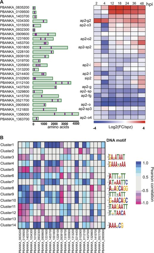
®
Plasmodium Liver-Stage Transcriptome

                                                                                                                                                                  Downloaded from http://mbio.asm.org/ on April 18, 2021 by guest
FIG 1 Experimental design for RNA-Seq of early and mid-stages of P. berghei liver infection. (A) Experimental design schematic. Female Anopheles mosquitoes
were dissected and GFP-expressing P. berghei sporozoites were harvested to infect HuH7 or HepG2 cells. Cells were harvested 2, 4, 12, 18, 24, 36, or 48 hpi and
FACS sorted to enrich viable P. berghei-infected cells for RNA collection. (B) Representative flow cytometry fluorescence dot plots indicating the population of
GFP⫹ Sytox Blue– cells that were collected at various time points. (C) Relative percentage of transcripts mapping to P. berghei or H. sapiens at various times
postinfection. Uninfected samples correspond to naive uninfected cells treated with debris from dissected male Anopheles mosquito salivary glands. The data
are medians of two to five biological replicates.

RESULTS
    RNA-Seq of early- and mid-P. berghei liver stages. During the course of the LS,
sporozoites undergo morphological changes and rapid replication. To investigate
differentially expressed transcripts that flux during this stage, HuH7 or HepG2 hepa-
toma cells were infected with green fluorescent protein (GFP)-expressing P. berghei
ANKA sporozoites. At various times postinfection, samples were harvested, and 1,000 to
3,000 P. berghei-infected cells were collected by fluorescence-activated cell sorting
(FACS) (Fig. 1A). A poor understanding exists for the early- and mid-LS; therefore,
greater sampling was acquired before 24 hpi at 2, 4, 12, and 18 hpi (early). Previously
analyzed mid-LS samples at 24 and 48 hpi were collected to enable comparison to
other studies, as well as 36 hpi, which has not been previously evaluated. Plasmodium
infection in liver cells is highly heterogeneous, with ⬃50% of sporozoites that invade
liver cells failing to establish productive infections (14, 15). We ensured selection of
populations enriched for productive infections within viable host cells by isolating cells
that are both infected and have an uncompromised membrane (GFP⫹ Sytox Blue–).

January/February 2020 Volume 11 Issue 1 e03234-19                                                                                              mbio.asm.org 3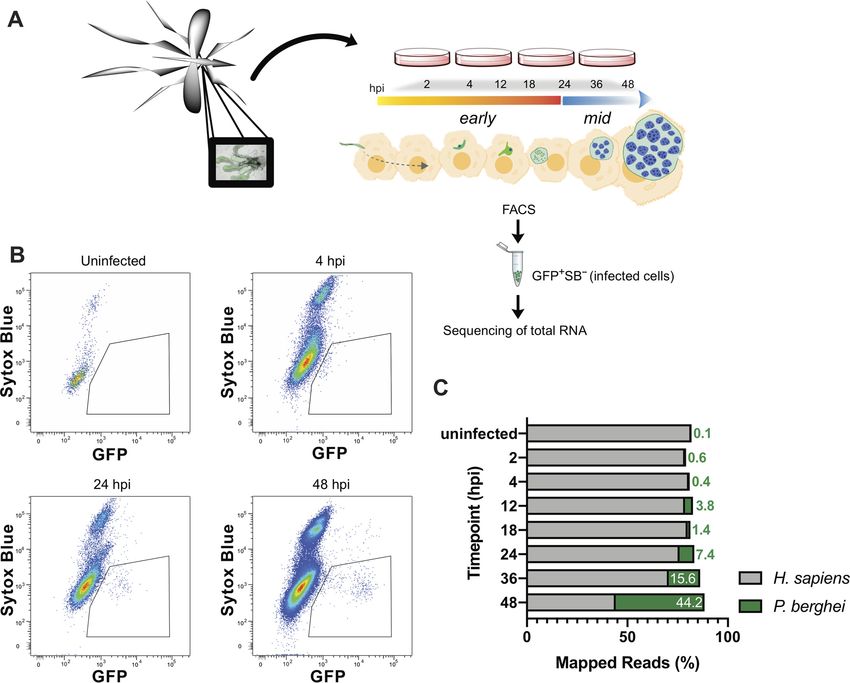
®
Toro-Moreno et al.

FACS analysis indicates that the population of infected cells (GFP⫹) shifts as a function
of time, consistent with proper intrahepatic parasite maturation (Fig. 1B). Further, our
gating excluded nonviable host cells (Sytox Blue⫹). In our method, we sorted directly
into lysis buffer. RNA was then extracted in each sample using a Clontech kit for
ultralow input RNA. Samples were evaluated for concentration and quality using a
Qubit and Bioanalyzer, respectively, and analyzed by RNA-Seq if they met quality
controls. To facilitate robust analysis, sample collection continued until a minimum of
three replicates per time point was acquired, which yielded a final range of three to
eight replicates.
    All samples were aligned to H. sapiens and P. berghei for analysis. Since parasite
nuclear division does not occur until mid-LS, ⬍4% of the reads mapped to P. berghei
before 24 hpi. This percentage rises continuously during mid-LS, when the parasite
undergoes nuclear division, and by 48 hpi ⬃45% of the reads correspond to P. berghei

                                                                                                                  Downloaded from http://mbio.asm.org/ on April 18, 2021 by guest
(Fig. 1C). Here, we are focused on parasite processes that control development within
hepatocytes; thus, principal-component analysis (PCA) was completed on P. berghei
data after the removal of batch effects. PCA revealed no major differences between the
parasite transcriptomes obtained by infecting HepG2 or Huh7 cells (see Fig. S1A in the
supplemental material), but a general clustering of replicates by genotype (time point)
was observed (Fig. S1B). Of note, PCA showed strong separation at 4 and 2 hpi, with the
latter grouping well with sporozoites, highlighting the parasite transformations that
must occur during these 2 h.
    To analyze our data set in the context of the entire Plasmodium life cycle, we
calculated Spearman correlations on our data, as well as previously published Plasmo-
dium transcriptomic data from sporozoites, the asexual blood stage (ABS), gametocytes,
ookinetes, hypnozoites, and the LS (see Table S1 in the supplemental material). This
analysis spanned data obtained from P. berghei, P. yoelii, P. cynomolgi, P. vivax, and P.
falciparum. Consistent with previous reports, the LS was more similar to the ABS than
to gametocytes and ookinetes (8). Indeed, we observe two general groups comprising
of (i) mostly metabolically active, intracellular stages (LS and ABS) and (ii) mostly motile,
extracellular stages (Fig. 2). Notably, early liver stages of P. berghei and (axenic) P. vivax
(LS_2h/4h) fell into the latter group, being more highly correlated to sporozoites,
ookinetes, and gametocytes than to other LS time points.
    Early liver-stage transcriptome of P. berghei. Thousands of statistically significant
differentially expressed transcripts were detected at early-LS time points, with most of
these transcripts being downregulated at 2 and 4 hpi and then upregulated at 12 hpi
with respect to sporozoites (Fig. 3A and B; Data Set S1). This shift suggests a change
from gene suppression to activation as the parasite exits the early stage of intrahepatic
development. As expected, genes important for host cell traversal and invasion, such as
CELTOS, SUB2, and CSP, were downregulated at 2 hpi, concurrent with the upregulation
of genes important for nutrient acquisition (ZIP1, TPT, and NT1), reflecting the estab-
lishment of the infection in the host cell. Unsurprisingly, at these early stages, we also
observed strong upregulation of EXP2 and PV2, which encode parasitophorous vacuole
membrane (PVM)-associated proteins, together with several predicted exported pro-
teins of unknown function, indicating that early (⬍4 hpi) establishment and remodeling
of the PVM is essential for parasite LS maturation. Interestingly, we observed that LYTB
(IspH), the last enzyme in the isoprenoid biosynthesis pathway in the apicoplast, is
among the most-upregulated genes at both 2 and 4 hpi (Table S2). Apicoplast path-
ways are important potential drug targets for the development of LS antimalarials but
are not known to be involved in early-LS processes.
    Translational regulation of Plasmodium transcripts has been extensively docu-
mented, and it is known to play a pivotal role during developmental transitions in the
life cycle. We found pervasive upregulation of most of the functionally characterized
translational regulators in Plasmodium, at the exclusion of PUF1 and PUF2, which
appeared to be dramatically downregulated compared to their high expression in
sporozoites. DOZI, ALBA1, ALBA2, and ALBA4 were upregulated as early as 4 hpi

January/February 2020 Volume 11 Issue 1 e03234-19                                                mbio.asm.org 4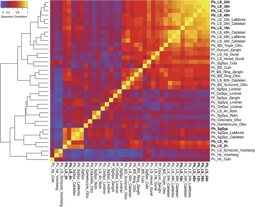
®
Plasmodium Liver-Stage Transcriptome

                                                                                                                                                               Downloaded from http://mbio.asm.org/ on April 18, 2021 by guest
FIG 2 Overview of Plasmodium transcriptome analyses. Hierarchical clustering of gene expression data sets from different stages of the
Plasmodium life cycle (7, 8, 25, 31, 33, 51–53). Data sets generated in this study are in bold. Clustering is based on Spearman correlation
coefficients calculated and plotted using R. Refer to Table S1 in the supplemental material for information regarding the data sets used
to generate this figure.

(log2-fold change [Log2FC] ⬍ 2, q ⬍ 0.01) (Fig. S2). Moreover, among the most differ-
entially expressed transcripts at 2 and 4 hpi, there was an enrichment of genes involved
in RNA-protein complexes and interactions, such as SR1, NOP10, CBF5, RPS12, and NAPL
(Table S3). Thus, translational regulation likely plays an important role in the early
stages of Plasmodium infection of the liver.
    At ⬃24 hpi and thereafter, the single-nucleated trophozoites replicate and subse-
quently mature into LS schizonts, each harboring tens of thousands of nuclei. Previous
work examining the LS transcriptome at these middle stages identified hundreds of
differentially expressed genes involved in translation, metabolism, protein trafficking,
and redox processes (5, 8). Since we saw a strong correlation between 12 hpi and
mid-LS (Spearman correlation ⫽ 0.837 to 0.949; Fig. 2), we sought to determine how
early a statistically significant upregulation of the core mid-LS transcriptome could be
observed in our data set. We found 1,197 genes in our data set that are significantly
upregulated at 24, 36, and 48 hpi compared to sporozoites (q ⬍ 0.01), constituting
about 20% of the P. berghei genome (Fig. 3A and B). Interestingly, we found that 87%
of transcripts that are upregulated throughout the mid-LS (24 hpi through 48 hpi) are
upregulated as early as 12 hpi (Fig. 3C). More specifically, 50% of the genes that are
upregulated in the mid-LS are first observed to be upregulated at 12 hpi (Fig. 3D).
    Coexpression analysis identifies functionally enriched gene clusters. To identify
coexpression patterns that may inform future functional studies, we performed a
clustering analysis of the k-means for all differentially expressed genes for all of the

January/February 2020 Volume 11 Issue 1 e03234-19                                                                                             mbio.asm.org 5
®
Toro-Moreno et al.
Downloaded from http://mbio.asm.org/ on April 18, 2021 by guest
FIG 3 Dynamic gene regulation throughout liver-stage P. berghei development. (A and B) Total (A) and upregulated (red)/downregulated
(blue) (B) differentially expressed (DE) transcripts (q ⬍ 0.01) are shown at each time point. (C) Expression profiles of 1,197 genes
upregulated at 24, 36, and 48 hpi ordered based on the time point they were first observed to be upregulated. Expression is shown as
the log2-fold change (Log2FC) versus sporozoite samples. (D) The proportions of genes upregulated throughout late-stage development
(24, 36, and 48 hpi) are divided by when they were first observed to be upregulated (2, 4, or 12 hpi).
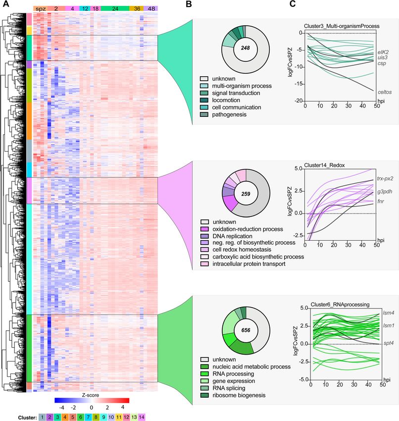
samples included in our data set. Fourteen clusters emerged from this hierarchical
clustering analysis (Fig. 4A; see Data Set S2 in the supplemental material). These clusters
could be further grouped within three major coexpression patterns when columns
were grouped by sample genotype (time point). The first major cluster group (clusters
3, 11, and 13) includes genes that are upregulated early during infection (sporozoite
[spz], 2 and 4 hpi) and are generally downregulated throughout the rest of LS infection,
such as ETRAMPs and SPELD. The second major cluster group (clusters 1, 2, 4, 7, 8, 9, 12,
and 14) includes genes that are downregulated during the early stages of infection but
are then consistently upregulated from 24 to 48 hpi. The third major cluster group
(clusters 5, 6, and 10) includes genes that are upregulated throughout the LS.
To investigate possible enrichment of biological processes of coexpressed genes, we
analyzed each cluster by gene ontology (GO). Such analyses revealed the enrichment
of various GO terms for each of the clusters (P ⬍ 0.01). We prioritized clusters for which
at least one GO term was enriched by a p-adj (Bonferroni) value of ⬍0.01. Cluster 3
stood out as highly enriched despite 142 of the total 248 genes in this cluster not being
annotated. For this cluster, enrichment analysis indicated significant enrichment of
“interspecies interaction” (GO:0044419, P ⬍ 1.91E– 07), as well as locomotion (GO:
0040011, P ⬍ 0.0005679) and signal transduction (GO:0007165, P ⬍ 0.00049572)
January/February 2020 Volume 11 Issue 1 e03234-19 mbio.asm.org 6®
Plasmodium Liver-Stage Transcriptome
Downloaded from http://mbio.asm.org/ on April 18, 2021 by guest
FIG 4 Coexpression analysis identifies enriched processes during P. berghei development in hepatocytes. (A) Hierarchical clustering using a correlation distance
with complete linkage of all genes significant (FDR ⱕ 5%) in at least one of the analyses. Gene expression is z-score transformed. (B) GO enrichment analysis
(biological process) of enriched clusters 3, 14, and 6. Representative GO terms (P ⬍ 0.01) and their respective number of genes (pie chart) are shown. The total
numbers of genes in each cluster are shown at the center of the pie chart. (C) Spline models of gene expression data for all the genes in the top-scoring GO
term in each cluster. Key genes in each group and their expression patterns are highlighted in red. Refer to Data Set S2 in the supplemental material for
complete GO analysis of all clusters.
(Fig. 4B). Genes in this cluster are highly expressed in sporozoites and thus appear to
be strongly downregulated during infection (Fig. 4C). In agreement with this result, this
cluster includes genes that have been previously shown to play an important role
during invasion (CELTOS, SPECT1, and TRAP), interactions with the host liver cell (UIS3,
January/February 2020 Volume 11 Issue 1 e03234-19 mbio.asm.org 7®
Toro-Moreno et al.
UIS4, CSP, p36, and p52), and translational control of LS-specific transcripts (UIS2, PUF1,
and PUF2) (16).
Cluster 14 was enriched for “oxidation-reduction process” (GO:0055114, P ⬍ 9.01E–
05), “DNA replication” (GO:0006260, P ⬍ 0.00164223), and “intracellular protein trans-
port” (GO:0006886, P ⬍ 0.00941436) (Fig. 4B). In this group, genes involved in redox-
regulatory processes (FNR and TRX-PX2), as well as biosynthetic genes such as G3PDH,
can be found. The expression of genes under the redox group appears to peak by ⬃12
hpi and then remains stably upregulated throughout infection. This expression pattern
highlights the need for this machinery to mitigate potential stress due to the dramatic
parasite replication and growth that is initiated at ⬃24 hpi (Fig. 4C). Little is known
about redox biology in Plasmodium parasites, particularly during the LS, but these
processes have historically been key pathways for drug discovery. Indeed, atovaquone,
Downloaded from http://mbio.asm.org/ on April 18, 2021 by guest
a drug for malaria prophylaxis in combination with proguanil, inhibits LS parasites in
vitro by impairing mitochondrial redox metabolism by targeting the cytochrome bc1
complex (17). This data set may serve as a starting point to discover more LS targets
involved in redox metabolism. Furthermore, although not enriched in our GO analysis,
we observed that several important liver-specific genes are found in this cluster, such
as IBIS1, LISP1, and LISP2. Finally, in cluster 6, we saw enrichment of core functions such
as “gene expression” (GO:0010467, P ⬍ 5.71E– 05) and “RNA processing” (GO:0006396,
P ⬍ 5.98E– 06), which contains 656 genes. As expected of housekeeping functions,
these genes appear to be expressed throughout LS infection.
Our analysis identified several clusters with enriched GO terms, some of which
accurately describe the known LS biology at different time points. Although GO
enrichment provided a useful assessment of differentially expressed processes, we note
that it is limited in its reach in Plasmodium compared to other model organisms since
⬃40% of the genome remains unannotated. Hence, to further explore the composition
of these coexpression clusters, we made use of the Rodent Malaria genetically modified
Parasite Database (RMgmDB) to provide phenotypic information about our clusters
throughout the life cycle (18). Interestingly, we observed that while most clusters have
a high proportion of genes for which disruption resulted in phenotypes across the
entire life cycle, only a few clusters had genes that displayed phenotypes exclusively in
sporozoite and/or liver stage (Fig. S3). Specifically, clusters 3 and 14 had the highest
percentage of spz/LS-specific genes (13 and 9%, respectively), reinforcing the potential
for identifying new LS drug and vaccine targets within these clusters.
Expression dynamics of AP2 transcription factors. Transcriptional regulation of
gene expression has been extensively studied in the intraerythrocytic developmental
cycle (IDC) and mosquito stages of P. berghei and P. falciparum. The AP2 transcription
factors (TFs), comprised of 26 genes in P. berghei, are the best-characterized family of
TFs in apicomplexans (Fig. 5A). AP2s are known to regulate Plasmodium transitions into
different developmental stages and have emerged as key factors leading to both sexual
commitment and sex differentiation (reviewed in reference 4). Unsurprisingly, we
observed that AP2 genes with established functions in mosquito stages (AP2-O and
AP2-O2) and those involved in sporozoite development (AP2-SPs) are downregulated
throughout the liver stages (Fig. 5A). The only ApiAP2 TF known to play a role in LS
development is AP2-L. AP2-L–/– parasites are able to traverse and invade liver cells but
arrest in the schizont stage (19). AP2-L transcripts are abundant in sporozoites and thus
appear to be strongly downregulated during infection, as early as 2 hpi (Fig. 5A).
We observe strong upregulation (⬃3-fold) of AP2-G2 at 2 and 4 hpi. AP2-G2 has
been shown to act as a repressor during the blood stage (BS) and gametocyte
development and to have different targets during these stages (20, 21). A group of such
targets corresponds to the liver-specific genes LISP1 and TREP, which are important for
LS schizont maturation and are expressed during late-LS infection (21). Interestingly, we
observe that AP2-G2 expression is negatively correlated to the average expression of
the main clusters harboring this set of genes, including clusters 1, 9, and 14 (see Data
Set S2 in the supplemental material). Thus, it is plausible that during the first hours of
January/February 2020 Volume 11 Issue 1 e03234-19 mbio.asm.org 8®
Plasmodium Liver-Stage Transcriptome
Downloaded from http://mbio.asm.org/ on April 18, 2021 by guest
FIG 5 Expression of P. berghei AP2 transcription factors in the liver stage. (A) Gene IDs of the 26 AP2
transcription factors in the P. berghei genome, their respective protein architecture schematic (with AP2
displayed in purple), and their corresponding expression as the log2-fold change versus spz at each time
point in the LS. (B) Heat map of Pearson correlations between AP2 transcription factors and the average
expression of all genes in each cluster (left). The top most enriched DNA motif for each cluster discovered
through the DREME pipeline is shown (right). Refer to Data Set S2 for a complete set of motifs and their
respective enrichment scores.
infection, AP2-G2 acts as a repressor of genes involved in later stages of LS develop-
ment, many of which remain uncharacterized.
Interestingly, we observed significant upregulation of the uncharacterized
ApiAP2s PBANKA_0835200 and PBANKA_0109500 throughout the LS starting at 12
hpi, in contrast to the early upregulation of AP2-G2. Although still functionally
uncharacterized, their orthologs in P. falciparum have been recently shown to
coexpress during differentiation in gametocytogenesis and to be inversely corre-
January/February 2020 Volume 11 Issue 1 e03234-19 mbio.asm.org 9®
Toro-Moreno et al.
lated to genes involved in ABS development. This expression pattern suggests they
may have a role as corepressors of genes involved in the ABS (22). In our data set,
we observe a strong correlation with clusters 11 and 12 (both negative) and with
cluster 8 (positive).
We sought to identify enriched DNA motifs in each of the coexpression clusters by
analyzing the 5= untranslated region sequences (1 kb) of their genes against the
upstream sequence for all of the genes in other clusters using DREME (Data Set S3) (23).
While genes in clusters 1, 2, 5, and 13 lacked any enriched DNA motifs, de novo
discovery uncovered hundreds of DNA motifs in the remaining clusters, with the
topmost enriched motif shown in Fig. 5B. We found that the most significant motif in
cluster 12 (T[G/C]TAACA) matched the motif recognized by ApiAP2 PBANKA_0521700
(GTGTTACAC, P ⬍ 1.28E– 05). This cluster included genes that are mostly downregu-
lated throughout the LS until the later time points in our time series, such as the BS
Downloaded from http://mbio.asm.org/ on April 18, 2021 by guest
schizont-specific genes SERA2 and SERA3. In addition, PBANKA_0521700 expression was
strongly correlated to cluster 12 (r ⫽ 0.83, P ⬍ 0.021), suggesting this cluster might
harbor previously unknown targets of this ApiAP2 (Fig. 5B).
DISCUSSION
Our data provide novel insights into gene expression fluxes throughout Plasmodium
development within hepatocytes. The transcriptional blueprints provided by our time
series enables comparison of early-, mid-, and late-LS parasite processes for the first
time. We found 146 genes exclusively upregulated early, such as EIF5, and 482 genes,
including SERA1 and LISP2, exclusively upregulated in the mid-LS (Fig. S4). Furthermore,
our data sets recapitulated well-established gene expression patterns of key LS genes
and overall were largely in agreement with recently reported data sets, supporting the
validity of our approach. Through our analysis, we identified a key shift in parasite gene
expression that occurs at 12 hpi and the role of transcription factors in driving LS
maturation. Specifically, we explored potential transcriptional regulation of coexpress-
ing genes by analyzing their upstream sequences for enrichment of potential DNA
binding motifs and their correlation to P. berghei AP2 transcription factors. Our results
revealed an association between the uncharacterized PBANKA_0521700 AP2 TF and
cluster 12. PBANKA_0521700 is preferentially expressed in the ring stages of the IDC
and is refractory to disruption in the BS (21, 24), hampering functional studies of this
gene. Our data, in conjunction with previously reported P. berghei RNA-Seq (8, 21, 24,
25) and single-cell studies covering the entire life cycle (9), could be useful to refine
hypotheses about the functions and targets of this TF, as well as other AP2 TFs.
Although AP2 TFs have been at the center of gene expression studies in Plasmo-
dium, novel “omics” approaches have begun uncovering other layers of gene regula-
tion. Indeed, posttranscriptional regulation, such as N6-methyladenosine (m6A) of
mRNA and alternative splicing, have recently been recognized as essential for fine-
tuning gene expression in blood and sexual stages (26, 27). In particular, disruption of
the splicing factor PbSR-MG was shown to perturb sex-specific alternative splicing, thus
demonstrating its role as a cellular differentiation regulator (28). Interestingly, we
observed a dramatic upregulation of the splicing factor SR1 coinciding with the
parasite’s metamorphoses in the LS, hinting at an important role for alternative splicing
during this stage. Future reverse genetic studies may help establish a role for alterna-
tive splicing in the LS.
A well-documented form of gene expression regulation in Plasmodium occurs at the
translational level. Translational repression (TR) of hundreds of transcripts has been
reported at most stages of the P. berghei life cycle (29). TR is particularly pervasive in the
sporozoite transition from the mosquito to the mammalian host (30, 31). During this
transition, hundreds of transcripts that are highly expressed in sporozoites are stored in
mRNA granules, until infection of the host relieves this repression, resulting in protein
translation. The extent to which a TR program operates in the LS is currently unknown.
However, we observed that ⬃50% of all transcripts upregulated after 24 hpi are also
upregulated at 12 hpi, including some with known roles in LS schizont maturation (IBIS1
January/February 2020 Volume 11 Issue 1 e03234-19 mbio.asm.org 10®
Plasmodium Liver-Stage Transcriptome
and BP2). Furthermore, we observed the upregulation of several known translational
regulators, e.g., DOZI and ALBA1, -2, and -4, which could potentially repress translation
of transcripts important for late LS development and/or the subsequent transition to
the ABS. Unfortunately, this possibility will be exceedingly difficult to test in the
absence of robust global proteomic analysis of the early LS parasite. Nonetheless, our
data, coupled with recent RNA-Seq and proteomic studies of the more accessible late
LS, can provide a starting point to address this question (8, 32).
Previous work examining the transcriptional changes of axenically grown early LS P.
vivax identified upregulation of calcium-related proteins (RACK1) and RNA-binding
proteins (ALBA1, -2 and -4) (33). We saw upregulation of the P. berghei orthologs of
these genes, as well as hundreds of other genes, dramatically expanding the data set
for genes upregulated at this stage (Fig. S5). For example, we found that LYTB is
upregulated at 2 and 4 hpi, indicating isoprenoids may be important at this time.
Downloaded from http://mbio.asm.org/ on April 18, 2021 by guest
Although the FASII and de novo heme biosynthesis pathways have been genetically and
chemically validated as essential to the late liver stages, less is known about isoprenoid
biosynthesis during the early liver stages (34, 35). When intracellular sporozoites
metamorphosize to replication-competent trophozoites, most organelles are expelled
at the exclusion of the nucleus, mitochondrion, and apicoplast (10). Thus, it is plausible
that the apicoplast serves an important metabolic role with isoprenoids in the liver
stages of infection. Unfortunately, the use of isoprenoid biosynthesis inhibitors has
yielded inconclusive results about its function during the LS (36, 37), emphasizing the
need for future genetic studies to elucidate the role of isoprenoid biosynthesis through-
out intrahepatic development. Thus, we anticipate our data will be useful to guide
future reverse genetic and functional studies to investigate the role of Plasmodium
genes with important early- and mid-LS functions.
Our understanding of Plasmodium LS biology still lags behind that of other parasite
life cycle stages, hindering the development of much-needed prophylactic measures to
combat malaria. Our work represents a window into the previously undescribed
transcriptome of the early LS upon host cell infection and offers a comprehensive view
of the Plasmodium LS. Future studies expanding on our analysis and validating time-
specific LS genes will further advance our molecular understanding of this critical step
in the Plasmodium life cycle.
MATERIALS AND METHODS
Parasites. Sporozoites were freshly harvested prior to experiments from dissected salivary glands of
Anopheles stephensi mosquitoes infected with P. berghei ANKA stably expressing a GFP purchased from
the New York University Langone Medical Center Insectary.
Cell culture. HepG2 were purchased from ATCC and HuH7 cells were kindly provided by Peter Sorger
(Harvard Medical School). Hepatocytes used for P. berghei infections were maintained in Dulbecco
modified Eagle medium with L-glutamine (Gibco) supplemented with 10% (vol/vol) heat-inactivated fetal
bovine serum (Sigma-Aldrich) and 1% antibiotic-antimycotic (Thermo Fisher Scientific) in a standard
tissue culture incubator (37°C, 5% CO2).
Sample collection for RNA-Seq. Infected hepatoma cells were collected as previously described
(38). Briefly, T25 flasks were seeded with 3 ⫻ 105 HepG2 or 8 ⫻ 104 HuH7 cells. About 24 h after seeding,
the cells were infected with 1 ⫻ 105 GFP-expressing P. berghei-ANKA sporozoites. Infected cells and
uninfected controls were sorted directly into RNA lysis buffer (Clontech) using a BD FACSAria II cell sorter
(BD Biosciences) at the Duke Human Vaccine Institute. Sytox Blue was used as a live/dead cell indicator
(Thermo Fisher Scientific). Infected cells were collected by sorting of the GFP and gated compared to
uninfected hepatoma cells. RNA was extracted using SMART-seq v4 Ultra Low Input RNA kit for
sequencing (Clontech), and libraries were prepared at the Duke Next Generation Sequencing Core
Facility and sequenced on the Illumina HiSeq 4000 as 50-bp single-end reads. Four or five samples were
pooled on each flow cell lane.
RNA-Seq and differential expression analysis. RNA-Seq data were processed using a TrimGalore
toolkit (39), which employs Cutadapt (40) to trim low-quality bases and Illumina sequencing adapters
from the 3= end of the reads. Only reads that were 20 nucleotides or longer after trimming were kept for
further analysis. Reads were mapped to a combination of the GRCh37v75 (41) version of the human
genome and the PbANKAv3 of the P. berghei genome using the STAR RNA-Seq alignment tool (42). Reads
were kept for subsequent analysis if they mapped to a single genomic location. All samples mapping ⬎1
million reads to the P. berghei genome were used for a preliminary analysis. Gene counts were compiled
using the HTSeq tool (43). Only P. berghei genes that had at least 10 reads in any given library were used
in subsequent analysis. Normalization and differential expression were carried out using the DESeq2 (44)
Bioconductor (45) package with the R statistical programming environment (46). The false discovery rate
January/February 2020 Volume 11 Issue 1 e03234-19 mbio.asm.org 11®
Toro-Moreno et al.
(FDR) was calculated to control for multiple hypothesis testing. When calculating the differential
expression between genes at each time point relative to the control, the cell type and sequencing batch
were included as cofactors in the model.
Spearman correlations between published P. berghei RNA-Seq data sets and our own were calculated
and plotted using the cor function in the stats R package.
Clustering analysis. To determine the different patterns of gene expression across all groups of
samples, we first identified genes that showed differential expression in at least one of the comparisons
performed (FDR ⱕ 5%). The genes were the clustered across all samples by a correlation distance using
complete linkage after z-score transformation. The NbClust (47) package was used to separate the gene
expression across all samples into distinct clusters.
De novo motif discovery was performed using DREME from the MEME suite (23). For each cluster, the
input data set was the upstream 1,000-kb region of each gene within that cluster, and the negative set
was the upstream region of genes that were not in that cluster. The analysis was run in discriminative
mode, scanning the given strand only, with the predicted motif size of 4 to 10 bp and a cutoff E value
of 0.05. The top most enriched motif for each cluster was then analyzed with TOMTOM (48) to compare
to previously in silico-discovered motifs (49).
Downloaded from http://mbio.asm.org/ on April 18, 2021 by guest
The correlation matrix was generated in Prism by calculating the Pearson correlation between each
AP2 transcription factor and the average fold change expression for all the genes in each cluster.
Gene ontology. GO analyses for each cluster were performed using the GO enrichment tool for
Biological Processes in PlasmoDB (50) with a cutoff of P ⬍ 0.01. The number of genes from the cluster
in each of the representative top-scoring GO terms (i.e., the lowest P values) were plotted.
SUPPLEMENTAL MATERIAL
Supplemental material is available online only.
FIG S1, TIF file, 0.3 MB.
FIG S2, TIF file, 1.1 MB.
FIG S3, TIF file, 1.2 MB.
FIG S4, TIF file, 0.1 MB.
FIG S5, TIF file, 2.7 MB.
TABLE S1, DOCX file, 0.02 MB.
TABLE S2, DOCX file, 0.01 MB.
TABLE S3, DOCX file, 0.01 MB.
DATA SET S1, XLSX file, 6.1 MB.
DATA SET S2, XLSX file, 0.1 MB.
ACKNOWLEDGMENTS
This study is funded by the National Institutes of Health (NIH; DP2AI138239; to
E.R.D.), the CM Hauser Fellowship (M.T.M.), and the NSF (DGE-1644868, to K.S.). The
content of this study is solely the responsibility of the authors and does not necessarily
represent the official views of the NIH.
We thank Ana Rodriguez and Sandra Gonzalez from the NYU Insectary for providing
Plasmodium-infected mosquitoes, David Corcoran from the Duke Genomic Analysis and
Bioinformatics (GCB) Core Facility, the DHVI Flow Cytometry Core Facility, and Joseph
Saelens. We also thank Photini Sinnis, Amanda Balaban, and the JHMRI Insectary
and Parasitology Core Facilities for their help. We thank Luisa Toro Moreno for data
managing support, and Jen-Tsan Ashley Chi and Steven Haase for useful discus-
sions.
Conception or design of the work: M.T.-M., K.S., D.P., and E.R.D.; data collection: K.S,
D.P., and E.R.D.; data analysis and interpretation: M.T.-M., K.S., and T.S.; drafting of the
article: M.T.-M.; critical revision and contributions to the article: K.S. and, E.R.D.; final
approval of the version to be published: M.T.-M., K.S., T.S., D.P., and E.R.D.
REFERENCES
1. Prudêncio M, Rodriguez A, Mota MM. 2006. The silent path to thousands 4. Josling GA, Williamson KC, Llinás M. 2018. Regulation of sexual commit-
of merozoites: the Plasmodium liver stage. Nat Rev Microbiol 4:849 – 856. ment and gametocytogenesis in malaria parasites. Annu Rev Microbiol
https://doi.org/10.1038/nrmicro1529. 72:501–519. https://doi.org/10.1146/annurev-micro-090817-062712.
2. Aly ASI, Vaughan AM, Kappe S. 2009. Malaria parasite development in 5. Tarun AS, Peng X, Dumpit RF, Ogata Y, Silva-Rivera H, Camargo N, Daly
the mosquito and infection of the mammalian host. Annu Rev Microbiol TM, Bergman LW, Kappe S. 2008. A combined transcriptome and pro-
63:195–221. https://doi.org/10.1146/annurev.micro.091208.073403. teome survey of malaria parasite liver stages. Proc Natl Acad Sci U S A
3. World Health Organization. 2019. World malaria report. World Health 105:305–310. https://doi.org/10.1073/pnas.0710780104.
Organization, Geneva, Switzerland. 6. Bertschi NL, Voorberg-Van der Wel A, Zeeman AM, Schuierer S, Nigsch F,
January/February 2020 Volume 11 Issue 1 e03234-19 mbio.asm.org 12®
Plasmodium Liver-Stage Transcriptome
Carbone W, Knehr J, Gupta DK, Hofman SO, van der Werff N, Nieuwen- Plasmodium falciparum sexual differentiation. bioRxiv https://doi.org/10
huis I, Klooster E, Faber BW, Flannery EL, Mikolajczak SA, Chuenchob V, .1101/633222.
Shrestha B, Beibel M, Bouwmeester T, Kangwanrangsan N, Sattabongkot 23. Bailey TL. 2011. DREME: motif discovery in transcription factor ChIP-seq
J, Diagana TT, Kocken CHM, Roma G. 2018. Transcriptomic analysis data. Bioinformatics 27:1653–1659. https://doi.org/10.1093/bioinformatics/
reveals reduced transcriptional activity in the malaria parasite Plasmo- btr261.
dium cynomolgi during progression into dormancy. Elife 7:e41081. 24. Otto TD, Böhme U, Jackson AP, Hunt M, Franke-Fayard B, Hoeijmakers
https://doi.org/10.7554/eLife.41081. WAM, Religa AA, Robertson L, Sanders M, Ogun SA, Cunningham D, Erhart
7. Cubi R, Vembar SS, Biton A, Franetich JF, Bordessoulles M, Sossau D, A, Billker O, Khan SM, Stunnenberg HG, Langhorne J, Holder AA, Waters AP,
Zanghi G, Bosson-Vanga H, Benard M, Moreno A, Dereuddre-Bosquet N, Newbold CI, Pain A, Berriman M, Janse CJ. 2014. A comprehensive evalua-
Le Grand R, Scherf A, Mazier D. 2017. Laser capture microdissection tion of rodent malaria parasite genomes and gene expression. BMC Biol
enables transcriptomic analysis of dividing and quiescent liver stages of 12:86. https://doi.org/10.1186/PREACCEPT-1233682211145405.
Plasmodium relapsing species. Cell Microbiol 19. https://doi.org/10.1111/ 25. LaMonte GM, Orjuela-Sanchez P, Calla J, Wang LT, Li S, Swann J, Cowell
cmi.12735. AN, Zou BY, Abdel-Haleem Mohamed AM, Villa Galarce ZH, Moreno M,
8. Caldelari R, Dogga S, Schmid MW, Franke-Fayard B, Janse CJ, Soldati- Tong Rios C, Vinetz JM, Lewis N, Winzeler EA. 2019. Dual RNA-Seq
Favre D, Heussler V. 2019. Transcriptome analysis of Plasmodium berghei identifies human mucosal immunity protein Mucin-13 as a hallmark of
during exo-erythrocytic development. Malar J 18:330. https://doi.org/10 Plasmodium exoerythrocytic infection. Nat Commun 10:488. https://doi
.1186/s12936-019-2968-7.
Downloaded from http://mbio.asm.org/ on April 18, 2021 by guest
.org/10.1038/s41467-019-08349-0.
9. Howick VM, Russell AJC, Andrews T, Heaton H, Reid AJ, Natarajan K, 26. Baumgarten S, Bryant JM, Sinha A, Reyser T, Preiser PR, Dedon PC, Scherf
Butungi H, Metcalf T, Verzier LH, Rayner JC, Berriman M, Herren JK, Billker A. 2019. Transcriptome-wide dynamics of extensive m6A mRNA meth-
O, Hemberg M, Talman AM, Lawniczak M. 2019. The malaria cell atlas: ylation during Plasmodium falciparum blood-stage development. Nat
single parasite transcriptomes across the complete Plasmodium life Microbiol 4:2246 –2259. https://doi.org/10.1038/s41564-019-0521-7.
cycle. Science 365:eaaw2619. https://doi.org/10.1126/science.aaw2619. 27. Reference deleted.
10. Jayabalasingham B, Bano N, Coppens I. 2010. Metamorphosis of the 28. Yeoh LM, Goodman CD, Mollard V, McHugh E, Lee VV, Sturm A, Cozi-
malaria parasite in the liver is associated with organelle clearance. Cell jnsen A, McFadden GI, Ralph SA. 2019. Alternative splicing is required for
Res 20:1043–1059. https://doi.org/10.1038/cr.2010.88. stage differentiation in malaria parasites. Genome Biol 20:151. https://
11. Bando H, Pradipta A, Iwanaga S, Okamoto T, Okuzaki D, Tanaka S, doi.org/10.1186/s13059-019-1756-6.
Vega-Rodríguez J, Lee Y, Ma JS, Sakaguchi N, Soga A, Fukumoto S, Sasai 29. Lasonder E, Rijpma SR, Van Schaijk BCL, Hoeijmakers WAM, Kensche PR,
M, Matsuura Y, Yuda M, Jacobs-Lorena M, Yamamoto M. 2019. CXCR4 Gresnigt MS, Italiaander A, Vos MW, Woestenenk R, Bousema T, Mair GR,
regulates Plasmodium development in mouse and human hepatocytes. Khan SM, Janse CJ, Bártfai R, Sauerwein RW. 2016. Integrated transcrip-
J Exp Med 216:1733–1748. https://doi.org/10.1084/jem.20182227. tomic and proteomic analyses of P Falciparum gametocytes: molecular
12. Kaiser K, Camargo N, Kappe S. 2003. Transformation of sporozoites into insight into sex-specific processes and translational repression. Nucleic
early exoerythrocytic malaria parasites does not require host cells. J Exp
Acids Res 44:6087– 6101. https://doi.org/10.1093/nar/gkw536.
Med 197:1045–1050. https://doi.org/10.1084/jem.20022100.
30. Muller I, Jex AR, Kappe SHI, Mikolajczak SA, Sattabongkot J, Patrapuvich
13. Doi Y, Shinzawa N, Fukumoto S, Okano H, Kanuka H. 2011. Calcium
R, Lindner S, Flannery EL, Koepfli C, Ansell B, Lerch A, Emery-Corbin SJ,
signal regulates temperature-dependent transformation of sporozoites
Charnaud S, Smith J, Merrienne N, Swearingen KE, Moritz RL, Petter M,
in malaria parasite development. Exp Parasitol 128:176 –180. https://doi
Duffy MF, Chuenchob V. 2019. Transcriptome and histone epigenome of
.org/10.1016/j.exppara.2011.02.011.
Plasmodium vivax salivary-gland sporozoites point to tight regulatory
14. Prudêncio M, Rodrigues CD, Ataíde R, Mota MM. 2008. Dissecting in vitro
control and mechanisms for liver-stage differentiation in relapsing ma-
host cell infection by Plasmodium sporozoites using flow cytometry.
laria. Int J Parasitol 49:501–513. https://doi.org/10.1016/j.ijpara.2019.02
Cell Microbiol 10:218 –224. https://doi.org/10.1111/j.1462-5822.2007
.007.
.01032.x.
31. Lindner SE, Swearingen KE, Shears MJ, Walker MP, Vrana EN, Hart KJ,
15. Risco-Castillo V, Topçu S, Marinach C, Manzoni G, Bigorgne AE, Briquet S,
Minns AM, Sinnis P, Moritz RL, Kappe S. 2019. Transcriptomics and
Baudin X, Lebrun M, Dubremetz J-F, Silvie O, Amino R, Giovannini D,
proteomics reveal two waves of translational repression during the matu-
Thiberge S, Gueirard P, Boisson B, Dubremetz J-F, Prévost M-C, Ishino T,
Yuda M, Ménard R, Bano N, Romano JD, Jayabalasingham B, Coppens I, ration of malaria parasite sporozoites. Nat Commun 10:4964. https://doi
et al. 2015. Malaria sporozoites traverse host cells within transient .org/10.1038/s41467-019-12936-6.
vacuoles. Cell Host Microbe 18:593– 603. https://doi.org/10.1016/j.chom 32. Shears MJ, Sekhar Nirujogi R, Swearingen KE, Renuse S, Mishra S, Jaipal
.2015.10.006. Reddy P, Moritz RL, Pandey A, Sinnis P. 2019. Proteomic analysis of
16. Vaughan AM, Kappe SHI, Vaughan AM, Kappe SHI. 2017. Malaria parasite Plasmodium merosomes: the link between liver and blood stages in
liver infection and exoerythrocytic biology. Cold Spring Harb Perspect malaria. J Proteome Res 18:3404 –3418. https://doi.org/10.1021/acs
Med 7:a025486. https://doi.org/10.1101/cshperspect.a025486. .jproteome.9b00324.
17. Siregar JE, Kurisu G, Kobayashi T, Matsuzaki M, Sakamoto K, Mi-Ichi F, 33. Roth A, Adapa SR, Zhang M, Liao X, Saxena V, Goffe R, Li S, Ubalee R,
Watanabe Y, Hirai M, Matsuoka H, Syafruddin D, Marzuki S, Kita K. 2015. Saggu GS, Pala ZR, Garg S, Davidson S, Jiang RHY, Adams JH. 2018.
Direct evidence for the atovaquone action on the Plasmodium cyto- Unraveling the Plasmodium vivax sporozoite transcriptional journey
chrome bc1 complex. Parasitol Int 64:295–300. https://doi.org/10.1016/j from mosquito vector to human host. Sci Rep 8:12183. https://doi.org/
.parint.2014.09.011. 10.1038/s41598-018-30713-1.
18. Janse CJ, Kroeze H, van Wigcheren A, Mededovic S, Fonager J, Franke- 34. Nagaraj VA, Sundaram B, Varadarajan NM, Subramani PA, Kalappa DM,
Fayard B, Waters AP, Khan SM. 2011. A genotype and phenotype data- Ghosh SK, Padmanaban G. 2013. Malaria parasite-synthesized heme is
base of genetically modified malaria parasites. Trends Parasitol 27:31–39. essential in the mosquito and liver stages and complements host heme
https://doi.org/10.1016/j.pt.2010.06.016. in the blood stages of infection. PLoS Pathog 9:e1003522. https://doi
19. Iwanaga S, Kaneko I, Kato T, Yuda M. 2012. Identification of an AP2- .org/10.1371/journal.ppat.1003522.
family protein that is critical for malaria liver stage development. PLoS 35. Rizopoulos Z, Matuschewski K, Haussig JM. 2016. Distinct prominent
One 7:e47557. https://doi.org/10.1371/journal.pone.0047557. roles for enzymes of Plasmodium berghei heme biosynthesis in sporo-
20. Yuda M, Iwanaga S, Kaneko I, Kato T. 2015. Global transcriptional zoite and liver-stage maturation. Infect Immun 84:3252–3262. https://
repression: an initial and essential step for Plasmodium sexual develop- doi.org/10.1128/IAI.00148-16.
ment. Proc Natl Acad Sci U S A 112:12824 –12829. https://doi.org/10 36. Baumeister S, Wiesner J, Reichenberg A, Hintz M, Bietz S, Harb OS,
.1073/pnas.1504389112. Roos DS, Kordes M, Friesen J, Matuschewski K, Lingelbach K, Jomaa H,
21. Modrzynska K, Pfander C, Chappell L, Yu L, Suarez C, Dundas K, Gomes Seeber F. 2011. Fosmidomycin uptake into Plasmodium and Babesia-
AR, Goulding D, Rayner JC, Choudhary J, Billker O. 2017. A knockout infected erythrocytes is facilitated by parasite-induced new permea-
screen of ApiAP2 genes reveals networks of interacting transcriptional bility pathways. PLoS One 6:e19334. https://doi.org/10.1371/journal
regulators controlling the Plasmodium life cycle. Cell Host Microbe .pone.0019334.
21:11–22. https://doi.org/10.1016/j.chom.2016.12.003. 37. Nair SC, Brooks CF, Goodman CD, Sturm A, Strurm A, McFadden GI,
22. van Biljon R, van Wyk R, Painter H, Orchard L, Reader J, Niemand J, Llinas Sundriyal S, Anglin JL, Song Y, Moreno SNJ, Striepen B. 2011. Apicoplast
M, Birkholtz L-M. 2019. Hierarchical transcriptional control regulates isoprenoid precursor synthesis and the molecular basis of fosmidomycin
January/February 2020 Volume 11 Issue 1 e03234-19 mbio.asm.org 13®
Toro-Moreno et al.
resistance in Toxoplasma gondii. J Exp Med 208:1547–1559. https://doi 47. Charrad M, Ghazzali N, Boiteau V, Niknafs A. 2015. NbClust: an R package
.org/10.1084/jem.20110039. for determining the relevant number of clusters in a data set. J Stat
38. Posfai D, Sylvester K, Reddy A, Ganley JG, Wirth J, Cullen QE, Dave T, Software 61:1–36. https://doi.org/10.18637/jss.v061.i06.
Kato N, Dave SS, Derbyshire ER. 2018. Plasmodium parasite exploits 48. Gupta S, Stamatoyannopoulos JA, Bailey TL, Noble W. 2007. Quantifying
host aquaporin-3 during liver-stage malaria infection. PLoS Pathog 14: similarity between motifs. Genome Biol 8:R24. https://doi.org/10.1186/
e1007057. https://doi.org/10.1371/journal.ppat.1007057. gb-2007-8-2-r24.
39. Krueger F. 2017. Babraham Bioinformatics: Trim Galore!, v044. https:// 49. Campbell TL, De Silva EK, Olszewski KL, Elemento O, Llinás M. 2010.
github.com/FelixKrueger/TrimGalore/releases/tag/0.4.4. Identification and genome-wide prediction of DNA binding specificities
40. Martin M. 2011. Cutadapt removes adapter sequences from high- for the ApiAP2 family of regulators from the malaria parasite. PLoS
throughput sequencing reads. Embnet J 17:10. https://doi.org/10.14806/ Pathog 6:e1001165. https://doi.org/10.1371/journal.ppat.1001165.
ej.17.1.200. 50. Collaborativea T. 2001. PlasmoDB: an integrative database of the Plas-
41. Kersey PJ, Staines DM, Lawson D, Kulesha E, Derwent P, Humphrey JC, modium falciparum genome. Tools for accessing and analyzing finished
Hughes DST, Keenan S, Kerhornou A, Koscielny G, Langridge N, McDow- and unfinished sequence data. Nucleic Acids Res 29:66 – 69.
all MD, Megy K, Maheswari U, Nuhn M, Paulini M, Pedro H, Toneva I, 51. Voorberg-van der Wel A, Roma G, Gupta DK, Schuierer S, Nigsch F,
Wilson D, Yates A, Birney E. 2012. Ensembl genomes: an integrative
Carbone W, Zeeman A-M, Lee BH, Hofman SO, Faber BW, Knehr J, Pasini
resource for genome-scale data from non-vertebrate species. Nucleic
E, Kinzel B, Bifani P, Bonamy GMC, Bouwmeester T, Kocken CHM, Di-
Acids Res 40:D91–D97. https://doi.org/10.1093/nar/gkr895.
Downloaded from http://mbio.asm.org/ on April 18, 2021 by guest
agana TT, Voorberg-van der Wel A, Roma G, Gupta DK, Schuierer S,
42. Dobin A, Davis CA, Schlesinger F, Drenkow J, Zaleski C, Jha S, Batut P,
Nigsch F, Carbone W, Zeeman A-M, Lee BH, Hofman SO, Faber BW, Knehr
Chaisson M, Gingeras TR. 2013. STAR: ultrafast universal RNA-seq aligner.
J, Pasini E, Kinzel B, Bifani P, Bonamy GMC, Bouwmeester T, Kocken CHM,
Bioinformatics 29:15–21. https://doi.org/10.1093/bioinformatics/bts635.
43. Anders S, Pyl PT, Huber W. 2015. HTSeq-A Python framework to work Diagana TT. 2017. A comparative transcriptomic analysis of replicating
with high-throughput sequencing data. Bioinformatics 31:166 –169. and dormant liver stages of the relapsing malaria parasite plasmodium
44. Love MI, Huber W, Anders S. 2014. Moderated estimation of fold change cynomolgi. Elife 6:e29605. https://doi.org/10.7554/eLife.29605.
and dispersion for RNA-seq data with DESeq2. Genome Biol 15:550. 52. Gural N, Mancio-Silva L, Miller AB, Galstian A, Butty VL, Levine SS,
https://doi.org/10.1186/s13059-014-0550-8. Patrapuvich R, Desai SP, Mikolajczak SA, Kappe SHI, Fleming HE, March
45. Huber W, Carey VJ, Gentleman R, Anders S, Carlson M, Carvalho BS, S, Sattabongkot J, Bhatia SN. 2018. In vitro culture, drug sensitivity, and
Bravo HC, Davis S, Gatto L, Girke T, Gottardo R, Hahne F, Hansen KD, transcriptome of Plasmodium vivax hypnozoites. Cell Host Microbe 23:
Irizarry RA, Lawrence M, Love MI, MacDonald J, Obenchain V, Oleś AK, 395– 406. https://doi.org/10.1016/j.chom.2018.01.002.
Pagès H, Reyes A, Shannon P, Smyth GK, Tenenbaum D, Waldron L, 53. Zanghì G, Vembar SS, Baumgarten S, Ding S, Guizetti J, Bryant JM, Mattei
Morgan M. 2015. Orchestrating high-throughput genomic analysis with D, Jensen ATR, Rénia L, Goh YS, Sauerwein R, Hermsen CC, Franetich JF,
Bioconductor. Nat Methods 12:115–121. https://doi.org/10.1038/nmeth Bordessoulles M, Silvie O, Soulard V, Scatton O, Chen P, Mecheri S,
.3252. Mazier D, Scherf A. 2018. A specific PfEMP1 is expressed in Plasmodium
46. The R Foundation. 2018 R: the R project for statistical computing. falciparum sporozoites and plays a role in hepatocyte infection. Cell Rep
https://www.r-project.org/. 22:2951–2963. https://doi.org/10.1016/j.celrep.2018.02.075.
January/February 2020 Volume 11 Issue 1 e03234-19 mbio.asm.org 14You can also read