The formation of a hatching line in the serosal cuticle confers multifaceted adaptive functions on the eggshell of a cicada
←
→
Page content transcription
If your browser does not render page correctly, please read the page content below
Moriyama et al. Zoological Letters    (2021) 7:8
https://doi.org/10.1186/s40851-021-00178-8

 RESEARCH ARTICLE                                                                                                                                   Open Access

The formation of a hatching line in the
serosal cuticle confers multifaceted
adaptive functions on the eggshell of a
cicada
Minoru Moriyama1,2* , Kouji Yasuyama3,4 and Hideharu Numata2,5

  Abstract
  Insect eggshells must meet various demands of developing embryos. These demands sometimes conflict with each
  other; therefore, there are tradeoffs between eggshell properties, such as robustness and permeability. To meet
  these conflicting demands, particular eggshell structures have evolved in diverse insect species. Here, we report a
  rare eggshell structure found in the eggshell of a cicada, Cryptotympana facialis. This species has a prolonged egg
  period with embryonic diapause and a trait of humidity-inducible hatching, which would impose severe demands
  on the eggshell. We found that in eggs of this species, unlike many other insect eggs, a dedicated cleavage site,
  known as a hatching line, was formed not in the chorion but in the serosal cuticle. The hatching line was
  composed of a fine furrow accompanied by ridges on both sides. This furrow-ridge structure formed in the
  terminal phase of embryogenesis through the partial degradation of an initially thick and nearly flat cuticle layer.
  We showed that the permeability of the eggshell was low in the diapause stage, when the cuticle was thick, and
  increased with degradation of the serosal cuticle. We also demonstrated that the force required to cleave the
  eggshell was reduced after the formation of the hatching line. These results suggest that the establishment of the
  hatching line on the serosal cuticle enables flexible modification of eggshell properties during embryogenesis, and
  we predict that it is an adaptation to maximize the protective role of the shell during the long egg period while
  reducing the barrier to emerging nymphs at the time of hatching.
  Keywords: Chorion, Cryptotympana facialis, Desiccation tolerance, Embryonic diapause, Hatching line, Serosal
  cuticle, Water loss

Background                                                                             face various challenges that potentially conflict with each
The development of the eggshell, a multilayered enve-                                  other. Because of their immobility, small size, and high
lope composed mainly of chorion, is considered indis-                                  surface-to-volume ratio, insect eggs are usually at risk
pensable for insect expansion and diversification in                                   for a variety of environmental stresses such as physical
terrestrial habitats [1–3]. As an interface between post-                              damage, predation, desiccation, pathogen intrusion, and
zygotic cells and external environments, eggshells must                                drowning. In this regard, physically robust and chem-
                                                                                       ically impermeable eggshells appear to be favored to
                                                                                       maximize their role as a barrier. At the same time, it is
* Correspondence: m-moriyama@aist.go.jp
1
 National Institute of Advanced Industrial Science and Technology (AIST),              necessary for eggshells to be permeable so that embryos
Tsukuba 305-8566, Japan
2
                                                                                       can access external resources through. The exchange of
 Graduate School of Science, Osaka City University, Osaka 558-8585, Japan
                                                                                       oxygen and carbon dioxide is essential for aerobic
Full list of author information is available at the end of the article

                                        © The Author(s). 2021 Open Access This article is licensed under a Creative Commons Attribution 4.0 International License,
                                        which permits use, sharing, adaptation, distribution and reproduction in any medium or format, as long as you give
                                        appropriate credit to the original author(s) and the source, provide a link to the Creative Commons licence, and indicate if
                                        changes were made. The images or other third party material in this article are included in the article's Creative Commons
                                        licence, unless indicated otherwise in a credit line to the material. If material is not included in the article's Creative Commons
                                        licence and your intended use is not permitted by statutory regulation or exceeds the permitted use, you will need to obtain
                                        permission directly from the copyright holder. To view a copy of this licence, visit http://creativecommons.org/licenses/by/4.0/.
                                        The Creative Commons Public Domain Dedication waiver (http://creativecommons.org/publicdomain/zero/1.0/) applies to the
                                        data made available in this article, unless otherwise stated in a credit line to the data.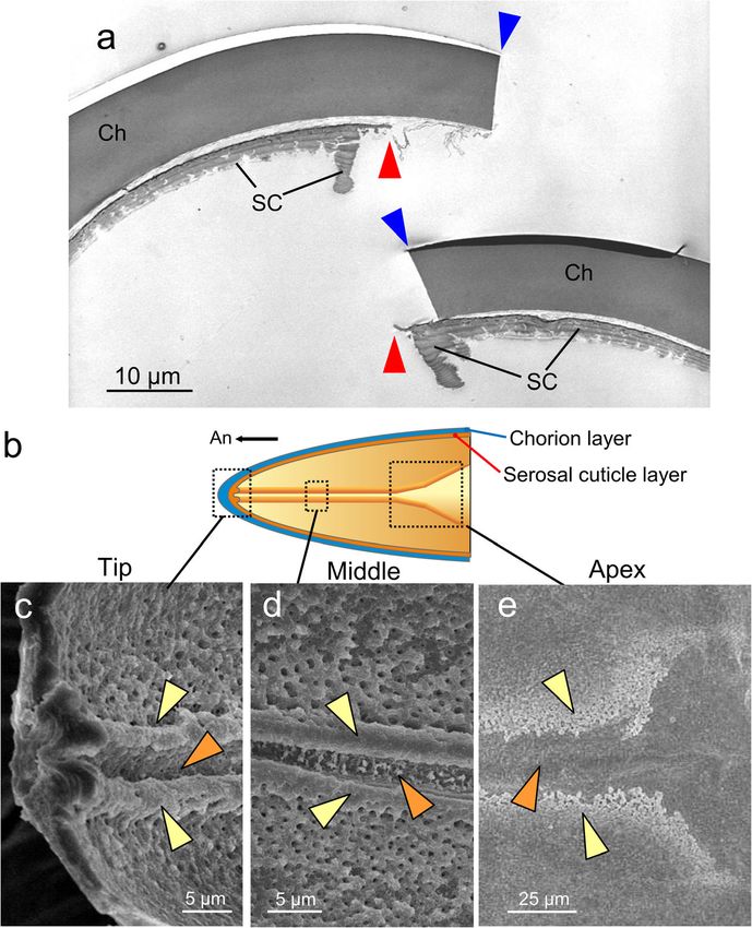
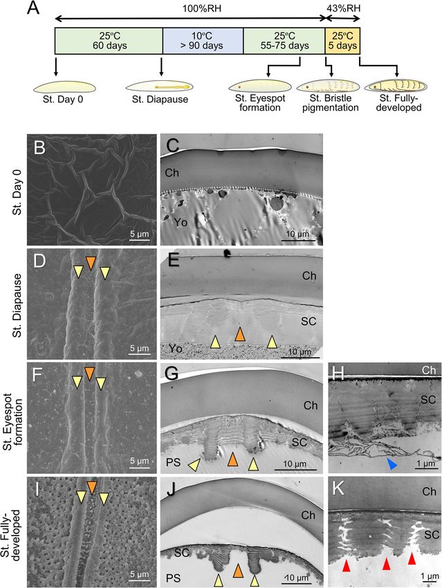
Moriyama et al. Zoological Letters   (2021) 7:8                                                              Page 2 of 13

respiration, but the ability of eggshells to perform it be-   peculiar structure rarely seen in insect eggs: a structure
comes a cause of water loss because of the small mo-          resembling a hatching line was built into the serosal cu-
lecular size of water [4, 5]. Many insects need to absorb     ticle. We further investigated the morphogenesis of this
environmental water in a liquid or vaporous state during      structure along with accompanying changes in eggshell
embryogenesis [6–9], and in some species, nutrients may       properties. The findings suggested that this peculiar
be absorbed from the surrounding substrate [10, 11]. In       structure contributes to flexible changes in eggshell
addition, robust eggshells can be a physical obstacle         properties that aid in fulfilling various conflicting de-
when neonates emerge from them [12, 13]. These de-            mands faced by cicada eggs.
mands favor permeable and fragile eggshells, although
such properties necessarily impair the protective func-       Methods
tions of the shell. Therefore, tradeoffs between these        Collection and maintenance of cicada eggs
properties have evolved and diversified under species-        Adult females of C. facialis were collected on the cam-
specific selection pressure [14, 15] and have been fine-      pus of Osaka City University, Osaka, Japan. Cryptotym-
tuned along with specific life cycle strategies even within   pana facialis normally lays eggs in dead twigs of woody
individual species [16–18]. In addition, innovative adap-     plants (Fig. 1a) [33]. In order to obtain loose eggs, the
tations have evolved in insect eggshells to make these        collected adult females were wrapped in a wet paper
conflicting demands compatible. For example, the chor-        towel as described previously [34]. Eggs laid in the folds
ion layer often possesses aeropyles or air cavities, which    of the paper towel were picked out every day and put in
restrict the surface area to minimize water loss while        a plastic Petri dish (6 cm diameter and 1 cm depth).
maximizing gas exchange capacity [1, 2]. Chorionic ex-        They were kept in a sealed plastic container (13 cm
tensions, such as respiratory horns and appendages,           diameter and 9 cm depth) humidified with pieces of wet
reinforce gas exchange, especially in air-limited environ-    cotton and maintained at 25 °C under a photoperiod of
ments [1, 19]. To facilitate eggshell rupture at hatching,    16 h of light and 8 h of dark (16L8D). After a 60-day in-
eggs of many species possess a special region of weak-        cubation under these conditions, all eggs reached the
ness on the chorion, called the operculum or hatching         diapause stage, and embryogenesis stopped [34]. Then,
line [2, 20–22]. During embryogenesis, the serosal cu-        embryonic diapause was terminated by keeping the eggs
ticle can be positioned beneath the chorion layer and         at 10 °C under a 12L12D photoperiod for more than 90
augment or replace functions of the eggshell [23, 24].        days. Post-diapause embryogenesis was resumed by in-
This additional membrane can confer superior desicca-         cubating the eggs at 25 °C under a 16L8D photoperiod.
tion tolerance [25–27]. In some species, the specialized      In order to prevent hatching of fully developed embryos
serosa and serosal cuticle, called the hydropyle, offer a     at high humidity, eggs containing embryos that devel-
route for water absorption [28–30].                           oped pigmented eyespots and bristles were transferred
   In this study, we present a unique feature that is found   to 43% relative humidity (RH) conditions [39], which
in eggshells of cicadas and enables these insects to meet     were established using a saturated potassium carbonate
the conflicting demands of their embryonic lives. Cicada      solution [40]. For the experiments utilizing post-
eggs are laid into a small hole made in plant tissues by a    diapause stages, eggs retrieved from the field-collected
spear-shaped ovipositor [31–33]. A remarkable feature         twigs were also used.
of the eggs of temperate cicadas is the extremely long
duration of the egg stage; in some species, the egg period    Eggshell observation using electron microscopy
reaches up to 12 months [31, 34, 35]. Thus, the eggs are      For scanning electron microscopy (SEM), eggs were
expected to face a sequence of environmental stresses         fixed with 4% paraformaldehyde solution containing 1%
over the course of the seasons. Another unique trait of       glutaraldehyde and were then treated with 1% osmium
cicada eggs is that even after the completion of embryo-      tetroxide. The samples were dehydrated using a series of
genesis, nymphs remain in the eggshell, rapidly emerging      ethanol solutions and finally treated with isoamyl acet-
from it in response to high humidity cues derived from        ate. After the samples were subjected to critical-point
rain [36, 37]. We speculated that such unique life cycle      drying and platinum coating (10–15 nm), the eggshells
traits impose severe demands for robustness and perme-        were observed with an SEM apparatus (JEOL, JSM-
ability on cicada eggshells and can be associated with a      6340F). For transmission electron microscopy (TEM),
functional specialization of eggshell structures to meet      eggs were fixed and dehydrated as described above.
these demands. In this study, we first examined the           Then, they were treated with propylene oxide and em-
ultrastructure of the eggshell during the course of the       bedded in an epoxy resin (LUVEAK-812). Ultrathin sec-
prolonged egg period in Cryptotympana facialis (Fig. 1a),     tions (90 nm) were prepared with an ultramicrotome
whose embryonic development and hatching traits have          (Leica, Ultracut), stained with uranyl acetate and lead
previously been investigated [34–38]. Then, we found a        citrate, and observed with an H-7100 (Hitachi). For
Moriyama et al. Zoological Letters      (2021) 7:8                                                                                     Page 3 of 13

 Fig. 1 Eggs of Cryptotympana facialis. a An adult female of C. facialis laying eggs in a dead twig. b An egg on the day of oviposition. c An egg
 with a fully developed embryo. An orange broken line indicates the site of eggshell cleavage (hatching line), and an orange arrow indicates the
 cleavage direction. Crossed arrows indicate the anterior (An) and dorsal (Do) directions

thickness measurement, we obtained transverse sections                      min. Next, they were reacted with Schiff’s reagent for 30
of eggshells 180–200 μm and 600–620 μm from the                             min and rinsed with a solution of 0.5% sodium pyrosul-
anterior tip. The precise position of these sections was                    fite in 50 mN HCl. The stained eggs were dehydrated
obtained from the cumulative counter of the ultramicro-                     using a series of ethanol solutions and finally transferred
tome. The thicknesses of the chorion and serosal cuticle                    to methyl salicylate for microscopic observation.
were calculated from analyses of TEM images using
Photoshop (6.0, Adobe).                                                     Measurement of eggshell permeability
                                                                            As a measure of eggshell permeability, we assessed
Nuclear staining                                                            water-loss rates under dry conditions. Eggs at various
To observe the localization of the serosal cells, we per-                   developmental stages were transferred from the moist-
formed whole-mount nuclear staining using the Feulgen                       ened chamber to a dry container adjusted to 43% RH
reaction [41]. The eggs were fixed with Carnoy’s fixative                   using a saturated potassium carbonate solution [39].
at 60 °C for 30 min. After being washed with 70% etha-                      Water-loss rates were calculated by measuring wet
nol, the eggs were hydrated and then subjected to hy-                       weights with an electronic microbalance (Mettler-To-
drolysis in 1 N hydrochloric acid (HCl) at 60 °C for 30                     ledo, MT5) before and after the desiccation treatments;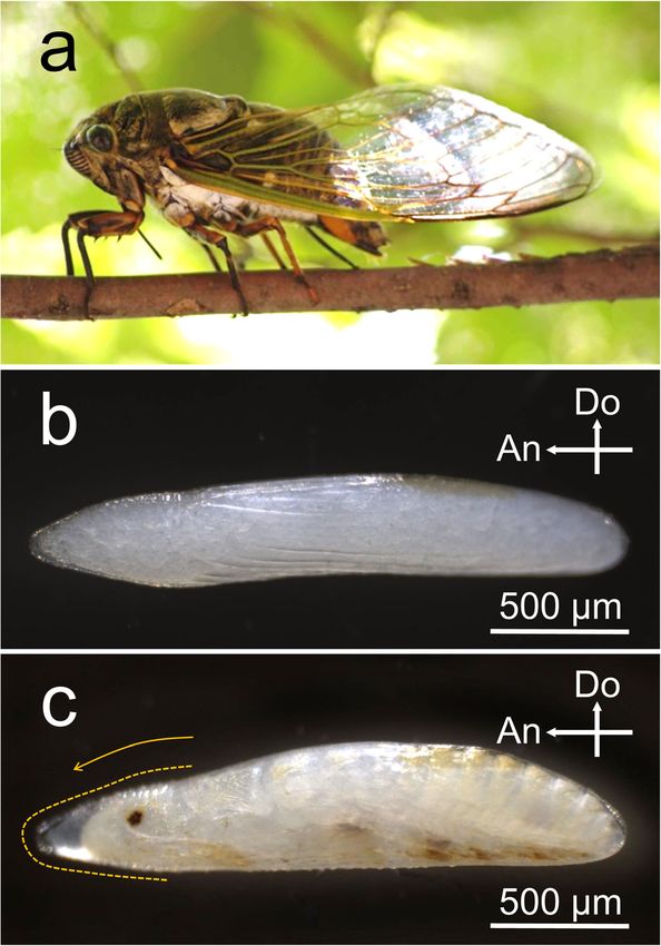
Moriyama et al. Zoological Letters   (2021) 7:8                                                                  Page 4 of 13

these quantities were expressed as weight loss per egg          a smooth surface with polygonal shapes (Fig. 2b), while
per day. In order to ensure accuracy in weighing, 10 eggs       the posterior region was covered with numerous tiny
were weighed collectively.                                      holes in addition to horizontal wrinkles with small knobs
                                                                (Fig. 2a, c). We did not find any distinct signs on the
Measurement of eggshell cleavability                            chorion surface at the future cleavage site (Fig. 2b).
We constructed a force-gauge system to measure the              TEM observation of the transverse sections revealed that
cleavability of the eggshells (Fig. S1). Pressing eggs from     the thick chorion layer was composed of a homoge-
outside generates turgor pressure, resulting in eggshell        neously solid substance in both the anterior and poster-
rupture and extrusion of the embryo. Eggs at various de-        ior regions (Fig. 2d, e). Tiny holes found in the posterior
velopmental stages were placed in the center of a small         part did not penetrate the chorion layer (Fig. 2e). In con-
chamber (42 mm × 60 mm × 17 mm) and fixed to the                trast to the chorion of many other insects [1, 2], no in-
bottom with double-sided adhesive tape. The center of           ternal air spaces or cave-like structures were found. The
the chamber lid had a hole (φ11 mm) to enable penetra-          transverse sections also verified that the eggshell had no
tion of a pushing probe attached to a digital force gauge       obvious hatching line structure at the time of oviposition
(Aikoh, RZ-1). The contact probe was made of resin and          (Fig. 2d).
had a cylindrical body (φ10 mm) with a columnar tip
(φ0.4 mm, Fig. S1). The probe was lowered vertically            Hatching line in the serosal cuticle
through the lid hole and gently pressed against the dor-        Next, we observed the fine structure of the cleavage site
sal center of the egg. The load values that caused egg-         in hatched eggshells (Fig. 3a). The chorion was abruptly
shell rupture were recorded. When the pushing force             broken out without any visible modification. In this
exceeded 1000 mN, the pressing was stopped because              stage, however, beneath the chorion layer, there was an
further pressing was predicted to cause the egg to burst        extraembryonic cuticular layer, namely, the serosal cu-
rather than cleave along the hatching line.                     ticle. The cleavage site in the serosal cuticle was remark-
                                                                ably thin and possessed protruding cuticular structures
Statistics                                                      on both sides. This ridge-and-furrow structure extended
All statistical analyses were performed using R (v 3.6.3)       along the midline of the anterior eggshell in accordance
[42]. We performed statistical analyses based on a gener-       with the cleavage line (Fig. 3b-e). At the dorsal apex site,
alized linear model (GLM) framework. A Gaussian error           the structure became wider and shallower and gradually
distribution and an inverse-Gaussian error distribution         disappeared (Fig. 3e). These observations indicated that
were employed for eggshell thickness and water-loss             the ridge-and-furrow structure in the serosal cuticle de-
rates, respectively. In order to assess the effects of devel-   fines the hatching line of C. facialis.
opmental stages on eggshell cleavability, a nonparamet-
ric test for multiple comparisons, the Steel-Dwass test,        Morphogenesis of the hatching line in the serosal cuticle
was selected. In this test, eggs that did not rupture under     To explore the process of formation of this peculiar
1000 mN pressure or that burst anywhere other than the          structure, we surveyed eggshells across various embry-
hatching line were assumed to have a maximum value              onic stages, as shown in Fig. 4a. As seen in Fig. 2, no se-
(1000 mN) and included in the tests.                            rosal cuticle was present on the day of oviposition (Fig.
                                                                4b, c). The deposition of the serosal cuticle was identifi-
Results                                                         able approximately 15–20 days after oviposition. By the
Eggshell of C. facialis                                         diapause stage, in which embryonic development is
The eggshell of C. facialis was colorless and transparent,      arrested at the early germ-band stage [34], a thick, la-
and the yolk granules and developing embryos were vis-          mellar serosal cuticle had formed beneath the chorion
ible from the outside (Fig. 1b, c). As embryogenesis pro-       layer (Fig. 4d, e). Interestingly, the future cleavage site
ceeded, the eggs became somewhat swollen at the                 (furrow) was covered with a thick cuticle layer, and the
posterior end by absorbing water (Fig. 1c), as reported in      future ridge positions slightly bulged. During post-
other cicada eggs [43]. At the time of hatching, the egg-       diapause development, the inside of the serosal cuticle
shell was always cleaved at a certain site that was located     layer was partially scraped away, and the furrow-like
on the midline of the anterior one-fourth of the eggshell       structure appeared (Fig. 4f, h). As the completion of em-
(Fig. 1c, orange broken line). Cleavage normally started        bryonic development approached, the cuticle layer was
from the dorsal apex and extended toward the ventral            further reduced, especially in the furrow position,
terminal via the tip region. SEM observation revealed           whereas the ridge positions persisted, and as a result,
that the anterior one-fourth of the eggshell has a distinct     distinct ridge and furrow structures were shaped (Fig. 4i,
surface structure different from that of the posterior re-      j). In this stage, many pores that penetrated the serosal
gion (Fig. 2a). The anterior region was characterized by        cuticle were observed, especially around the hatching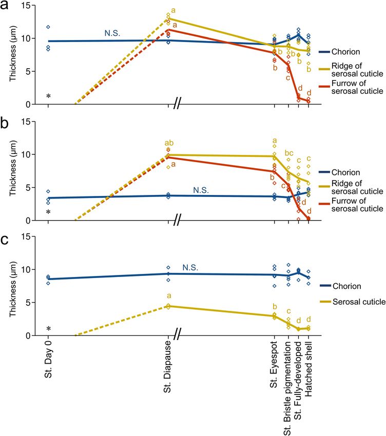
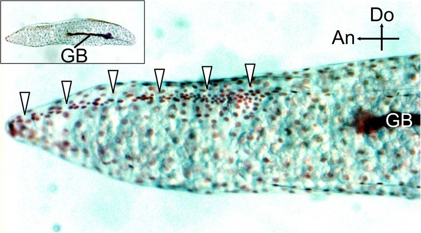
Moriyama et al. Zoological Letters      (2021) 7:8                                                                                     Page 5 of 13

 Fig. 2 Fine structure of the eggshell on the oviposition date. a A whole SEM image of an eggshell. Arrows indicate the anterior (An) and dorsal
 (Do) directions. b A magnified SEM image of the dorsal middle region of the anterior part, which contains the future cleavage site. c An SEM
 image of the lateral region in the posterior part. d A TEM image of (b). e A TEM image of (c). In this sample, the egg contents have been
 removed (*). Abbreviations: Ch, chorion; VM, vitellin membrane: Yo, yolk

line (Fig. 4k). The developmental changes in the                             These results demonstrated that the structure of the se-
thickness of the chorion and serosal cuticle were                          rosal cuticle dynamically changes during embryogenesis
quantified at the middle dorsal site (Fig. 5a) and the                     and that the hatching line on the serosal cuticle is shaped
dorsal apex site (Fig. 5b) within the hatching line and                    by cutting from a thick cuticle layer rather than by casting
at a lateral site away from the hatching line (Fig. 5c).                   ab initio. A base for the ridge-and-furrow structure was
The thickness of the chorion in these three positions                      embedded in advance during the formation of the thick
did not change across developmental stages. The se-                        cuticle. In the serosal cuticle layer at the diapause stage,
rosal cuticle reached its maximum thickness at the                         the outlines of the developing ridge-and-furrow structure
diapause stage. The relatively thick cuticle layer per-                    were distinguishable as slightly electron-dense regions
sisted until approximately the time of eyespot forma-                      (Fig. 4e, orange and yellow arrowheads). In addition, at
tion, but thereafter, the rate of cuticle degradation                      the stage of serosal cuticle deposition (20 days after ovi-
was accelerated toward the end of embryogenesis, es-                       position), the serosal cells were densely gathered along the
pecially in the furrow position. At the time of hatch-                     future hatching line (Fig. 6), suggesting that they were pre-
ing, the serosal cuticle at the furrow position was                        paring special architectures in this stage.
reduced to less than 5% of maximum thickness,
whereas the ridge positions retained approximately                         Functional change during the formation of the hatching
60% of maximum thickness. Degradation of the se-                           line
rosal cuticle took place throughout the eggshell and                       Do these dynamic morphological changes in the serosal
was not restricted to the hatching line (Fig. 5c).                         cuticle lead to functional consequences? As an index of
Moriyama et al. Zoological Letters      (2021) 7:8                                                                                     Page 6 of 13

 Fig. 3 The hatching line in the serosal cuticle. a A TEM image of the anterodorsal cleavage site of a hatched eggshell. Blue and red arrowheads
 indicate the cleavage sites in the chorion and serosal cuticle, respectively. b A schematic representation of the inner surface structure of the
 anterodorsal part of the eggshell. c-e Interior SEM images of the eggshell of the fully developed eggs at the represented sites shown in (b). A
 furrow (orange arrowheads) flanked by two ridges (yellow arrowheads) runs along the midline of the anterior part. Abbreviations: Ch, chorion; SC,
 serosal cuticle

eggshell permeability, we assessed the changes in water-                   increased in tandem with cuticle degradation (see Fig.
loss rates with serosal cuticle degradation. Weight-loss                   5), and the desiccation treatment caused 7.0 μg (2.1%)
rates during a 5-day desiccation period at 43% RH were                     and 12.1 μg (3.7%) loss/day in the eyespot formation
compared among eggs in the diapause, eyespot forma-                        stage and the bristle pigmentation stage, respectively.
tion, and bristle pigmentation stages. It should be noted                    Next, we investigated the changes in the cleavability of
that eggs in the diapause stage had heavier initial                        the eggshell. In this experiment, we measured the push-
weights because the eggs absorbed water during the                         ing force necessary to generate sufficient turgor pressure
early stages of the post-diapause development (Fig. 7a).                   for the hatching line to rupture (see Methods and Fig.
Water-loss rates in the diapause stage, in which the se-                   S1 for details). In the eyespot formation stage, the hatch-
rosal cuticle reached maximum thickness, stayed under                      ing line was rarely cleaved by less than 1000 mN of force
2.2 μg/day (comparable to 0.8% of total egg weight/day).                   on the egg (Fig. 7b). At the bristle pigmentation stage,
As embryogenesis proceeded, water-loss rates drastically                   the majority of eggshells were cleaved at the hatching
Moriyama et al. Zoological Letters      (2021) 7:8                                                                                     Page 7 of 13

 Fig. 4 The developmental process of hatching-line formation in the serosal cuticle. a Time course of embryonic development and sampling
 points. b, d, f, i SEM images of the inner surface around the dorsal hatching line at each developmental stage. c, e, g, j Transverse TEM images
 of the area around the dorsal hatching line. Orange and yellow arrowheads indicate furrow and ridge positions, respectively. h A magnified
 image of the degrading serosal cuticle at the stage of eyespot formation (blue arrowhead). k A magnified image of pores that appeared in fully
 developed eggs (red arrowheads). Abbreviations: Ch, chorion; SC, serosal cuticle; PS, periembryonic space; Yo, yolk

line when compressed, but the necessary amount of                           Discussion
force was still high. In the fully developed eggs with                      The hatching line in the serosal cuticle
complete ridge-and-furrow structures, the hatching line                     To facilitate the safe hatching of neonates, many insect
became fragile enough to be cleaved by a force of less                      eggshells have special structures, known as hatching lines
than 500 mN. These results demonstrated that eggshell                       and opercula, that create weakness at the site to be sepa-
properties such as robustness and permeability dynamic-                     rated [1, 2]. These structures are usually part of the mater-
ally changed in association with hatching-line formation                    nally derived chorion layer and are therefore carried
accompanying serosal cuticle degradation.                                   throughout the egg period. In the present study, we found
Moriyama et al. Zoological Letters       (2021) 7:8                                                                                        Page 8 of 13

 Fig. 5 Developmental changes in eggshell thickness during embryogenesis. The thickness of the chorion and the serosal cuticle was measured
 on the dorsal hatching line 180–200 μm (a) or 600–620 μm (b) from the tip or at a lateral non-hatching-line site 180–200 μm from the tip (c). The
 horizontal axis indicates stages of embryonic development with a scale reflecting typical developmental time (see Fig. 4a), except that the period
 of low temperature needed for diapause termination is not included (//). Lines connect the mean values of each point in development, and
 diamonds show individual values (N = 3–5). No serosal cuticle was present on Day 0 (*). Although the cuticle layer became recognizable at
 approximately Day 15–20, we did not quantify the thickness change until the start of diapause (broken line). Different letters indicate statistically
 significant differences among developmental stages (likelihood ratio test of a generalized linear model, P < 0.05), while N.S. indicates no
 significance (P > 0.05)

that cicada eggs had a hatching line in the serosal cuticle,                  but the fine structure and developmental process of this
whereas no obvious weakness-causing structures were                           hatching line have not been investigated. Here, we found
found in the chorion. The serosal cuticle, a membrane                         that the hatching line of cicada eggs is composed of a fine
made of chitin and proteins, is deposited beneath the chor-                   furrow accompanied by two ridges, one on each side (Fig.
ion by the extraembryonic serosa during embryonic devel-                      3). We also demonstrated that the ridge-and-furrow pat-
opment [23, 24, 44, 45]. This secondary cuticle layer is                      tern is excavated from an initially thick lamellar cuticle ra-
commonly observed in most insect taxa and is considered                       ther than being piled up on a thin base layer (Figs. 4, 5).
to augment or replace the protective functions of the chor-                   The degradation of the serosal cuticle occurred to some
ion. Until now, the construction of hatching lines in the se-                 extent during the middle to late phase of embryonic devel-
rosal cuticle was reported only in a carabid beetle, Carabus                  opment after diapause termination. However, substantial
insulicola [46]. In that beetle, picric acid–stainable lines in               excavation of the hatching line occurred in a relatively
the serosal cuticle were found to become the rupture site,                    short time at the terminal phase of embryogenesis (Fig. 5).
Moriyama et al. Zoological Letters        (2021) 7:8                                                                                        Page 9 of 13

 Fig. 6 Prior casting of the hatching line by serosal cells during early embryogenesis. Nuclei in the germ-band (GB) elongation stage (20 days after
 oviposition) were visualized by Feulgen staining. An image of the whole egg is displayed in the inset. The nuclei of serosal cells were distinctly
 gathered along the developing hatching line (arrowheads). Crossed arrows indicate the anterior (An) and dorsal (Do) directions

  Degradation of the serosal cuticle toward the comple-                        the following embryonic stages [53]. This suggests that
tion of embryogenesis has been explicitly described in                         the pleuropodia are unlikely to be the main structures
some orthopterans [12, 47] and coleopterans [48].                              involved in serosal cuticle degradation in cicadas. It has
Chitin-degrading enzymes, known as hatching enzymes,                           been reported that hatching enzymes affect the inner
are believed to be secreted from the pleuropodia [12, 49,                      but not the outer layers of the multilayered serosal cu-
50] or serosa [51, 52]. The pleuropodia of a periodical ci-                    ticle, which have distinctly different properties of color
cada, Magicicada cassini, develop in mid-embryonic                             and stainability [12, 48]. Although all layers of the se-
stages around the time of katatrepsis and degenerate in                        rosal cuticle of C. facialis eggs are colorless and

 Fig. 7 Dynamic changes in eggshell properties with serosal cuticle degradation. a Change in water-loss rates. Box plots indicate water-loss rates
 after 5 days of exposure to 43% relative humidity conditions; these rates were assessed gravimetrically in eggs at various developmental stages.
 The initial weights at each developmental stage are also shown by line plots. Different letters indicate statistically significant differences (N = 12
 samples (each containing 10 eggs), likelihood ratio test of a generalized linear model, P < 0.05). b Change in hatching-line cleavability. The
 pushing force needed to cause a rupture of the hatching line was measured. The number of eggs that were not ruptured by less than 1000 mN
 and the number that burst in locations other than the hatching line are shown in the rightmost bars. Median values for each developmental
 stage are indicated by red triangles, and different letters indicate significant differences (Steel-Dwass test, P < 0.05)
Moriyama et al. Zoological Letters   (2021) 7:8                                                               Page 10 of 13

transparent, we found that the developing furrow-and-         invagination (Fig. 6), as is typical in hemimetabolous in-
ridge structure embedded in the thick lamellar cuticle is     sects [58–60], and the thick cuticle layer eventually
distinguishable by TEM observation at the time of cu-         formed by the time the egg reached the diapause stage.
ticle deposition. This implies that the hatching line is      We verified that water-loss rates, as a measure of egg-
made of qualitatively different materials resistant to di-    shell permeability, remained at remarkably low levels in
gestion by hatching enzymes.                                  this stage compared to the later stages, when the cuticle
   Although the exact roles of the furrow-and-ridge           was degraded (Fig. 7a). Because of low metabolic de-
shape of the hatching line are not clear at present, we       mand during diapause, the production of thick, imper-
speculate about its contribution to hatching. First, we       meable eggshells is likely to be an adaptation against
could not find any sign of weakness in the chorion layer      water loss and penetration of pathogens during pro-
throughout the egg period. Nevertheless, the chorion          longed developmental periods at the expense of oxygen
always ruptures along the hatching line of the serosal cu-    supply. This idea is supported by the finding that
ticle at hatching. We observed that, except during the        diapause-destined eggs have eggshells with lower perme-
terminal phase of embryogenesis, pressing the egg with a      ability to oxygen and water than non-diapause-destined
strong force caused the eggshell to rupture in locations      eggs in grasshoppers [6, 61] and silkworms [16].
other than the hatching line, even before serosal cuticle       In the late phase of embryogenesis, the oxygen re-
formation. This implies that the chorion around the           quirement increases to support development and neo-
hatching line also becomes fragile toward the comple-         nate activity [5], and a tough eggshell can be an obstacle
tion of embryogenesis. Therefore, the furrow part of the      to hatching [12]. The degradation of the serosal cuticle
hatching line serves as a site of weakness itself and may     and the excavation of the furrow-and-ridge structure in
also affect the chorion by secreting unknown effectors.       C. facialis appear to fulfill these demands. The perme-
In fact, the serosal cuticle around the hatching line was     ability of the eggshell was shown to increase toward
observed to possess numerous fine canals (Fig. 4i-k),         hatching (Fig. 7a). We also demonstrated that eggshells
which might potentially serve as a route of secretion. On     underwent a physical change to facilitate rupture at the
the other hand, the ridge structure may support the abil-     hatching line (Fig. 7b). Taken together, our findings sug-
ity of nymphs to cleave the eggshell. At the time of          gest that the formation of the hatching line in the serosal
hatching, pharate nymphs that bear an embryonic cu-           cuticle enables the eggshell to change its properties flex-
ticle, called pronymphs, start forward movement by ver-       ibly in order to reconcile conflicting demands during
micular motion, but they lack any apparent structure          embryogenesis, i.e., to be impermeable during early to
with which to open the eggshell, for example, a structure     mid-embryogenesis, including diapause, and permeable
resembling an egg burster or egg tooth [13, 20, 54]. The      or fragile in the late embryonic phase.
two ridges of the serosal cuticle are presumed to receive
the force applied by the movement of a nymph and ef-          Evolution of the hatching line in the serosal cuticle of
fectively focus it on the cleavage line; otherwise, the       cicadas
force might disperse across the eggshell.                     One question that our findings raise is why this unique
                                                              structure has evolved in cicadas. In carabid eggs, the
The hatching line in the serosal cuticle allows dynamic       other known example, the chorion is sometimes peeled
changes in eggshell properties                                off and replaced with the serosal cuticle [46]. Therefore,
The complex process of hatching-line formation in C.          the situation would not be the same as in the cicada
facialis may provide an advantage in changing the prop-       eggs, in which the robust chorion persists until hatching.
erties of the eggshell during embryogenesis. Modulation       The primary reason in cicadas may be related to the ex-
of eggshell functions by production and degradation of        tremely long egg period. The egg period of temperate ci-
the serosal cuticle has been demonstrated in several in-      cadas that overwinter in embryonic diapause typically
sect species [24]. The underlying serosal cuticle has been    ranges from 9 to 12 months, while a period of 1–3
shown to reduce the water and oxygen penetrability of         months is still needed even in cicada species without
eggshells in coleopteran and lepidopteran species [16,        embryonic diapause [31, 35]. Our previous study ad-
55, 56]. Desiccation tolerance in mosquito eggs is            dressing C. facialis embryonic development revealed that
attained concomitantly with serosal cuticle deposition        its prolonged egg duration is attributable not only to a
[25–27]. Moreover, a clear contribution of the serosal        long overwintering diapause but also to an extraordinar-
cuticle to desiccation tolerance was experimentally dem-      ily slow rate of embryonic development [34]. Therefore,
onstrated in the red flour beetle, Tribolium castaneum,       the oxygen demand during the non-diapause period is
by RNAi-based knockdown of genes involved in serosal          predicted to be low compared to that of other insects, al-
cuticle synthesis [52, 57]. In C. facialis eggs, deposition   though it would decrease further in the diapause period,
of the serosal cuticle could be observed after germ-band      as mentioned above. If a hatching line is formed in the
Moriyama et al. Zoological Letters (2021) 7:8 Page 11 of 13
chorion, although its area is limited, it can be a nonne- Authors’ contributions
gligible cause of water loss or pathogen intrusion for MM carried out all of the experiments and statistical analyses; KY supervised
the SEM and TEM observation; MM and HN designed the study and wrote
eggs with extremely long egg periods. We found that ci- the manuscript. All authors read and approved the final manuscript.
cada eggs do not bear chorionic structures specialized
for gas exchange, such as aeropyles and air cavities, Funding
which are common to a diverse range of insects [1, 2, This study was supported by KAKENHI Grant Number 18657027 (to HN and
KY) and, in part, by 17 K15399 and 19H02973 (to MM). The funding bodies
20]. This suggests that cicadas favor eggshells serving as played no role in the design of the study; the collection, analysis, and
a robust barrier at the expense of gas exchange. On the interpretation of data; or the writing of the manuscript.
other hand, some cicada species, including C. facialis,
Availability of data and materials
need to rapidly emerge from the eggshell in response to
The datasets used and/or analyzed during the current study are available
rain to ensure that the nymphs can burrow into wet, soft from the corresponding author on reasonable request.
ground [36–38]. These opposite needs at hatching ver-
sus earlier stages may be fulfilled with a robust shell by Declarations
locally varying the destructibility of the serosal cuticle, Ethics approval and consent to participate
rather than the chorion, to form a hatching line. Not applicable.
In this study, we focused on the eggshell of C. facialis,
which undergoes embryonic diapause and humidity- Consent for publication
Not applicable.
inducible hatching. In contrast to C. facialis, some tem-
perate cicada species use a strategy of hatching within Competing interests
the oviposition year, without overwintering in diapause The authors declare that they have no competing interests.
[31, 53, 62, 63]. In addition, although C. facialis lays eggs
Author details
in dead twigs [33], some other cicadas choose live plant 1
National Institute of Advanced Industrial Science and Technology (AIST),
tissues for oviposition [64, 65]. Their eggs appear to en- Tsukuba 305-8566, Japan. 2Graduate School of Science, Osaka City University,
counter various moist environments and are unlikely to Osaka 558-8585, Japan. 3Kawasaki Medical School, Kurashiki 701-0192, Japan.
4
Kawasaki University of Medical Welfare, Kurashiki 701-1093, Japan. 5Graduate
hatch in response to humidity cues, implying that they School of Science, Kyoto University, Kyoto 606-8502, Japan.
might face different types of adaptive demands than C.
facialis. Such variation in oviposition site selection is Received: 16 February 2021 Accepted: 30 April 2021
known even within the genus Cryptotympana [64, 66].
Therefore, to clarify the adaptive significance and evolu- References
tionary origin of hatching lines in the serosal cuticle, 1. Hinton HE. Biology of insect eggs. Oxford: Pergamon Press; 1981.
comparative studies must examine these cicada species 2. Margaritis LH. Structure and physiology of the eggshell. In: Kerkut GA,
Gilbert LI, editors. Comprehensive insect physiology biochemistry and
with various diapause and hatching traits and address pharmacology, vol. 1. Oxford: Pergamon Press; 1985. p. 153–230.
other Auchenorrhyncha species. 3. Zeh DW, Zeh JA, Smith RL. Ovipositors, amnions and eggshell architecture
in the diversification of terrestrial arthropods. Q Rev Biol. 1989;64(2):147–68.
https://doi.org/10.1086/416238.
4. Hinton HE. Respiratory systems of insect egg shells. Annu Rev Entomol.
Conclusions 1969;14(1):343–68. https://doi.org/10.1146/annurev.en.14.010169.002015.
This study demonstrated that the development of a pe- 5. Woods HA. Water loss and gas exchange by eggs of Manduca sexta: trading
off costs and benefits. J Insect Physiol. 2010;56(5):480–7. https://doi.org/10.1
culiar structure in the serosal cuticle of Cryptotympana 016/j.jinsphys.2009.05.020.
facialis enables flexible modification of eggshell proper- 6. Browning TO. Permeability to water of the shell of the egg of Locusta
ties during the long embryonic period. These findings migratoria migratorioides, with observations on the egg of Teleogryllus
commodus. J Exp Biol. 1969;51(1):99–105. https://doi.org/10.1242/jeb.51.1.99.
reveal a novel mode of environmental adaptation 7. Browning TO. The permeability of the shell of the egg of Teleogryllus
through sophisticated insect eggshells. commodus measured with the aid of tritiated water. J Exp Biol. 1969;51(2):
397–405. https://doi.org/10.1242/jeb.51.2.397.
8. Yoder JA, Denlinger DL. Water vapour uptake by diapausing eggs of a
tropical walking stick. Physiol Entomol. 1992;17(1):97–103. https://doi.org/1
Supplementary Information 0.1111/j.1365-3032.1992.tb00995.x.
The online version contains supplementary material available at https://doi. 9. Niikawa K, Takeda M. Water absorption by diapause and nondiapause eggs
org/10.1186/s40851-021-00178-8. in two Velanfictorus species (Orthoptera: Gryllidae). Appl Entomol Zool.
1996;31(1):105–10. https://doi.org/10.1303/aez.31.105.
Additional file 1: Fig. S1. A schematic illustration of the system to 10. Rotheram S. The surface of the egg of a parasitic insect. I. The surface of the
measure hatching linebreaking forces. egg and first instar larva of Nemeritis. Proc R Soc London - Biol Sci. 1973;183:
179–94.
11. Buckner JS, Freeman TP, Ruud RL, Chu CC, Henneberry TJ. Characterization
and functions of the whitefly egg pedicel. Arch Insect Biochem Physiol.
2002;49(1):22–33. https://doi.org/10.1002/arch.10006.
Acknowledgments 12. Slifer EH. The origin and fate of the membranes surrounding the
We would like to thank Taiji Suda for his technical support with electron grasshopper egg; together with some experiments on the source of the
microscopy. We also thank Elizabeth Nakajima for linguistic corrections. hatching enzyme. J Cell Sci. 1937;s2–79:493–506.Moriyama et al. Zoological Letters (2021) 7:8 Page 12 of 13
13. Pérez-de la Fuente R, Engel MS, Azar D, Peñalver E. The hatching 35. Moriyama M, Numata H. Ecophysiological responses to climate change in
mechanism of 130-million-year-old insects: an association of neonates, egg cicadas. Physiol Entomol. 2019;44(2):65–76. https://doi.org/10.1111/phen.12283.
shells and egg bursters in Lebanese amber. Palaeontology. 2019;62:547–59. 36. Moriyama M, Numata H. Induction of egg hatching by high humidity in the
14. Woods HA, Singer MS. Contrasting responses to desiccation and starvation cicada Cryptotympana facialis. J Insect Physiol. 2006;52(11-12):1219–25.
by eggs and neonates of two Lepidoptera. Physiol Biochem Zool. 2001; https://doi.org/10.1016/j.jinsphys.2006.09.005.
74(4):594–606. https://doi.org/10.1086/322169. 37. Moriyama M, Numata H. A cicada that ensures its fitness during climate
15. Jagadeeshan S, Singh RS. Rapid evolution of outer egg membrane proteins warming by synchronizing its hatching time with the rainy season. Zool Sci.
in the Drosophila melanogaster subgroup: a case of ecologically driven 2011;28(12):875–81. https://doi.org/10.2108/zsj.28.875.
evolution of female reproductive traits. Mol Biol Evol. 2007;24(4):929–38. 38. Moriyama M, Numata H. Urban soil compaction reduces cicada diversity.
https://doi.org/10.1093/molbev/msm009. Zool Lett. 2015;1(1):19. https://doi.org/10.1186/s40851-015-0022-3.
16. Sonobe H, Matsumoto A, Fukuzaki Y, Fujiwara S. Carbohydrate metabolism and 39. Moriyama M, Numata H. Desiccation tolerance in fully developed embryos of
restricted oxygen supply in the eggs of the silkworm, Bombyx mori. J Insect two cicadas, Cryptotympana facialis and Graptopsaltria nigrofuscata. Entomol
Physiol. 1979;25(5):381–8. https://doi.org/10.1016/0022-1910(79)90003-9. Sci. 2010;13(1):68–74. https://doi.org/10.1111/j.1479-8298.2010.00365.x.
17. Kim SE. Changes in eggshell permeability to oxygen during the early 40. Winston PW, Bates DH. Saturated solutions for the control of humidity in
developmental stages in diapause eggs of Bombyx mori. J Insect Physiol. biological research. Ecology. 1960;41(1):232–7. https://doi.org/10.2307/1931961.
1987;33(4):229–35. https://doi.org/10.1016/0022-1910(87)90042-4. 41. Lyon HO, Schulte EK, Prento P, Barer MR, Béné MC. Standardized staining
18. Zrubek B, Woods HA. Insect eggs exert rapid control over an oxygen-water methods: Feulgen-Rossenbeck reaction for desoxyribonucleic acid and
tradeoff. Proc R Soc B Biol Sci. 2006;273(1588):831–4. https://doi.org/10.1 periodic acid-Schiff (PAS) procedure. Biotech Histochem. 2002;77(3):121–5.
098/rspb.2005.3374. https://doi.org/10.1080/bih.77.3.121.125.
19. Margaritis LH, Kafatos FC, Petri WH. The eggshell of Drosophila melanogaster. 42. R Core Team. R: A language and environment for statistical computing. R
Fine structure of the layers and regions of the wild-type eggshell. J Cell Sci. Foundation for Statistical Computing, Vienna, Austria. https://www.R-project.
1980;43(1):1–35. https://doi.org/10.1242/jcs.43.1.1. org/. Accessed 15 Feb 2021.
20. Cobben RH. Evolutionary trends in Heteroptera part I: eggs, architecture of 43. White J, Lloyd M. On the stainability and mortality of periodical cicada eggs.
the shell, gross embryology and eclosion. Wageningen: Centre for Am Midl Nat. 1981;1062:219–28.
Agricultural Publishing and Documentation; 1968. 44. Machida R, Ikeda Y, Tojo K. Evolutionary changes in developmental
21. Matesco VC, Fürstenau BBRJ, Bernardes JLC, et al. Morphological features of potentials of the embryo proper and embryonic membranes in hexapoda: a
the eggs of Pentatomidae (Hemiptera: Heteroptera). Zootaxa. 2009;30:1–30. synthesis revised. Proc Arthropod Embryol Soc Japan. 2002;37:1–11.
22. Fukui M, Fujita M, Tomizuka S, Mashimo Y, Shimizu S, Lee CY, et al. Egg 45. Schmidt-Ott U, Kwan CW. Morphogenetic functions of extraembryonic
structure and outline of embryonic development of the basal mantodean, membranes in insects. Curr Opin Insect Sci. 2016;13:86–92. https://doi.org/1
Metallyticus splendidus Westwood, 1835 (Insecta, Mantodea, Metallyticidae). 0.1016/j.cois.2016.01.009.
Arthropod Struct Dev. 2018;47(1):64–73. https://doi.org/10.1016/j.asd.201 46. Kobayashi Y, Niikura K, Oosawa Y, Takami Y. Embryonic development of
7.11.001. Carabus insulicola (Insecta, Coleoptera, Carabidae) with special reference to
23. Machida R. Evidence from embryology for reconstructing the relationships external morphology and tangible evidence for the subcoxal theory. J
of hexapod basal clades. Arthropod Syst Phylogeny. 2006;64:95–104. Morphol. 2013;274(12):1323–52. https://doi.org/10.1002/jmor.20181.
24. Panfilio KA. Extraembryonic development in insects and the acrobatics of 47. Furneaux PJ, James CR, Potter S. The egg shell of the house cricket (Acheta
blastokinesis. Dev Biol. 2008;313(2):471–91. https://doi.org/10.1016/j.ydbio.2 domesticus): an electronmicroscope study. J Cell Sci. 1969;5(1):227–49.
007.11.004. https://doi.org/10.1242/jcs.5.1.227.
25. Rezende GL, Martins AJ, Gentile C, Farnesi LC, Pelajo-Machado M, Peixoto 48. Kobayashi Y. Development of the pleuropodia in the embryo of the
AA, et al. Embryonic desiccation resistance in Aedes aegypti: presumptive glowworm Rhagophthalmus ohbai (Rhagophthalmidae, Coleoptera, Insecta),
role of the chitinized serosal cuticle. BMC Dev Biol. 2008;8(1):82. https://doi. with comments on their probable function. Proc Arthropod Embryol Soc
org/10.1186/1471-213X-8-82. Japan. 2003;38:19–26.
26. Goltsev Y, Rezende GL, Vranizan K, Lanzaro G, Valle D, Levine M. 49. Slifer EH. A cytological study of the pleuropodia of Melanoplus differentialis
Developmental and evolutionary basis for drought tolerance of the (Orthoptera, Acrididae) which furnishes new evidence that they produce
Anopheles gambiae embryo. Dev Biol. 2009;330(2):462–70. https://doi.org/1 the hatching enzyme. J Morphol. 1938;63(1):181–205. https://doi.org/10.1
0.1016/j.ydbio.2009.02.038. 002/jmor.1050630109.
27. Vargas HCM, Farnesi LC, Martins AJ, Valle D, Rezende GL. Serosal cuticle 50. Konopová B, Buchberger E, Crisp A. Transcriptome of pleuropodia from
formation and distinct degrees of desiccation resistance in embryos of the locust embryos supports that these organs produce enzymes enabling the
mosquito vectors Aedes aegypti, Anopheles aquasalis and Culex larva to hatch. Front Zool. 2020;17:1–22.
quinquefasciatus. J Insect Physiol. 2014;62:54–60. https://doi.org/10.1016/j. 51. Zhu Q, Arakane Y, Beeman RW, Kramer KJ, Muthukrishnan S. Functional
jinsphys.2014.02.001. specialization among insect chitinase family genes revealed by RNA
28. Slifer EH. The formation and structure of a special water absorbing area in the interference. Proc Natl Acad Sci U S A. 2008;105(18):6650–5. https://doi.org/1
membranes covering the grasshopper egg. J Cell Sci. 1938;s2–80:437–57. 0.1073/pnas.0800739105.
29. Madhavan MM. Structure and function of the hydropyle of the egg of the 52. Jacobs CGC, Braak N, Lamers GEM, van der Zee M. Elucidation of the serosal
bug, Sphaerodema molestum. J Insect Physiol. 1974;20(7):1341–9. https://doi. cuticle machinery in the beetle Tribolium by RNA sequencing and
org/10.1016/0022-1910(74)90237-6. functional analysis of Knickkopf1, Retroactive and Laccase2. Insect Biochem
30. Mtow S, Machida R. Development and ultrastructure of the thickened Mol Biol. 2015;60:7–12. https://doi.org/10.1016/j.ibmb.2015.02.014.
serosa and serosal cuticle formed beneath the embryo in the stonefly 53. Strauß J, Lakes-Harlan R. Embryonic development of pleuropodia of the
Scopura montana Maruyama, 1987 (Insecta, Plecoptera, Scopuridae). cicada, Magicicada cassini. J Insect Sci. 2006;6(27):1–6. https://doi.org/10.1
Arthropod Struct Dev. 2018;47(6):643–54. https://doi.org/10.1016/j.asd.2018. 673/2006_06_27.1.
09.002. 54. Rakitov R. Pronymphs, hatching, and proboscis assembly in leafhoppers and
31. Beamer R. Studies on the biology of Kansas Cicadidae. Univ Kansas Sci Bull. froghoppers (Hemiptera, Cicadellidae and Aphrophoridae). Arthropod Struct
1928;18:155–263. Dev. 2018;47(5):529–41. https://doi.org/10.1016/j.asd.2018.06.001.
32. Myers JG. Insect singers: a natural history of the cicadas. London: George 55. Lincoln DCR. The oxygen and water requirements of the egg of Ocypus
Routledge and Sons; 1929. olens Müller (Staphylinidae, Coleoptera). J Insect Physiol. 1961;7(3-4):265–72.
33. Moriyama M, Matsuno T, Numata H. Dead-twig discrimination for https://doi.org/10.1016/0022-1910(61)90077-4.
oviposition in a cicada, Cryptotympana facialis (Hemiptera: Cicadidae). Appl 56. Woods HA, Bonnecaze RT, Zrubek B. Oxygen and water flux across
Entomol Zool. 2016;51(4):615–21. https://doi.org/10.1007/s13355-016-0438-z. eggshells of Manduca sexta. J Exp Biol. 2005;208(7):1297–308. https://doi.
34. Moriyama M, Numata H. Diapause and prolonged development in the org/10.1242/jeb.01525.
embryo and their ecological significance in two cicadas, Cryptotympana 57. Jacobs CGC, Rezende GL, Lamers GEM, van der Zee M. The extraembryonic
facialis and Graptopsaltria nigrofuscata. J Insect Physiol. 2008;54(12):1487–94. serosa protects the insect egg against desiccation. Proc Biol Sci. 2013;280:
https://doi.org/10.1016/j.jinsphys.2008.08.008. 20131082.Moriyama et al. Zoological Letters (2021) 7:8 Page 13 of 13
58. Tojo K, Machida R. Embryogenesis of the mayfly Ephemera japonica
McLachlan (Insecta: Ephemeroptera, Ephemeridae), with special reference to
abdominal formation. J Morphol. 1997;234(1):97–107. https://doi.org/10.1
002/(SICI)1097-4687(199710)234:13.0.CO;2-K.
59. Masumoto M, Machida R. Development of embryonic membranes in the
silverfish Lepisma saccharina Linnaeus (Insecta: Zygentoma, Lepismatidae).
Tissue Cell. 2006;38(3):159–69. https://doi.org/10.1016/j.tice.2006.01.004.
60. Suzuki K, Watanabe Y, Tojo K. Embryogenesis of the damselfly Euphaea
yayeyamana Oguma (Insecta: Odonata: Euphaeidae), with special reference
to the formation of their larval abdominal “gill-like” appendages. Entomol
Sci. 2020;23(3):280–93. https://doi.org/10.1111/ens.12421.
61. Gehrken U, Doumbia YO. Diapause and quiescence in eggs of a tropical
grasshopper Oedaleus senegalensis (Krauss). J Insect Physiol. 1996;42(5):483–
91. https://doi.org/10.1016/0022-1910(95)00128-X.
62. Azuma S. Biological studies of the sugar cane cicada, Mogannia minuta
Matsumura, with special reference to its occurrence in relation to changes
of commercial sugar cane varieties in Okinawa. Sci Bull Fac Agric Univ
Ryukyus. 1976;23:125–40 (in Japanese with English abstract).
63. Williams KS, Simon C. The ecology, behavior, and evolution of periodical
cicadas. Annu Rev Entomol. 1995;40(1):269–95. https://doi.org/10.1146/a
nnurev.en.40.010195.001413.
64. Hou Z, Zhong H, Nansen C, Wei C. An integrated analysis of hyperspectral
and morphological data of cicada ovipositors revealed unexplored links to
specific oviposition hosts. Zoomorphology. 2019;138(2):265–76. https://doi.
org/10.1007/s00435-019-00433-9.
65. Clay K, Shelton AL, Winkle C. Differential susceptibility of tree species to
oviposition by periodical cicadas. Ecol Entomol. 2009;34(2):277–86. https://
doi.org/10.1111/j.1365-2311.2008.01071.x.
66. Hayashi M, Saisho Y. The Cicadidae of Japan. Tokyo: Seibundo Shinkosha;
2011. (in Japanese)
Publisher’s Note
Springer Nature remains neutral with regard to jurisdictional claims in
published maps and institutional affiliations.You can also read