Muscle metabolic remodelling patterns in Duchenne muscular dystrophy revealed by ultra high resolution mass spectrometry imaging
←
→
Page content transcription
If your browser does not render page correctly, please read the page content below
www.nature.com/scientificreports
OPEN Muscle metabolic remodelling
patterns in Duchenne
muscular dystrophy revealed
by ultra‑high‑resolution mass
spectrometry imaging
Ivana Dabaj1,2,9, Justine Ferey3,9, Florent Marguet2,4, Vianney Gilard3,5, Carole Basset4,
Youssef Bahri6, Anne‑Claire Brehin7, Catherine Vanhulle1, France Leturcq8,
Stéphane Marret1,2, Annie Laquerrière2,4, Isabelle Schmitz‑Afonso6, Carlos Afonso6,
Soumeya Bekri2,3* & Abdellah Tebani3
Duchenne muscular dystrophy (DMD) is a common and severe X-linked myopathy, characterized
by muscle degeneration due to altered or absent dystrophin. DMD has no effective cure, and the
underlying molecular mechanisms remain incompletely understood. The aim of this study is to
investigate the metabolic changes in DMD using mass spectrometry-based imaging. Nine human
muscle biopsies from DMD patients and nine muscle biopsies from control individuals were subjected
to untargeted MSI using matrix-assisted laser desorption/ionization Fourier-transform ion cyclotron
resonance mass spectrometry. Both univariate and pattern recognition techniques have been used for
data analysis. This study revealed significant changes in 34 keys metabolites. Seven metabolites were
decreased in the Duchenne biopsies compared to control biopsies including adenosine triphosphate,
and glycerophosphocholine. The other 27 metabolites were increased in the Duchenne biopsies,
including sphingomyelin, phosphatidylcholines, phosphatidic acids and phosphatidylserines. Most of
these dysregulated metabolites are tightly related to energy and phospholipid metabolism. This study
revealed a deep metabolic remodelling in phospholipids and energy metabolism in DMD. This systems-
based approach enabled exploring the metabolism in DMD in an unprecedented holistic and unbiased
manner with hypothesis-free strategies.
Duchenne muscular dystrophy (DMD) is the most common muscular dystrophy in children, with an incidence of
1 in 5000 live-born boys annually and an estimated prevalence of 15.9 per 100,000 boys in the USA and 19.5 per
100,000 boys in the U K1–3. This lethal X-linked recessive neuromuscular disorder is caused by mutations in the
DMD gene resulting in absent or reduced functional dystrophin (DYS). As in many genetic diseases, DMD exhib-
its a continuum of disease severity, which may be correlated to the presence or absence of a functional protein.
Becker muscular dystrophy is a milder form of dystrophinopathy in which DYS levels are reduced, but a residual
partially functional protein r emains3. The dystrophin-associated protein complex underlies the link between
the extracellular matrix and the cytoskeleton, thus ensuring the skeletal muscle strength. Dystrophin dysfunc-
tion results in severe architectural muscle changes with subsequent degeneration and impaired r egeneration4,5.
1
Department of Neonatal Pediatrics, Intensive Care and Neuropediatrics, Rouen University Hospital, 76031 Rouen,
France. 2Normandie Univ, UNIROUEN, CHU Rouen, INSERM U1245, 76000 Rouen, France. 3Department of
Metabolic Biochemistry, Rouen University Hospital, 76031 Rouen, Cedex, France. 4Department of Pathology,
Rouen University Hospital, Rouen, France. 5Department of Neurosurgery, Rouen University Hospital, Rouen,
France. 6Normandie Univ, COBRA UMR 6014 Et FR 3038 Univ Rouen; INSA Rouen; CNRS IRCOF, 1 Rue TesnieÌre,
76821 Mont‑Saint‑Aignan Cedex, France. 7Department of Genetics and Reference Center for Developmental
Disorders, Normandy Center for Genomic and Personalized Medicine, Normandie Univ, UNIROUEN, Inserm U1245
and Rouen University Hospital, 76000 Rouen, France. 8APHP, Laboratoire de Génétique Et Biologie Moléculaire,
HUPC Cochin, Paris, France. 9These authors contributed equally: Ivana Dabaj and Justine Ferey. *email:
soumeya.bekri@chu‑rouen.fr
Scientific Reports | (2021) 11:1906 | https://doi.org/10.1038/s41598-021-81090-1 1
Vol.:(0123456789)
www.nature.com/scientificreports/
Dystrophin has been shown to be temporo-spatially expressed by discrete neuronal populations, mainly in the
pyramidal cells of the hippocampus, in the amygdala, in Purkinje cells and granular neurons of the cerebellum.
Thus, DYS is involved in several cognitive functions and human brain development, although the precise mecha-
nisms remain unclear6. Muscle weakness and motor delay are usually early symptoms. In the absence of steroid
treatment, the ambulation loss occurs at about 10 years of age and near paralysis by age 20, with scoliosis, loss
of upper limb function, cardiac involvement, and respiratory insufficiency. Brain dysfunction also occurs at the
start of various neurobehavioral developmental disorders, such as intellectual disability, or more specific cogni-
tive disorders, delayed speech and coordination acquisition, executive dysfunction, attention deficit disorder and
autism spectrum disorders1–3,7. To date, there is no cure for DMD, but the life expectancy of DMD patients has
improved owing to steroid treatments and better care. DMD patients lose the ability to walk around 13–14 years
of age and eventually die in their 30 s due to cardiopulmonary c omplications1,3. However, numerous clinical trials
and experimental approaches are ongoing, such as exon skipping, gene therapy, myostatin inhibitors, utrophin
modulation, CRISPR/Cas9 suppression of stop codons and stem cell t herapy8. Validated outcome measures are
required to assess the potential benefits of these treatments. Currently, several functional tests are used such as
the 6 min walk test, the North Star ambulatory assessment, and the performance of upper limb test or time items
in addition to muscular imaging. However, these tests are often ineffective, and their results do not necessarily
correlate with the patient’s condition9. Owing to wide phenotypic variability, reliable outcome measurements
that would enable assessing treatment efficacy remain tentative, and new biomarkers must be identified to better
understand the DMD pathophysiology and more accurately evaluate patient prognosis and follow-up1. Metabolic
impairments have been associated with DMD since the 1970s10–13. However, the advent of new omics technologies
allows exploring the metabolism and its components in an unprecedented holistic and unbiased manner with
hypothesis-free strategies14. Metabolites are small organic molecules involved in enzymatic reactions to form the
metabolism. The “metabolome” refers to all metabolites present in a given biological system, fluid, cell, or tissue.
Metabolomics is the omics technology that enables exploring the metabolome by comprehensively measuring
metabolite levels in a given biological sample15. Its main goal is to parse the biochemical changes in a biological
system by probing the metabolite variations related to genetic, environmental, drug, dietary and other factors
or interventions. Mass spectrometry is a widely used metabolomics technology because of its high detection
sensitivity, metabolome coverage and rapid data acquisition t urnover16. The spatial distribution of metabolites
can be tracked holistically and systematically, which is highly valuable for gaining a mechanistic understanding of
biological processes. Mass spectrometry imaging (MSI) achieves this by simultaneously revealing the spatial dis-
tribution of multiple molecules in a single experiment from various biological samples, particularly tissue Sects.17.
Thus, this technology shows promising translational potential in the biomedical field18,19. MSI has been applied
to study DMD muscle biopsies using matrix-assisted laser desorption/ionization time-of-flight (MALDI-TOF)
imaging and TOF secondary ion mass spectrometry. Previous investigations highlighted lipid compositional
changes20,21 or Krebs cycle intermediates22 in muscles from DMD mouse models and DMD patients23. However,
the relatively low mass resolution of the TOF–MS instruments makes peak annotation very challenging. Thus,
powerful new mass spectrometry instruments based on Fourier-transform ion cyclotron resonance (FTICR)
offer better analytical performance in terms of resolution, sensitivity and s pecificity24–26. Thus, this technology
offers unprecedented exhaustive metabolome coverage and non-ambiguous molecular formula a ssignments26.
This work presents the first study ever published using MSI based on MALDI coupled with FTICR to explore
muscle metabolic remodelling in DMD patients. Given the scope of the study and the high analytical superiority
of the platform, this investigation allowed holistically parsing the complex metabolic remodelling in DMD tis-
sues, revealing the DMD-associated molecular metabolic impairments and deregulations, aiming to open new
avenues for deeper biological investigations to uncover new therapeutic targets, biomarkers and diagnostic tools.
Results
Histological findings. Morphological lesions meeting DMD diagnostic criteria were observed in muscle
biopsies including fiber necrosis with inflammatory response, diffuse variation in fiber size with rounded and
hypercontracted fibers, basophilic regenerative fibers, and endomysial and perimysial fibrosis. Immunohisto-
chemical studies revealed that six patients lacked DYS1, and three had severely decreased and irregular DYS1
immunoreactivity, all patients but one having no DYS2 and DYS3 (Fig. 1, Supplementary Tables 1 and 2). These
expression patterns were confirmed by western blot analyses. Immunohistochemical or western blot techniques
showed that absent or decreased DYS was associated with absent or decreased proportions of α-, β-, γ- and δ
sarcoglycans (Fig. 1, Supplementary Tables 1 and 2). Molecular analyses allowed for identifying different vari-
ants consisting of large deletions in the DMD gene sequence in 8 patients and a small frameshift deletion in one
patient (Supplementary Tables 1 and 2).
Mass spectrometry‑based metabolomics imaging. This work explored the differential metabolic
patterns between Duchenne and control biopsies by analyzing the spectral fingerprints extracted in MSI experi-
ments from the analyzed muscle biopsies. These fingerprints were formed from ions generated by the mass spec-
trometry analysis used for data analysis and metabolite identification. Average spectra are presented in Supple-
mentary Fig. 1 The first statistical analysis yielded 52 discriminant ions with significant differences between the
Duchenne and control biopsies. Further annotation steps allowed filtering and cleaning this list to include only
unambiguously identified metabolites. In positive-ion mode, the combination of high mass accuracy and MS/
MS experiments highlighted phosphatidylcholine (PC) and sphingomyelin (SM) lipids due to their fragmenta-
tion patterns. PC and SM lipids can be detected in positive-ion mode owing to their quaternary amine groups.
The fragmentation ions observed upon collisional activation of both [M + Na]+ and [M + K]+ of PC and SM
lipids included the loss of trimethylamine (59.073499 Da) and phosphocholine (183.066045 Da). Supplementary
Scientific Reports | (2021) 11:1906 | https://doi.org/10.1038/s41598-021-81090-1 2
Vol:.(1234567890)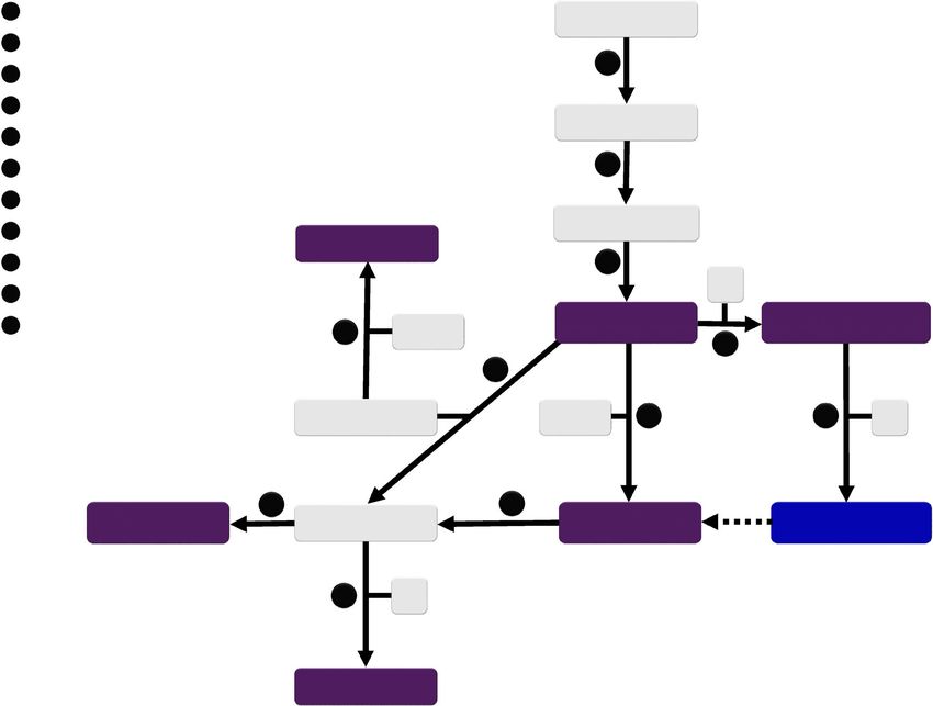
www.nature.com/scientificreports/
Figure 1. Morphological and immunohistochemical characteristics of DMD patients’ muscles. Left column:
Haematoxylin–Eosin (HE) (P1, P3, P4, P5, P6, P7, P8, P9) or Hemalun-Eosin-Safran (HES) (P2) displaying
endo-and perimysial fibrosis, necrotic and atrophic fibres along with inflammatory infiltrates suggestive of
progressive musclar dystrophy. Second to seventh columns : dystrophin immunolabeling (dys 1, dys 2 and
dys 3 labeling the core, NH2-terminal and COOH-terminal domains of the protein respectively) showing
severely decreased or absent immunoreactivity in patients compared to control muscle apposed on the same
slide. Eighth to eleventh columns : α- and γ-sarcoglycan immunolabelings showing either a decrease or
normal immunoreactivity in patients compared to muscle control opposed on the same slide. Horizontal lines
correspond to patients 1–9; scale bars: 20 µ.
Fig. 2A shows the fragmentation pattern of C28H50NO7P at m/z 566.32166 [M + Na]+. Given the FTICR high
mass accuracy, a loss of m/z 59.07351 and m/z 183.06608 corresponded to trimethylamine and phosphocholine
with mass errors of 186 ppb and 191 ppb, respectively. Therefore, the ion at m/z 566.32166 with the molecu-
lar formula, C28H50NO7P, was attributed to lysoPC(20:4) [M + Na]+. Other PC and SM lipids were similarly
unambiguously assigned. In negative-ion mode, other lipids included phosphatidic acid and phosphatidylserine.
Adenosine triphosphate (ATP) at m/z 505.98833 was annotated using MS/MS analysis by the loss of an adenine
group at m/z 272.95698 ( C5H8O9P2, [M-H]- with an error of 362 ppb; Supplementary Fig. 2B). Other metabolites
and lipids were similarly assigned with the mass accuracy. The final list included 34 unambiguously identified
features with 21 and 13 in positive- and negative-ionization modes, respectively. Supplementary Table 3 presents
the data matrix; Supplementary Table 4 presents the related statistics of the identified metabolites along with
their annotation metrics. Supplementary Fig. 3 presents the boxplots of the different metabolites. More mass
spectra are presented in Supplementary Figs. 4–10. Seven metabolites were decreased in the Duchenne biop-
sies: adenosine tetraphosphate, adenosine triphosphate, cytidine monophosphate, glycerophosphocholine, ino-
sitol pentakisphosphate, inositol tetraphosphate, and phosphoribosyl pyrophosphate. The other 27 metabolites
were increased in the Duchenne biopsies, including SM, phosphatidylcholines, phosphatidic acids and phos-
phatidylserines. To explore the distribution of this signature and its expression across samples, we performed
a clustering analysis using Euclidean distance, a similarity metric, between samples. Figure 2A shows two dis-
tinct clusters between the DMD and control biopsies due to the differential expression of the above-mentioned
metabolites across samples. We also explored the covariation of these metabolites using Spearman correlation
analysis. Figure 2B shows two main co-expression clusters that include upregulated and downregulated metab-
olites. Intraclass subclusters are observed such as a module that includes phosphatidylcholines, phosphatidic
acids and phosphatidylserines. Another module includes SMs and lysophosphatidylcholines. Figure 2C presents
the directional changes of these metabolites in the DMD samples along with their statistical significance. This
figure reports adjusted p-values and estimate which indicates metabolite change direction. Figure 3 presents
boxplots of the glycerophosphocholine, lysophosphatidylcholine, phosphatidylcholine, phosphatidic acid, SM,
phosphatidylserine, along with their tissue distributions in the DMD and control biopsies with their related
Scientific Reports | (2021) 11:1906 | https://doi.org/10.1038/s41598-021-81090-1 3
Vol.:(0123456789)www.nature.com/scientificreports/
A Control
Duchenne
Decreased in Duchenne
Increased in Duchenne
Group
Cytidine monophosphate
Glycerophosphocholine
Inositol pentakisphosphate
Inositol tetraphosphate
Adenosine triphosphate
Adenosine tetraphosphate
Phosphoribosyl pyrophosphate
Glutathione
PC(34:0)
Triacylglycerol (52:1)
Deoxyadenosine diphosphate
SM (18:0/16:0)
SM(d18:1/14:0)
SM (18:1/18:1)
LysoPC(20:4)
LysoPC(16:0)
LysoPC(18:0)
Hypoxanthine riboside
Nicotinamide adenine dinucleotide
PA(38:2)
PA(40:4)
PC(32:0)
Arachidonoylcarnitine
PC(38:3)
2 PC(38:4)
PC(38:5)
1 PC(36:3)
Scaled intensity
PC (34:1)
0
PC(36:4)
Pyrophosphate
−1
Adenosine diphosphate
−2 PA(38:3)
PA(40:5)
−3 PS(36:1)
P1 P2 P3 P4 P6 P7 P8 P9 P5 C1 C3 C4 C5 C6 C7 C8 C9 C2
Variation
B Decreased in Duchenne
Increased in Duchenne
Cytidine monophosphate
Glycerophosphocholine
Inositol pentakisphosphate
Inositol tetraphosphate
Adenosine tetraphosphate
Adenosine triphosphate
Phosphoribosyl pyrophosphate
Glutathione
Hypoxanthine riboside
Nicotinamide adenine dinucleotide
Pyrophosphate
PA(38:2)
PA(40:4)
Adenosine diphosphate
PA(40:5)
Correlation PS(36:1)
1 Arachidonoylcarnitine
PA(38:3)
PC (34:1)
PC(36:3)
PC(38:3)
0.5
PC(38:5)
PC(36:4)
PC(38:4)
Triacylglycerol (52:1)
0 LysoPC(20:4)
PC(34:0)
Deoxyadenosine diphosphate
SM (18:0/16:0)
-0.5 SM(d18:1/14:0)
LysoPC(16:0)
LysoPC(18:0)
PC(32:0)
-1 SM (18:1/18:1)
Variation
Cytidine monophosphate
Glycerophosphocholine
Inositol pentakisphosphate
Inositol tetraphosphate
Adenosine tetraphosphate
Adenosine triphosphate
Phosphoribosyl pyrophosphate
Glutathione
Hypoxanthine riboside
Nicotinamide adenine dinucleotide
Pyrophosphate
PA(38:2)
PA(40:4)
Adenosine diphosphate
PA(40:5)
PS(36:1)
Arachidonoylcarnitine
PA(38:3)
PC (34:1)
PC(36:3)
PC(38:3)
PC(38:5)
PC(36:4)
PC(38:4)
Triacylglycerol (52:1)
LysoPC(20:4)
PC(34:0)
Deoxyadenosine diphosphate
SM (18:0/16:0)
SM(d18:1/14:0)
LysoPC(16:0)
LysoPC(18:0)
PC(32:0)
SM (18:1/18:1)
C 1.0
Decreased in Duchenne Increased in Duchenne
0.5
Estimate
0.0
−0.5
−1.0
Arachidonoylcarnitine
Nicotinamide adenine dinucleotide
Triacylglycerol (52:1)
Phosphoribosyl pyrophosphate
PA(38:2)
Pyrophosphate
Glycerophosphocholine
PA(38:3)
PA(40:4)
PA(40:5)
LysoPC(20:4)
LysoPC(18:0)
Adenosine triphosphate
Adenosine tetraphosphate
Cytidine monophosphate
Inositol pentakisphosphate
Inositol tetraphosphate
Glutathione
PC (34:1)
PS(36:1)
PC(36:3)
PC(32:0)
PC(38:4)
PC(36:4)
PC(38:3)
PC(34:0)
SM (18:1/18:1)
Adenosine diphosphate
PC(38:5)
SM(d18:1/14:0)
SM (18:0/16:0)
Deoxyadenosine diphosphate
Hypoxanthine riboside
LysoPC(16:0)
−log adj p
7
6
5
4
Figure 2. Metabolome variations between DMD and control tissues. (A) Unsupervised hierarchical clustering
based on metabolic profiles (34 metabolites) detected in 9 DMD and 9 control tissues. C: Control, P: Patient; (B)
Spearman correlation heatmap of the 34 differentially expressed metabolites between control and DMD tissues;
(C) Barplots of the differentially expressed metabolites and their directional changes in the DMD tissues. Bar
color is proportional to –log (adjusted p-value). Red denotes high significance.
Scientific Reports | (2021) 11:1906 | https://doi.org/10.1038/s41598-021-81090-1 4
Vol:.(1234567890)www.nature.com/scientificreports/
Figure 3. Tissue distribution of selected discriminant metabolites. Left) Tissue metabolite distribution in
the control tissue. Right) Tissue metabolite distribution in the DMD tissue. Middle) Boxplot of the selected
metabolites between DMD and control tissues with related adjusted p-values. The y-axis shows the log-scaled
average intensity. Ion images were generated using SCiLs Lab software.
Scientific Reports | (2021) 11:1906 | https://doi.org/10.1038/s41598-021-81090-1 5
Vol.:(0123456789)www.nature.com/scientificreports/
1 Choline Kinase (EC 2.7.1.32)
Choline
2 Phosphocholine cytidyltransferase (EC 2.7.7.15)
1
3 Phosphocholine transferase (EC 2.7.8.2)
4 Phospholipase C (EC 3.1.4.3)
Phosphocholine
5 Phospholipase D (EC 3.1.4.4)
6 Phospholipases A1 & A2 (EC 3.1.1.32/EC 3.1.1.4) 2
7 Lysophospholipase (EC 3.1.1.5)
CDP-Phosphocholine
8 Phosphatidate phosphatase (EC 3.1.3.4)
Sphingomyelin
9 Diacylglycerol acyltransferase (EC 2.3.1.20) 3
FA
10 Phosphatidylserine synthase (EC 2.7.8.-/EC 2.7.8.29)
Phosphatidylcholine Lysophosphatidylcholine
11 Sphingomyelin synthase (EC 2.7.8.27)
11 Ceramide (2FA) (1FA)
6
4
Phosphocholine Choline 5 7 FA
10 8
Phosphatidylserine Phosphatidic acid Glycerophosphocholine
Diacylglycerol
(2FA) (2FA) (0FA)
9 FA
Triacylglycerol
Figure 4. Phospholipid and triacylglycerol metabolism. Purple: increased metabolites in DMD. Blue: decreased
metabolites in DMD. EC: Enzyme Commission number. FA: fatty acids.
adjusted p-values. Even though the figures don’t show a cellular, sub-cellular or fiber level resolution, they high-
light the heterogenous distribution of intensities across the tissue section that mirrors the distribution of the
related metabolites.
Discussion
Defective dystrophin is known to be the main alteration in the DMD etiology, however, metabolic impairment
rain27. Here, we explored
has been reported in several tissues such as the skeletal and cardiac muscles, liver and b
the differential metabolic patterns of muscle biopsies from DMD patients and control samples using a hypothesis-
free strategy based on MSI. Our results showed profound metabolic pathway remodelling in the muscles of
DMD patients compared with the controls grouped in two distinct clusters corresponding to the upregulated
and downregulated metabolites. The most discriminative metabolites were primarily phospholipids and energy
metabolites, suggesting that both these metabolic pathways are important players in muscle pathology and pos-
sibly by extension cognitive/behavioral disabilities.
Phospholipids (PLs) are major components of all cell membranes, and glycerophospholipids (GLs) are the
most abundant membrane PLs. GLs are composed of a glycerol backbone linked to a phosphate group in posi-
tion 3. Acyl groups may be attached at positions 1 and 2 of the glycerol. Different moieties, such as choline,
inositol, ethanolamine and serine, are linked to the phosphate group, contributing to GL diversity (e.g., PC,
phosphatidylinositol [PI], phosphatidylethanolamine (PE), PS, and phosphatidylglycerol). PCs are the most
abundant GL in mammalian cells. Other lipid classes, such as cholesterol and glycosphingolipids, play structural
embranes28,29. Disruption of PL metabolism has been previously reported in D
roles in cell m MD10,11,13,20,21,23,30,31.
We showed that several compounds belonging to PC, lysophosphatidylcholine (LPC), phosphatidic acid (PA),
PS and SM classes, as well as triacylglycerols, are increased, while glycerophosphocholine (GPC) is decreased
in DMD muscles compared with control muscles (Fig. 3). This metabolic remodelling could be attributed to a
putative decrease in lysophospholipase activity (Fig. 4). Lysophospholipase catalyzes lysophosphatidylcholine
deacylation and mediates GPC p roduction32. Sharma et al. considered DMD to be mainly a GPC d eficiency33.
Consequently, this impairment could lead to an accumulation in phosphatidylcholine, which is redirected toward
other metabolic pathways with an increase in PA, SM, phosphatidylserine and triacylglycerol (Fig. 4). It has
Scientific Reports | (2021) 11:1906 | https://doi.org/10.1038/s41598-021-81090-1 6
Vol:.(1234567890)www.nature.com/scientificreports/
been shown that lysophospholipase activity in erythrocyte membranes of DMD patients was lower than that of
healthy age-matched subjects32. Chalovich et al. compared the31P-NMR spectra of muscles from DMD patients,
Werdnig-Hoffman (Spinal Muscular Atrophy Type 1) patients and controls. These authors observed that GPC
levels were lower or near absent in DMD muscles compared with control muscles and dramatically increased
(15-fold) in one Werdnig-Hoffman p atient30. Increased triacylglycerol and SM in DMD mouse muscles have
been interpreted as resulting from increased phospholipase C a ctivity13. High phospholipid levels and high
phospholipid-to-cholesterol ratios have been documented u sing1H-NMR spectroscopy, which helps distinguish
DMD patients from healthy subjects34. Phospholipase D activity is reported to be increased in DMD muscles.
Calcium homeostasis has been shown to be altered in DMD patients, with calcium concentration being increased
in dystrophin-deficient muscle. Phospholipase D activity is enhanced under high concentrations of free calcium,
resulting in increased PA l evels35,36. In summary, profound phospholipid metabolism remodelling is associated
with DYS deficiency; thus, DMD has been considered a glycerophosphocholine d eficiency33,37. Morphological
alterations of the mitochondria have been reported in DMD muscles with higher rates of swollen mitochondria
compared with that of normal muscle38. PL alterations in DMD and a dysregulation of Ca2+ homeostasis may
induce mitochondrial membrane fragility and associated morphological changes with subsequent energy and
oxidative metabolism d isruptions39–42.
Energy metabolism impairments in DMD are major contributors to DYS-deficient muscle d egeneration43 as
illustrated by mitochondrial function improvement upon the partial restoration of DYS e xpression44. Accord-
ingly, we showed a significant increase in ADP and decreased ATP levels in DMD muscles compared with those
of the control muscles (Supplementary Fig. 3). Several preclinical and clinical studies aimed at promoting energy
metabolism are ongoing to treat DMD45. The need for studies with higher cellular or fiber level resolution would
unveil deeper metabolic insights and further explore these results. Van Pelt et al. performed a multi-omics study
in a Duchenne mouse model. They reported the impairment of glycolytic metabolism and phospholipids46. Two
metabolites overlap with our results adenosine diphosphate and PC(36:3) (Supplementary Table 5). Exploring the
potential release of the metabolites in less invasive biological fluids is relevant. Spitali et al. reported serum-based
metabolomics results in patients affected by several multiple forms of muscular dystrophy. Fifteen metabolites
have been reported belonging to energy, amino acid and testosterone m etabolisms47. Lindsay et al. performed a
urine-based metabolomics study in a Duchenne mouse model. They reported that five of seven detected Krebs
cycle metabolites were depleted in these mice consistent with an impaired energy metabolism22. No overlap has
been observed between our results and those reported in these studies (Supplementary Table 5).
In conclusion, although metabolic impairments have been reported in DMD, this work describes for the
first time the use of ultra-high-resolution MSI in DMD, which enabled more systematically and integratively
exploring on-tissue metabolic disturbances. This powerful technique allowed investigating the different metabolic
components in a single experiment. The present results highlight the potential use of MSI technology coupled
with systems biology approaches to holistically explore metabolic impairments in DMD. This work lays the
foundation for more mechanistic investigations of DMD and other metabolic diseases.
Methods
The overall workflow is presented in Supplementary Fig. 11.
Patients. Nine patients with molecularly proven DMD over the last 10 years were selected for the study. All
patients were referred to our neurology department, and biopsies were performed as a part of the diagnostic
workup when the disease was suspected. Samples were collected before initiating treatment. Medical charts were
reviewed for age at disease onset, age at diagnosis, age at walking unaided, age at walking loss, and age at last
visit. Symptoms at disease onset were recorded as calf hypertrophy, muscle weakness, contractures, behavioral
problems, speech delay, respiratory distress and cardiac arrhythmia. Disease activity parameters consisted of
maximal motor function, maximal CPK, age of initial joint contractures and current joint contractures, surgical
therapy and long-term physical therapy. Other parameters consisted of scoliosis occurrence or any other spinal
deformities, along with their surgical and nonsurgical therapies, occurrence of bone fractures and other ortho-
pedic surgeries, and mobility at last visit. Evaluated complications were respiratory insufficiency (e.g., IPPB,
ventilation, tracheotomy), existing cardiac complications (e.g., rhythm abnormalities or heart abnormalities
on ultrasound), digestive complications (e.g., nutritional problems, gastrostomy) and iatrogenic complications.
Inclusion in therapeutic protocols, hospitalization frequency, cognitive delays and behavioral problems, includ-
ing autism and school attendance, were also recorded.
The mean patient age at disease onset was 4 ± 1.5 years, at last visit was 10.5 ± 2.2 years, and at time of biopsy
was 5.5 ± 1.9 years. All patients are presently alive, and six (P1, P5, P6, P7, P8, and P9) were ambulant at the last
visit. One non-ambulant patient (P2) lost the ability to walk at age 7; he was not given steroids, nor included in
any therapeutic protocol. The other two patients (P3, P4) lost the ability to walk at 11 and 13 years of age. CK
levels ranged from 5418–52,000. Symptoms at disease onset were muscle weakness with difficulty walking and
climbing stairs and frequent falls. Eight patients had calf hypertrophy. Six patients presented no motor features
at onset but had speech delays (P2, P5, P8) and/or behavioral/autistic disturbances (P5, P8, P9) that had likely
led to delayed diagnosis. Four patients had cognitive delays (P2, P5, P8, P9). Two patients (P2, P5) developed
behavioral abnormalities. Six patients had learning difficulties affecting speech, writing, attention and memory
(P1, P2, P5, P7, P8, P9). Three patients (P4, P5, P9) had nutritional and feeding problems, with anorexia in two
and obesity in one (P9). All but two patients were given steroids (P1, P2); one of these two patients were lost
to follow up one year after diagnosis, and the other received no treatment owing to poor family compliance.
Four patients (P1, P5, P7, P9) could run, the others could walk without help. Two patients had mild respiratory
insufficiency that did not require ventilation. Eight patients had no significant cardiac arrhythmia or functional
Scientific Reports | (2021) 11:1906 | https://doi.org/10.1038/s41598-021-81090-1 7
Vol.:(0123456789)www.nature.com/scientificreports/
Weakness at onset Present (7) Absent (2)
Contractures at onset Present (2) Absent (7)
Calf hypertrophy at onset Present (7) Absent (2) Motor
Maximal motor function Walk alone (5) Run (4)
Joint contractures last visit Present (9)
Behavioral problem at onset Present (3) Absent (6)
Speech delay at onset Present (3) Absent (6)
Cognitive delay at onset Present (1) Absent (8)
ASD Present (1) Absent (8)
Neurodevelopment
Behavioral problem other Present (3) Absent (6)
Cognitive delay Present (4) Absent (5)
Learning dysability Present (6) Absent (3)
Regular school Present (6) Absent (3)
Scoliosis last visit Present (1) Absent (8)
Other spine deformities last visit Present (4) Absent (5)
Orthopedics
Fracture Present (3) Absent (6)
Joint contractures surgery Present (2) Absent (7)
Respiratory insufficiency last visit Present (1) Absent (8) Pulmonary
Cardiac rythm abnormalities last visit Present (1) Absent (8)
US abnormality Absent (9) Cardiology
Heart medication Present (8) Absent (1)
Feeding problem Present (3) Absent (6) Gastrointestinal
DYS1 IHC Severely decreased (3) Absent (6)
Severely
DYS2 IHC decreased (1)
Severely decreased (1) Absent (8)
Severely Severely decreased (1)
DYS3 IHC decreased (1)
Absent (8)
Slightly Severely
alpha−SG IHC decreased (1) decreased (1)
Decreased (5) Absent (2) Biopsy
beta−SG IHC Normal (2) Decreased (4) Absent (3)
delta−SG IHC Normal (2) Decreased (4) Absent (3)
gamma−SG IHC Slightly decreased (7) Normal (2)
Steroids Present (7) Absent (2) Treatment
Figure 5. Clinical characteristics of DMD patients. ASD: autistic spectrum disorders, DYS: dystrophin, IHC:
immunohistochemistry, US: ultrasound, SG: sarcoglycan.
abnormalities. Patient P4 had rhythm abnormalities consisting of supraventricular extrasystoles 3.9% of the
time, which did not require specific treatment. All but one patient received a prophylactic antiarrhythmic drug
(perindopril), and one (P1) was included in a perindopril protocol (2-year double-blind treatment protocol).
Three patients (P4, P5, P7) were included in exon-skipping protocols, and one (P6) was included in a givinostat
(histone deacetylase inhibitor) protocol. Clinical data overview is presented in Fig. 5 and detailed clinical data
are listed in Supplementary Tables 1 and 2.
Muscle biopsies. Each patient underwent a muscle biopsy at a mean age of 5.5 ± 1.9 years, which was
assessed according to standardized histochemical and histoenzymological m ethods48. For immunohistochemi-
cal studies, 6-μm frozen sections were immunolabeled with antibodies against β-spectrin (diluted 1:50; Novo-
castra Leica Biosystems, Nanterre, France), dystrophin (DYS1, DYS2 and DYS3 correspond to amino acids
1181–1388, 3669–3685 and 321–494, of the dystrophin molecule and are diluted respectively at 1:10, 1:8 and
1:6—Novocastra Leica Biosystems), and α-, β-, γ- and δ sarcoglycans (diluted 1:100, 1:100, 1:100 and 1:50,
respectively; Novocastra Leica Biosystems). Dystrophinopathy was morphologically diagnosed per the criteria
of Dubowitz et al.48.
As age-matched control muscles were not available, nine male adults aged 28–58 years were selected as con-
trols for comparative analyses. These patients presented myalgias, muscle weakness, and/or muscle fatigability.
In one case, a polymyositis was suspected; Buschke scleroderma was suspected in another. In all control patients,
routine biological tests, CK levels, EMGs, cardiac ultrasonography, pulmonary testing and muscle MRI were
considered normal. Their muscle biopsies were also normal, and they were concluded to have probable fibromy-
algia. One obvious potential limit of this work that needs to be highlighted consists in absence of age-matched
control muscles could hamper interpretation of the results, as biochemical properties of muscles change over
the lifetime. Even though paediatric muscle samples could be obtained during orthopaedic surgery, the location
of the biopsy may change and may not entirely match with that of Duchenne patients.
Reagents and chemicals. Methanol, acetonitrile, ethanol and water of LC–MS grade were purchased
from Fisher Scientific (Loughborough, UK); 2.5-dihydroxybenzoic acid (DHB) and 9-aminoacridine hydro-
chloride monohydrate (9-AA) MALDI matrices were purchased from Sigma-Aldrich (St. Louis, MO, USA).
Sodium trifluoroacetate (NaTFA, Sigma-Aldrich) at 0.1 mg mL-1 (ACN/H2O 50:50 [v/v]) was used as a calibrant
before each analysis.
Scientific Reports | (2021) 11:1906 | https://doi.org/10.1038/s41598-021-81090-1 8
Vol:.(1234567890)www.nature.com/scientificreports/
Sample preparation. Fresh frozen 10-µm sections of human muscle were mounted on conductive ITO-
coated glass slides 75 × 25 mm (Bruker, Bremen, Germany) and stored at -80 °C until analysis. Matrix solutions
were applied with an automatic microsprayer HTX TM-Sprayer (HTX Imaging, Chapel Hill, NC, USA) as previ-
ously described49. In the positive-ion mode, DHB matrix (30 mg mL-1 in MeOH/H2O 50:50 [v/v]) was deposited
with the following parameters: nozzle temperature 80 °C, nozzle velocity 1200 mm min-1, N2 pressure 10 psi,
N2 flow rate 2 L min-1, number of passes 12, flow rate 100 µL min-1 and track spacing 3 mm. In negative-ion
mode, 9-AA matrix (10 mg mL-1, EtOH/H2O 70:30 [v/v]) was sprayed using the following parameters: nozzle
temperature 90 °C, nozzle velocity 1200 mm min-1, N2 pressure 10 psi, N2 flow rate 3 L min-1, number of passes
2, flow rate 120 µl min-1, track spacing 3 mm and drying time 30 s between passes. Each slide was vacuum-dried
before analysis.
Mass spectrometry instrumentation and data processing. Data were acquired on a FTICR instru-
ment (SolariX XR, Bruker, Bremen, Germany) equipped with a 12-T superconducting magnet and a dynami-
cally harmonized ICR cell. This instrument is also equipped with both a laser desorption ionization source
(Smartbeam II, Nd:YAG × 3 laser at 355 nm, Bruker, Bremen, Germany) and an electrospray (ESI) source. Each
MALDI spectrum for each position is the result of 1 scan and 500 consecutive laser shots. Spectra were acquired
over an 80 µm raster. Before imaging analyses, the instrument was externally calibrated in the required mode
by NaTFA infusion via ESI source, then internally calibrated by assigning known metabolites from m/z 150–
1000 via MALDI source. The instrument was autocalibrated during image acquisition. In positive-ionization
mode, calibration was performed by assigning C 7H6O4 (m/z 155.033885 [M + H]+, matrix peak), C
7H15NO3 (m/z
162.112470 [M + H]+, carnitine), C 7H6O4 (m/z 177.015829, [M + Na]+, matrix peak), C
5H14NO4P (m/z 184.073321,
[M + H]+, phosphocholine), C9H17NO4 (m/z 204.123034, [M + H]+, acetylcarnitine), C14H8O6 (m/z 273.039364,
[M + H]+, matrix peak), C21H12O9 (m/z 409.055408, [M + H]+, matrix peak), C40H80NO8P (m/z 734.569432,
[M + H]+, lipid), and C42H82NO8P (m/z 798.54096, [M + K]+, lipid). In negative-ion mode the assigned peaks
were C13H10N2 (m/z 193.77122, [M-H]-, matrix peak), C17H26N6O6 (m/z 409.184106, [M-H]-, amino acids),
C10H15N5O10P2 (m/z 426.022139, [M-H]-, adenosine diphosphate), C10H16N5O13P3 (m/z 505.988470, [M-H]-,
adenosine triphosphate), C23H39N7O7 (m/z 524.283820, [M-H]-, amino acids), C30H46N6O8 (m/z 599.319871,
[M-H]-, lipid) and C 39H77O9P (m/z 701.512680, [M-H]-, lipid). Data size was set at 2 million points for a tran-
sient length of 0.87 s, and spectra were acquired with a 97% data file reduction. A single MSI measurement has
been performed by specimen. Images were captured using FTMS control and FlexImaging (v 5.0, Bruker) soft-
ware. Images were processed with SCiLS Lab Pro software (Bruker Daltonics, Bremen, Germany). The total ion
current method was used for normalization, and m/z intervals were automatically set at ± 1 ppm. Images were
viewed using both FlexImaging and SCiLS Lab software (Bruker Daltonics, Bremen, Germany).
Data analyses. Ion intensities have been log-transformed. Univariate analyses were performed using t-tests
to identify discriminatory ionic species between the assessed groups. Age has been taken into account by adding
it as covariate. Spearman correlation analysis was performed using R software. Euclidean distance was used as
a similarity measure in the clustering analysis. False discovery rates were corrected using the Benjamini–Hoch-
berg–Yekutieli method, and p < 0.05 was considered statistically significant.
Metabolite annotation and identification. Preliminary assignments based on accurate mass measure-
ments were performed using the mass spectrometry databases, METLIN 50 and HMDB 51, using a threshold
of ± 2 ppm. For some metabolites, the precise raw formula led to one hit. Others were identified via “on-tissue”
tandem mass spectrometry experiments using MALDI tandem MS/MS. Ions of interest were first isolated using
a window of ± 1 Da, then fragmented by collision-induced dissociation with energy levels between 10 and 40 eV.
For each MS/MS analysis, 50 scans were accumulated for better sensitivity. Spectra were reprocessed using Data
Analysis 4.4 software (Bruker Daltonics, Bremen, Germany) and recalibrated with the single-point calibration
option.
Ethical statement. All muscle samples used in this study belong to a collection declared to the French
Ministry of Health (collection number DC-2015–2468, accession number AC-2015–2467) located in the Pathol-
ogy Department (Prof. Annie Laquerrière) of Rouen University Hospital in accordance with the relevant guide-
lines and regulations and with permission of the local authorities. Written informed consents were obtained
from the parents when the patient is under 18 or from the adult patient in order to perform any investigation
related to their pathology. The study protocols have been approved by ethics committee of NORD WEST1 –
Rouen University Hospital (collection number DC-2015–2468, accession number AC-2015–2467).
Data availability
All the data that support the findings are presented in the manuscript and the supplementary material.
Received: 14 September 2020; Accepted: 4 January 2021
References
1. Al-Khalili Szigyarto, C., & Spitali, P. Biomarkers of Duchenne muscular dystrophy: current findings. Degenerative Neurological
and Neuromuscular Disease 8, 1–13, https://doi.org/10.2147/dnnd.s121099 (2018).
Scientific Reports | (2021) 11:1906 | https://doi.org/10.1038/s41598-021-81090-1 9
Vol.:(0123456789)www.nature.com/scientificreports/
2. Birnkrant, D. J. et al. Diagnosis and management of Duchenne muscular dystrophy, part 1: diagnosis, and neuromuscular, reha-
bilitation, endocrine, and gastrointestinal and nutritional management. The Lancet. Neurol. 17, 251–267. https://doi.org/10.1016/
s1474-4422(18)30024-3 (2018).
3. Mercuri, E., Bonnemann, C. G. & Muntoni, F. Muscular dystrophies. Lancet 394, 2025–2038. https://doi.org/10.1016/s0140
-6736(19)32910-1 (2019).
4. Guiraud, S. et al. The pathogenesis and therapy of muscular dystrophies. Annu. Rev. Genomics Hum. Genet. 16, 281–308. https://
doi.org/10.1146/annurev-genom-090314-025003 (2015).
5. Guiraud, S. & Davies, K. E. Regenerative biomarkers for Duchenne muscular dystrophy. Neural Regen. Res. 14, 1317–1320. https
://doi.org/10.4103/1673-5374.253534 (2019).
6. Doorenweerd, N. et al. Timing and localization of human dystrophin isoform expression provide insights into the cognitive
phenotype of Duchenne muscular dystrophy. Sci. Rep. 7, 12575. https://doi.org/10.1038/s41598-017-12981-5 (2017).
7. Signorelli, M. et al. Longitudinal serum biomarker screening identifies malate dehydrogenase 2 as candidate prognostic biomarker
for Duchenne muscular dystrophy. J. Cachexia, Sarcopenia Muscle, https://doi.org/10.1002/jcsm.12517 (2019).
8. Hoxha, M. Duchenne muscular dystrophy: Focus on arachidonic acid metabolites. Biomed. Pharmacother. 10, 796–802, https://
doi.org/10.1016/j.biopha.2018.12.034 (2019).
9. Muntoni, F. et al. Categorising trajectories and individual item changes of the north star ambulatory assessment in patients with
Duchenne muscular dystrophy. PLoS ONE 14, e0221097. https://doi.org/10.1371/journal.pone.0221097 (2019).
10. Chio, C., Peterson, D. & Kratzer, F. Lipid composition and synthesis in the muscles of normal and dytrophic chickens. Can. J.
Biochem. 50, 1267–1272 (1972).
11. Hughes, B. P. Lipid changes in Duchenne muscular dystrophy. J. Neurol. Neurosurg. Psychiatry 35, 658–663. https: //doi.org/10.1136/
jnnp.35.5.658 (1972).
12. Kwok, C. T., Kuffer, A. D., Tang, B. Y. & Austin, L. Phospholipid metabolism in murine muscular dystrophy. Exp. Neurol. 50,
362–375 (1976).
13. Kwok, C. T. & Austin, L. Phospholipid composition and metabolism in mouse muscular dystrophy. Biochem. J. 176, 15–22. https
://doi.org/10.1042/bj1760015 (1978).
14. Tebani, A., Afonso, C., Marret, S. & Bekri, S. Omics-Based Strategies in Precision Medicine: Toward a Paradigm Shift in Inborn
Errors of Metabolism Investigations. International journal of molecular sciences 17, https://doi.org/10.3390/ijms17091555 (2016).
15. Nicholson, J. K., Lindon, J. C. & Holmes, E. “Metabonomics”: understanding the metabolic responses of living systems to patho-
physiological stimuli via multivariate statistical analysis of biological NMR spectroscopic data. Xenobiotica For. Compound. Biol.
Syst. 29, 1181–1189. https://doi.org/10.1080/004982599238047 (2008).
16. Tebani, A., Afonso, C. & Bekri, S. Advances in metabolome information retrieval: turning chemistry into biology. Part I: analytical
chemistry of the metabolome. J Inherit Metab Dis, https://doi.org/10.1007/s10545-017-0074-y (2017).
17. Spengler, B. Mass spectrometry imaging of biomolecular information. Anal. Chem. 87, 64–82. https://doi.org/10.1021/ac504543v
(2015).
18. Zhang, J. et al. Nondestructive tissue analysis for ex vivo and in vivo cancer diagnosis using a handheld mass spectrometry system.
Sci Transl Med 9, https://doi.org/10.1126/scitranslmed.aan3968 (2017).
19. Xiao, Y. et al. Recent advances of ambient mass spectrometry imaging for biological tissues: A review. Analytica Chimica Acta
(2020).
20. Benabdellah, F., Yu, H., Brunelle, A., Laprevote, O. & De La Porte, S. MALDI reveals membrane lipid profile reversion in MDX
mice. Neurobiol Dis 36, 252–258. https://doi.org/10.1016/j.nbd.2009.07.013 (2009).
21. Touboul, D. et al. Changes of phospholipid composition within the dystrophic muscle by matrix-assisted laser desorption/ioniza-
tion mass spectrometry and mass spectrometry imaging. Eur. J. Mass Spectrom. (Chichester, England) 10, 657–664. https://doi.
org/10.1255/ejms.671 (2004).
22. Lindsay, A., Chamberlain, C. M., Witthuhn, B. A., Lowe, D. A. & Ervasti, J. M. Dystrophinopathy-associated dysfunction of Krebs
cycle metabolism. Hum. Mol. Genet. 28, 942–951. https://doi.org/10.1093/hmg/ddy404 (2019).
23. Tahallah, N., Brunelle, A., De La Porte, S. & Laprevote, O. Lipid mapping in human dystrophic muscle by cluster-time-of-flight
secondary ion mass spectrometry imaging. J. Lipid Res. 49, 438–454. https://doi.org/10.1194/jlr.M700421-JLR200 (2008).
24. Shaw, J. B. et al. 21 Tesla Fourier transform ion cyclotron resonance mass spectrometer greatly expands mass spectrometry toolbox.
J. Am. Soc. Mass Spectrom. 27, 1929–1936 (2016).
25. Kooijman, P. C. et al. Increased throughput and ultra-high mass resolution in DESI FT-ICR MS imaging through new-generation
external data acquisition system and advanced data processing approaches. Sci. Rep. 9, 1–11 (2019).
26. Ferey, J. et al. A new optimization strategy for MALDI FTICR MS tissue analysis for untargeted metabolomics using experimental
design and data modeling. Anal. Bioanal. Chem. 411, 3891–3903. https://doi.org/10.1007/s00216-019-01863-6 (2019).
27. Esposito, G. & Carsana, A. Metabolic alterations in cardiomyocytes of patients with duchenne and becker muscular dystrophies.
Journal of Clinical Medicine 8, 2151 (2019).
28. Hishikawa, D., Hashidate, T., Shimizu, T. & Shindou, H. Diversity and function of membrane glycerophospholipids generated by
the remodeling pathway in mammalian cells. J. Lipid Res. 55, 799–807. https://doi.org/10.1194/jlr.R046094 (2014).
29. Vance, J. E. Phospholipid synthesis and transport in mammalian cells. Traffic (Copenhagen, Denmark) 16, 1–18. https://doi.
org/10.1111/tra.12230(2015).
30. Chalovich, J. M., Burt, C. T., Danon, M. J., Glonek, T. & Barany, M. Phosphodiesters in muscular dystrophies. Ann N Y Acad Sci
317, 649–669. https://doi.org/10.1111/j.1749-6632.1979.tb56585.x (1979).
31. Pearce, P., Johnsen, R., Wysocki, S. & Kakulas, B. Muscle lipids in Duchenne muscular dystrophy. Aust. J. Exp. Biol. Med. Sci. 59,
77–90 (1981).
32. Podolski, J. L. et al. Erythrocyte membrane lysophospholipase activity in muscular dystrophy. J. Neurol. Sci. 59, 423–429. https://
doi.org/10.1016/0022-510x(83)90027-8 (1983).
33. Sharma, U., Atri, S., Sharma, M. C., Sarkar, C. & Jagannathan, N. R. Skeletal muscle metabolism in Duchenne muscular dys-
trophy (DMD): an in-vitro proton NMR spectroscopy study. Magn. Reson. Imaging 21, 145–153. https://doi.org/10.1016/s0730
-725x(02)00646-x (2003).
34. Srivastava, N. K., Pradhan, S., Mittal, B. & Gowda, G. A. High resolution NMR based analysis of serum lipids in Duchenne muscular
dystrophy patients and its possible diagnostic significance. NMR Biomed. 23, 13–22. https://doi.org/10.1002/nbm.1419 (2010).
35. McDermott, M. I., Wang, Y., Wakelam, M. J. O. & Bankaitis, V. A. Mammalian phospholipase D: Function, and therapeutics.
Progress in Lipid Res., 101018, https://doi.org/10.1016/j.plipres.2019.101018 (2019).
36. Walter, M. et al. Involvement of phospholipase D in store-operated calcium influx in vascular smooth muscle cells. FEBS Lett. 479,
51–56. https://doi.org/10.1016/S0014-5793(00)01880-9 (2000).
37. Infante, J. P. & Huszagh, V. A. Mechanisms of resistance to pathogenesis in muscular dystrophies. Mol. Cell. Biochem. 195, 155–167.
https://doi.org/10.1023/a:1006972315739 (1999).
38. Pellegrini, C. et al. Melanocytes–a novel tool to study mitochondrial dysfunction in Duchenne muscular dystrophy. J. Cell. Physiol.
228, 1323–1331. https://doi.org/10.1002/jcp.24290 (2013).
39. Dubinin, M. V. et al. Duchenne muscular dystrophy is associated with the inhibition of calcium uniport in mitochondria and an
increased sensitivity of the organelles to the calcium-induced permeability transition. Biochimica et biophysica acta. Molecular
basis of disease 1866, 165674, https://doi.org/10.1016/j.bbadis.2020.165674 (2020).
Scientific Reports | (2021) 11:1906 | https://doi.org/10.1038/s41598-021-81090-1 10
Vol:.(1234567890)www.nature.com/scientificreports/
40. Hughes, M. C. et al. Early myopathy in Duchenne muscular dystrophy is associated with elevated mitochondrial H2 O2 emission
during impaired oxidative phosphorylation. J. Cachexia, Sarcopenia Muscle 10, 643–661. https: //doi.org/10.1002/jcsm.12405 (2019).
41. Kuno, A. et al. Resveratrol ameliorates mitophagy disturbance and improves cardiac pathophysiology of dystrophin-deficient mdx
mice. Sci. Rep. 8, 15555. https://doi.org/10.1038/s41598-018-33930-w (2018).
42. Lin, Y. F. et al. A novel mitochondrial micropeptide MPM enhances mitochondrial respiratory activity and promotes myogenic
differentiation. Cell Death Disease 10, 528. https://doi.org/10.1038/s41419-019-1767-y (2019).
43. Timpani, C. A., Hayes, A. & Rybalka, E. Revisiting the dystrophin-ATP connection: How half a century of research still implicates
mitochondrial dysfunction in Duchenne Muscular Dystrophy aetiology. Med. Hypotheses 85, 1021–1033. https: //doi.org/10.1016/j.
mehy.2015.08.015 (2015).
44. Vila, M. C. et al. Mitochondria mediate cell membrane repair and contribute to Duchenne muscular dystrophy. Cell Death Differ.
24, 330–342. https://doi.org/10.1038/cdd.2016.127 (2017).
45. Rybalka, E., Timpani, C. A., Stathis, C. G., Hayes, A. & Cooke, M. B. Metabogenic and nutriceutical approaches to address energy
dysregulation and skeletal muscle wasting in duchenne muscular dystrophy. Nutrients 7, 9734–9767. https: //doi.org/10.3390/nu712
5498 (2015).
46. Van Pelt, D. W. et al. Multiomics analysis of the mdx/mTR mouse model of Duchenne muscular dystrophy. Connective Tissue
Research, 1–16, https://doi.org/10.1080/03008207.2020.1791103 (2020).
47. Spitali, P. et al. Cross-sectional serum metabolomic study of multiple forms of muscular dystrophy. J. Cell Mol. Med. 22, 2442–2448.
https://doi.org/10.1111/jcmm.13543 (2018).
48. Dubowitz, V., Sewry, C. & Oldfors, A. Muscle Biopsy: A Practical Approach (Elsevier, Amsterdam, 2013).
49. Ferey, J. et al. A new optimization strategy for MALDI FTICR MS tissue analysisfor untargeted metabolomics using experimental
design and datamodeling. ABC 411, 3891–3903 (2019).
50. Smith, C. A. et al. METLIN: a metabolite mass spectral database. Ther. Drug Monit. 27, 747 (2005).
51. Wishart, D. S. et al. HMDB 3.0--The Human Metabolome Database in 2013. Nucleic Acids Res. 41, 801 (2013).
Acknowledgements
This work was supported by the European Regional Developement Fund (ERDF) N° HN0001343, the European
Union’s Horizon 2020 Research Infrastructures program (Grant Agreement 731077), the Région Normandie,
and the Laboratoire d’Excellence (LabEx) SunOrg (ANR-11-LABX-0029). Access to a CNRS FTICR research
infrastructure (FR3624) is gratefully acknowledged.
Author contributions
SB and AT conceived and designed the study. AL and FM provided the samples and histopathological data. CB
tissue handling and technical assistance. CV, ID, SM and AL provided clinical data. ACB provided genetic data.
FL performed Western blot experiments. JF, YB, VG, ISA and CA performed the mass spectrometry imaging data
acquisition and raw data processing. AT performed statistical analyses, data visualization, illustrations and data
interpretation. AT, ID, JF, FM, AL and SB wrote the manuscript. All authors read and approved the manuscript.
Competing interests
The authors declare no competing interests.
Additional information
Supplementary information The online version contains supplementary material available at https://doi.
org/10.1038/s41598-021-81090-1.
Correspondence and requests for materials should be addressed to S.B.
Reprints and permissions information is available at www.nature.com/reprints.
Publisher’s note Springer Nature remains neutral with regard to jurisdictional claims in published maps and
institutional affiliations.
Open Access This article is licensed under a Creative Commons Attribution 4.0 International
License, which permits use, sharing, adaptation, distribution and reproduction in any medium or
format, as long as you give appropriate credit to the original author(s) and the source, provide a link to the
Creative Commons licence, and indicate if changes were made. The images or other third party material in this
article are included in the article’s Creative Commons licence, unless indicated otherwise in a credit line to the
material. If material is not included in the article’s Creative Commons licence and your intended use is not
permitted by statutory regulation or exceeds the permitted use, you will need to obtain permission directly from
the copyright holder. To view a copy of this licence, visit http://creativecommons.org/licenses/by/4.0/.
© The Author(s) 2021
Scientific Reports | (2021) 11:1906 | https://doi.org/10.1038/s41598-021-81090-1 11
Vol.:(0123456789)You can also read