Aspects of transition cow metabolomics-Part II: Histomorphologic changes in the liver parenchyma throughout the transition period, in cows with ...
←
→
Page content transcription
If your browser does not render page correctly, please read the page content below
J. Dairy Sci. 104:9227–9244
https://doi.org/10.3168/jds.2020-19057
© 2021, The Authors. Published by Elsevier Inc. and Fass Inc. on behalf of the American Dairy Science Association®.
This is an open access article under the CC BY-NC-ND license (http://creativecommons.org/licenses/by-nc-nd/4.0/).
Aspects of transition cow metabolomics—Part II: Histomorphologic changes
in the liver parenchyma throughout the transition period, in cows
with different liver metabotypes and effects of a metaphylactic
butaphosphan and cyanocobalamin treatment
F. Pietsch,1 M. Schären,1* T. Snedec,1 K. B. Theinert,1 A.-S. Leonhardt,1 A. Kaiser,1 F. Rachidi,1 D. Böttcher,2
J. Scheinert,2 H.-A. Schoon,2 P. Wohlsein,3 J. Spilke,4 A. Haudum,5 W. Baumgartner,6 and A. Starke1
1
Clinic for Ruminants and Swine, Faculty of Veterinary Medicine, Leipzig University, An den Tierkliniken 11, 04103 Leipzig, Germany
2
Institute of Veterinary Pathology, Faculty of Veterinary Medicine, Leipzig University, An den Tierkliniken 33, 04103 Leipzig, Germany
3
Department of Pathology, University of Veterinary Medicine Hanover, Foundation, Bünteweg 17, 30559 Hanover, Germany
4
Biometrics and Informatics in Agriculture Group, Institute of Agricultural and Nutritional Sciences, Martin-Luther University, Halle-Wittenberg, Karl-
Freiherr-von-Fritsch-Str. 4, 06108 Halle (Saale), Germany
5
Veterinary Practice, Herrnschlag 3, 4170 St. Stefan am Walde, Austria
6
University Clinic for Ruminants, University of Veterinary Medicine, Veterinärplatz 1, 1210 Vienna, Austria
ABSTRACT There was mild to moderate fat infiltration in the liver
of 37% of cows in the last 2 wk AP, and moderate
The aims of this study were to evaluate histopatho- to severe fat infiltration in 66% of cows in the first
logic changes during the transition period, describe the days PP. The degree of fat infiltration increased from
histopathological features of the metabotypes identified 2 wk AP until the end of the first week PP, and then
in Part I (Schären et al., 2021b), and investigate effects decreased until the end of the study period, at which
of a metaphylactic treatment with butaphosphan and time about 25% of cows had moderate to severe fatty
cyanocobalamin (BCC) on the liver parenchyma. Eighty infiltration. Lipidosis was positively correlated with the
German Holstein cows (mean 305-d production: 10,957 severity of liver cell degeneration, and negatively cor-
kg, range: 6,480–15,193 kg; mean lactation number: 3.9, related with the degree of glycogen deposits. Complete
range: 2–9) from a commercial dairy farm in Saxony, glycogen depletion of hepatocytes was not observed in
Germany, were enrolled in a randomized, prospective, cows, even in the presence of severe hepatic lipidosis.
triple-blinded study. Two groups received a treatment Moderate to severe lymphocytic hepatitis was seen in
with BCC (5 or 10 mL/100 kg of body weight 10% 39% of cows throughout the study period, and cows
butaphosphan and 0.005% cyanocobalamin, Catosal, with lactation numbers 5 or greater had perisinusoidal
Bayer Animal Health, n = 20 each) and one group a fibrosis more often than younger cows. Severe fibrosis
placebo treatment (NaCl 0.9%, n = 40). Liver biopsy and cirrhosis of the liver did not occur. Metabotype B
specimens were collected 14 d antepartum (AP) and animals exhibited a higher chance of fatty infiltration,
7, 28, and 42 d postpartum (PP), routinely processed lower glycogen storage, and perisinusoidal fibrosis and
for histologic examination, and stained with hema- for this metabotype positive correlations were calcu-
toxylin and eosin, Sudan III, periodic acid-Schiff, and lated between increased fat deposition in the liver and
picrosirius red stains. The sections were assessed for marked glycogen depletion, and increased degenerative,
fat and glycogen content and degenerative, inflamma- inflammatory, fibrotic, and proliferative changes of he-
tory, fibrotic, and proliferative changes. The statistical patic tissue. For the treatment with BCC, no significant
analysis included the effects of the sampling day, the effect was observed. In summary, during the transition
lactation number, the treatment, and the metabotype period, the liver of dairy cows is characterized by fat
(A = medium, B = minor, C = large alterations in the accumulation and glycogen depletion and histologic
liver metabolome profile between AP and PP status). signs of hepatitis and hepatocyte degeneration. These
histomorphologic changes were accentuated in animals
exhibiting little alterations in their liver metabolome
profile across the transition period (metabotype B)
and support the assumption of a decreased grass silage
Received June 9, 2020.
Accepted March 18, 2021. quality as a causative factor.
*Corresponding author: melanie.schaeren@vetmed.uni-leipzig.de Key words: fatty liver, fibrosis, glycogen, hepatitis
9227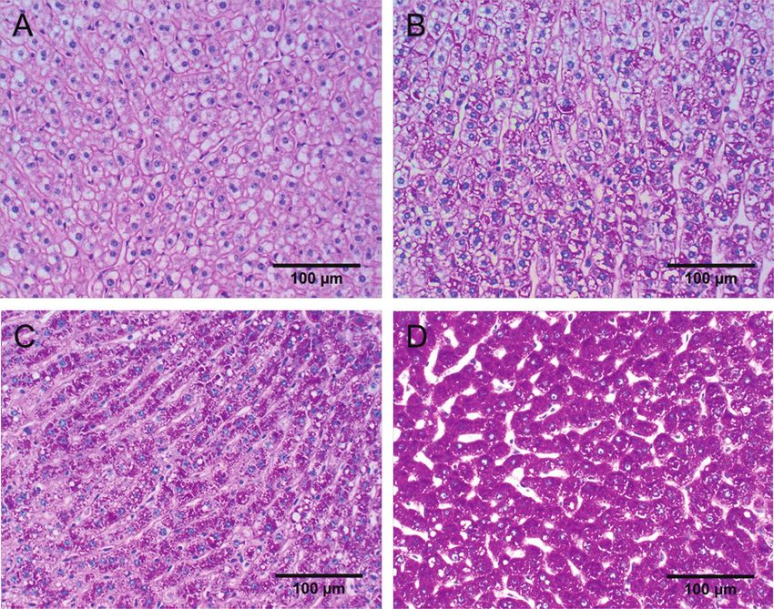
Pietsch et al.: TRANSITION COW METABOLOMICS–PART II                          9228
                        INTRODUCTION                               (2) investigate the histopathological features of the
                                                                   liver metabotypes identified in Part I (Schären et al.,
   Moderate to severe hepatic lipidosis (fatty liver) may          2021b), and (3) investigate effects of a metaphylactic
occur in 40 to 66% of dairy cows during early lacta-               treatment with BCC on the liver parenchyma.
tion (Reid, 1980; Gerloff et al., 1986; Jorritsma et al.,
2001). The risk of liver failure in cows has been shown
                                                                                 MATERIALS AND METHODS
to increase up to 5.3-fold with increasing fat content
(Rehage et al., 1996). Hepatic fatty infiltration, liver           Study Design
cell degeneration (Reid, 1980; Reid and Collins, 1980;
Johannsen et al., 1988), and the infiltration of reactive             The study design is described in detail in Materi-
inflammatory cells (Fürll, 1989; Mertens, 1992) were               als and Methods of Part I (Schären et al., 2021b). In
thought to be interrelated and result in impaired liver            summary, an on-farm randomized, prospective, triple-
function.                                                          blinded study was performed on a 660-cow dairy in
   In addition to fat, glycogen plays an important role            Saxony, Germany, between November 23, 2015, and
in the metabolism of dairy cows, providing a readily               December 3, 2016. The cows were housed in a freestall
available source of energy (Gardner et al., 2014). Un-             system with deep bedding boxes (except for a part of
like monogastric animals, hepatic gluconeogenesis is               the dry period) and received a TMR-based (grass- and
crucial for glucose metabolism in ruminants (Herdt,                corn-silage as main components) ration. The herd was
1988; Aschenbach et al., 2010). Cows are therefore ca-             characterized by a rolling average milk production of
pable of meeting about 75% of daily energy demands                 10,747 kg and fat and protein contents of 3.73 and
with the ruminal production of VFA, including acetate,             3.33%, respectively [official monthly milk control re-
propionate, and butyrate (65:20:15; Bergman, 1990).                port by the local state control association (Landeskon-
However, the energy demand of high-producing dairy                 trollverband, Sachsen) on December 5, 2016]. Eighty
cows constitutes a considerable challenge. In addition             animals in second or higher lactation were included in
to about 200 g of glucose required for maintenance,                the study and followed from 14 d antepartum (AP, ex-
the daily production of 40 to 50 kg of milk requires               pected calving date) until 49 d postpartum (PP), with
another approximately 3.8 kg of glucose (Elliot, 1976),            a thorough and close documentation of the production
which is an enormous demand on the liver in terms of               and clinical traits and clinical chemistry. Their average
gluconeogenesis and illustrates the role of glycogen as a          lactation number was 3.9 (range: 2–9, lactation number
critical energy substrate and reserve.                             at calving in trial), the BCS was 2.91 [range: 2.00–3.75;
   We are not aware of current studies on the concur-              14 d AP, according to 5-point scale of Edmonson et al.
rent dynamic of fat and glycogen metabolism in terms               (1989)] and the 305-d milk production in previous lac-
of histomorphologic and histochemical changes in the               tation was 10,957 kg (range: 6,480–15,193). To analyze
liver of dairy cows during the transition period and               a possible effect of age, the cows were allocated to one
their interrelation with inflammatory, fibrotic, and               of 3 lactation groups: second lactation (L1), third and
proliferative changes. Therefore, in the scope of a large          fourth lactations (L2), and fifth and higher lactations
study, investigating pathomechanisms of the fatty liver            (L3).
syndrome in dairy cows, multiple liver biopsies were                  To evaluate a metaphylactic treatment protocol
collected in 80 German Holstein cows throughout the                with butaphosphan and cyanocobalamin, the following
transition period. Aside the sound documentation and               treatment groups were established: 2 groups with a
interrelation of the production state, clinical traits and         Catosal treatment (Bayer Animal Health GmbH) with
liver pathohistological features, the trial design and             the recommended dose (as registered in Germany) of 5
aims included the interrelation of these data with the             mL/100 kg of BW (VER5, n = 20, parity: 4.2 ± 2.0,
liver and blood metabolome and the investigation of                mean ± SD) or the double dose of 10 mL/100 kg of
the effects of a metaphylactic treatment with butaphos-            BW (VER10, n = 20, parity: 3.4 ± 1.3), and 2 placebo
phan and cyanocobalamin (BCC, described in detail in               groups with 5 or 10 mL of NaCl 0.9%/100 kg of BW
Part I; Schären et al., 2021b).                                    (for analysis, the 2 groups were merged to one control
   Hence, the aims of Part II of the study presented here          group, CON, n = 40, parity: 4.0 ± 1.9). The group as-
were to (1) describe liver histopathological alterations           signment occurred randomly using a lottery procedure.
across the transition period, regarding fat and glycogen           The animals were treated at 6 time points: 7, 6, and 5
storage, as well as degenerative, inflammatory, fibrotic,          d AP, and 1, 2, and 3 d PP.
and proliferative changes, with a particular interest on              Three different metabotypes were identified in a
whether a relationship exists between the dynamics of              follow-up analysis based on their metabolic alterations
fat storage and the concurrent depletion of glycogen,              in the liver over the course of the transition period

Journal of Dairy Science Vol. 104 No. 8, 2021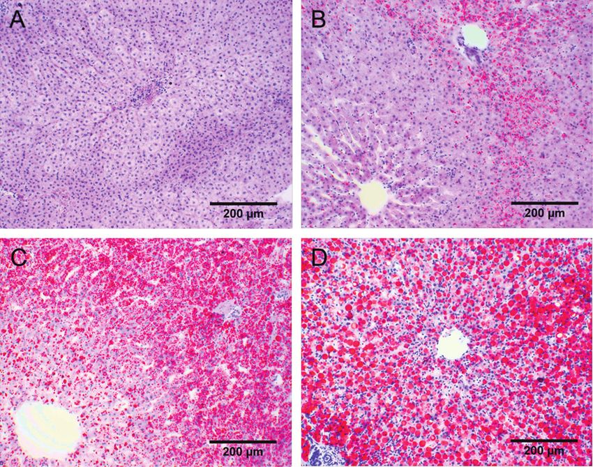
Pietsch et al.: TRANSITION COW METABOLOMICS–PART II                            9229

[based on patterns observed in the partial least squares           et al., 2019) and stained with hematoxylin and eosin
discriminant analysis (PLS-DA) plots of the liver me-              (HE) according to Mayer (1920) and picrosirius red
tabolome, A = medium, B = minor, C = large altera-                 (PSR) according to Constantine (1969) and modified
tions in the liver metabolic profiles between AP and PP            by Grüninger (1996). A cryotome (Reichert-Jung) was
status, details in Part I; Schären et al., 2021]. It was           used to cut the second sample of formaldehyde-fixed
further discovered that the metabotypes corresponded               tissue into 10- to 12-µm sections, which were stained
with the time the animals entered the study: animals               with Sudan III-hematoxylin (SIII; Riedelsheimer and
calving in either period A: November 23, 2015 to Feb-              Büchl-Zimmermann, 2015) and mounted on microscope
ruary 6, 2016 (n = 15), B: February 7 to May 31, 2016              slides. The ethanol-fixed liver sample was embedded in
(n = 28), or C: June 1 to December 3, 2016 (n = 37).               Paraplast (Vogel), cut into 3- to 4-µm sections using a
The treatment groups (VER5, VER10, and CON) were                   sliding microtome, and stained with periodic acid-Schiff
almost equally distributed within the metabotypes (A:              reaction (PAS) as described by Hotchkiss (1948), as
n = 6, 4, and 5; B: n = 13, 8, and 7; C: n = 19, 10, and           well as an amylase control stain (saliva test). The
8 for VER5, VER10, and CON, respectively). Metabo-                 stained sections were stored in the dark at 5 to 7 °C to
type B animals were characterized by: lower milk pro-              preserve stain intensity.
tein percentage and higher milk fat percentage, higher
AP and stronger peripartum decrease in BCS, higher                 Histopathologic Evaluation
risk to be diseased, and higher PP blood bilirubin, fatty
acids, gamma-glutamyltransferase, and triglyceride                    Liver samples were evaluated histologically for the
levels. Reasons for this increased lipomobilization and            following characteristics: fatty infiltration (severity of
clinical sequelae presumably lie in a decreased grass              fat deposition and size of lipid droplets), glycogen stor-
silage quality fed in this period (elaborated in the dis-          age (degree of glycogen storage and size of glycogen
cussion of Part I; Schären et al., 2021).                          particles), and degenerative, inflammatory, fibrotic, and
                                                                   proliferative changes, which included liver cell degen-
Liver Biopsies and Sample Preparation                              eration, severity of hepatitis, total severity of fibrosis,
                                                                   severity of perisinusoidal fibrosis, and the number of
  Each cow underwent 4 liver biopsy procedures at 14               bile ducts. The liver samples were evaluated “blindly”
d AP and 7, 28, and 42 d PP using the technique de-                by a trained veterinarian using a light microscope
scribed by Gohlke et al. (2013). Difficulty in predicting          (CETI-AC 22V IN, TOPIC-B, magnification 40× to
parturition, variable farm management schedules, and               400×). Five liver lobules were randomly evaluated in
the high workload demands on the operators resulted in             each sample using the 10×, 20×, and 40× objectives.
deviations from the scheduled sampling days; T1: mean              Histologic changes were scored as focal (seen in one
12 d AP, range 1 to 26 d; T2: mean 7 d PP, range 4                 liver lobule only), multifocal (seen in 2 or more liver
to 13 d; T3: mean 28 d PP, range 23 to 34 d; and T4:               lobules), or diffuse (seen in 5 liver lobules). A standard-
mean 42 d PP, range 37 to 50 d. The deviations from                ized assessment key was used to evaluate individual
the sampling protocol were taken into account during               histologic changes (Tables 1 and 2).
statistical analysis.                                                 The severity of fatty infiltration of the liver samples
                                                                   was determined using the SIII-stained sections, and a
Sample Preparation                                                 modified scoring system described by Mertens (1992)
                                                                   to grade each sample. In contrast to the system de-
   The samples of fresh liver tissue were carefully rinsed         scribed by Mertens (1992), classification of cellular fat
with sterile isotonic saline solution (0.9% sodium                 infiltration was reduced from 6 to 4 classes and the
chloride solution; Serumwerk Bernburg AG), and any                 scoring modified accordingly. In each of the liver lobule
remaining blood was removed with sterile gauze pads.               zones, which included the centrilobular, intermediate,
The liver samples were cut into 0.5 × 0.5 × 2 cm pieces            and peripheral or periportal zones (Rappaport et al.,
using a scalpel blade and a minimal amount of pressure             1954; Jungermann and Katz, 1989), size of lipid drop-
to avoid crushing the tissue. Two pieces were placed               lets was scored as follows: no lipid droplets seen (0),
in 4% neutral buffered formaldehyde solution, and one              small droplets (1), medium-sized droplets (2), and large
piece was placed in 96% ethanol for a minimum of 24                droplets (3), and the severity of lipid infiltration of he-
hours. The samples were stored in the fixative solutions           patocytes was scored as none (0), mild (1), moderate
at room temperature in the dark.                                   (2), and severe (3). For each of the 3 zones, the total
   One aliquot of formaldehyde-fixed tissue was rou-               number of points was multiplied and the result added
tinely processed for histologic evaluation (Kabisch                [Σ (size of lipid droplets × severity of lipid infiltration

Journal of Dairy Science Vol. 104 No. 8, 2021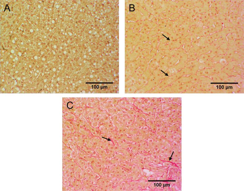
Pietsch et al.: TRANSITION COW METABOLOMICS–PART II                                              9230
Table 1. Applied histologic interpretation key for lesion types in liver tissue samples

Criterion                               Degree                             Definition
Size of lipid droplets                  No droplets                        No droplets seen in section
                                        Small droplets                     Droplets (red to orange) smaller than nucleus of hepatocytes
                                        Medium-sized droplets              Droplets similar in size to nucleus of hepatocytes
                                        Large droplets                     Droplets larger than nucleus of hepatocytes
Size of glycogen particles              No PAS-positive material1          No PAS-positive material
 (according to Mertens, 1992)           Dust-like                          Dust-like magenta to light violet colored glycogen particles
                                        Dust-like to granular              Dust-like and granular glycogen particles
                                        Granular                           Predominantly granular glycogen particle size
                                        Granular to coarse                 Granular and coarse glycogen particles
                                        Coarse                             Predominantly coarse glycogen particles
Hepatocyte degeneration                 No degeneration                    No nuclear death apparent
                                        Degenerated cell nuclei            Nucleus (blue) displaced peripherally by fat vacuole; nuclear death
                                                                           apparent
1
PAS = periodic acid-Schiff.

of hepatocytes)c,i,pl, where c = centrilobular zone, i =                   cell degeneration. When regressive nuclear changes
intermediate zone, and pl = peripheral or periportal                       were seen in only a few hepatocytes, the sample was
zones]. The severity of lipid infiltration was calculated                  considered to have no degeneration. However, when
from the mean of the 5 liver lobules and scored as no                      similar nuclear changes were seen in multiple fields of
lipid infiltration (0), mild (1–6 points), moderate (7–15                  view, hepatic degenerative changes were considered to
points), or severe (16–27 points; Figure 1). The size of                   be present.
lipid droplets was compared with the size of the hepa-                        Inflammatory cell infiltration (severity of hepatitis)
tocyte nuclei in SIII-stained sections. The predominant                    was determined in HE-stained sections by evaluating
size of the lipid droplets was used for analysis.                          the entire tissue section for the number of lymphocytes,
   The degree of glycogen storage and its distribution                     plasma cells, neutrophils, and macrophages in inflam-
within liver lobules were determined in PAS-reaction                       matory foci. The largest number of inflammatory cells
tissue sections (Figure 2). Furthermore, in contrast to                    in a given focus was used for analysis.
the degree of glycogen storage, the predominant size of                       The PSR-stained sections were used to evaluate col-
glycogen particles, according to the terminology used                      lagen deposits and to determine the overall severity of
by Mertens (1992) rather than its percentage or distri-                    fibrosis. This included the presence of collagen deposits
bution, was evaluated in PAS-reaction tissue sections                      in the perisinusoidal space (space of Disse), as well as
(Figure 3).                                                                around the central vein or portal fields.
   The structure of hepatocyte nuclei was evaluated in                        The severity of perisinusoidal fibrosis was also de-
HE-stained sections to determine the presence of liver                     termined using PSR-stained sections. Perisinusoidal

Table 2. Applied histologic interpretation key for the severity of lesions in liver tissue samples

                  Hepatocyte                                                                   Overall                 Perisinusoidal
Degree            vacuolation             Glycogen                  Hepatitis                  fibrosis                fibrosis
No lesion         No vacuoles (red to     No PAS-positive           Fewer than 10              No fibrosis, no red-    No red-staining material
                  orange)                 material1                 inflammatory cells2        staining material       in perisinusoidal area
Mild              Up to 25%3              Dust-like/dust-like       10–30 inflammatory         Connective tissue       Hint of light red staining
                                          and granular, up to       cells2                     proportion up to        in perisinusoidal area
                                          25%4                                                 25%4
Moderate          25% up to 50%3          Granular, up to 50%4      31–60 inflammatory         Connective tissue       Distinct with 10×
                                                                    cells2                     proportion up to        objective, light to dark
                                                                                               50%4                    red, very delicate to
                                                                                                                       several thicker strands,
                                                                                                                       partially diffuse
Severe            More than 50%3          Granular to coarse        More than 60               Connective tissue       Massive, distinct, thick
                                          or coarse, more than      inflammatory cells2        proportion more than    strands, diffuse
                                          50%4                                                 50%4
1
 PAS = periodic acid-Schiff.
2
 Accumulated in foci.
3
 Relative to the parenchymal area of the zone under investigation (centrilobular, intermediate, peripheral or periportal).
4
 Relative to the parenchymal area of the entire section.

Journal of Dairy Science Vol. 104 No. 8, 2021
Pietsch et al.: TRANSITION COW METABOLOMICS–PART II 9231
fibrosis was part of the overall fibrosis but focused on with ηijklm = μ + Gi + Tj + metabotypek + LCl + b1 ×
collagen deposits in the space of Disse (Figure 4). The xijklm + aiklm, where ηijklm is the linear predictor, Gi =
mean number of bile ducts was determined on PSR- treatment group effect (fix; i = 1,2,3 where 1 = CON,
stained sections. The number of bile ducts was counted 2 = VER5, and 3 = VER10), Tj = fixed sampling day
in 5 portal fields and the mean calculated. effect (j = 1,2,3,4, where 1 = 14 d AP, 2 = 7 d PP, 3 =
28 and 42 d PP), metabotypek = metabotype effect (fix;
Data and Statistical Analysis k = 1,2,3, where 1 = A, 2 = B, and 3 = C), LCl =
lactation class effect (fix; l = 1, 2, and 3), and aiklm =
Histopathologic Scores. For analysis of the ordinal random effect of animal m in group i, metabotype k,
variable histopathologic score with repeated measure- and lactation class l, with aiklm ~ N (0, σa2 ) . The differ-
ments within cows, the cumulative logit (random ef- ence between effective and planned sampling time
fect) model based on the threshold concept was used points (xijklm) is accounted for by the linear regression
(McCulloch and Searle, 2001). Let Yijklm be the ordinal coefficient b1. A possible interaction of the factors
response (with r categories) of animal m in treatment metabotype and treatment was tested, but no signifi-
group i, sampling day j, metabotype k, and lactation cance was observed (data not shown). Because of the
class l. Further, let Zijklm be the underlying continuous existing model complexity attributable to the main ef-
latent variable and θn cutpoints (thresholds with n = fects, we did not examine other interactions when con-
1,…, r − 1). Then, at given animal effect, the cumula- sidering the sample size.
tive logit random effect model is given by: For estimation of the model parameters, the
maximum likelihood method implemented in PROC
logit[P(Yijklm ≤ n)] = logit[P(Zijklm ≤ θn)] = θn – ηijklm, GLIMMIX of SAS 9.4 (SAS Institute) was used. The
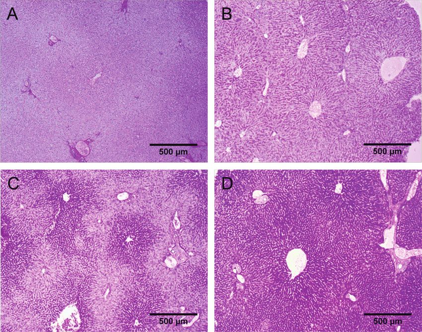
Figure 1. Degree of fatty infiltration of the liver in sections stained with Sudan III-hematoxylin: (A) no fatty infiltration (score 0 points);
(B) mild fatty infiltration (score 3 points); (C) moderate fatty infiltration (score 13 points); and (D) severe fatty infiltration (score 27 points).
Journal of Dairy Science Vol. 104 No. 8, 2021Pietsch et al.: TRANSITION COW METABOLOMICS–PART II 9232
implementation provides F-test (related to the effects Number of Bile Ducts. This trait was quantita-
in the linear predictor), cumulative probabilities, and tive. Accordingly, we evaluated this trait by applying a
odds ratio (OR). For statistical evaluation of the OR, linear mixed model (PROC MIXED of SAS 9.4). The
simultaneous confidence intervals were provided using effect structure was identical to the evaluation of the
the Bonferroni correction. scores as described above. However, a log-transforma-
Correlations. The reported correlation coefficients tion had to be done to ensure normal distribution of
between the histopathologic scores were pooled Spear- the residuals. The least squares means communicated
man correlations (PROC COR of SAS 9.4). The re- for this feature were back-transformed to facilitate as-
ported 2 evaluations levels were: sessment of the results.
1) Evaluation over all data: Reported correlations RESULTS
are based on the pooled estimates within all
combinations of sampling day × metabotype The variables fatty infiltration, glycogen storage, and
(these fixed effects were particularly important, liver cell degeneration changed during the study period
see results F-test). and were also significantly influenced by the metabo-
2) Evaluation per metabotype: Reported correla- type (P ≤ 0.05; Table 3). Metaphylactic treatment
tions were based on the pooled estimates within with butaphosphan and cyanocobalamin (VER5 and
metabotype over all time periods. VER10) had no effect on histomorphologic variables,
and lactation number only affected the incidence of
We chose this form of estimation to avoid a bias of the perisinusoidal fibrosis (P ≤ 0.05). The difference be-
correlation coefficient estimates due to the fixed effects. tween scheduled and effective sampling day of the liver
Figure 2. Degree of glycogen storage in sections with periodic acid-Schiff reaction: (A) no glycogen storage; (B) mild glycogen storage; (C)
moderate glycogen storage; and (D) pronounced glycogen storage.
Journal of Dairy Science Vol. 104 No. 8, 2021Pietsch et al.: TRANSITION COW METABOLOMICS–PART II 9233
biopsies had an effect on the variable fatty infiltration. was also apparent from T2 to T4 (ORT2/T4 = 0.17) and
The OR and 95% confidence intervals (CI) were cal- from T3 to T4 (ORT3/T4 = 0.26). The changes in the
culated to describe differences among different levels distribution of the size of lipid droplets in the liver were
of the effects sampling day, metabotype, and lactation similar; lipid droplets were scant and mostly small at
number. The OR was considered to be significantly dif- T1 (Figure 5B) but considerably larger at T2, T3, and
ferent from the value one when the 95% CI did not T4, and lipid droplets were seen in almost all PP tis-
overlap with the value one. Only significant OR are sue sections. The chance of smaller droplets at T1 was
shown in the text hereafter. The complete set of OR is 27.5 times the chance at T2 (ORT1/T2), 36.6 times the
depicted in Appendix Table A1. chance at T3 (ORT1/T3), and 13.9 times the chance at
T4 (ORT1/T4). This pattern of decreasing lipid droplets
Fatty Infiltration size was furthermore apparent from T3 to T4 (ORT3/T4
= 0.38).
The degree of fatty infiltration of the liver was mild The metabotypes had an effect (P ≤ 0.05) on fatty
to minimal before parturition (T1; Figure 5A) and in- infiltration and lipid droplet size (Table 3). The chance
creased after parturition; it was most severe at T2 (d of a lower degree of fatty infiltration in metabotype
4–13 PP). The chance of lower fatty infiltration at T1 A (1% severe, 11% moderate, 61% mild, 27% no fatty
was 63.8 times the chance at T2 (ORT1/T2). Fatty infil- infiltration) was 7.7 times the chance in metabotype
tration decreased with increasing DIM; the chance of B (ORA/B; 6% severe, 45% moderate, 44% mild, 5%
lower fatty infiltration at T1 was 42.2 times the chance no fatty infiltration). In contrast, the chance of lower
at T3 (ORT1/T3) and 11.1 times the chance at T4 fatty infiltration in metabotype B was only 0.38 times
(ORT1/T4). This pattern of decreasing fatty infiltration (ORB/C) the chance observed in metabotype C (3%
Figure 3. Size of glycogen particles in sections with periodic acid-Schiff reaction: (A) no glycogen; (B) dust-like glycogen particles; (C) dust-
like to granular glycogen particles; and (D) coarse glycogen particles.
Journal of Dairy Science Vol. 104 No. 8, 2021Pietsch et al.: TRANSITION COW METABOLOMICS–PART II 9234
Figure 4. Degree of perisinusoidal fibrosis in sections stained with picrosirius red stain. Arrows point to perisinusoidal fibrosis: (A) no fibro-
sis; (B) mild fibrosis; and (C) moderate fibrosis. Severe fibrosis did not occur and is therefore not shown.
severe, 26% moderate, 60% mild, 11% no fatty infiltra- Glycogen Storage
tion). Additionally, the chance of a lower degree of fatty
infiltration in metabotype A was 2.9 (ORA/C) times the The amount of glycogen stored in the liver was
chance in metabotype C. The chance of smaller lipid comparatively large at T1 (Figure 5C, D), reached a
droplets sizes was 3.8 (ORA/B) and 4.0 (ORA/C) times minimum at T2, and then increased toward T3 (Figure
the chance in metabotype A, compared with metabo- 5C). The chance of a lower degree of glycogen storage
type B and C, respectively. at T1 was 0.02 (ORT1/T2) and 0.20 times (ORT1/T3), the
Table 3. P-values for the F-test for the fixed effects of the model [sampling day, metabotype, treatment group, lactation number, and regression
for the correction of the difference between scheduled and effective sampling day of biopsy, Regr Corr Diff T (b1)] for the histologic assessment
of liver biopsy samples from 80 German Holstein cows during the transition period
Degenerative, inflammatory, fibrotic,
Fat Glycogen and proliferative lesion2
Droplet Particle Hepat Overall
Fixed effect1 Degree size Degree size deg Hep fib PSF NBD
Sampling dayPietsch et al.: TRANSITION COW METABOLOMICS–PART II 9235 Figure 5. Probability estimation for the rating of the degree of fatty liver infiltration (A); size of fat droplets (B); degree of glycogen stor- age (C); and size of glycogen particles (D) in 80 German Holstein cows during the transition period. The columns show the probabilities at the sampling day of liver biopsy: T1 [12 d antepartum (AP)], T2 [7 d postpartum (PP)], T3 (28 d PP), and T4 (42 d PP). A and C, level of infiltra- tion/storage: no, mild, moderate, and severe infiltration; B, size of fat droplets: no, small, medium, large droplets; D, particle size of deposits: G1, no or dust-like; G2, dust-like to granular; G3, granular; G4, granular to coarse; G5, coarse. For significance between the sampling days, see Table 3 and Appendix Table A1. chance at T2 and T3, respectively (Appendix Table in metabotype C (51% severe, 44% moderate, 5% mild, A1). This relation changed after T2; the chance of low- ORA/C; Appendix Table A1). Similarly, the chance for er glycogen storage at T2 was 8.1 (ORT2/T3) and 14.6 smaller glycogen particles was 0.25 (ORA/B) and 0.19 times (ORT2/T4) the chance at T3 and T4, respectively. (ORA/C) times the chance observed in metabotype B With respect to the size of glycogen particles, the and C, respectively. classes “no glycogen” and “dust-like” were pooled be- cause of their low frequencies and because it facilitated Degenerative, Inflammatory, Fibrotic, presentation of the results (Figure 5D). The structure and Proliferative Changes of the glycogen deposits was mostly larger glycogen particle size at T1, which had decreased to a minimum The degree of liver cell degeneration was the only at T2, after which time the particle size increased again variable that was affected by sampling day (P ≤ 0.05; at T3. The chance of smaller glycogen particles at T1 Table 3). Regressive nuclear changes in hepatocytes was 0.05 (ORT1/T2) and 0.30 times (ORT1/T3) the chance were rare (1%) at T1 but occurred in 24, 26, and 13% at T2 and T3, respectively (Appendix Table A1). Gly- of hepatocytes, respectively, at T2, T3, and T4. The cogen particle size increased with increasing DIM; the chance of normal hepatocyte nuclei at T1 was 28.0 chance of smaller glycogen particles deposits at T2 was (ORT1/T2), 31.1 (ORT1/T3), and 13.5 times (ORT1/T4) 6.5 (ORT2/T3) and 9.4 times (ORT2/T4) the chance at T3 the chance at T2, T3, and T4, respectively (Appen- and T4, respectively. dix Table A1). With respect to the metabotypes, the The metabotype also had an effect on the degree of chance of normal hepatocyte nuclei in metabotype A glycogen storage and the size of glycogen particle de- (5% degenerative) was 4.43 times (ORA/B) the chance posits. The chance of lower glycogen storage in metabo- in B (19% degenerative). Both metabotype A and B type A (77% severe, 21% moderate, 2% mild) was 0.29 did not differ from metabotype C (11% degenerative). times the chance in metabotype B (49% severe, 45% Metabotypes also affected the degree of perisinu- moderate, 6% mild, ORA/B) and 0.31 times the chance soidal fibrosis. The chance of low-grade perisinusoidal Journal of Dairy Science Vol. 104 No. 8, 2021
Pietsch et al.: TRANSITION COW METABOLOMICS–PART II                                  9236

fibrosis in metabotype A (6% moderate, 45% mild,                            Correlations Among Variables
49% absent) was 3.7 times the chance in metabotype
B (18% moderate, 61% mild, 21% absent; ORA/B; Ap-                              There were several correlations (P ≤ 0.05) between
pendix Table A1). Both metabotypes A and B did not                          the histologic variables (Appendix Table A2); however,
differ from C (16% moderate, 61% mild, 23% absent).                         the estimates varied depending on the metabotypes.
   Degree of perisinusoidal fibrosis was the only variable                  The severity of fatty infiltration and size of lipid drop-
affected (P ≤ 0.05) by lactation number; this lesion                        lets were positively correlated (P ≤ 0.05) with large
was more common in cows with lactation numbers 5 or                         correlation coefficients. The same was true for the re-
greater (L3) than in younger cows. The chance of less                       lationship between degree and particle size of glycogen
severe perisinusoidal fibrosis in cows of L1 and L2 were                    deposits (Appendix Table A2).
4.2 (ORL1/L3) and 4.4 times (ORL2/L3) the chance in                            There was a negative correlation (P ≤ 0.05) between
cows of L3 (Appendix Table A1, Figure 6).                                   the degree of glycogen storage and severity of fatty
   For the number of bile ducts in hepatic tissue, there                    infiltration in metabotype B and C, between degree of
was a trend for differences among sampling days (F-                         glycogen deposits and size of lipid droplets in metabo-
test, P = 0.085; Table 3); the largest difference oc-                       type C, and between the size of glycogen particles and
curred between T2 and T4 (Tukey-test, P = 0.11). The                        severity of fatty infiltration in metabotype B and C
numbers for T1, T2, T3, and T4 obtained after back-                         (Appendix Table A2).
transformation were 1.76 ± 0.05 (LSM ± SE), 1.77 ±                             Fatty infiltration was positively correlated (P ≤ 0.05)
0.04, 1.80 ± 0.05, and 1.92 ± 0.06, respectively.                           with hepatocyte degeneration in all metabotypes. He-
   To assess the level of hepatitis, the severity of lym-                   patocyte degeneration was also negatively correlated (P
phocytic infiltration of the liver tissue was determined.                   ≤ 0.05) with the degree of glycogen and size of glycogen
Among the 4 biopsy time points, severe infiltration was                     particles; the correlations were strongest for the degree
seen in 10 to 15%, moderate infiltration in 19 to 25%,                      of deposits in metabotype B and C, and strongest for
mild infiltration in 50 to 55%, and no infiltration in 11                   the size of glycogen particles in metabotype B. The
to 16% of cows. Not all specimens had inflammatory                          overall degree of fibrosis was positively correlated with
cell infiltration but periportal lymphocytic infiltration                   lipid droplet size in metabotype A and negatively corre-
was most common and neutrophils and macrophages                             lated with the size of glycogen particles in metabotype
were rare. The level of hepatitis was also assessed based                   C.
on the overall degree of fibrosis. Among the sampling                          The overall degree of fibrosis was positively corre-
days, the overall degree of fibrosis was moderate in less                   lated with the degree of hepatitis and with the degree
than 1% and mild in 44 to 64% of cows, and 36 to 56%                        of perisinusoidal fibrosis in all metabotypes, whereas
of cows had no fibrosis. Severe fibrosis was not seen.                      the degree of hepatitis was positively correlated (P ≤
The degrees of cellular infiltration and fibrosis were not                  0.05) with hepatocyte degeneration only in metabotype
affected by any of the effects studied (Table 3).                           B. In metabotype A the degree of perisinusoidal fibrosis
                                                                            was positively correlated (P ≤ 0.05) with the degree
                                                                            of hepatitis, whereas the overall degree of fibrosis was
                                                                            positively correlated with hepatocyte degeneration.
                                                                            There were positive correlations (P ≤ 0.05) between
                                                                            the number of bile ducts and several variables that were
                                                                            limited to certain metabotypes including the degree of
                                                                            fatty infiltration (metabotype A and B), lipid droplet
                                                                            size (metabotype A), degree of hepatitis (metabotype
                                                                            A), and overall degree of fibrosis (metabotype A and
                                                                            B). The degree of glycogen deposits was negatively cor-
                                                                            related (P ≤ 0.05) with the overall degree of fibrosis in
                                                                            metabotype B.

                                                                                                 DISCUSSION
   Figure 6. Probability estimation for the rating of the degree of
perisinusoidal fibrosis in 80 German Holstein cows during the transi-       Usefulness of the Experimental Design
tion period. The columns show the probabilities for cows in different       for the Histologic Assessment
lactation groups at the sampling day of liver biopsy: second lactation
(L1); third and fourth lactations (L2); fifth lactation and higher (L3).
Degree of fibrosis: no, mild, moderate, and severe fibrosis. For signifi-    The biopsy protocol used in our study for the assess-
cance between the sampling days, see Table 3 and Appendix Table A1.         ment of histomorphologic changes in liver tissue during

Journal of Dairy Science Vol. 104 No. 8, 2021
Pietsch et al.: TRANSITION COW METABOLOMICS–PART II 9237 the transition period of dairy cows was similar to that analysis of substances such as total lipids (TL), triacyl- used by others (Rukkwamsuk et al., 1999; Tharwat et glycerols, and glycogen would also be useful. al., 2012; Gohlke et al., 2013). Strict adherence to the The interpretation key we chose was based on pre- experimental protocol was difficult in this commercial vious studies (Mertens, 1992; Rehage et al., 1996; dairy herd for a variety of reasons but temporal devia- Haudum, 2009) and was useful for the histologic and tions from the protocol were taken into account in the histochemical assessment of liver changes in dairy cows. statistical analysis. We feel that our experimental pro- Our findings relating to fatty infiltration of the liver tocol was sound because the histomorphologic findings (Reid, 1980; Gerloff et al., 1986; Jorritsma et al., 2001) largely reflected the metabolic changes observed during and glycogen deposits (Rukkwamsuk et al., 1999; We- the transition period (see discussion section below). ber et al., 2013; McCarthy et al., 2015) were in good The biopsy technique (Gohlke et al., 2013) provided agreement with previous studies. The characterization tissue samples of adequate size. Liver biopsy specimens of the size of glycogen particles (Mertens, 1992; Figure are representative of the whole liver regarding lipid 3) allowed a more detailed description of the structural content, fatty acid composition (Gaál and Husvéth, processes during glycogen storage and metabolism 1983; Gerspach et al., 2017), and glycogen concentra- than quantitative descriptions alone. Until now, there tion (Duplessis et al., 2020) in dairy cows. We did not have been no studies on the dynamics of inflammatory, see progressive liver fibrosis and therefore ruled out degenerative, fibrotic, and proliferative hepatic lesions an adverse effect of repeated biopsy sample collection. in dairy cows during the transition period. Finally, it In contrast, progressive fibrosis was seen in human should be remembered that the use of any interpreta- medicine after repeat liver biopsy was performed for tion key is subjective and that its validity depends on several years (Okanoue et al., 2005). In dairy cows, the experience of the investigator; in the present study, repeated liver biopsies do not seem to have an effect on the histologic specimens were assessed and classified by the acute-phase proteins haptoglobin, serum amyloid a closely supervised postgraduate veterinarian. Over- A, fibrinogen, and IL-6, which have high sensitivity, or all, we feel that the experimental design of the present on body temperature, DMI, milk yield, and total white study was suitable for the assessment of histopathologic blood cell count (Vels et al., 2009; Jawor et al., 2016). liver changes in dairy cows during the transition period. Fixation and preservation of tissue samples in for- malin and alcohol, as well as storage at room tempera- Fat Accumulation in the Liver as a Reflection ture, provided high-quality and artifact-free histologic of a Negative Energy Balance sections. Different stains and histochemical reactions allowed good differentiation of histologic and histo- We observed fat deposits in the liver of cows as early chemical characteristics of the tissue. Staining with as 2 wk AP. This was most likely due to a reduction HE provided a good overview and differentiation of in- in DMI, which starts before parturition and causes an flammatory cells but did not allow distinction between increase in free fatty acids (FFA; Bertics et al., 1992; hydropic degeneration of hepatocytes, hepatocellular Rukkwamsuk et al., 1999; Gross et al., 2013). Fatty in- glycogen deposits, and small fat droplets (Levene et filtration of the liver reached a maximum at T2, which al., 2012); PAS reactions and SIII stains are required to corresponded with the time of maximum mobilization stain glycogen and fat, respectively. The PAS reaction of body fat. At 7 d (4–13 d; T2) PP, 66% of the cows must be combined with diastase to allow for the differ- had moderate to severe fatty infiltration, which was entiation of easily digestible carbohydrates (glycogen) higher than frequencies reported earlier (Reid, 1980; and other PAS-positive substances such as glycolipids Gerloff et al., 1986; Jorritsma et al., 2001). Even though or neutral mucopolysaccharides (Riedelsheimer and fatty liver has been reported to increase the morbidity Büchl-Zimmermann, 2015). We were able to identify and mortality rate of production diseases (Breukink an increase in collagen and elastic fibers in the PSR- and Wensing, 1997) with a mortality rate of 25% for stained sections. Electron microscopic and immuno- so-called fat cows (Morrow et al., 1979), all cows of histochemical examinations would offer additional the present study survived the transition period. Our information about nuclear changes including nuclear findings strongly suggest that hepatic lipidosis of dairy hyperchromasia and karyopyknosis, changes in cell cows is reversible (Johannsen et al., 1988; Rukkwamsuk organelles (mitrochondria and endoplasmic reticulum), et al., 1999; Fiore et al., 2017) and that fat deposits in and the detection of other substances such as specific the liver may disappear later in lactation. Thus, the collagen fiber types (Collins and Reid, 1980; Konomi histologic findings of the liver reflect the normal course et al., 1981; Johannsen et al., 1993). The biochemical of a negative energy balance (NEB) in dairy cows, Journal of Dairy Science Vol. 104 No. 8, 2021
Pietsch et al.: TRANSITION COW METABOLOMICS–PART II                             9238

which was also reflected in our clinical chemistry data            This was in agreement with previous reports of inflam-
(illustrated in Part I; Schären et al., 2021b).                    matory lesions in the liver and in body fat of dairy cows
                                                                   in the first few weeks of lactation (Fürll, 1989; Mertens,
Negative Correlation Between Fat                                   1992; Kalaitzakis et al., 2007). The insufficient energy
and Glycogen Storage                                               availability, for instance during the transition period,
                                                                   impairs the ability of dairy cows to mount a sufficient
  The first biopsy sample collected PP showed a                    immune response (McCarthy et al., 2016). In the
massive increase in fatty infiltration of the liver ac-            context of fat mobilization, BHB (Suriyasathaporn et
companied by a decrease in glycogen content. This was              al., 2000), and FFA (Lacetera et al., 2004) impair the
evidenced histologically by an increase in large lipid             immune response or lead to a higher incidence of infec-
droplets and a decrease in the particle size of glyco-             tions, which can predispose to inflammatory diseases,
gen. This means that the dynamics of fat and glycogen              such as metritis, mastitis, or hepatitis, as observed in
metabolism contrasted one another. Our findings of                 our study. Overall, inflammatory signs were not cor-
a negative correlation between fat and glycogen me-                related with fatty infiltration of the liver, which was
tabolism confirmed earlier studies, which were based               in agreement with a previous study (Bradford et al.,
on only one or 2 biopsy sampling times (West, 1990;                2015); however, the occurrence of moderate to severe
Mertens, 1992; Gerspach et al., 2017) and therefore                lymphocytic infiltration of the liver in almost 40% of
unable to document histologic features throughout the              the cows appears to justify the diagnosis of steatohepa-
transition period. Abatement of the NEB over time                  titis.
caused a decrease in fat content and a recovery of the                In contrast, hepatocyte degeneration was highly cor-
glycogen deposits in the liver parenchyma by the end of            related with fatty infiltration of the liver. It is believed
the study period at d 42 (37–50 d; T4).                            that previously formed small lipid droplets coalesce
                                                                   (Mertens, 1992; Walther and Farese, 2012) to form large
The Effect of Metabotypes on Energy Metabolism                     droplets, which displace and suppress the cell nucleus,
                                                                   possibly giving rise to a mechanical form of nuclear
   A pronounced difference in energy metabolism was                degeneration (Johannsen et al., 1988, Fürll, 1989). The
observed between the metabotypes. An overview of the               proportion of triacylglycerol of TL in liver tissue of
effects on production and clinical traits, and clinical            German Holstein cows remains almost constant when
chemistry is given in Part I (Schären et al., 2021b). In           the content of TL is 100 mg/g of fresh liver tissue or
metabotype B pre-fresh cows had a higher BCS than                  greater (Starke et al., 2010), and therefore, one can
metabotype A and C, decreasing by one score from                   assume that severe hepatic lipidosis is characterized
3.11 at T1 to 2.19 at 14 d PP, which was consistent                by the accumulation of other lipid fractions, such as
with a much more pronounced fat mobilization than in               cholesterol, cholesterol ester, and FFA in hepatocytes
the cows of the other metabotypes. Parallelly, cows of             (Reid et al., 1977; Collins and Reid, 1980; Bobe et al.,
metabotype B had higher milk fat content and higher                2004). In human medicine, it has been shown that FFA
blood concentrations of bilirubin, FFA and triacylglyc-            have a toxic effect on hepatocytes and that fat vacuoles
erols, higher activities of gamma-glutamyl transferase,            have a protective effect (Cusi, 2009; Garbarino and
and a higher change to be diseased during the first 30             Sturley, 2009; Neuschwander-Tetri, 2010). Whether this
DIM compared with the cows of the other metabotypes.               also applies to cows with fatty infiltration of the liver
These clinical variables are consistent with the observed          is not known but is currently under investigation [K.
increased fatty infiltration of the liver in metabotype B.         B. Theinert (Leipzig University, Germany), T. Snedec
                                                                   (Leipzig University, Germany), F. Pietsch (Leipzig Uni-
Findings Suggestive of Steatohepatitis and Potential               versity, Germany), S. Theile (Leipzig University, Ger-
Fibrous Liver Changes in Dairy Cows                                many), A.-S. Leonhardt (Leipzig University, Germany),
                                                                   J. Spilke (Martin-Luther University, Halle-Wittenberg,
  When inflammatory cells were present in liver                    Germany), E. Bannert (Leipzig University, Germany),
samples, the main finding in the transition period was             S. Pichelmann (University of Fribourg, Switzerland),
lymphocytic, periportal hepatitis; 39% of the cows had             H. Fuhrmann (Leipzig University, Germany), W.
moderate to severe hepatitis. In all likelihood, this was          Baumgartner (University of Veterinary Medicine, Vi-
nonspecific, reactive hepatitis characterized by focal or          enna, Austria), M. Schären (Leipzig University, Ger-
diffuse lymphohistiocytic infiltration of the portal area          many), and A. Starke (Leipzig University, Germany);
of the liver (Neumann and Danner, 1998; Rothuizen                  unpublished data]. Fatty infiltration of the liver is a
and van den Ingh, 1998; Cullen and Stalker, 2016).                 transient occurrence in dairy cows, and therefore, the

Journal of Dairy Science Vol. 104 No. 8, 2021
Pietsch et al.: TRANSITION COW METABOLOMICS–PART II 9239 potential for a lipotoxic effect remains when FFA are when the triacylglycerol content exceeded 150 mg/g mobilized from triacylglycerol (Schaffer, 2003). fresh liver tissue. We did not observe a correlation between fatty Interestingly, there were trends toward the devel- infiltration and fibrosis of the liver, possibly because opment of steatohepatitis and fibrotic changes in of differences in glycogen metabolism in people and metabotype B animals, which suggests that inflamma- ruminants. In human beings, glycogen is generated pri- tory, degenerative, fibrotic, and proliferative reactions marily in musculature in response to physical activity, in the liver are possible during periods of inadequate whereas in ruminants, it occurs predominantly in the feed quality. Number of bile ducts was correlated with liver (Gardner et al., 2014). Of note, we were able to degree of hepatic lipidosis, extent and size of glycogen document glycogen deposits in the biopsy specimens particles, and overall degree of fibrosis, and there was throughout the transition period, and there were sever- a trend toward a correlation for perisinusoidal fibrosis. al negative trends in the relationships between glycogen Furthermore, cows of metabotype B had more severe deposits and degree of fibrosis of the liver (Appendix NEB and a higher morbidity rate. Possibly, the more Table A2). This could be interpreted as a regenerative pronounced NEB in metabotype B led to an increase effect on the bovine liver with the potential to limit the in lipolysis and in turn to inflammatory dysregulation adverse effect of fatty infiltration. Perisinusoidal fibro- affected by FFA (Contreras et al., 2018). These ob- sis was more common in older cows than in younger servations are in all likelihood related to poor silage. cows. Perisinusoidal fibrosis is thought to have an ad- Silage with elevated butyrate concentrations could verse effect on liver function in humans (Schaffner et increase BHB production accompanied by a decrease al., 1963) and the loss of microvilli and characteristic in feed intake (Stöber and Scholz, 1991). The first pass fenestrations in the space of Disse in animals. The sinu- effect, which is the uptake of an absorbed substance soids are transformed functionally and morphologically by the liver before the substance reaches the systemic into capillaries (so-called capillarization of sinusoids), circulation, could also have played a role because it which is thought to impair the exchange of metaboli- may have caused flooding of the liver with toxins and in cally important substances between the hepatocytes turn, the development of inflammatory, degenerative, and the blood plasma (Braet and Wisse, 2002; Cul- fibrotic, and proliferative liver lesions. Mycotoxins, for len and Stalker, 2016). We were unable to determine instance aflatoxins, are thought to favor the develop- whether perisinusoidal fibrosis had an adverse effect on ment of liver fibrosis, bile duct proliferation (Newberne liver function. We suspect that perisinusoidal fibrosis and Butler, 1969; Lynch et al., 1971), and immunosup- is the result of increased blood pressure because blood pression (Sultana and Hanif, 2009). More severe injury, flow to the liver is increased in high-producing dairy or conditions that inhibit replication of mature cells, cows (Sangsritavong et al., 2002), leading to increased causes proliferation of bipotential progenitor cells (or pressure in the liver sinusoids. Milk yield increases with oval cells), referred to as ductular reaction. These cells increasing lactation number to a maximum in the fifth can mature into hepatocytes or bile duct epithelium lactation, and this could have had an effect on the de- (Sell, 1994; Cullen and Stalker, 2016). This could pro- gree of perisinusoidal fibrosis seen in our study. vide an alternative explanation for the proliferation of The swelling of the liver parenchyma causes an in- bile ducts seen in cows in metabotype B. Moreover, crease in intrahepatic pressure resulting in compression aflatoxins, fumonisin, and pyrrolizidine alkaloids are of the liver sinusoids (Orrego et al., 1981). In human hepatotoxic and cancerogenic in cattle (Sultana and medicine, it is hypothesized that sinusoidal pressure in- Hanif, 2009; Petzinger, 2011). creases because of tensile and shear forces accompanied by compression, which is caused by volume increase CONCLUSIONS attributable to fat and inflammatory cell infiltration, ultimately stimulating fibrosis (Mueller, 2016). Reid The liver biopsy technique, the histopathologic in- and Collins (1980) reported an increased liver cell vol- terpretation key, and the histologic stains used were ume and compression of hepatic sinusoids caused by suitable for addressing the goals of this study and for fat-laden liver cells in cows with fatty liver. Haudum et the histopathologic assessment of liver tissue from dairy al. (2011) and Starke et al. (2011) described the adverse cows. Electron microscopic and immunohistochemical effects of the degree of hepatic lipidosis on the venous examinations were not done but could increase the pulsatility index and blood flow velocity and thus on information regarding liver lesions in the transition liver function in dairy cows. This observation was based period at the cellular level. The NEB in dairy cows on a reduction in elasticity of blood vessels, which oc- during the transition period caused massive accumu- curred as a result of swelling of the liver parenchyma lation of fat in the liver accompanied by pronounced Journal of Dairy Science Vol. 104 No. 8, 2021
Pietsch et al.: TRANSITION COW METABOLOMICS–PART II 9240
glycogen depletion. This negative correlation between Constantine, V. S. 1969. A combined tissue stain for the selective
staining of collagen, elastic fibers and acidic carbohydrates. J. In-
fat and glycogen metabolism was reversible. The oc- vest. Dermatol. 52:353–356. https://doi.org/10.1038/jid.1969.60.
currence of numerous inflammatory cells in liver tissue Contreras, G. A., C. Strieder-Barboza, and J. De Koster. 2018. Sym-
and hepatocyte degeneration suggested a diagnosis of posium review: Modulating adipose tissue lipolysis and remodeling
to improve immune function during the transition period and early
steatohepatitis in German Holstein cows. We observed lactation of dairy cows. J. Dairy Sci. 101:2737–2752. https://doi
a striking increase in perisinusoidal fibrosis in older .org/10.3168/jds.2017-13340.
cows, which could be a precursor of liver fibrosis. The Cullen, J. M., and M. J. Stalker. 2016. Liver and Biliary System. Pages
258–352 in Jubb, Kennedy, and Palmer's Pathology of Domestic
histopathological features show a strong interrelation Animals. M. G. Maxie, ed. 6th ed. Vol. 2. Elsevier Health Sciences.
with the metabotypes identified in Part I (Schären et Cusi, K. 2009. Role of insulin resistance and lipotoxicity in non-alco-
al., 2021b). Cows with little alterations in their liver holic steatohepatitis. Clin. Liver Dis. 13:545–563. https://doi.org/
10.1016/j.cld.2009.07.009.
metabolome across the transition period (metabotype Duplessis, M., L. Blais, W. Poisson, and C. L. Girard. 2020. Extrapo-
B) exhibited an increased risk of fatty infiltration of the lation of hepatic glycogen concentration of the whole organ by
liver, lower glycogen storage and an interrelation with performing a liver biopsy. J. Dairy Sci. 103:4858–4862. https://doi
.org/10.3168/jds.2019-17905.
degenerative, inflammatory, fibrotic, and proliferative Edmonson, A. J., I. J. Lean, L. D. Weaver, T. Farver, and G. Web-
traits. These findings support the assumption of a bad ster. 1989. A body condition scoring chart for Holstein dairy
grass silage quality as a causative factor. cows. J. Dairy Sci. 72:68–78. https://doi.org/10.3168/jds.S0022
-0302(89)79081-0.
Elliot, J. M. 1976. The glucose economy of the lactating dairy cow.
Pages 59–66 in Proc. Cornell Nutr. Conf. Feed Manuf., Ithaca,
ACKNOWLEDGMENTS NY. Cornell University.
Fiore, E., G. Piccione, L. Perillo, A. Barberio, E. Manuali, M. Mor-
We thank the management and employees of gante, and M. Gianesella. 2017. Hepatic lipidosis in high-yielding
Wirtschaftshof Sachsenland Standort Bräunsdorf for dairy cows during the transition period: Haematochemical and
histopathological findings. Anim. Prod. Sci. 57:74–80. https://doi
their help during the study and afterwards, the team .org/10.1071/AN15262.
at the Clinic for Ruminants and Swine and at the Fürll, M. 1989. Vorkommen, Ätiologie, Pathogenese, Diagnostik und
Institute of Veterinary Pathology for their help with medikamentelle Beeinflussung von Leberschäden beim Rind, DVM
Thesis, Veterinärmedizinische Fakultät, Universität Leipzig, Ger-
the design and execution of the study. Furthermore, we many. [in German]
thank Bayer Animal Health GmbH and ESAOTE Bio- Gaál, T., and F. Husvéth. 1983. Comparison of the liver biopsy sample
medica Deutschland GmbH for the financial support. and the “whole liver” in respect of lipid content and fatty acid
composition of lipids. Acta Vet. Hung. 31:51–56.
The authors have not stated any conflicts of interest. Garbarino, J., and S. L. Sturley. 2009. Saturated with fat: New per-
spectives on lipotoxicity. Curr. Opin. Clin. Nutr. Metab. Care
12:110–116. https://doi.org/10.1097/MCO.0b013e32832182ee.
REFERENCES Gardner, G. E., P. McGilchrist, and D. W. Pethick. 2014. Ruminant
glycogen metabolism. Anim. Prod. Sci. 54:1575–1583. https://doi
Aschenbach, J. R., N. B. Kristensen, S. S. Donkin, H. M. Hammon, .org/10.1071/AN14434.
and G. B. Penner. 2010. Gluconeogenesis in dairy cows: The secret Gerloff, B. J., T. H. Herdt, and R. S. Emery. 1986. Relationship of
of making sweet milk from sour dough. IUBMB Life 62:869–877. hepatic lipidosis to health and performance in dairy cattle. J. Am.
https://doi.org/10.1002/iub.400. Vet. Med. Assoc. 188:845–850.
Bergman, E. N. 1990. Energy contributions of volatile fatty acids from Gerspach, C., S. Imhasly, R. Klingler, M. Hilbe, S. Hartnack, and M.
the gastrointestinal tract in various species. Physiol. Rev. 70:567– Ruetten. 2017. Variation in fat content between liver lobes and
590. https://doi.org/10.1152/physrev.1990.70.2.567. comparison with histopathological scores in dairy cows with fatty
Bertics, S. J., R. R. Grummer, C. Cadorniga-Valino, and E. E. Stod- liver. BMC Vet. Res. 13:98. https://doi.org/10.1186/s12917-017
dard. 1992. Effect of prepartum dry matter intake on liver triglyc- -1004-9.
eride concentration and early lactation. J. Dairy Sci. 75:1914–1922. Gohlke, A., C. J. Ingelmann, G. Nürnberg, J. M. Weitzel, H. M. Ham-
https://doi.org/10.3168/jds.S0022-0302(92)77951-X. mon, S. Görs, A. Starke, S. Wolffram, and C. C. Metges. 2013.
Bobe, G., J. W. Young, and D. C. Beitz. 2004. Invited review: Pa- Influence of 4-week intraduodenal supplementation of quercetin
thology, etiology, prevention, and treatment of fatty liver in dairy on performance, glucose metabolism, and mRNA abundance of
cows. J. Dairy Sci. 87:3105–3124. https://doi.org/10.3168/jds genes related to glucose metabolism and antioxidative status in
.S0022-0302(04)73446-3. dairy cows. J. Dairy Sci. 96:6986–7000. https://doi.org/10.3168/
Bradford, B. J., K. Yuan, J. K. Farney, L. K. Mamedova, and A. J. jds.2013-6852.
Carpenter. 2015. Invited review: Inflammation during the transi- Gross, J. J., F. J. Schwarz, K. Eder, H. A. van Dorland, and R. M.
tion to lactation: new adventures with an old flame. J. Dairy Sci. Bruckmaier. 2013. Liver fat content and lipid metabolism in dairy
98:6631–6650. https://doi.org/10.3168/jds.2015-9683. cows during early lactation and during a mid-lactation feed re-
Braet, F., and E. Wisse. 2002. Structural and functional aspects of striction. J. Dairy Sci. 96:5008–5017. https://doi.org/10.3168/jds
liver sinusoidal endothelial cell fenestrae: A review. Comp. Hepa- .2012-6245.
tol. 1:1. https://doi.org/10.1186/1476-5926-1-1. Grüninger, B. U. 1996. Zur Pathogenese von Angiopathien im Endo-
Breukink, H. J., and T. Wensing. 1997. Pathophysiology of the liver metrium der Stute – Morphologisch-funktionelle Untersuchungen,
in high yielding dairy cows and its consequences for health and DVM Thesis, Univ. Leipzig, Germany. [in German]
production. Isr. J. Vet. Med. 52:66–72. Haudum, A. 2009. Echotexturanalyse des Lebergewebes zur nicht-in-
Collins, R. A., and I. M. Reid. 1980. A correlated biochemical and vasiven Bestimmung des Leberfettgehaltes bei Milchrindern, DVM
stereological study of periparturient fatty liver in the dairy Thesis, Tierärztliche Hochschule Hannover, Germany. [in German]
cow. Res. Vet. Sci. 28:373–376. https://doi.org/10.1016/S0034 Haudum, A., A. Starke, M. Beyerbach, P. Wohlsein, and J. Rehage.
-5288(18)32725-5. 2011. Ultrasonographic assessment of liver dimensions in dairy
Journal of Dairy Science Vol. 104 No. 8, 2021You can also read