Structural polymorphism and substrate promiscuity of a ribosome-associated molecular chaperone - MR
←
→
Page content transcription
If your browser does not render page correctly, please read the page content below
Magn. Reson., 2, 375–386, 2021                                                                            Open Access
https://doi.org/10.5194/mr-2-375-2021
© Author(s) 2021. This work is distributed under
the Creative Commons Attribution 4.0 License.

         Structural polymorphism and substrate promiscuity of
              a ribosome-associated molecular chaperone
               Chih-Ting Huang1 , Yei-Chen Lai2 , Szu-Yun Chen1 , Meng-Ru Ho1 , Yun-Wei Chiang2 , and
                                              Shang-Te Danny Hsu1,3
                          1 Instituteof Biological Chemistry, Academia Sinica, Taipei 11529, Taiwan
                      2 Department   of Chemistry, National Tsing Hua University, Hsichu 30013, Taiwan
                     3 Institute of Biochemical Sciences, National Taiwan University, Taipei 106, Taiwan

                             Correspondence: Shang-Te Danny Hsu (sthsu@gate.sinica.edu.tw)

                                  Received: 16 January 2021 – Discussion started: 25 January 2021
                              Revised: 12 April 2021 – Accepted: 2 May 2021 – Published: 4 June 2021

       Abstract. Trigger factor (TF) is a highly conserved multi-domain molecular chaperone that exerts its chaperone
       activity at the ribosomal tunnel exit from which newly synthesized nascent chains emerge. TF also displays
       promiscuous substrate binding for a large number of cytosolic proteins independent of ribosome binding. We
       asked how TF recognizes a variety of substrates while existing in a monomer–dimer equilibrium. Paramagnetic
       nuclear magnetic resonance (NMR) and electron spin resonance (ESR) spectroscopy were used to show that
       dimeric TF displays a high degree of structural polymorphism in solution. A series of peptides has been generated
       to quantify their TF binding affinities in relation with their sequence compositions. The results confirmed a
       previous predication that TF preferentially binds to peptide fragments that are rich in aromatic and positively
       charged amino acids. NMR paramagnetic relaxation enhancement analysis showed that TF utilizes multiple
       binding sites, located in the chaperone domain and part of the prolyl trans–cis isomerization domain, to interact
       with these peptides. Dimerization of TF effectively sequesters most of the substrate binding sites, which are
       expected to become accessible upon binding to the ribosome as a monomer. As TF lacks ATPase activity, which is
       commonly used to trigger conformational changes within molecular chaperones in action, the ribosome-binding-
       associated disassembly and conformational rearrangements may be the underlying regulatory mechanism of its
       chaperone activity.

1   Introduction                                                   thesized nascent polypeptide chains emerge during transla-
                                                                   tion (Ferbitz et al., 2004; Lakshmipathy et al., 2007; Merz
Molecular chaperones are pivotal in facilitating protein fold-     et al., 2008; Rutkowska et al., 2008). Unlike most molecular
ing and maintaining proteostasis in vivo (Hartl, 2016; Hartl       chaperones, which display ATPase activity to confer chap-
and Hayer-Hartl, 2002). In prokaryotes, trigger factor (TF) is     erone activities, TF does not have ATPase activity. Instead,
a highly conserved multi-domain molecular chaperone, con-          TF forms a dragon-like cradle at the ribosomal tunnel exit
sisting of a ribosome binding domain (RBD), substrate bind-        to sequester emerging nascent chains and continues to hold
ing domain (SBD), and a prolyl peptidyl trans–cis isomer-          onto its substrates after being released from the ribosome un-
ization domain (PPI) (Hesterkamp and Bukau, 1996; Hoff-            til folding is complete (Ferbitz et al., 2004; Hesterkamp and
mann et al., 2010). TF is a unique molecular chaperone in          Bukau, 1996). Therefore, TF is considered as a holdase to
that it is the first molecular chaperone that all newly syn-       delay protein (mis)folding events. TF binds to the ribosome
thesized nascent polypeptide chains encounter (Hoffmann            with a dissociation constant (Kd ) of ca. 1 µM, and free TF
et al., 2012; Kaiser et al., 2006). TF binds to the riboso-        self-dimerizes in solution with a comparable Kd . Given that
mal protein L23 through the RBD in a 1 : 1 stoichiometry           the cellular concentrations of the ribosome and TF are ca. 15
at the exit of the ribosomal tunnel from which newly syn-

Published by Copernicus Publications on behalf of the Groupement AMPERE.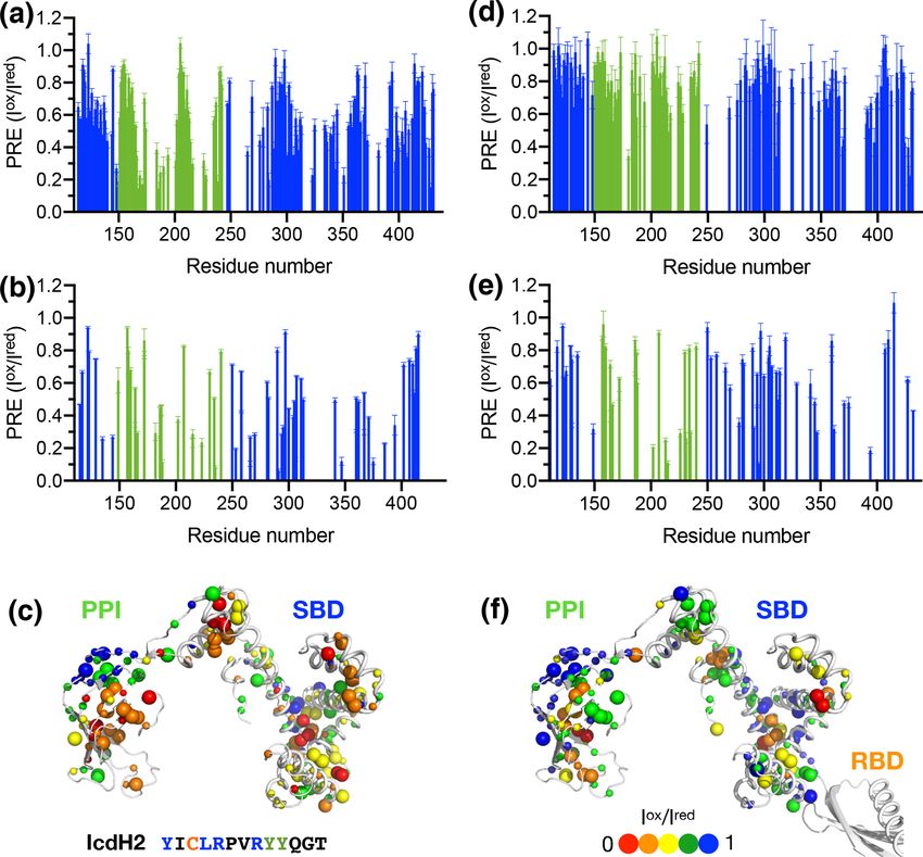
376                                  C.-T. Huang et al.: Structure polymorphism and substrate promiscuity of trigger factor

and 50 µM, respectively, most ribosomes are likely to be oc-        In this study, we sought to evaluate the predictive power
cupied by one TF molecule, leaving ca. 35 µM of free TF in       of the empirical scoring function for TF binding motifs pro-
monomer–dimer equilibrium. In the presence of ribosome-          posed by Bukau and co-workers (Deuerling et al., 2003;
bound nascent chains, the binding affinity of TF to the ribo-    Patzelt et al., 2001). Combining methyl NMR and electron
some can be enhanced by up to 2 orders of magnitudes; the        spin resonance (ESR) spectroscopy, we confirmed the dy-
off rate of TF from the ribosome is also markedly slowed         namic and polymorphic nature of the TF dimer. We generated
down remarkable (Kaiser et al., 2006).                           a collection of fluorescein isothiocyanate (FITC)-labeled
   Although TF primarily acts on the ribosome in a co-           peptide to validate by fluorescence polarization (FP) the pre-
translational manner, TF has been shown to exhibit chap-         dictive power of the proposed TF binding scoring function.
erone activity in the absence of the ribosome to promis-         Two TF binding peptides were subsequently spin-labeled for
cuously facilitate folding of cytosolic proteins. The crystal    paramagnetic relaxation enhancement (PRE) measurements
structure of a thermophilic TF in complex with the ribo-         to identify multiple substrate binding sites within the SBD
somal protein L9 shows that TF binds to its substrate in         and PPI. Importantly, we demonstrated that dimerization of
a 2 : 2 stoichiometry, demonstrating the multifaceted sub-       TF can sequester these binding motifs and that the dynamic
strate recognition modes of TF (Martinez-Hackert and Hen-        equilibrium between monomer and dimer is essential for sub-
drickson, 2009). While there is good nuclear magnetic res-       strate recognition. Collectively, our findings illustrated the
onance (NMR) evidence to demonstrate that a ribosome-            functional importance of TF dimerization in the context of
bound nascent chain can fold into its native conformation        co-translational folding.
without the aid of TF (Cabrita et al., 2016, 2009; Hsu et
al., 2007; Waudby et al., 2019), there are indeed a handful of
                                                                 2     Materials and methods
proteins within the E. coli proteome that require the contri-
butions of TF to fold correctly (Niwa et al., 2012). Although    2.1    Purification of recombinant TF variants
the number of obligatorily substrates of TF within the E. coli
proteome is limited, TF plays an important role in working       The open reading frame of E. coli TF was obtained from
with SecA and SecB to regulate the membrane protein se-          the Nara E. coli ORF collection (Otsuka et al., 2015) and
cretory pathway, as nascent membrane proteins need to be         had been subcloned into a pET-21d plasmid with a His6
correctly sorted by TF and SecA/B as they emerge from the        tag at the N-terminus. The constructs of the SBD and the
ribosome (Buskiewicz et al., 2004; Gelis et al., 2007; Huang     RBD-truncated TF construct corresponding to residues 113–
et al., 2016). It therefore raises the question as to how TF     432 (hereafter PPI+SBD) were kind gifts from Helen Jane
recognizes specific substrates when faced with a multitude       Dyson (Scripps Institute, USA). The single-cysteine mutants,
of sequence variations of the bacterial proteome during co-      Arg14Cys (14C), Thr150Cys (150C), Glu326Cys (326C),
translational and post-translational folding. To this end, an    and Ser376Cys (376C) (Kaiser et al., 2006), were kind gifts
empirical scoring function for predicting TF binding motifs      from Franz-Ulrich Hartl (Max Planck Institute for Biochem-
has been proposed based on peptide array analyses: a puta-       istry, Germany). The plasmids of all TF variants were ampli-
tive TF binding motif should be at least eight amino acids       fied using an E. coli DH5α strain (Sigma-Aldrich, USA) with
in length and contain both aromatic (phenylalanine, tyrosine     appropriate antibiotics selection, and their sequences were
and tryptophan) and positively charged lysine or arginine        subsequently confirmed by standard DNA sequencing (Ge-
residues (Patzelt et al., 2001). In a landmark study, Kalodi-    nomics, Taipei, Taiwan).
mos and co-workers demonstrated that it requires three TF           Unlabeled, uniformly 15 N labeled, or uniformly 15 N/13 C
molecules to bind to one fully unfolded PhoA, which con-         labeled protein samples were expressed by growing the trans-
tains multiple TF binding sites with low-micrometer binding      formed cells in Luria-Bertani (LB) medium or M9 minimal
affinities (Saio et al., 2014). TF exhibits multiple substrate   medium containing 15 NH4 Cl (1 g L−1 ) and 13 C D-glucose
binding sites in SBD and PPI, which are evolutionarily con-      (2 g L−1 ) for uniformly 15 N/13 C labeling in the presence of
served. The additivity of substrate binding affinities in the    kanamycin or ampicillin for antibiotics selection. Selective
multiple binding sites on SBD and PPI results in much higher     13 C and 1 H labeling at methyl groups of isoleucine (δ1),

binding affinity and specificity for long PhoA fragments with    leucine, valine, methionine, and/or Ala β positions, i.e., U-
an apparent Kd in the nanometer range. The strong bind-          [15 N, 2 H], Ile-[δ1−13 Cm , 1 Hm ], Leu/Val-[13 Cm , 1 Hm ], Met-
ing enables the determination of the solution structures of      [13 C, 1 H], and/or Ala-[β−13 Cm , 1 Hm ], was achieved by
TF in complex with the three different PhoA fragments by         growing E. coli culture in perdeuterated M9 medium con-
solution-state NMR spectroscopy complex. The structural in-      taining 99.9 % D2 O, 15 NH4 Cl (1 g L−1 ), and 2 H D-glucose
formation of TF in complex with various PhoA fragments           (2 g L−1 ) followed by addition of selectively 13 C and 1 H la-
indicated that TF preferentially binds to aromatic residues as   beled metabolic precursors and 100 mg L−1 13 C-labeled me-
well as large hydrophobic residues. There is no indication       thionine (Cambridge Isotope Laboratory, USA) 30 min prior
of the preference for positively charged lysine or arginine      to IPTG induction, as described previously. For selective
residues as previously predicted.                                methyl-group-labeled samples, protein overexpression was

Magn. Reson., 2, 375–386, 2021                                                         https://doi.org/10.5194/mr-2-375-2021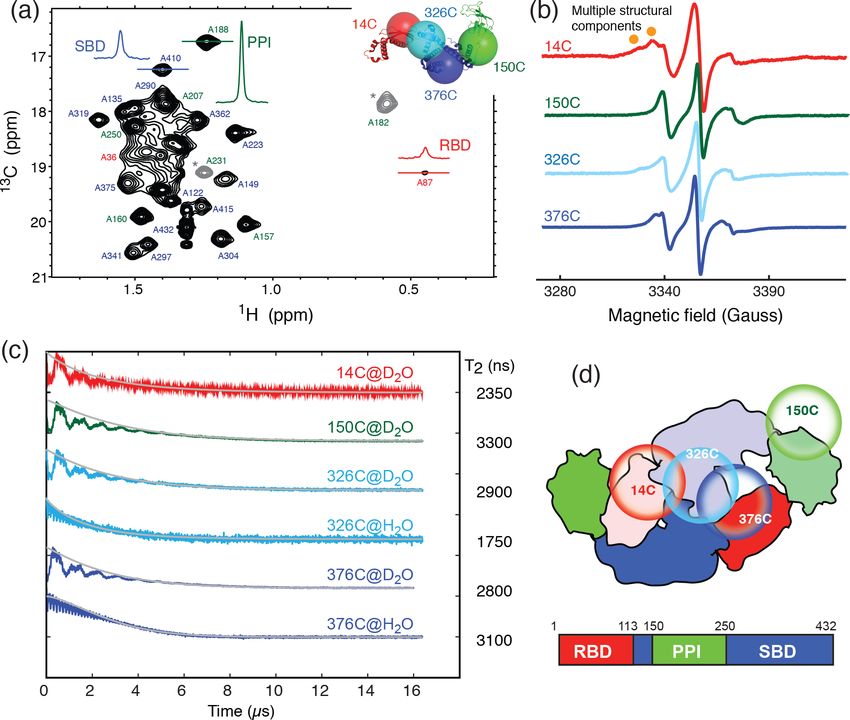
C.-T. Huang et al.: Structure polymorphism and substrate promiscuity of trigger factor                                    377

carried out at 37 ◦ C for 4 h after the addition of IPTG. For    protein concentration were fit to a one-site binding model
the other samples, the overexpression was induced by the ad-     using the software Prism (GraphPad, USA) to extract ap-
dition of 0.5 mM IPTG when the cell density reached OD600        parent association constants associated with different com-
of 0.6–0.8 followed by overnight growth at 16 ◦ C.               binations of FITC-labeled peptides and TF constructs (Lou
   The cells were harvested by centrifugation using a Beck-      et al., 2014).
mann J20XP centrifuge with a JLA 8.1K rotor for 30 min
with 6000 rpm at 4 ◦ C and resuspended in buffer contain-        2.3   NMR paramagnetic relaxation enhancement
ing 50 mM potassium phosphate (pH 8.0) and 300 mM NaCl.                analysis
The harvested cells were disrupted using a sonicator, and
the cell debris and supernatant were separated by a second       A quantity of 1 mg of IcdH2 and IcdH3 peptides was
centrifugation step at 45 000 × g for 30 min at 4 ◦ C. The su-   individually dissolved in 1 mL deionized water and pH-
pernatant was loaded onto a prepacked 5 mL His-Trap HP           adjusted to 7.6. A stock solution of S-(1-oxyl-2,2,5,5-
column (GE Healthcare Life Science) followed by extensive        tetramethyl-2,5-dihydro-1H-pyrrol-3-yl)methyl methanesul-
washing using a buffer containing 20 mM imidazole to re-         fonothioate (MTSL) was prepared by dissolving MTSL pow-
move protein impurities and prevent non-specific binding.        der in dimethyl sulfoxide (DMSO) to reach 150 mM. For
Target fusion protein was eluted using 250 mM imidazole          overnight reaction, 10-fold MTSL was added to the pep-
with the same buffer background. The eluted fractions were       tide solution at 4 ◦ C in the dark. MTSL-labeled peptides
pooled and subject to size-exclusion chromatography (SEC;        were purified by high-performance liquid chromatography
HiLoad 26/60 Superdex 75, GE Healthcare Life Sciences)           (HPLC) and validated by MALDI-TOF mass spectrome-
with 20 mM sodium phosphate (pH 7.4) and 100 mM NaCl             try against their expected molecular weights. The eluents
to remove impurities to yield a purity of higher than 95 %       were lyophilized and resuspended in the SEC buffer to
based on visual inspection of the Coomassie Brilliant Blue-      reach a concentration of 20 mM. U-[15 N, 2 H], Ile-[δ1−13 Cm ,
stained sodium dodecyl sulfate polyacrylamide gel (SDS-          1 H ], Leu/Val-[13 C , 1 H ], Met-[13 C, 1 H], Ala-[β−13 C ,
                                                                    m                 m     m                               m
PAGE). The protein solution was aliquoted, flash-frozen by       1 H ] PPI+SBD, and full-length TF were used for PRE
                                                                    m
liquid nitrogen, and stored at −80 ◦ C until further use. Un-    measurements by recording the backbone 15 N−1 H trans-
less otherwise specified, the SEC buffer was used for all the    verse relaxation optimized spectroscopy (TROSY) and side-
biophysical characterizations described herein.                  chain methyl 13 C−1 H band-selective optimized flip angle
                                                                 short transient heteronuclear multiple quantum correlation
2.2   Fluorescence polarization analysis of FITC-labeled
                                                                 (SOFAST-HMQC) spectroscopy in oxidized and reduced
      peptides
                                                                 states. The NMR spectra were collected using NMR spec-
                                                                 trometers operating at a proton Larmor frequency of 850
Five peptides corresponding to fragments of isocitrate de-       or 600 MHz, equipped with a cryogenic triple-resonance
hydrogenase (ICDH; Table 1) were synthesized in-house. A         TCI probe (Bruker, Germany), processed by NMRPipe and
fraction of all these peptides were subsequently labeled with    analyzed by NMRFAM-SPARKY (https://nmrfam.wisc.edu/
FITC at the N-terminus for fluorescence polarization (FP)        nmrfam-sparky-distribution/, last access: 2 June 2021). The
measurements. All peptides (with and without FITC label-         nitroxide of MTSL was reduced by adding an aliquot of
ing) were purified by high-performance liquid chromatogra-       ascorbic acid to yield a final concentration of 1 mM. The
phy (HPLC) and validated by MALDI-TOF (matrix-assisted           observed PREs were expressed as the ratio of the peak in-
laser desorption ionization-time of flight) mass spectrome-      tensities of the oxidized (paramagnetic state) to the reduced
try against their expected molecular weights. For FP analy-      (diamagnetic state) state (I ox /I red ).
sis, FITC-labeled peptides were dissolved in DMSO to yield
a stock solution of 4 M. They were subsequently diluted by       Continuous-wave (CW) and pulsed ESR measurements
20 mM Tris (pH 7.4) and 100 mM NaCl to yield a molar con-
centration of 100 µM. 200 µL of TF variants was transferred      Introduction of MTSL into single-cysteine TF variants, i.e.,
into a 96-well plate followed by serial dilution by the same     14C, 150C, 326C, and 376C, was achieved by incubating
buffer in a 1 : 1 dilution ratio. The final protein concentra-   the protein samples with 10 mM DTT, which was removed
tions were between 0.1 and 1000 µM. After serial dilution,       using a desalting column (PD-10, GE Healthcare, USA).
50 µL of protein solutions of various protein concentrations     Immediately after the removal of DTT, 10-fold molar ex-
was transferred to new wells by an eight-channel pipette and     cess of MTSL was added, and the mixtures were incu-
was mixed with FITC-labeled peptide solution to yield a pep-     bated overnight at 4 ◦ C in the dark. Free MTSL was sub-
tide concentration of 1 µM. FP measurements of these sam-        sequently removed using the same desalting column, and
ples were carried out using a plate reader (Paradigm, Molec-     complete MTSL incorporation was confirmed by mass spec-
ular Device, USA) with an excitation wavelength of 485 nm        trometry (Rezwave, Taiwan). A Bruker ELEXSYS E580-400
and an emission wavelength of 535 nm. The integration time       X-band CW/pulsed spectrometer, equipped with a split-ring
was set to 250 ms. The observed FP values as a function of       resonator (EN4118X-MS3) and a helium gas flow system

https://doi.org/10.5194/mr-2-375-2021                                                      Magn. Reson., 2, 375–386, 2021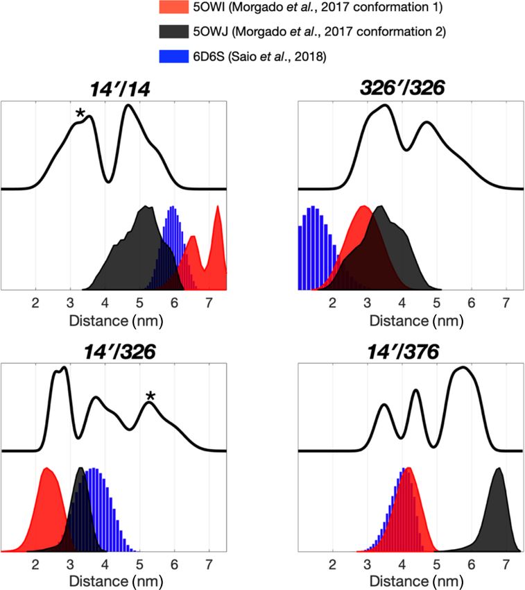
378                                             C.-T. Huang et al.: Structure polymorphism and substrate promiscuity of trigger factor

Table 1. List of ICDH-derived peptides and their properties associated with TF binding.

           Peptide      Sequence∗                                         Length (residue         Predicted       Length-normalized                       Kd
           name                                                              number)                  score             score                           (µM)
           IcdH1        55 KAYKGERKISWMEIYT70                                     16                   −8.9               −0.56                    28.2 ± 1.0
           IcdH2        125 YICLRPVRYYQGT137                                      13                  −14.0               −1.08                     8.6 ± 0.3
           IcdH3        177 KFLREEMGVKKIRFPEHC194                                 18                   −9.9               −0.55                      132 ± 9
           IcdH4        230 KGNIMKFTEGAFK242                                      13                   −5.6               −0.40                     293 ± 49
           IcdH5        340 GTAPKYAGQDK350                                        11                   −1.6               −0.13                  1466 ± 2117
         ∗ The starting and ending residue numbers corresponding to the sequence of ICDH of individual peptides are indicated as superscripts.

(4118CF and 4112HV), was used. CW ESR spectra were                                      distribution of the DEER spectroscopy was performed us-
recorded at temperature 310 K, with an operating frequency                              ing a home-written program operating in MATLAB (Math-
of 9.4 GHz, 100 kHz field modulation, and 1.5 mW incident                               Works) as previously described and demonstrated (Lai et
microwave power. 0.25–0.6 mM TF variants in deuterated                                  al., 2019; Sung et al., 2015; Tsai et al., 2015). Basically,
buffer were loaded in 3 mm (o.d.) quartz tubes. d8-glycerol                             the data were analyzed using Tikhonov regularization based
was supplemented to achieve a final glycerol concentration                              on the L-curve method (Chiang et al., 2005b), followed
of 30 % (v/v). The total volume is approximately 20 µL. For                             by a data refinement process using the maximum entropy
the electron spin echo (ESE) measurements, the sample tube                              method (MEM) to obtain the non-negative distance distribu-
was plunge-cooled in liquid nitrogen and then transferred                               tions (Chiang et al., 2005b; Li et al., 2020).
into the ESR probe head, which was precooled to 50 K. ESE
experiments were performed using the two-pulse Hahn echo
                                                                                        3     Results
sequence, consisting of a π/2 pulse along the x axis, fol-
lowed by a delay τ and a train of π pulses, separated by                                TF exists in solution in a monomer–dimer equilibrium with
inter-pulse delays 2τ (Lai et al., 2013; Zecevic et al., 1998).                         a low-micrometer Kd (Kaiser et al., 2006). The RBD is re-
The field was adjusted to optimize the spin echo, and the du-                           sponsible for the dimerization (Patzelt et al., 2002). Isolated
ration times of π/2 and π pulses were set to 16 and 32 ns.                              RBD, SBD, and PPI exhibit well-resolved 2D 15 N−1 H back-
As previously described [2], the ESE signals were fitted to a                           bone amide and 13 C−1 H side-chain methyl correlation spec-
stretched exponential function to extract T2 values from the                            tra, whose chemical shifts assignments have been previously
ESE data using the MATLAB software.                                                     reported at a residue-specific level (Huang and Hsu, 2016;
   For double electron–electron resonance (DEER) measure-                               Yao et al., 2008). These assignments serve as the basis to
ments, samples were prepared either by single-labeled TF                                complete the backbone and side-chain methyl NMR chemi-
variants or 1 : 1 mixture of two different single-labeled TF                            cal shift assignments through a divide-and-conquer assign-
variants with the final protein concentration of 0.25 mM. A                             ment strategy despite the apparent high-molecular weight
quantity of 30 % (v/v) d8-glycerol was added to the sam-                                of full-length TF of approximately 100 kDa (Morgado et
ple as cryoprotectants in all DEER measurements. DEER                                   al., 2017; Saio et al., 2014, 2018). Detailed structural and
experiments were performed using the typical four-pulse                                 dynamic analysis of full-length TF by solution-state NMR
constant-time DEER sequence (Jeschke, 2012). The detec-                                 spectroscopy remains very challenging, not least because of
tion pulses were set to 32 and 16 ns for π and π/2 pulses,                              the large dynamics range of the peak intensities correspond-
respectively, and the pump frequency was set to approxi-                                ing to different domains of TF. This can be exemplified by
mately 65 MHz lower than the detection pulse frequency.                                 the very large dynamic range of the cross-peak intensities
The pulse amplitudes were chosen to optimize the refocused                              of the alanine methyl resonances of full-length TF under a
echo. The π/2-pulse was employed with +x/−x phase cy-                                   perdeuterated background (Fig. 1a). The methyl resonances
cles to eliminate receiver offsets. The duration of the pump-                           corresponding to the alanine residues within the RBD were
ing pulse was 32 ns, and its frequency was coupled into the                             severely broadened, whereas those of PPI remained very
microwave bridge by a commercially available setup from                                 sharp, and those of the SBD were intermediate. The trun-
Bruker. All pulses were amplified via a pulsed traveling wave                           cation of the RBD significantly reduced the dynamic range
tube (TWT) amplifier (E580-1030). The field was adjusted                                of the observed alanine methyl resonances for both the SBD
such that the pump pulse is applied to the maximum of the                               and PPI+SBD (Fig. S1 in the Supplement).
nitroxide spectrum, where it selects the central mI = 0 transi-                            To further probe the dynamics of individual domains in
tion of hyperfine Azz together with the mI = ±1 transitions.                            the context of a dimeric TF, we employed ESR spectroscopy
The accumulation time for each set of data was about 10 h at                            with site-specific spin labels. We individually introduced a
a temperature of 50 K. Determination of inter-spin distance                             spin label to one of the four sites in TF, namely residue 14 on

Magn. Reson., 2, 375–386, 2021                                                                                       https://doi.org/10.5194/mr-2-375-2021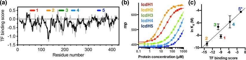
C.-T. Huang et al.: Structure polymorphism and substrate promiscuity of trigger factor                                                     379

Figure 1. Domain dynamics of dimeric TF in solution. (a) 13 C−1 H heteronuclear multi-quantum correlation (HMQC) spectrum of U-
[15 N, 2 H], Ala-[β−1 Hm /13 Cm ] TF. Selected proton line shapes of cross-peaks originated from the RBD, SBD, and PPI are shown in
red, blue, and green, respectively, with their corresponding residue identities indicated. Aliased cross-peaks are shown in grey and labeled
with asterisks. Inset: cartoon representation of a monomeric TF (PDB ID: 1w26) with matching coloring for the individual domains. The Cα
atoms of residues 14, 150, 327, and 376, which were individually mutated into cysteine for MTSL spin labeling, are shown in semitransparent
spheres with a radius of 25 Å and indicated with their residue identities. (b) CW-ESR spectra of spin-labeled TF variants recorded at 310 K
as a function of magnetic field. The spectrum of 14C exhibits multiple side peaks (indicated by filled orange circles) that are indicative of
multiple structural components. (c) ESE measurements of spin-labeled TF variants. The transverse relaxation times (T2 ) of the nitroxide were
deduced from the fitting shown by solid grey lines, and their values are indicated on the right. Comparison of the relaxation characteristics of
the same samples in H2 O and D2 O for 326C and 376C indicated the solvent-exposed nature of the spin labels. (d) Schematic representation
of the domain arrangement of dimeric TF, with the locations of the spin labels indicated by open circles. The domain organization of TF is
shown below, with the boundaries of individual domains indicated.

the RBD (14C), residue 150 on PPI (150C), and residues 326                those of 14C, while 376C did not exhibit the same signals,
or 376 on the SBD (326C or 376C), by covalently attaching                 implying that the conformational heterogeneity of the RBD is
a MTSL to the mutated cysteine side-chain (Fig. 1a, inset).               more pronounced than that of the SBD. In the case of 150C,
The spin-labeled TF variants were analyzed by CW-ESR to                   there was no indication of conformational heterogeneity as
probe the domain dynamics manifested in the line shapes.                  the ESR lines were sharp without a side band. ESE analysis
Comparison of the CW-ESR spectra the TF variants showed                   was subsequently used to deduce the transverse relaxation
distinct side bands for 14C at 310 K, suggesting the presence             time (T2 ) of the free radical, i.e., the nitroxide of MTSL, at
of multiple conformations (Fig. 1b). In contrast, the CW-ESR              individual sites (Fig. 1c). In line with the methyl NMR line
spectrum of 326C showed minor signals that were similar to                width analysis, the T2 of 14C was the shortest (2350 ns), fol-

https://doi.org/10.5194/mr-2-375-2021                                                                   Magn. Reson., 2, 375–386, 2021
380                                   C.-T. Huang et al.: Structure polymorphism and substrate promiscuity of trigger factor

lowed by 376C (2800 ns), 326C (2900 ns), and that of 150C
was the longest (3300 ns). Further comparison of the time
domain spin-echo ESR spectra of 326C and 376C in H2 O
and D2 O showed a clear impact of solvent on the relaxation
of the spin labels. The results indicated that both spin labels
were solvent-exposed despite their implication in dimer for-
mation. Collectively, the NMR and ESR analyses suggested
distinct domain dynamics of a dimeric TF, with PPI being the
least restricted and the RBD being the most heterogeneous.
Although the SBD also forms part of the dimer interface, its
dynamics is less restricted than that of the RBD.
   The severely broadened methyl proton resonances of the
RBD residues and faster T2 relaxation of the spin label at 14C
likely correspond to the conformational heterogeneity within
the dimer interface. Indeed, a number of different TF dimer
structures have been reported by two independent studies
based on different NMR restrains (Morgado et al., 2017;
Saio et al., 2018). To investigate the TF dimer conformations
through ESR spectroscopy, we carried out double electron–
electron resonance (DEER) measurements to determine the
inter-spin distance distributions of different combinations of
spin-labeled TF samples. These included the uniformly sin-
gle species or the 1 : 1 mixture of two variants (denoted as
site A0 /site B). Figure 2 shows the distance distributions ex-
                                                                  Figure 2. Multiple dimeric TF conformations revealed from the
tracted from the DEER time-domain data (Fig. S2) using the
                                                                  DEER measurements. DEER samples were prepared by either the
Tikhonov-based regulation methods (Lai et al., 2019; Chi-
                                                                  single species or the 1 : 1 mixture (denoted as site A0 /site B) of the
ang et al., 2005a). The DEER distance distributions (solid        three single-cysteine variants, 14C, 326C, and 376C. DEER dis-
lines in Fig. 2) are compared with the predicted inter-spin       tance distributions of TF dimer (solid line) were compared with the
distance distribution (shaded areas in Fig. 2) calculated from    distance distributions calculated from the previously determined TF
the three previously reported NMR structures (Morgado et          dimer structure (PDB codes: 5OWI, red; 50WJ, black; and 6D6S,
al., 2017; Saio et al., 2018) using the MtsslWizard program       blue). There are a few discrepancies between the DEER and NMR
(Hagelueken et al., 2015). In general, the DEER distance dis-     results, as indicated by asterisks.
tributions show multiple distinct populations indicating con-
formational heterogeneity in the TF dimer. While the major-
ity of the DEER-derived peak distributions could find cor-        array study based on the sequence of isocitrate dehydroge-
respondences from the NMR structures, a few discrepancies         nase (ICDH), several surface-immobilized peptides showed
did exist. They are indicated by asterisks in Fig. 2. Specifi-    prominent TF binding (Deuerling et al., 2003). Together with
cally, the DEER measurements identified a shorter distance        the results derived from other peptide arrays, an empirical
pair for 140 /14 centered at approximately 3 nm, when all re-     scoring function for predicting the potential TF binding site
ported NMR structures showed corresponding distances at           along a given protein sequence was proposed (Deuerling et
4 nm and above. Likewise, the DEER-derived distance dis-          al., 2003; Patzelt et al., 2001). Nevertheless, the predictive
tribution of 3260 /326 showed an additional peak at approx-       power of such a scoring function has not been experimen-
imately 4.7 nm, but it was not present in the NMR struc-          tally verified thus far. According to the prediction, an ideal
tures. Furthermore, the DEER-derived distance distributions       TF binding motif should be at least eight residues long and
of 140 /326 and 140 376 showed three distinct populations,        rich in aromatic residues and positively charged lysine or
which were in agreement with the conclusion drawn by the          arginine. The requirement for the coexistence of hydrophobic
CW-ESR analysis that the RBD exhibits abundant structural         and charged residues is an intriguing feature. Nevertheless,
heterogeneity. Overall, the RBD (14C) exhibited a higher          the relatively loose definition can lead to a huge number of
level of conformational heterogeneity than what was previ-        potential binding sites within the bacterial proteomes. As a
ously determined by NMR spectroscopy. Collectively, our           model system, we correlated the previously reported peptide
ESR analyses clearly demonstrated the abundant structural         array data of TF binding to ICDH (Deuerling et al., 2003)
polymorphism of the TF dimer in solution.                         and the predicated TF binding score as a function of ICDH
   Having established the ground work of characterizing the       sequence (Fig. 3a). By visual inspection of the blotting den-
dynamics of TF in its apo form, we next set to characterize       sities of the peptide array, we identified five segments within
how TF recognizes its substrates. According to the peptide        the ICDH sequence that showed strong TF binding and ful-

Magn. Reson., 2, 375–386, 2021                                                            https://doi.org/10.5194/mr-2-375-2021
C.-T. Huang et al.: Structure polymorphism and substrate promiscuity of trigger factor 381
Figure 3. Experimental validation of the scoring function of predicted TF binding motifs. (a) Predicted TF binding score of as a function
of residue number of ICDH. Running averages with a window size of 8 and 13 residues are shown in grey and black, respectively. A
segment with a predicted binding score of lower than −0.5 is considered a potential binding site. Five peptides were chemically synthesized
corresponding to the regions indicated above, numbered from 1 to 5. (b) Fluorescence polarization (FP) analysis of TF binding to the synthetic
peptides labeled with FITC. The FP of the peptides IcdH1–5 as a function of TF concentration are colored from red to blue as indicated in
inset, which correspond to the segments indicated in (a). (c) Linear regression of the natural logarithm of the binding constant (Kd ) as a
function of the predicted TF binding score. The resulting function is y = 0.0856x + 0.772 with R 2 = 0.88. The error bars were derived from
three technical replicates of the FP analysis. The error of IcdH5 was larger than the mean value, which is omitted in this plot.
filled the requirement of peptide length and composition (Ta- to full-length TF (Kd = 132 ± 9 µM) is weaker than that of
ble 1). We adjusted the window sizes of the selected se- IcdH2 (Kd = 8.6 ± 0.3 µM), the observed PREs in IcdH3
quences to maximize the amount of preferred amino acid were more prominent than that of IcdH2 when PPI+SBD,
types and chemically synthesized these peptides followed which is a truncated and monomeric form of TF, was used in
by introducing a FITC moiety at the N-termini individual the NMR PRE analysis (Figs. 4 and 5). When full-length TF
peptides to facilitate fluorescence polarization (FP) measure- was used for the same NMR PRE analysis under the TF con-
ments to determine the binding affinities of these peptides centration that it is predominantly dimeric, the PREs were
to TF. Except for IcdH3 and IcdH4, whose sequences are significantly reduced (Figs. 4d–e, 5d–e), and the remaining
partly helical in the crystal structure (Bolduc et al., 1995), PREs were mostly localized within PPI that is not part of
all the remaining sequences correspond to loop regions that the TF dimer interface (Figs. 4f and 5f). The loss of PRE
do not adopt particular secondary structures. The resulting was much more pronounced for IcdH3 compared to that of
dissociation constants (Kd ) ranged between low micrometer IcdH2, in line with the FP analysis that showed a weaker
and low millimolar, spanning more than 2 orders of mag- TF binding for IcdH3 compared to IcdH2. The implication
nitudes (Fig. 3b). Importantly, the natural logarithms of the of this finding is that the dimerization of TF sequesters the
observed Kd values showed a good correlation with the pre- substrate binding sites within the SBD and to a lesser extent
dicted binding score (an R 2 value of 0.88 was obtained from the binding site in PPI. Dynamic equilibrium between the
the linear regression), demonstrating the predictive power of monomeric and dimeric TF is therefore expected to play an
the empirical scoring function (Fig. 3c). important role in regulating its chaperone activity.
To further examine the structural basis of substrate recog-
nition by TF, we chose IcdH2 and IcdH3 (Table 1), which
had an endogenous cysteine residue within their sequences 4 Discussion
that can be spin-labeled with MTSL for paramagnetic re-
laxation enhancement (PRE) measurements. We first used In this study, we employed methyl NMR and ESR spec-
U-[15 N, 2 H], Ile-[δ1−13 Cm , 1 Hm ], Leu/Val-[13 Cm , 1 Hm ], troscopy to characterize the dynamics of full-length TF in
Ala-[β−13 Cm , 1 Hm ], Met-[13 C, 1 H] PPI+SBD, which is its dimeric form. Although TF exists in equilibrium be-
monomeric, to collected 2D 15 N−1 H backbone amide and tween monomer and dimer, the experimental conditions un-
13 C−1 H side-chain methyl correlation spectra in the pres-
der which the NMR and ESR experiments were conducted,
ence of the MTSL-labeled IcdH2 or IcdH3 under oxidized i.e., protein concentrations well above 0.25 mM, ensured that
(paramagnetic) and reduced (diamagnetic) states to deter- TF is predominantly dimeric, as has been established pre-
mine the PREs originated from the interaction with MTSL- viously (Morgado et al., 2017; Saio et al., 2018). Our find-
labeled IcdH2 or IcdH3 defined by the resonance inten- ings indicated that the TF dimer interface is not well de-
sity ratios between the oxidized and reduced states, I ox /I red fined, which may exist in several distinct configurations, as
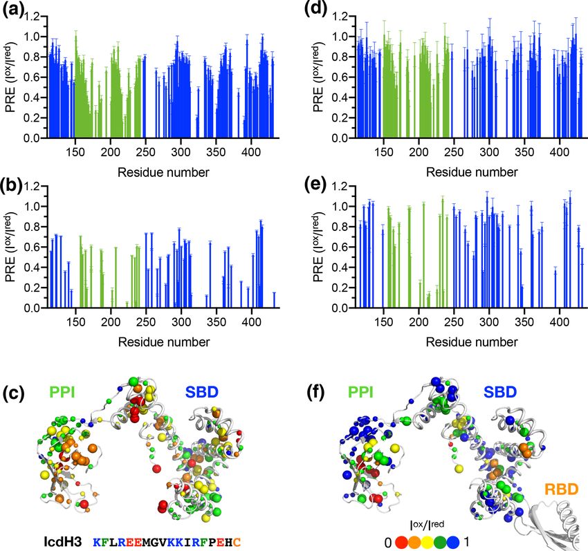
(Fig. S3). The observed PREs were mapped onto the struc- evidenced by the severely broadened linewidths of the ala-
ture of PPI+SBD, which revealed multiple hotspots within nine methyl group within the RBD (Fig. 1a). The conforma-
the SBD and one cluster within PPI that showed strong PREs tional heterogeneity within the RBD also manifested in the
(Figs. 4a–c, 5a–c). Although the binding affinity of IcdH3 additional side bands of the CW-ESR spectra of 14C, which
https://doi.org/10.5194/mr-2-375-2021 Magn. Reson., 2, 375–386, 2021382 C.-T. Huang et al.: Structure polymorphism and substrate promiscuity of trigger factor Figure 4. Structural mapping of the PREs induced by the MTSL-labeled IcdH2 peptide on TF without and with the RBD. The backbone amide-based PREs of PPI+SBD (a) and full-length TF (d). The side-chain methyl-based PREs of PPI+SBD (b) and full-length TF (e). Structural mapping of the observed PREs onto the structure of PPI+SBD (c) and full-length TF (f). The backbone amide nitrogen atoms and side-chain methyl carbon atoms are shown by small and large spheres and are colored with a gradient from red to blue, corresponding to small and larger PREs as indicated by the filled circles below. The observed PREs expressed as the ratio of the peak intensities of the oxidized (paramagnetic state) to the reduced (diamagnetic state) states (I ox /I red ) as a function of residue number between 113 and 432. The PRE values corresponding to PPI and SBD are colored in green and blue, respectively. The residues corresponding to the RBD are omitted due to the severe line broadening that precludes reliable data analysis. probe the environment around the MTSL moiety at the RBD (Fig. 3). Unlike the nuclear Overhauser effect (NOE) or PRE (Fig. 1b). Furthermore, there is a good correlation between that provide averaged distance information that is heavily the NMR methyl linewidth analysis and the T2 analysis of weighted by shorter contacts, ESR-based DEER data can be the MTSL-labeled TF variants, which showed that the PPI is converted into distance distributions of inter-spin distances, the most dynamic, consistent with the previous findings that thereby informing us on the degree of conformational hetero- TF forms an antiparallel dimeric assembly where PPI makes geneity. We compared the distance distributions derived from limited contacts with other domains (Morgado et al., 2017; the DEER measurements and the simulated distances based Saio et al., 2018). on the reported NMR structures and showed that the NMR We next collected DEER data on singly MTSL-labeled structures only reflect part of the conformations observed TFs or 1 : 1 mixture of TFs that were separately MTSL- by DEER measurements (Fig. 3). Morgado et al. (2017) de- labeled to measure long-range inter-spin distances up to 7 nm termined two distinct dimeric assemblies of TF based on Magn. Reson., 2, 375–386, 2021 https://doi.org/10.5194/mr-2-375-2021
C.-T. Huang et al.: Structure polymorphism and substrate promiscuity of trigger factor                                              383

Figure 5. Structural mapping of the PREs induced by MTSL-labeled IcdH2 peptide on TF without and with the RBD. The backbone amide-
based PREs of PPI+SBD (a) and full-length TF (d). The side-chain methyl-based PREs of PPI+SBD (b) and full-length TF (e). Structural
mapping of the observed PREs onto the structure of PPI+SBD (c) and full-length TF (f). Structural mapping of the PRE of MTSL-labeled
IcdH3 peptide on TF variants. The color scheme of the bar charts and the cartoon representations of the structural models are the same as
those of Fig. 4.

PRE-derived distances, whereas Saio et al. (2018) used a               two major peaks centered at 3.5 and 4.7 nm, respectively
large number of NOEs to determine a single ensemble of                 (3260 /326 in Fig. 2). The 3.5 nm peak distribution was in
TF dimer. Except for the intermolecular distance between               agreement with the two distinct PRE-derived TF conforma-
14C and 376C (140 /376 in Fig. 2), where two of the three              tions. On the other hand, each of the two PRE-based NMR
NMR-derived TF dimers show identical inter-spin distances,             structures yielded a very long inter-spin distance distribution
which also agreed with the DEER measurements, essentially              for singly labeled 14C and the 1 : 1 mixture of 14C and 376C
all three reported NMR structures probe distinct subsets of            (140 /14 and 140 /376 in Fig. 2) that were not probed by the
conformations within a large conformational space that was             DEER measurements. Collectively, our ESR analyses under-
probed by ESR-based DEER measurements. Nevertheless,                   scored the structural polymorphism of the TF dimer and the
there were notable discrepancies between the NOE-derived               similarity and difference between the reported NMR struc-
NMR structure and DEER measurements. On the one hand,                  tures themselves and in relation to the DEER-derived struc-
the NOE-based NMR structure yielded a very short theoret-              tural information.
ical inter-spin distance distribution of singly labeled 326C              We next generated five FITC-labeled peptides derived
centered at 1 nm, whereas the DEER measurement showed                  from ICDH to demonstrate the predicted power of the empir-

https://doi.org/10.5194/mr-2-375-2021                                                              Magn. Reson., 2, 375–386, 2021
384                                   C.-T. Huang et al.: Structure polymorphism and substrate promiscuity of trigger factor

ical scoring function for TF binding based on the sequence        association, it is not surprising that the TF dimerization can
composition (Table 1 and Fig. 3). Two peptides that harbor        outcompete peptide binding at a relatively high TF concen-
an endogenous cysteine within the sequences, namely IcdH2         tration (100 µM). Nevertheless, cytosolic TF concentration is
and IcdH3, were spin-labeled with MTSL to map their bind-         estimated to be in the range of 35 µM, while that of the ri-
ing sites on TF by PRE measurements. We identified three          bosome is about 1 µM. TF binds to the ribosome in a 1 : 1
distinct binding sites within the SBD and one binding site        stoichiometry, and the associated binding affinity is strongly
within the PPI (Figs. 4 and 5). The locations of these binding    modulated by the presence and compositions of fledgling
sites are consistent with the previous study in which four dis-   nascent chains.
ordered fragments of PhoA are used to map the binding sites
on TF (Saio et al., 2014). The authors also reported multiple
binding sites within the PPI and SBD when short peptides          5   Conclusion
were used to map the binding sites by chemical shift per-
turbations and intermolecular NOEs. When a longer peptide         The intricate interplay between TF, nascent chains and the
fragment of PhoA is used as a substrate, each of the substrate    ribosome can be modulated by the sequence compositions of
binding sites within the PPI and SBD is occupied by a spe-        the nascent chains. Our NMR, ESR, and biophysical analyses
cific TF binding motif, thereby leading to a unique binding       confirmed the structural polymorphism of the TF dimer and
mode that enables structure determination of the substrate-       the multivalency of substrate binding, which is sensitive to
bound TF. By determining the microscopic Kd values for in-        TF dimer formation. These results led us to propose that the
dividual binding sites, which fall within the low-micrometer      relatively strong ribosome binding affinity serves as the key
range, the authors demonstrate by relaxation dispersion anal-     regulatory mechanism to modulate monomer–dimer equilib-
ysis that the multivalency of substrate recognition signifi-      rium and therefore the accessibility of the substrate binding
cantly increases the binding affinity to a nanometer range.       sites, which are fully exposed when TF binds to the ribo-
Note that in the previous study, the RBD-truncated TF vari-       some through its RBD. The observed binding affinities of
ant, PPI+SBD, was used to determine the solution structures       the selected peptides from ICDH indeed fit well within the
of TF in complex with different PhoA fragments based on in-       dynamic range of these binding events.
termolecular NOEs, while full-length TF was used to demon-
strate that full-length PhoA in its unfolded form can be oc-
                                                                  Data availability. Data are available upon request.
cupied by multiple TF molecules by the attenuation of peak
intensities of PhoA.
   According to the ESR analysis, the spin labels within the
                                                                  Supplement. The supplement related to this article is available
RBD and SBD are mostly solvent-exposed (Fig. 1c). Further-
                                                                  online at: https://doi.org/10.5194/mr-2-375-2021-supplement.
more, the dimer interface appeared to be quite heterogeneous
and dynamic, according to methyl NMR and CW-ESR line
shape analyses (Fig. 1) and the more robust DEER measure-         Author contributions. STDH conceived and designed the exper-
ments (Fig. 2). The unique domain architecture of TF sug-         iments with contributions from YWC for ESR. CTH prepared the
gests that the dimer interface does not form a properly encap-    NMR and ESR samples and assisted in NMR data collection and
sulated cavity to accommodate its substrates. Additionally,       analysis. YCL and YWC collected and analyzed the ESR data. CTH
the distributions of sparsely negatively charged surfaces sur-    and SYC contributed to the FITC-labeled peptide binding analyses
rounded by small patches of neutral (hydrophobic) surfaces        supported by MRH. STDH wrote the manuscript with inputs from
within the SBD and PPI coincide with the observed pep-            all authors.
tide binding sites, which may explain why positively charged
residues and aromatic residues are both favored for TF bind-
ing. Unlike GroEL/GroES, which has an efficient nucleotide-       Competing interests. The authors declare that they have no con-
dependent regulatory mechanism to mechanically control the        flict of interest.
exposure of its substrate binding sites, TF may utilize the
self-dimerization to achieve the same regulation (Hartl and
                                                                  Special issue statement. This article is part of the special issue
Hayer-Hartl, 2002).
                                                                  “Robert Kaptein Festschrift”. It is not associated with a conference.
   Here we compared the peptide-binding-induced PREs in
PPI+SBD and full-length TF and showed that dimeriza-
tion of TF effectively sequesters the binding sites within the
                                                                  Acknowledgements. This article is dedicated to Robert Kaptein
SBD from peptide binding. Although PPI is not involved in         on the occasion of his 80th birthday as part of the special is-
dimer formation, the peptide-induced PREs in the PPI are          sue “Robert Kaptein Festschrift”. We thank Franz-Ulrich Hartl at
also diminished potentially due to steric hindrance. Consid-      Max Planck Institute of Biochemistry, Martinsried, Germany, for
ering that the effective peptide binding affinities are rela-     stimulating discussions and sharing the constructs of the single-
tively weak compared to the dissociation constant of TF self-     cysteine TF variants. We also thank Helen Jane Dyson at Scripps

Magn. Reson., 2, 375–386, 2021                                                           https://doi.org/10.5194/mr-2-375-2021
C.-T. Huang et al.: Structure polymorphism and substrate promiscuity of trigger factor 385
Institute, San Diego, USA, for sharing the constructs of the SBD Deuerling, E., Patzelt, H., Vorderwülbecke, S., Rauch, T., Kramer,
and PPI+SBD. The NMR data were collected at the High Field G., Schaffitzel, E., Mogk, A., Schulze-Specking, A., Langen, H.,
NMR Center at Academia Sinica, which is funded by Academia and Bukau, B.: Trigger Factor and DnaK possess overlapping
Sinica Core Facility and Innovative Instrument Project (AS-CFII- substrate pools and binding specificities, Mol. Microbiol., 47,
108-112), the Instrumentation Center at the National Tsing Hua 1317–1328, https://doi.org/10.1046/j.1365-2958.2003.03370.x,
University (NTHU), supported by the Ministry of Science and Tech- 2003.
nology, Taiwan (MOST) and NTHU, and the Instrument Center at Ferbitz, L., Maier, T., Patzelt, H., Bukau, B., Deuerling, E., and
the National Taiwan University. The ESR data were collected at Ban, N.: Trigger factor in complex with the ribosome forms
the Instrumentation Center at NTHU. The FITC-labeled peptides a molecular cradle for nascent proteins, Nature, 431, 590–596,
were synthesized by and analyzed at the Synthesis facility and Bio- https://doi.org/10.1038/nature02899, 2004.
physics facility, respectively, at the Institute of Biological Chem- Gelis, I., Bonvin, A. M., Keramisanou, D., Koukaki, M., Gouridis,
istry, Academia Sinica. G., Karamanou, S., Economou, A., and Kalodimos, C. G.:
Structural basis for signal-sequence recognition by the translo-
case motor SecA as determined by NMR, Cell, 131, 756–769,
Financial support. This research has been supported by the Min- https://doi.org/10.1016/j.cell.2007.09.039, 2007.
istry of Science and Technology (MOST), Taiwan (grant nos. Hagelueken, G., Abdullin, D., and Schiemann, O.:
100-2113-M-001-031-MY2 and 102-2113-M-001-017-MY2 – to MtsslSuite: Probing biomolecular conformation by spin-
Shang-Te Danny Hsu) and the intramural fund from Academia labeling studies, Method. Enzymol., 563, 595–622,
Sinica, Taiwan, to Shang-Te Danny Hsu. https://doi.org/10.1016/bs.mie.2015.06.006, 2015.
Hartl, F. U.: Cellular Homeostasis and Aging, Ann. Rev.
Biochem., 85, 1–4, https://doi.org/10.1146/annurev-biochem-
Review statement. This paper was edited by Hashim Al-Hashimi 011116-110806, 2016.
and reviewed by three anonymous referees. Hartl, F. U. and Hayer-Hartl, M.: Molecular chaperones in the cy-
tosol: from nascent chain to folded protein, Science, 295, 1852–
1858, https://doi.org/10.1126/science.1068408, 2002.
Hesterkamp, T. and Bukau, B.: The Escherichia coli trigger
factor. FEBS Lett., 389, 32–34, https://doi.org/10.1016/0014-
References 5793(96)00582-0, 1996.
Hoffmann, A., Bukau, B., and Kramer, G.: Struc-
Bolduc, J. M., Dyer, D. H., Scott, W. G., Singer, P., ture and function of the molecular chaperone Trig-
Sweet, R. M., Koshland Jr., D. E., and Stoddard, B. ger Factor, Biochim. Biophys. Acta, 1803, 650–661,
L.: Mutagenesis and Laue structures of enzyme interme- https://doi.org/10.1016/j.bbamcr.2010.01.017, 2010.
diates: isocitrate dehydrogenase, Science, 268, 1312–1318, Hoffmann, A., Becker, A. H., Zachmann-Brand, B., Deuer-
https://doi.org/10.1126/science.7761851, 1995. ling, E., Bukau, B., and Kramer, G.: Concerted action of
Buskiewicz, I., Deuerling, E., Gu, S. Q., Jockel, J., Rodnina, M. the ribosome and the associated chaperone trigger factor
V., Bukau, B., and Wintermeyer, W.: Trigger factor binds to confines nascent polypeptide folding, Mol. Cell, 48, 63–74,
ribosome-signal-recognition particle (SRP) complexes and is ex- https://doi.org/10.1016/j.molcel.2012.07.018, 2012.
cluded by binding of the SRP receptor, P. Natl. Acad. Sci. USA, Hsu, S.-T. D., Fucini, P., Cabrita, L. D., Launay, H., Dob-
101, 7902–7906, https://doi.org/10.1073/pnas.0402231101, son, C. M., and Christodoulou, J.: Structure and dynam-
2004. ics of a ribosome-bound nascent chain by NMR spec-
Cabrita, L. D., Hsu, S.-T. D., Launay, H., Dobson, C. M., and troscopy, P. Natl. Acad. Sci. USA, 104, 16516–16521,
Christodoulou, J.: Probing ribosome-nascent chain complexes https://doi.org/10.1073/pnas.0704664104, 2007.
produced in vivo by NMR spectroscopy, P. Natl. Acad. Sci. USA, Huang, C., Rossi, P., Saio, T., and Kalodimos, C. G.: Structural ba-
106, 22239–22244, https://doi.org/10.1073/pnas.0903750106, sis for the antifolding activity of a molecular chaperone, Nature,
2009. 537, 202–206, https://doi.org/10.1038/nature18965, 2016.
Cabrita, L. D., Cassaignau, A. M. E., Launay, H. M. M., Waudby, Huang, C.-T. and Hsu, S.-T. D.: NMR assignments of the
C. A., Wlodarski, T., Camilloni, C., Karyadi, M.-E., Robertson, peptidyl-prolyl cis-trans isomerase domain of trigger fac-
A. L., Wang, X., Wentink, A. S., Goodsell, L. S., Woolhead, C. tor from E. coli, Biomol. NMR Assign., 10, 149–152,
A., Vendruscolo, M., Dobson, C. M., snd Christodoulou, J.: A https://doi.org/10.1007/s12104-015-9655-6, 2016.
structural ensemble of a ribosome-nascent chain complex during Jeschke, G.: DEER Distance Measurements on Proteins, Annu.
cotranslational protein folding, Nat. Struct. Mol. Biol., 23, 278– Rev. Phys. Chem., 63, 419–446, https://doi.org/10.1146/annurev-
285, https://doi.org/10.1038/nsmb.3182, 2016. physchem-032511-143716, 2012
Chiang, Y. W., Borbat, P. P., and Freed, J. H.: The deter- Kaiser, C. M., Chang, H. C., Agashe, V. R., Lakshmipa-
mination of pair distance distributions by pulsed ESR us- thy, S. K., Etchells, S. A., Hayer-Hartl, M., Hartl, F. U.,
ing Tikhonov regularization, J. Magn. Reson., 172, 279–295, and Barral, J. M.: Real-time observation of trigger fac-
https://doi.org/10.1016/j.jmr.2004.10.012, 2005a. tor function on translating ribosomes, Nature, 444, 455–460,
Chiang, Y. W., Borbat, P. P., and Freed, J. H.: Maximum entropy: https://doi.org/10.1038/nature05225, 2006.
A complement to Tikhonov regularization for determination of Lai, Y. C., Chen, Y. F., and Chiang, Y. W.: ESR study of interfa-
pair distance distributions by pulsed ESR, J. Magn. Reson., 177, cial hydration layers of polypeptides in water-filled nanochan-
184–196, https://doi.org/10.1016/j.jmr.2005.07.021, 2005b.
https://doi.org/10.5194/mr-2-375-2021 Magn. Reson., 2, 375–386, 2021386 C.-T. Huang et al.: Structure polymorphism and substrate promiscuity of trigger factor nels and in vitrified bulk solvents, PLoS ONE, 8, e68264, Patzelt, H., Rüdiger, S., Brehmer, D., Kramer, G., Vorder- https://doi.org/10.1371/journal.pone.0068264, 2013. wülbecke, S., Schaffitzel, E., Waitz, A., Hesterkamp, T., Lai, Y. C., Kuo, Y. H., and Chiang, Y. W.: Identifying protein con- Dong, L., Schneider-Mergener, J., Bukau, B., and Elke formational dynamics using spin-label ESR, Chem. Asian J., 14, Deuerling, E.: Binding specificity of Escherichia coli trig- 3981–3991, https://doi.org/10.1002/asia.201900855, 2019. ger factor, P. Natl. Acad. Sci. USA, 98, 14244–14249, Lakshmipathy, S. K., Tomic, S., Kaiser, C. M., Chang, H.-C., https://doi.org/10.1073/pnas.261432298, 2001. Genevaux, P., Georgopoulos, C., Barral, J. M., Johnson, A. E., Patzelt, H., Kramer, G., Rauch, T., Schonfeld, H. J., Bukau, Hartl, F. U., and Etchells, S. A.: Identification of nascent chain B., and Deuerling, E.: Three-state equilibrium of Es- interaction sites on trigger factor, J. Biol. Chem., 282, 12186– cherichia coli trigger factor, Biol. Chem., 383, 1611–1619, 12193, https://doi.org/10.1074/jbc.M609871200, 2007. https://doi.org/10.1515/BC.2002.182, 2002. Li, C. C., Kao, T. Y., Cheng, C. C., and Chiang, Y. W.: Structure Rutkowska, A., Mayer, M. P., Hoffmann, A., Merz, F., and regulation of the BsYetJ calcium channel in lipid nanodiscs, Zachmann-Brand, B., Schaffitzel, C., Ban, N., Deuerling, P. Natl. Acad. Sci. USA, 117, 30126–30134, 2020. E., and Bukau, B.: Dynamics of trigger factor interaction Lou, Y.-C., Wang, I., Rajasekaran, M., Kao, Y.-F., Ho, M.-R., Hsu, with translating ribosomes, J. Biol. Chem., 283, 4124-4132, S.-T.D., Chou, S.-H., Wu, S.-H., and Chen, C.: Solution structure https://doi.org/10.1074/jbc.M708294200, 2008. and tandem DNA recognition of the C-terminal effector domain Saio, T., Guan, X., Rossi, P., Economou, A., and Kalodi- of PmrA from Klebsiella pneumoniae, Nucleic. Acids Res., 42, mos, C. G.: Structural basis for protein antiaggregation ac- 4080–4093, https://doi.org/10.1093/nar/gkt1345, 2014. tivity of the trigger factor chaperone, Science, 344, 1250494, Martinez-Hackert, E. and Hendrickson, W. A.: Promiscu- https://doi.org/10.1126/science.1250494, 2014. ous substrate recognition in folding and assembly activi- Saio, T., Kawagoe, S., Ishimori, K., and Kalodimos, C. G.: ties of the trigger factor chaperone, Cell, 138, 923–934, Oligomerization of a molecular chaperone modulates its activ- https://doi.org/10.1016/j.cell.2009.07.044, 2009. ity, eLife, 7, e35731, https://doi.org/10.7554/eLife.35731, 2018. Merz, F., Boehringer, D., Schaffitzel, C., Preissler, S., Hoffmann, Sung, T.-C., Li, Ching-Y., Lai, Yei-C., Hung, C.-L., Shih, A., Maier, T., Rutkowska, A., Lozza, J., Ban, N., Bukau, B. and O., Yeh, Y.-Q., Jeng, U-S., and Chiang, Y.-W.: Solu- Deuerling, E.: Molecular mechanism and structure of Trigger tion structure of apoptotic BAX oligomer: Oligomerization Factor bound to the translating ribosome, EMBO J., 27, 1622– likely precedes membrane insertion, Structure, 23, 1878–1888, 1632, https://doi.org/10.1038/emboj.2008.89, 2008. https://doi.org/10.1016/j.str.2015.07.013, 2015. Morgado, L., Burmann, B. M., Sharpe, T., Mazur, A., and Hiller, Tsai C.-J., Liu, S., Hung, C.-L., Jhong, S.-R., Sung, T.-C., and S.: The dynamic dimer structure of the chaperone Trigger Fac- Chiang, Y.-W.: BAX-induced apoptosis can be initiated through tor, Nat. Commun., 8, 1992, https://doi.org/10.1038/s41467-017- a conformational selection mechanism, Structure, 23, 139–148, 02196-7, 2017. https://doi.org/10.1016/j.str.2014.10.016, 2015. Niwa, T., Kanamori, T., Ueda, T., and Taguchi, H.: Global anal- Waudby, C. A., Dobson, C. M., and Christodoulou, J.: Nature and ysis of chaperone effects using a reconstituted cell-free trans- regulation of protein folding on the ribosome, Trends Biochem. lation system, P. Natl. Acad. Sci. USA, 109, 8937–8942, Sci., 44, 914–926, https://doi.org/10.1016/j.tibs.2019.06.008, https://doi.org/10.1073/pnas.1201380109, 2012. 2019. Otsuka, Y., Muto, A., Takeuchi, R., Okada, C., Ishikawa, M., Yao, Y., Bhabha, G., Kroon, G., Landes, M., and Dyson, H. Nakamura, K., Yamamoto, N., Dose, H., Nakahigashi, K., J.: Structure discrimination for the C-terminal domain of Es- Tanishima, S., Suharnan, S., Nomura, W., Nakayashiki, T., cherichia coli trigger factor in solution, J. Biomol. NMR, 40, Aref, W.G., Bochner, B. R., Conway, T., Gribskov, M., Ki- 23–30, https://doi.org/10.1007/s10858-007-9207-1, 2008. hara, D., Rudd, K. E., Tohsato, Y., Wanner, B. L., and Zecevic, A., Eaton, G. R., Eaton, S. S., and Lindgren, M.: Dephas- Mori, H.: GenoBase: comprehensive resource database of Es- ing of electron spin echoes for nitroxyl radicals in glassy solvents cherichia coli K-12, Nucleic Acids Res., 43, D606–D617, by non-methyl and methyl protons, Mol. Phys., 95, 1255–1263, https://doi.org/10.1093/nar/gku1164, 2015. https://doi.org/10.1080/00268979809483256, 1998. Magn. Reson., 2, 375–386, 2021 https://doi.org/10.5194/mr-2-375-2021
You can also read