Long-Term Survival of Chlamydomonas Reinhardtii During Conditional Senescence
←
→
Page content transcription
If your browser does not render page correctly, please read the page content below
Long-Term Survival of Chlamydomonas Reinhardtii
During Conditional Senescence
Djihane Yushrina Damoo
UBC: The University of British Columbia
Dion Durnford ( durnford@unb.ca )
University of New Brunswick https://orcid.org/0000-0001-5232-2333
Research Article
Keywords: Chlamydomonas reinhardtii, conditional senescence, longevity, ageing, photoprotection
DOI: https://doi.org/10.21203/rs.3.rs-614363/v1
License: This work is licensed under a Creative Commons Attribution 4.0 International License.
Read Full License
Page 1/21Abstract
Chlamydomonas reinhardtii undergoes conditional senescence when grown in batch culture due to
nutrient limitation. Here we explored plastid and photo-physiological adaptations in
Chlamydomonasreinhardtiiduring a long-term ageing experiment by methodically sampling them over 22
weeks. Following exponential growth, Chlamydomonas entered an extended declining growth phase
where cells continued to divide, though at a lower rate. Ultimately this ongoing division was fueled by the
recycling of macromolecules, that was obvious in the rapidly declining protein and chlorophyll content in
the cell during this phase. This process was sufficient to maintain a high level of cell viability as the
culture entered stationary phase. Beyond that, cell viability starts to plummet. During the turnover of
macromolecules after exponential growth that saw RuBisCO levels drop, the LHCII antenna was relatively
stable. This, along with the upregulation of the light stress-related proteins (LHCSR), contributes to an
efficient energy dissipation mechanism to protect the ageing cells from photooxidative stress during the
senescence process. Ultimately, viability dropped to about 7%at 22 weeks in a batch culture. We
anticipate that this research will help further understand the various acclimation strategies carried out by
Chlamydomonas to maximize survival under conditional senescence.
Introduction
Senescence is the age-dependent deterioration process at the cellular, tissue, organ, or organismal level,
ultimately leading to death. In plants, senescence is illustrated by the dramatic color changes that occur
in the fall in temperate regions of the world. It is not a chaotic and unorganized breakdown but rather an
orderly, active loss of normal function (Lim and Nam 2007). While this process is well studied in plants,
the molecular responses associated with senescence in unicellular microorganisms is not well
established.
Despite being “immortal” when grown in favourable conditions, symmetrically dividing unicellular
organisms are known to undergo conditional senescence as a result of starvation-induced conditions
(Nyström 2003; Lillie and Pringle 1980). In bacteria, conditional senescence leads to a reduction in cell
size, often a loss in membrane integrity, and a drop in the overall rate of protein synthesis (Nyström
2003), but also the induction of a distinct set of proteins involved in stress responses (Kolter et al. 1993).
Similarly, when yeast enter stationary phase and start senescence, the cells undergo a variety of changes
to enhance longevity, including changes in the cell wall composition, storage carbohydrate accumulation,
and induction of stress-related genes (Werner-Washburne et al. 1993). This indicates that unicellular
microorganisms do age under the appropriate environmental conditions (Florea 2017).
Photosynthetic microorganisms also show signs of conditional senescence. In dinoflagellates and
diatoms, there is a loss in chlorophyll during chloroplast degradation, similar to plant senescence (Messer
and Ben-Shaul 1972; Louda et al. 1998). During conditional senescence in green microalgae, thylakoid
Page 2/21membranes become more compacted and with reduced density, chloroplast size declines, and there is an
accumulation of lipid globules (McLean 1968; Humby et al. 2013), which also commonly occurs in leaves
during senescence (Hurkman 1979). The microalga, Spongiochloris typica, acquires thickened cell walls
as they enter stationary phase as a prelude to the formation of resting cells (Mclean 1969). Indeed, some
microalgae can undergo a transition to stress-resistant forms such as resting cells or cysts, that are
arrested in the cell cycle as an alternative program to conditional senescence (Ellegaard and Ribeiro
2018a).
Conditional senescence in photosynthetic microorganisms is a unique challenge because of the potential
damage from excess light. If cultures become nutrient-depleted while the light conditions remain the
same, this increases the chances of a photooxidative stress. The reduction of reductant-consuming
pathways, like reproduction and catabolic reactions, can lead to a build-up metabolic intermediates and
can cause an indirect light stress. This stress can lead to an increase in reactive oxygen species (ROS)
production. This energy would have to be diverted to other pathways or the energy effectively dissipated
to reduce the cell from inadvertently producing ROS. While this ROS stress would certainly damage
proteins and lipids if produced in excess of the detoxification capabilities of the cell, the unique
combination of stressors during conditional senescence could produce a unique ROS signature that may
signal the cells to acclimate appropriately (Choudhury et al. 2017). In the case of microalgae during
conditional senescence, cells under high-light stress rapidly depleted PSII complexes and induced a pH-
dependent quenching mechanism focused on detached LHCII antenna in a way that differs from a
similar stress under nutrient-replete conditions (Meagher et al. 2021). Overall, during conditional
senescence the photosynthetic apparatus is being disassembled and recycled on a large scale, yet certain
components are maintained to have an ongoing photoprotection potential that could affect longevity.
How microalgae like Chlamydomonas survive and extend lifespan under chronic, nutrient depleted
conditions is poorly understood. While there are many nutrient-depletion approaches that focus on
individual nutrients over a short-time frame, conditional senescence is different in the time-frame and
order of physiological and biochemical changes in the cell. Even in nutritional depletion studies of
Chlamydomonas, there are differences in the response to individual and combined nutrient stresses
(Kamalanathan et al. 2016; Yang et al. 2018). Presumably in conditional senescence, the combination of
nutrient stresses is even more complex, though less well defined and dependent of the exact growth
conditions. The objective of our study is to follow the long-term physiological and photo-physiological
changes in Chlamydomonas reinhardtii during conditional senescence in a batch culturing system that is
commonly used in many laboratories. We followed established senescence markers such as chlorophyll
content, protein levels, and photochemical efficiency to examine the general approach to survival under
these growth conditions. Our aim is to better understand strategies used by Chlamydomonas reinhardtii
at extending their lifespan during conditional senescence and to use this baseline study to further explore
the determinants of longevity under conditional senescent conditions.
Materials And Methods
Page 3/21Culturing and induction of Conditional Senescence
Chlamydomonas reinhardtii strain CC-125 was used for these experiments. A culture of CC125 was
inoculated in TAP (Tris-Acetate-Phosphate) (Harris 2013) and grown for four days under low light on an
orbital shaker at 30 rpm, ensuring that the culture remained at or below 5-6x10⁶ cells ml− 1. The starting
culture was then diluted with fresh TAP to a final density of 3x10⁵ cells ml− 1 and transferred to forty, 50
ml Erlenmeyer flasks with 22 ml of culture. This approach avoided any lag phase (Phase 1) from the
growth curve that one typically observes when cultures are inoculated directly from plates or older
cultures. This reduced variability in the timing of conditional senescence. The flasks were covered with a
foam stopper and the top wrapped in aluminum foil. The flasks were then set in continuous low light of
about 70 µmol photons. m− 2. s− 1 at 22°C on an orbital shaker at 30 rpm for a period of 152 days.
Sampling was done on the 4th, 11th, 25th and, 40th day post inoculation (DPI) and every two weeks after
that until day 152.
Cell abundance and viability
Cells were counted by hand using a Neubauer ultraplane (1/400 SQ. MM) hemocytometer under a light
microscope and the viability assessed using SYTOX® orange nucleic acid (Life technologies, CA, USA) as
described by Meagher et al (2021).
Cumulative survival curves were calculated by multiplying the percent viability of the one sampling day
with the viability of the sampling day that it precedes. However, because dead cells don’t decay
immediately and can potentially be counted as dead in successive timepoints, we applied a correction
factor to better estimate a survival curve. The total number of dead cells counted at a time point was
corrected by accounting for the cell decay rate, determined from the slope of cell abundance cure post-
stationary (from the curve in Fig. 1A). Beyond stationary, the maximum number of cells were used in the
calculation of viability with the assumption that there was limited growth after that point, and only cell
death, which is obviously a simplification but a reasonable assumption.
Chlorophyll quantification and fluorescence
Chlorophyll was extracted from 0.5 ml of culture in 80% acetone according to Meagher et al (2021). For
fluorescence, samples were kept in the dark for about 15 minutes then 0.5ml of sample was filtered onto
a 25 mm glass microfiber filters (GF/C™, Whatman ™, GE healthcare life sciences, UK). The chlorophyll
fluorescence was then measured using a PAM101 pulse amplitude modulated fluorometer (Walz,
Germany). The samples were exposed to a saturating light pulse (0.6s) of 2500–3000 µmol photons. m−
2
. s− 1 to “close” the PSII reaction center to measure the maximum fluorescence in the dark-adapted state
(Fm). The samples were then exposed to actinic light of 700 µmol photons. m− 2. s− 1 for 13 pulses to
assess their ability to deal with short-term light stress. The actinic light was then turned off and the
recovery process was followed for about 20 minutes in the dark.
Page 4/21Fluorescence parameters calculated after 5 mins into the actinic light period included the quantum yield
of NPQ ; the quantum yield of non-regulated energy dispersion of PSII,
, which represents all the heat dissipating mechanisms that are non-photo-protective; and
the quantum efficiency of PSII , which represents the redox state of PSII (Kramer et
al. 2004). Also, we calculated the non-photochemical quenching using the classic equation
and also the corrected NPQ (Tietz et al. 2017) was also used to account for the
possibility of unrelaxed quenching according, as implemented by Meagher et al (2021).
Protein extraction and analyses
Total cell protein was isolated from 5ml of culture. Processing of the cells, quantitation of the proteins,
fractionation of the proteins on 12% SDS-Polyacrylamide gels, and Western blotting were done as
described by Meagher et al (2021).
Results
Declining Growth Phase-Phase 3
The experiment was designed so that measurements started just as the cells were exiting exponential
phase (Phase 2), which occured at about 4 days post-inoculation (Fig. 1a). However, cells continued to
grow for at least 40 days in batch culture at a reduced growth rate of 0.038 doublings per day, compared
to about 1 doubling per day from 0–4 days; a phase we call the declining growth phase (DGP – phase 3,
Fig. 1a). At the end of the DGP the final cell abundance was 1.6 x10⁷ cell ml− 1. There were no significant
differences in cell viability in this phase (p < 0.01), with over 95% of the cells being viable. Cumulative
survival was estimated at 90% over the entire DGP (Fig. 1b).
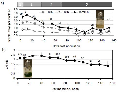
The DGP marked the beginning of the degradation processes where total chlorophyll levels per viable cell
dropped significantly by about 44%, from a high of 5.5 to 3.2 pg Chl cell ml− 1 at day 40 (p < 0.01, Fig. 2a),
though there was no change in the ratio of chlorophyll a to b (Fig. 2b). Total cell protein levels dropped
significantly by over 70% on a viable cell basis (Fig. 3A, p < 0.01). RuBisCO, one of the most abundant
proteins in the cell, showed large declines during this phase (Fig. 3B). The major light harvesting complex
proteins also declined, but by a small amount, and remained otherwise stable through the DGP. The
stress-related protein LHCSR, however, increased substantially during the DGP, as expected (Fig. 3B).
Fluorescence parameters used to assess light-usage efficiency in the cells, showed changes in how
excess light was handled by the cell. There were no significant changes in the maximum quantum
efficiency of PSII (Fv/Fm) or the Fo/Fm parameter during the declining growth phase (Fig. 4). However,
Page 5/21when faced with a light stress, PSII efficiency (φPSII) is zero, meaning that the excess light is being
dissipated rather than being used to drive photosynthesis (Fig. 5a). The proportion of light dissipated by
non-photochemical quenching (φNPQ) increases significantly during the DGP (Fig. 5b), but a portion of
the light is still dissipated by non-regulated means (φNO) (Fig. 5c). We also calculated NPQ 5 minutes
into actinic light exposure using the classic equation based on the Stern-Volmer relationship (NPQ= (Fm-
Fm’)/Fm’). This NPQ calculation peaked at day 25 cultures and remained high for the remainder of the
DGP (Fig. 6, supplementary material), similar to the φNPQ calculation of Karmer et al (2004). The higher
NPQ capacity roughly correlates with the induction of LHCSR during the DGP.
Stationary Phase—Phase 4:
Stationary phase would normally be defined by the lack of additional cell growth, though it is
questionable whether that definition would accurately describe Chlamydomonas cultures under these
conditions. Nevertheless, Phase 4 was defined by an apparent lack of increase in cell abundance, though
it wasn’t always straight-forward to define at what point the cell number plateaued. However, there are
other physiological parameters that help define this stage between days 40 and 68. The onset of
stationary phase coincides with smaller reductions in the mean cell viability over 4 weeks, but the
variability at this stage was such that the differences were not significant (Fig. 1a). Cumulative survival,
however, was different from the DGP where the day 68 average cumulative survival was at 0.57 (Fig. 1b).
The general cell decline continued in stationary: total chlorophyll levels drop an additional 30% (60%
decline from exponential levels) and total protein content per cell reaches its lowest levels of 2.3 pg per
viable cell, a significant 84% drop from exponential phase (Fig. 3a). Amongst these declines in total
protein, the LHCII antenna proteins are more-or-less stable (Fig. 3b). Somewhat unexpectedly, the amount
of LHCSR in the cell transiently drops at the end of stationary phase (day 68, Fig. 3b).
Under relatively low-light conditions, stationary phase is where there are clearly significant changes in the
photosynthetic apparatus. There is a drop in the maximum quantum efficiency of PSII in stationary
phase, reaching a low of 0.3 at day 68 (Fig. 4a). This drop in Fv/Fm is driven by an increase in the Fo,
which is obvious by the increase in the Fo/Fm value starting at day 54 and remaining high throughout
stationary (Fig. 4b). There are apparent variations in the way excitation energy is handled in stationary
phase with declines in φNPQ and a corresponding increase in φNO (Fig. 5b, c). However, using the classic
NPQ equation, the NPQ returns to low levels. Because this could be due to residual quenching following
dark adaptation, thus underestimating the true Fm, we calculated corrected NPQ (Tietz et al 2017), which
showed that NPQ levels remain high during this phase (Fig. 6, supplementary material).
Death Phase—Phase 5.
The march of death for Chlamydomonas begins in earnest after 68 days in culture with an exponential
decline in viability that continues to day 124. By day 124, only 15% of the cells are viable, with the rate of
cell death between day 68 and 124 being roughly 2.5x10⁵ cells per day (Fig. 1a). Beyond day 124, the
cells remaining die at a slower rate and by day 152, about 7% of the cells in the culture remain viable.
Page 6/21Cumulative survival at the end of this experiment (day 152) was 0.012 (Fig. 1b). By the start of the death
phase, the protein levels per viable cell are at their lowest but gradually start to rise, and by the end of the
death phase reach a level that is roughly 4 times higher than cells in exponential. This late increase is
potentially an artifact since it is difficult to account for residual protein from dead cells inflating the
estimates. During this phase chlorophyll levels gradually drop approximately 80% from exponential
phase maximum, such that the culture looks white (Fig. 2a). While the concentration of LHCII proteins
remained fairly constant up to day 96, beyond that, the levels of the protein declined until only trace
amounts were present at day 138. The Chl a/b ratio after day 96 was around 1.5, implying that most of
the remaining chlorophyll was likely associated with the antenna rather than reaction centers, though this
wasn’t directly assessed (Fig. 2B). LHCSR, however, rebounded from its transient decline between days 96
and 110, a period that coincided with drops in LHCII content (Fig. 3B). LHCSR levels declined to lower
levels on days 124 and 138. Beyond day 138, protein samples were challenging to work with and proteins
did not migrate well on gels, so they were abandoned.
During death phase, the fluorescence characteristics remain similar for the cells in stationary phase. The
high Fo/Fm remained a feature along with the suppressed Fv/Fm (Fig. 4a,b). φNPQ and corrected NPQ
calculations suggest that the remaining cells continued to have a capacity for dissipation of light with the
remaining LHCIIs and LHCSR proteins in the membrane. Fluorescence signatures beyond day 138 were
difficult to resolve owing to the exceedingly low levels of chlorophyll, so interpretation of these values is
limited.
As the cultures aged, it was increasingly difficult to distinguish between intact cells and cell debris.
Therefore, to verify the accuracy of our data in the ageing cultures, we also determined the number of
viable cells using the viable plate counts method (VPC). We performed the VPC on the samples as from
day 138. There were no significant differences between the data assessed using SYTOX® Orange and
the data obtained from the VPC method (P = 0.130, data not shown). Another issue with such long-term
cultures is they can be susceptible to evaporation, even though there are foam stoppers and aluminum
foil covering them. We estimated that there was 57µl of water lost through evaporation per day. This
could skew the cell abundance slightly toward higher values in the older cultures, but with the cell viability
estimates, that remains a minor complication.
Discussion
This study was initiated after finding an old culture of Chlamydomonas from a long-departed student
(about a year earlier) on a shaker was completely bleached (and presumed dead) yet recovered when
plated out on fresh media. The strategy for surviving this length of time in the absence of nutrients, and
presumably an ongoing stress, brought up many interesting questions about the long-term survival under
adverse conditions. Understanding how Chlamydomonas ages in batch culture and its strategy for
survival is not only a relevant ecological question, but one relevant for biotechnology for the production
of high-value products in cell culture (Scranton et al. 2015).
Page 7/21We would normally predict that Chlamydomonas doesn’t age under benign, nutrient-sufficient conditions,
continuing to divide akin to an immortal cell line. Budding yeast (Saccharomyces cerevisiae), can also be
propagated indefinitely, but individuals within these asymmetrically dividing unicells do eventually age
and die. The budding yeast is a model system for investigating both replicative lifespan (number of cell
divisions before senescence) and chronological lifespan (longevity in a non-dividing state) (Denoth
Lippuner, Julou, and Barral 2014). While a chronological lifespan concept is more appropriate for
Chlamydomonas, the symmetrically dividing fission yeast (Schizosaccharomyces pombe) may be a
better system to compare concepts of ageing in microalgae. In fission yeast, there is no evidence of
ageing in cells grown under favourable conditions; however, if these yeast cultures are stressed, evidence
of cellular ageing can be detected (Coelho et al 2013). In particular, heat shock protein-associated protein
aggregates are asymmetrically distributed between “symmetrically” dividing cells such that there is an old
and young product of cell division (Coelho et al. 2013; 2014). The younger of these cells would be free of
the protein aggregates, putting it on the path to recovery. Even the bacterium, Escherichia coli, that
apparently divides symmetrically, the offspring can differentially possess new or old cell components
that can affect its subsequent growth rate (Stewart et al. 2005; Lindner et al. 2008). While there are other
studies looking at ageing in unicellular organisms (Florea 2017), it’s unknown whether something similar
occurs with Chlamydomonas cells during cell division. However, culture heterogeneity has been described
where there are different populations of cells with distinct growth-rates in the same culture (Damodaran
et al. 2015), which may suggest that there is some type of ageing occurring in these cells even though
these symmetrically dividing cells were grown under favourable conditions.
Our system deals more with conditional senescence, where cells senesce as nutrient levels are depleted
and there is a transition from favourable to unfavourable conditions. It’s under these conditions where
cell division is limited that we would predict cellular ageing to accelerate. The transition to conditional
senescent conditions is first evidenced by the changing growth rate as cells run out of nutrients. This
declining growth phase where cells continued to grow, though at a much-reduced rate, lasted for about 40
days. The declining growth phase is able to support limited growth by recycling internal macromolecules.
In particular, protein levels per cell decline by close to 70%. This is a long-observed pattern in bacteria
where protein levels decline upon entry into stationary phase when other stored nutrients are depleted
(Wanner and Egli 1990). Similar protein declines are also observed in Chlamydomonas reinhardtii during
nitrogen, phosphorus, or sulphur deprivation experiments (Cakmak et al. 2012; Kamalanathan et al.
2016). While part of the mechanism for this reduction in protein during senescence is dilution through cell
division (Meagher et al. 2021), the activation of autophagy is likely an essential process regulating the
turnover of proteins to enhance nutrient recycling. Evidence for the importance of autophagy in
senescence includes the induction of ATG8 in Chlamydomonas cultures approaching stationary phase
(Pérez-Pérez, Florencio, and Crespo 2010; Meagher et al. 2021). ATG8 is a ubiquitin-like protein that is
conjugated to a phospholipid in a step required for autophagosome formation (Mizushima, Yoshimori,
and Ohsumi 2011; Pérez-Pérez and Crespo 2010). Autophagy was found required for turnover of
ribosomal proteins and regulation of lipid metabolism (Couso et al. 2018), so this process is important
for metabolic restructuring in the cell. This nutrient recycling likely supported a limited cell division, but it
Page 8/21also extended lifespan during conditional senescence— cell viability remained high during the declining
growth phase.
It’s clear that the duration and characteristics of the declining growth phase is variable depending on how
the cells are grown, and perhaps the strain used. For instance, Humby et al. (2103) used a cell wall-less
strain under low gas-exchange conditions and observed a DGP that was very short; cells entered
stationary shortly after the exponential phase. Under active bubbling for maximum gas exchange using
the same strain as in this study (CC125), the declining growth phase lasted for at least 10 days following
the exit from logarithmic growth (Meagher et al. 2021). The declining growth phase is often ignored or
not identified in many culturing systems. In fact, these extended declining growth phases are a hallmark
of batch culture growth of bacteria (Wanner and Egli 1990) and fungi (Borrow et al. 1964; Vrabl et al.
2019). It’s been proposed that entry in this phase is due to a limiting nutrient that curbs cell growth, the
slope of which gets lower as other nutrients become depleted (Monod 1949; Vrabl et al. 2019). This is
likely the case in the variation we observed in different studies, where there is depletion of CO2, acetate,
nitrogen, phosphorous, or a micronutrient. It’s clear that by altering the culturing conditions, particularly
changing the potential for gas exchange, the progression of the standard growth curve can shift
dramatically.
Chlorophyll degradation/dilution in conditional senescence starts shortly after exiting exponential phase
and continues through the declining growth, stationary, and death phases. The degradation of chlorophyll
during senescence is a common response in photosynthetic organisms and the result of activation of
several enzymes that break down chlorophyll to a variety of catabolites (Hörtensteiner 2006), and in
some ways resembles the loss of chlorophyll during nutrient deficiency (Plumley and Schmidt 1989;
Kamalanathan et al. 2016). Under stress conditions when the normal sink capacities are limited,
chlorophyll can be a potential cellular photo-toxin when the absorbed light energy is diverted
inappropriately to oxygen, leading to the production of reactive oxygen species (ROS) (Hörtensteiner and
Kräutler 2011). The large and rapid drop in RuBisCO content after exiting exponential growth phase
(Fig. 3B) is indicative of a lost sink capacity and the risk associated with maintaining the light harvesting
machinery. Excess light in the absence of an appropriate sink could lead to an oxidative stress where ROS
can damage macromolecules (Apel and Hirt 2004). It’s interesting that our earlier study using a cell wall-
less strain showed no decline in chlorophyll levels as cells aged in a more closed culturing system,
despite the fact that thylakoid membrane density and chloroplast size declined dramatically (Humby et
al. 2013). While this was unexpected, we proposed that chlorophyll may have accumulated in the
enlarging oil bodies as cells aged. However, it’s possible that this was an artifact of the cell wall-less
strain that was used. These cells lyse shortly after death, perhaps leaving plastid fragments containing
chlorophyll that were still harvestable in later steps given the more accelerated senescence timeline.
The drop in the Chl a/b ratio in the death phase (after day 68), and the relatively stable maintenance of
LHCII levels implies there is a preferential maintenance of the LHC antenna over reaction centres during
conditional senescence (Humby et al. 2013; Meagher et al. 2021). This was also observed in the leaves of
senescing Arabidopsis (Nath et al. 2013). The Chl a/b ratio started to decline at about 54 days post
Page 9/21inoculation, which corresponded with the drop in Fv/Fm and increase in the Fo/Fm, that remained high
throughout stationary and death phases. As Meager et al (2021) argued, this is likely due to the
detachment of the antenna from the PSII reaction centre, leading to an increase in the Fo, and subsequent
drop in the Fv/Fm. In that study, the increase in the Fo/Fm was only apparent in a HL-stress culture in the
DGP, not the low-light culture, indicative of a light-stress response. In this study, the Fo/Fm increase was
pushed to the stationary phase under low-light conditions, indicating an indirect-light stress caused by
limited sink capacity. Of course, LHCII is a major antenna complex having a dual function; light-
harvesting and photo-protection (Natali and Croce 2015). LHCII is able to switch to a quenched
conformation in light stress conditions thereby dissipating excess light in the form of heat (Tian et al.
2015). The abundance of LHCII well into the death phase could suggest its importance during conditional
senescence, most likely in photo-protection that is triggered by LHCSR.
The ability of Chlamydomonas to deal with excess light changed throughout conditional senescence.
When faced with an excess-light challenge, the quantum efficiency of PSII (φPSII) was always close to
zero, indicating the reaction centers were fully reduced and all of the incoming light was being dissipated
when challenged with excess light. Initially, this energy was dissipated in a manner that was not
photoprotective (high φNO) when non-photochemical quenching (φNPQ) capacity was low. It is indicative
of cells that are unable to properly protect themselves from excess light (Klughammer and Schreiber
2008), that could invariably lead to photoinhibition. However, φNPQ started to increase at day 11,
reaching a maximum at 25 days post inoculation. φNPQ corresponds to the fraction of energy dissipated
in the form of heat via regulated, nonphotochemical quenching mechanisms (Kramer et al. 2004). This
increase is very likely due to the upregulation of LHCSR at day 11, which is known to be a major player in
NPQ in Chlamydomonas (Peers et al. 2009).
The stress-related LHC proteins (collectively LHCSR1 and 3) were notable in their upregulation in
response to senescence, peaking shortly after exponential growth. This stress-related protein is
upregulated during high-light stress conditions and responsible, in part, for the induction of non-
photochemical quenching in Chlamydomonas (Peers et al. 2009). LHCSR also responds to a variety of
stresses, such as nutrient limitation, not just light (Toepel et al. 2013), so its induction during senescence
is not surprising and previously observed (Humby et al. 2013; Meagher et al. 2021). The induction of
LHCSR likely facilitates the conversion of the antenna into light-quenching centers, acting as a photo-
protective mechanism for the reaction centers (Girolomoni et al. 2019). LHCSR is able to sense pH
variations in the thylakoid lumen and can reversibly switch its conformation from a light-harvesting one
to a dissipative one (Peers et al. 2009). This would essentially assist in neutralizing the incoming light
energy to minimize the production of ROS under these stress conditions. The transient decline in LHCSR
levels at days 68 and 82 was very consistent between replicates, yet a rather curious response. It’s
possible that recovered nutrients from dying cells in stationary contributed to the decline, but otherwise,
the signal triggering the reduction is unknown. However, the drop in LHCSR does roughly correlate to the
transit decline of φNPQ during these timepoints, and the increase in φNO. This reinforces the known link
of LHCSR with quenching potential (Peers et al. 2009).
Page 10/21This work highlights strategies used by Chlamydomonas to extend their lifespan under conditional
senescence. Following the exit from exponential growth, cells maintain a low-level of cell division for an
extended period through recycling macromolecules and reducing protein content and the size of the
photosynthetic apparatus. During this time, photoprotective mechanisms are induced, and these remain
high throughout the period, to minimize the production of reactive oxygen species. The prolonged
maintenance of LHCII over a period of almost 124 days and the induction of LHCSR has an important
role in this process. While we proposed a shift in the photo-protective mechanisms involving bulk
fluorescence, LHCII could also potentially have a structural role in maintaining the integrity of the
thylakoid membranes in ageing cultures, which might be essential for the recovery process when the
conditions improve.
An important question is whether there are adaptations in the surviving cells that encourage long-term
survival under nutrient depleted conditions. Certainly, sexual reproduction is activated in Chlamydomonas
under nutrient stress and the resulting zygote (hypnozygotes or zygospores) is effectively a resting stage
resistant to environmental stress (Daniel, Henley, and VanWinkle-Swift 2007; Ellegaard and Ribeiro
2018b). However, there are also non-sexual resting cells that are more comparable to this study. Many
microalgae can form resting cells, cysts, or akinetes with a similar environmental stress resistance
(Coleman 1983). In the green alga, zygonema, for instance, increasing age of the culture leads to the
formation of what are called “pre-akinetes” that are the result of the accumulation of storage products
and thickening of the cell wall. In zygonema, there is a metabolic restructuring that pushes metabolism
toward production of storage products (Arc et al. 2020), something that also occurs in Chlamydomonas
with age and nutrient deprivation (Siaut et al. 2011). We don’t have any clear evidence for formation of
stress-resistant resting cells, or its equivalent. But, for instance, we did observe a gradual increase in
protein per viable cell beyond 68 days. This may represent a collective partial recovery from scavenged
nutrients from dying cells and perhaps these cells are more resistant to the stress condition due to some
change in the cell wall or other intracellular structures. However, it is questionable whether this increase in
protein is real or an artifact of the cell collection process where protein from dead cells could clearly be
included, but it is nevertheless an intriguing question whether Chlamydomonas can produce something
akin to a resting cell or akinete, indicating a programmed response.
Declarations
Acknowledgments:
This research was funded through the Discovery Grant program from the National Sciences and
Engineering Research Council (NSERC) of Canada. The authors thank Ghaith Zamzam and Adrian-Reyes
Prieto for reading an earlier version of this manuscript.
Funding:
Discovery Grant funding provided to DGD from the National Sciences and Engineering Research Council
(NSERC) of Canada.
Page 11/21Conflicts of Interest/Competing Interests:
None.
Availability of data and material:
Available by request to the corresponding author.
Code availability:
Not applicable
Author’s Contributions:
DD conducted the research, analyzed data and participated in the writing of the manuscript. DGD
designed the experiment, analyzed the data, and wrote the manuscript.
Ethics Approval:
not applicable
Consent to participate:
Not applicable
Consent for publication:
Both authors approved of submission.
References
1. Apel, Klaus, and Heribert Hirt. 2004. “REACTIVE OXYGEN SPECIES: Metabolism, Oxidative Stress, and
Signal Transduction.” Annual Review of Plant Biology 55 (1): 373–99.
https://doi.org/10.1146/annurev.arplant.55.031903.141701
2. Arc, Erwann, Martina Pichrtová, Ilse Kranner, and Andreas Holzinger. 2020. “Pre-Akinete Formation in
Zygnema Sp. from Polar Habitats Is Associated with Metabolite Re-Arrangement.” Edited by Henrik
Buschmann. Journal of Experimental Botany 71 (11): 3314–22.
https://doi.org/10.1093/jxb/eraa123
3. Borrow, A., Sheila Brown, E. G. Jefferys, R. H. J. Kessell, Eithne C. Lloyd, P. B. Lloyd, A. Rothwell, B.
Rothwell, and J. C. Swait. 1964. “THE KINETICS OF METABOLISM OF GIBBERELLA FUJIKUROI IN
STIRRED CULTURE.” Canadian Journal of Microbiology 10 (3): 407–44.
https://doi.org/10.1139/m64-054
4. Cakmak, Turgay, Pinar Angun, Alper D Ozkan, Zeynep Cakmak, Tolga T Olmez, and Turgay Tekinay.
2012. “Nitrogen and Sulfur Deprivation Differentiate Lipid Accumulation Targets of Chlamydomonas
Page 12/21Reinhardtii.” Bioengineered, August. https://doi.org/10.4161/bioe.21427
5. Choudhury, Feroza K., Rosa M. Rivero, Eduardo Blumwald, and Ron Mittler. 2017. “Reactive Oxygen
Species, Abiotic Stress and Stress Combination.” The Plant Journal 90 (5): 856–67.
https://doi.org/10.1111/tpj.13299
6. Coelho, Miguel, Aygül Dereli, Anett Haese, Sebastian Kühn, Liliana Malinovska, Morgan E. DeSantis,
James Shorter, Simon Alberti, Thilo Gross, and Iva M. Tolić-Nørrelykke. 2013. “Fission Yeast Does Not
Age under Favorable Conditions, but Does So after Stress.” Current Biology 23 (19): 1844–52.
https://doi.org/10.1016/j.cub.2013.07.084
7. Coelho, Miguel, Steven J. Lade, Simon Alberti, Thilo Gross, and Iva M. Tolić. 2014. “Fusion of Protein
Aggregates Facilitates Asymmetric Damage Segregation.” Edited by Peter Walter. PLoS Biology 12
(6): e1001886. https://doi.org/10.1371/journal.pbio.1001886
8. Coleman, Annette W. 1983. “The Roles of Resting Spores and Akinetes m Chlorophyte Survival.” In
Survival Strategies of the Algae, 1–21. Cambridge University Press.
9. Couso, Inmaculada, María Esther Pérez-Pérez, Enrique Martínez-Force, Hee-Sik Kim, Yonghua He,
James G Umen, and José L Crespo. 2018. “Autophagic Flux Is Required for the Synthesis of
Triacylglycerols and Ribosomal Protein Turnover in Chlamydomonas.” Edited by Peter Bozhkov.
Journal of Experimental Botany 69 (6): 1355–67. https://doi.org/10.1093/jxb/erx372
10. Damodaran, Shima P., Stephan Eberhard, Laurent Boitard, Jairo Garnica Rodriguez, Yuxing Wang,
Nicolas Bremond, Jean Baudry, Jérôme Bibette, and Francis-André Wollman. 2015. “A Millifluidic
Study of Cell-to-Cell Heterogeneity in Growth-Rate and Cell-Division Capability in Populations of
Isogenic Cells of Chlamydomonas Reinhardtii.” Edited by Rajagopal Subramanyam. PLOS ONE 10
(3): e0118987. https://doi.org/10.1371/journal.pone.0118987
11. Daniel, Patricia, Jessica Henley, and Karen VanWinkle-Swift. 2007. “Altered Zygospore Wall
Ultrastructure Correlates With Reduced Abiotic Stress Resistance in a Mutant Strain of
Chlamydomonas monoica (Chlorophyta).” Journal of Phycology 43 (1): 112–19.
https://doi.org/10.1111/j.1529-8817.2006.00313.x
12. Denoth Lippuner, Annina, Thomas Julou, and Yves Barral. 2014. “Budding Yeast as a Model
Organism to Study the Effects of Age.” FEMS Microbiology Reviews 38 (2): 300–325.
https://doi.org/10.1111/1574-6976.12060
13. Ellegaard, Marianne, and Sofia Ribeiro. 2018a. “The Long-Term Persistence of Phytoplankton Resting
Stages in Aquatic ‘Seed Banks.’” Biological Reviews 93 (1): 166–83.
https://doi.org/10.1111/brv.12338
14. ———. 2018b. “The Long-Term Persistence of Phytoplankton Resting Stages in Aquatic ‘Seed Banks’:
Persistence of Phytoplankton Resting Stages.” Biological Reviews 93 (1): 166–83.
https://doi.org/10.1111/brv.12338
15. Florea, Michael. 2017. “Aging and Immortality in Unicellular Species.” Mechanisms of Ageing and
Development 167 (October): 5–15 https://doi.org/10.1016/j.mad.2017.08.006
Page 13/2116. Girolomoni, Laura, Stefano Cazzaniga, Alberta Pinnola, Federico Perozeni, Matteo Ballottari, and
Roberto Bassi. 2019. “LHCSR3 Is a Nonphotochemical Quencher of Both Photosystems in
Chlamydomonas Reinhardtii.” Proceedings of the National Academy of Sciences 116 (10): 4212–17.
https://doi.org/10.1073/pnas.1809812116.
17. Hörtensteiner, S. 2006. “Chlorophyll Degradation During Senescence.” Annual Review of Plant
Biology 57 (1): 55–77. https://doi.org/10.1146/annurev.arplant.57.032905.105212
18. Hörtensteiner, Stefan, and Bernhard Kräutler. 2011. “Chlorophyll Breakdown in Higher Plants.”
Biochimica et Biophysica Acta (BBA) - Bioenergetics 1807 (8): 977–88.
https://doi.org/10.1016/j.bbabio.2010.12.007
19. Humby, Penny L, Ellen C R Snyder, and Dion G Durnford. 2013. “Conditional Senescence in
Chlamydomonas Reinhardtii (Chlorophyceae).” Journal of Phycology 49 (2): 389–400.
https://doi.org/10.1111/jpy.12049
20. Hurkman, W J. 1979. “Ultrastructural Changes of Chloroplasts in Attached And Detached, Aging
Primary Wheat Leaves.” Amer. J. Bot. 66 (1): 64–70
21. Kamalanathan, Manoj, Mattia Pierangelini, Lauren Ann Shearman, Roslyn Gleadow, and John
Beardall. 2016. “Impacts of Nitrogen and Phosphorus Starvation on the Physiology of
Chlamydomonas Reinhardtii.” Journal of Applied Phycology 28 (3): 1509–20.
https://doi.org/10.1007/s10811-015-0726-y
22. Klughammer, Christof, and Ulrich Schreiber. 2008. “Complementary PS II Quantum Yields Calculated
from Simple Fluorescence Parameters Measured by PAM Fluorometry and the Saturation Pulse
Method,” PAM Application Notes 1: 27-35
23. Kolter, Roberto, Deborah A. Siegele, and Antonio Tormo. 1993. “The Stationary Phase of the Bacterial
Life Cycle.” Annual Review of Microbiology 47 (1): 855–74.
https://doi.org/10.1146/annurev.mi.47.100193.004231
24. Kramer, David M., Giles Johnson, Olavi Kiirats, and Gerald E. Edwards. 2004. “New Fluorescence
Parameters for the Determination of Q A Redox State and Excitation Energy Fluxes.” Photosynthesis
Research 79 (2): 209–18. https://doi.org/10.1023/B:PRES.0000015391.99477.0d
25. Lillie, S H, and J R Pringle. 1980. “Reserve Carbohydrate Metabolism in Saccharomyces Cerevisiae:
Responses to Nutrient Limitation.” Journal of Bacteriology 143 (3): 1384–94
26. Lim, Pyung Ok, and Hong Gil Nam. 2007. “Aging and Senescence of the Leaf Organ” J. Plant Biol. 50
(3): 10. https://doi.org/10.1007/BF03030657
27. Lindner, A. B., R. Madden, A. Demarez, E. J. Stewart, and F. Taddei. 2008. “Asymmetric Segregation of
Protein Aggregates Is Associated with Cellular Aging and Rejuvenation.” Proceedings of the National
Academy of Sciences 105 (8): 3076–81. https://doi.org/10.1073/pnas.0708931105
28. Louda, J.William, Jie Li, Lei Liu, M.Nancy Winfree, and Earl W. Baker. 1998. “Chlorophyll-a
Degradation during Cellular Senescence and Death.” Organic Geochemistry 29 (5–7): 1233–51.
https://doi.org/10.1016/S0146-6380(98)00186-7.
Page 14/2129. McLean, Robert J. 1968. “Ultrastructure of Spongiochloris Typica During Senescence12.” Journal of
Phycology 4 (4): 277–83. https://doi.org/10.1111/j.1529-8817.1968.tb04696.x
30. Mclean, Robert J. 1969. “Rejuvenation of Senescent Cells of Spongiochloris typica.” Journal of
Phycology, no. 5: 32–37
31. Meagher, Emily, Pattarasiri Rangsrikitphoti, Babar Faridi, Ghaith Zamzam, and Dion G Durnford.
2021. “Photoacclimation to High-Light Stress in Chlamydomonas Reinhardtii during Conditional
Senescence Relies on Generating PH-Dependent, High-Quenching Centres | Elsevier Enhanced
Reader.” Plant Physiology and Biochemistry 158: 136–45.
https://doi.org/10.1016/j.plaphy.2020.12.002.
32. Messer, Glenda, and Yehuda Ben-Shaul. 1972. “Changes in Chloroplast Structure during Culture
Growth of Peridinium Cinctum Fa. Westii (Dinophyceae).” Phycologia 11 (3–4): 291–99.
https://doi.org/10.2216/i0031-8884-11-3-291.1.
33. Mizushima, Noboru, Tamotsu Yoshimori, and Yoshinori Ohsumi. 2011. “The Role of Atg Proteins in
Autophagosome Formation.” Annual Review of Cell and Developmental Biology 27 (1): 107–32.
https://doi.org/10.1146/annurev-cellbio-092910-154005.
34. Monod, Jacques. 1949. “The Growth of Bacterial Cultures”. Ann. Rev. Microbio. 3: 371–94.
https://doi.org/10.1146/annurev.mi.03.100149.002103
35. Natali, Alberto, and Roberta Croce. 2015. “Characterization of the Major Light-Harvesting Complexes
(LHCBM) of the Green Alga Chlamydomonas reinhardtii.” PLOS ONE 10 (2): e0119211.
https://doi.org/10.1371/journal.pone.0119211
36. Nath, Krishna, Bong-Kwan Phee, Suyeong Jeong, Sun Yi Lee, Yoshio Tateno, Suleyman I.
Allakhverdiev, Choon-Hwan Lee, and Hong Gil Nam. 2013. “Age-Dependent Changes in the Functions
and Compositions of Photosynthetic Complexes in the Thylakoid Membranes of Arabidopsis
Thaliana.” Photosynthesis Research 117 (1–3): 547–56. https://doi.org/10.1007/s11120-013-9906-
2
37. Nyström, Thomas. 2003. “Conditional Senescence in Bacteria: Death of the Immortals.” Molecular
Microbiology 48 (1): 17–23. https://doi.org/10.1046/j.1365-2958.2003.03385.x
38. Peers, Graham, Thuy B. Truong, Elisabeth Ostendorf, Andreas Busch, Dafna Elrad, Arthur R.
Grossman, Michael Hippler, and Krishna K. Niyogi. 2009. “An Ancient Light-Harvesting Protein Is
Critical for the Regulation of Algal Photosynthesis.” Nature 462 (7272): 518–21.
https://doi.org/10.1038/nature08587
39. Pérez-Pérez, María Esther, and José L. Crespo. 2010. “Autophagy in the Model Alga Chlamydomonas
Reinhardtii.” Autophagy 6 (4): 562–63. https://doi.org/10.4161/auto.6.4.11822
40. Pérez-Pérez, María Esther, Francisco J. Florencio, and José L. Crespo. 2010. “Inhibition of Target of
Rapamycin Signaling and Stress Activate Autophagy in Chlamydomonas Reinhardtii.” Plant
Physiology 152 (4): 1874–88. https://doi.org/10.1104/pp.109.152520
41. Petrov, Veselin, Jacques Hille, Bernd Mueller-Roeber, and Tsanko S. Gechev. 2015. “ROS-Mediated
Abiotic Stress-Induced Programmed Cell Death in Plants.” Frontiers in Plant Science 6 (February): 1–
Page 15/2116. https://doi.org/10.3389/fpls.2015.00069
42. Plumley, F. G., and G. W. Schmidt. 1989. “Nitrogen-Dependent Regulation of Photosynthetic Gene
Expression.” Proceedings of the National Academy of Sciences 86 (8): 2678–82.
https://doi.org/10.1073/pnas.86.8.2678
43. Scranton, Melissa A., Joseph T. Ostrand, Francis J. Fields, and Stephen P. Mayfield. 2015.
“Chlamydomonas as a Model for Biofuels and Bio‐products Production.” The Plant Journal 82 (3):
523–31. https://doi.org/10.1111/tpj.12780
44. Siaut, Magali, Stéphan Cuiné, Caroline Cagnon, Boris Fessler, Mai Nguyen, Patrick Carrier, Audrey
Beyly, et al. 2011. “Oil Accumulation in the Model Green Alga Chlamydomonas Reinhardtii:
Characterization, Variability between Common Laboratory Strains and Relationship with Starch
Reserves.” BMC Biotechnology 11 (1): 7. https://doi.org/10.1186/1472-6750-11-7
45. Stewart, Eric J, Richard Madden, Gregory Paul, and François Taddei. 2005. “Aging and Death in an
Organism That Reproduces by Morphologically Symmetric Division.” Edited by Thomas Kirkwood.
PLoS Biology 3 (2): e45. https://doi.org/10.1371/journal.pbio.0030045
46. Tian, Lijin, Emine Dinc, and Roberta Croce. 2015. “LHCII Populations in Different Quenching States
Are Present in the Thylakoid Membranes in a Ratio That Depends on the Light Conditions.” Journal
of Physical Chemistry Letters 6 (12): 2339–44. https://doi.org/10.1021/acs.jpclett.5b00944
47. Tietz, Stefanie, Christopher C. Hall, Jeffrey A. Cruz, and David M. Kramer. 2017. “NPQ (T) : A
Chlorophyll Fluorescence Parameter for Rapid Estimation and Imaging of Non-Photochemical
Quenching of Excitons in Photosystem-II-Associated Antenna Complexes: New, Rapid Probe of Non-
Photochemical Quenching.” Plant, Cell & Environment 40 (8): 1243–55.
https://doi.org/10.1111/pce.12924
48. Toepel, Jörg, Maike Illmer-Kephalides, Sebastian Jaenicke, Jasmin Straube, Patrick May, Alexander
Goesmann, and Olaf Kruse. 2013. “New Insights into Chlamydomonas Reinhardtii Hydrogen
Production Processes by Combined Microarray/RNA-Seq Transcriptomics.” Plant Biotechnology
Journal 11 (6): 717–33. https://doi.org/10.1111/pbi.12062
49. Vrabl, Pamela, Christoph W. Schinagl, Desirée J. Artmann, Benedikt Heiss, and Wolfgang Burgstaller.
2019. “Fungal Growth in Batch Culture – What We Could Benefit If We Start Looking Closer.”
Frontiers in Microbiology 10 (October): 2391. https://doi.org/10.3389/fmicb.2019.02391
50. Wanner, Ursula, and Thomas Egli. 1990. “Dynamics of Microbial Growth and Cell Composition in
Batch Culture.” FEMS Microbiology Letters 75 (1): 19–43. https://doi.org/10.1016/0378-
1097(90)90521-Q
51. Werner-Washburne, M, E Braun, G C Johnston, and R A Singer. 1993. “Stationary Phase in the Yeast
Saccharomyces Cerevisiae.” Microbiological Reviews 57 (2): 383–401.
https://doi.org/10.1093/nar/gkr782
52. Yang, Lei, Jun Chen, Shan Qin, Min Zeng, Yongguang Jiang, Lang Hu, Peng Xiao, et al. 2018. “Growth
and Lipid Accumulation by Different Nutrients in the Microalga Chlamydomonas Reinhardtii.”
Biotechnology for Biofuels 11 (1): 40. https://doi.org/10.1186/s13068-018-1041-z
Page 16/21Figures Figure 1 Growth and survival of Chlamydomonas during long-term culturing. a) Cell abundance (left axis, filled circles) and cell viability assessed using SYTOX® Orange (right axis, open diamonds). b) Cumulative survival curve data showing the probability of a cell surviving to a specific day (each replicate shown). The growth phases identified are the exponential growth (2), the declining relative growth phase (3), the stationary phase, (4) and the death phase (5). n=3, error bars =2SE. Means that do not share a letter are significantly different (p
Figure 2

Chlorophyll content of cells during long-term batch culture growth. a) The average chlorophyll a and b
content in pg per viable cell: total chlorophyll (filled circles), chl a (grey circles), chl b (open circles).b) The
average ratio of chlorophyll a/b, which reflects the ratio between the antenna and reaction centers. n=3,
error bars =2SE. Photographs show the cultures at day 4 (bottom) and day 166 (top). Means that do not
share a letter are significantly different (p
Figure 3

Protein changes in Chlamydomonas during long-term batch culture growth. a) Total protein per viable cell
(pgcell-1). Offset graph shows the last three timepoints in the series .n=3, error bars =2SE. Means that do
not share a letter are significantly different (p
Figure 4

The fluorescence parameters of Chlamydomonas during long-term batch culture growth. A) Maximum
quantum efficiency of PSII (Fv/Fm). B) The dark-adapted, minimum fluorescence (Fo) normalized to the
maximal fluorescence (Fm) C). n=3, error bars =2SE. Means that do not share a letter are significantly
different (p
Figure 5

The fluorescence parameters of Chlamydomonas during long-term batch culture growth when challenged
with excess light. A) Fraction of light used in photochemistry (ϕPSII), B) fraction of light dissipated by
non-photochemical quenching (ϕNPQ), and C) the fraction of light dissipated by other, non-regulated
processes (ϕNO). n=3, error bars =2SE. Means that do not share a letter are significantly different (p
You can also read


