The Retinoblastoma Protein Tumor Suppressor Is Important for Appropriate Osteoblast Differentiation and Bone Development
←
→
Page content transcription
If your browser does not render page correctly, please read the page content below
The Retinoblastoma Protein Tumor Suppressor Is
Important for Appropriate Osteoblast Differentiation
and Bone Development
Seth D. Berman, Tina L. Yuan, Emily S. Miller, Eunice Y. Lee,
Alicia Caron, and Jacqueline A. Lees

David H. Koch Institute for Integrative Cancer Research at MIT, Massachusetts Institute of Technology,
Cambridge, Massachusetts

Abstract                                                                             sporadic human tumors, but there is strong correlation with
Mutation of the retinoblastoma (RB) tumor suppressor                                 certain tumor types. Specifically, RB mutations are observed in
gene is strongly linked to osteosarcoma formation.                                   almost all retinoblastomas (1) and also in a large percentage of
This observation and the documented interaction                                      osteosarcomas and small cell lung carcinomas. For patients who
between the retinoblastoma protein (pRb) and Runx2                                   carry germ line RB mutations, osteosarcoma is the second most
suggests that pRb is important in bone development.                                  common tumor type after retinoblastoma (2). Overall, >70% of
To assess this hypothesis, we used a conditional                                     osteosarcomas show a molecular change or mutation at the RB
knockout strategy to generate pRb-deficient embryos                                  locus (3, 4).
that survive to birth. Analysis of these embryos shows                                  The gene product, pRb, belongs to a family of proteins,
that Rb inactivation causes the abnormal development                                 including p107 and p130, termed the pocket proteins, although
and impaired ossification of several bones, correlating                              only pRb has been shown to possess significant tumor-
with an impairment in osteoblast differentiation.                                    suppressive properties (5). The best characterized role of pRb
We further show that Rb inactivation acts to promote                                 is its regulation of cell cycle progression. Overexpression of
osteoblast differentiation in vitro and, through                                     pRb causes G1 cell cycle arrest (6), whereas acute ablation
conditional analysis, establish that this occurs in a                                of pRb induces cell cycle re-entry in quiescent cells (7). To
cell-intrinsic manner. Although these in vivo and in vitro                           execute its cell cycle – inhibitory function, hypophosphorylated
differentiation phenotypes seem paradoxical, we find                                 pRb binds to and inhibits the E2F family of transcription factors
that Rb-deficient osteoblasts have an impaired ability                               (8). During G1, pRb becomes hyperphosphorylated by the
to exit the cell cycle both in vivo and in vitro that can                            cyclin D-cdk4/6 complex and subsequently by cyclin E-cdk2.
explain the observed differentiation defects. Consistent                             This phosphorylation releases the E2Fs from pRb to induce the
with this observation, we show that the cell cycle and                               transcription of cellular genes essential for S phase entry and
the bone defects in Rb-deficient embryos can be                                      cell division.
suppressed by deletion of E2f1, a known proliferation                                   The analyses of in vivo mouse models and in vitro
inducer that acts downstream of Rb. Thus, we conclude                                experiments show that pRb is required for the differentiation
that pRb plays a key role in regulating osteoblast                                   of specific tissues. In erythropoiesis, the loss of Rb results in
differentiation by mediating the inhibition of E2F                                   inefficient enucleation and incomplete terminal differentiation
and consequently promoting cell cycle exit.                                          of erythroid cells (9, 10). In skeletal muscle, pRb is required for
(Mol Cancer Res 2008;6(9):1440 – 51)                                                 proper cell cycle exit and differentiation (11). Conditional
                                                                                     deletion of Rb in the intestine causes increased proliferation and
                                                                                     abnormal expression of differentiation markers (12, 13). The
Introduction
                                                                                     loss of pRb affects the normal expression of differentiation
   The first tumor suppressor to be cloned was the retinoblas-
                                                                                     genes, such as h- and g-crystallines, in the lens (14). These
toma gene, RB. RB is mutated in approximately one-third of all
                                                                                     deficiencies in differentiation seem to be due, at least partially,
                                                                                     to a defect in cell cycle exit, a step believed to be required
                                                                                     in most differentiation pathways. However, this does not rule
Received 4/10/08; revised 6/12/08; accepted 6/13/08.                                 out the possibility that pRb contributes to differentiation in
Grant support: National Cancer Institute grants GM53204 and CA121921 (J.A.
Lees). S. Berman is a David H. Koch Graduate Fellow, E.Y. Lee received a Pearl
                                                                                     a more distinct and specific manner. Notably, pRb binds to
Staller Graduate Fellowship, and J.A. Lees is a Ludwig Scholar at Massachusetts      NRP/B, a protein up-regulated during neuronal differentiation
Institute of Technology.                                                             and involved in neuronal process formation (15). Relevant to
The costs of publication of this article were defrayed in part by the payment of
page charges. This article must therefore be hereby marked advertisement in          this, other markers of neuronal differentiation are decreased in
accordance with 18 U.S.C. Section 1734 solely to indicate this fact.                 the Rb-deficient embryo (16). With respect to fat cells, pRb
Note: Supplementary data for this article are available at Molecular Cancer
Research Online (http://mcr.aacrjournals.org/).
                                                                                     physically interacts with CAAT/enhancer binding protein-h,
Requests for reprints: Jacqueline A. Lees, David H. Koch Institute for Integrative   and the loss of this interaction inhibits adipocyte differentia-
Cancer Research at MIT, Massachusetts Institute of Technology, Cambridge, MA.        tion (17).
Phone: 617-252-1972; Fax: 617-253-9863. E-mail: jalees@mit.edu
Copyright D 2008 American Association for Cancer Research.                              Several studies implicate a role for pRb in osteoblast
doi:10.1158/1541-7786.MCR-08-0176                                                    differentiation. SV40-derived large T-antigen, which targets the

1440                                                                                                     Mol Cancer Res 2008;6(9). September 2008
            Downloaded from mcr.aacrjournals.org on February 8, 2021. © 2008 American Association for Cancer
                                                      Research.
Rb Regulates Proper Bone Development        1441

pocket proteins, prevents the differentiation of stromal cell lines    process of the sternum, were appropriately ossified but showed
into osteoblasts (18). The adenoviral E1A 12S protein also             an abnormal structure (Fig. 1C and D). These abnormal
represses osteoblast differentiation, and this is dependent on         structures were observed in all 13 Rbc /c embryos examined.
a functional E1A pocket protein binding domain (19). Most              Finally, several other bones such as the long bones of the fore-
striking is the finding that in immortalized cell lines, pRb           limbs and hind limbs did not exhibit any differing phenotypes
physically interacts with Runx2/CBFA1, one of the transcrip-           between the Rbc /c and wild-type embryos. It is possible that
tion factors essential for osteoblast differentiation (20, 21). This   certain embryonic bones, such as the limbs, are less susceptible
latter observation suggests that pRb may play a role in osteob-        to the effects of Rb loss than others, perhaps due to the
last differentiation that is independent of cell cycle regulation.     compensation effects of p107 and p130. Alternatively, the
    Determining the role of pRb in osteoblast differentiation          Mox2-Cre transgene may be less efficient in some settings.
in vivo may ultimately provide some important insights con-                To further explore the defects that were observed in the
cerning the high prevalence of Rb mutations in osteosarcoma.           Rbc /c embryos, we examined the skeletons of mutant
However, murine embryos deficient for pRb die between                  embryos at other developmental stages. At earlier time points,
embryonic days 13.5 and 15.5 (22-24). This early lethality has         embryonic days 15.5 and 16.5, the Rbc /c embryos displayed
thus far precluded the study of pRb in bone development,               all of the bone defects described above (data not shown). At the
which primarily does not occur until embryonic day 15.5 in             later time points, embryonic days 18.5 and 19.5/birth, the
mice. To circumvent this problem, we generated a conditional           phenotype was altered somewhat: we still observed aberrantly
Rb mouse strain that allows pRb-deficient embryos to survive           developed bones, such as the pterygoid, palatine process,
until birth. This mouse model has enabled us to perform in vitro       and xiphoid process (Fig. 1C and D; data not shown) with
and in vivo studies to determine the effects of pRb loss in            nearly complete penetrance (seven of eight embryonic day 18.5
osteoblast differentiation and bone development.                       Rbc /c embryos). However, we observed a similar alizarin
                                                                       red staining in the calvaria and hyoid bone of Rbc /c embryos
                                                                       versus wild-type littermate controls (Fig. 1A and B; data not
Results                                                                shown). We considered two explanations for this latter
pRb-Deficient Embryos Exhibit Bone Defects during                      observation. The first possibility was that pRb loss initially
Development                                                            impaired or delayed bone differentiation, but this defect was
    The retinoblastoma gene, RB, is mutated in a large                 then corrected by acceleration in the rate of bone deposition
proportion of osteosarcomas. In vitro studies suggest that Rb          after embryonic day 17.5. The second possibility was that pRb
may play a direct role in bone development (18-20), but this has       loss impaired bone differentiation at all developmental stages,
not been examined in vivo. The germ line Rb / mice die in              but this impairment was not apparent at later time points
mid-gestation (between embryonic days 13.5 and 15.5), prior            because the alizarin red detection method is more qualitative
to the formation of most bones. However, recent studies show           than quantitative. In other words, by embryonic day 18.5, there
that this mid-gestational lethality results from a placental defect    was some ossification in the appropriate regions of the Rbc /c
(25, 26). Thus, we generated a conditional mouse strain that           calvaria and hyoid bone but the level of deposited bone was
allows Rb mutant embryos to develop in the presence of a wild-         still lower than in the wild-type controls. To distinguish
type placenta. Specifically, we crossed an Rb mutant mouse line        between these two possibilities, we directly assessed the rate
with loxP sites flanking the third exon of Rb (Rbc/c ; ref. 7) with    of new bone formation after embryonic day 18.5 using calcein
a Mox2-Cre transgenic line (Mox2 +/Cre ) that expresses the Cre        incorporation. Calcein is a fluorescent compound that can be
recombinase in the embryo proper, but not in the placenta,             injected into an animal and is then incorporated into newly
beginning approximately at embryonic day 6.5 (27). The                 forming bones. We analyzed the amount of calcein incorpora-
resulting Mox2 +/Cre conditionally null Rb embryos (Rbc /c )           tion into the frontal bone of embryonic day 18.5 embryos
survive until birth, allowing us to assess pRb’s role in bone          12 hours after the calcein injection of pregnant females.
development. Importantly, we observed no difference between            Notably, the Rbc /c frontal bones incorporated significantly
wild-type embryos or cells (Rb+/c; Mox2 +/+) and heterozygous          less calcein compared with wild-type littermates (Fig. 1E).
animals or cells (Rb+/c ; Mox2+/Cre ) in any of our in vivo or         Similar results were obtained when calcein was injected
in vitro experiments, and therefore have used wild-type animals        f12 hours prior to birth (data not shown). These data indicate
as controls in our study.                                              that pRb loss does not cause an acceleration in frontal bone
    Initially, we examined skeletons of wild-type and Rbc /c           formation in the late stages of gestation. Instead, the rate of
embryos at embryonic day 17.5 by alizarin red staining of bone         ossification remains considerably lower than that observed in
and Alcian blue staining of cartilage. Compared with wild-type         wild-type embryos. Taken together, our data indicate that the
littermate controls, the embryonic day 17.5 Rbc /c embryos             loss of pRb causes a defect in the rate of ossification and/or
displayed less ossification in a variety of bones (Fig. 1). These      proper formation of several bones throughout embryonic
include the frontal and parietal calvarial bones of the skull          skeletal development.
(Fig. 1A) that arise through intramembranous ossification and
the hyoid bone (Fig. 1B) that develops by endochondral                 The Loss of pRb Affects an Early Step in the Differentia-
ossification. These defects were partially penetrant, as 9 of 13       tion of Osteoblasts In vivo
Rbc /c embryos exhibited the decreased ossification. More-                 Notably, pRb loss impairs the development of bones that
over, other bones in the Rbc /c embryos, including the ptery-          arise through two distinct mechanisms, termed endochondral
goid bone and palatine process in the head and the xiphoid             (e.g., the hyoid) and intramembranous (e.g., the calvaria)

Mol Cancer Res 2008;6(9). September 2008
          Downloaded from mcr.aacrjournals.org on February 8, 2021. © 2008 American Association for Cancer
                                                    Research.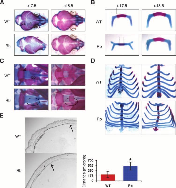
1442 Berman et al.
FIGURE 1. Deletion of Rb
causes defects in embryonic
bone development. A to D.
Alizarin red (bone) and Alcian
blue (cartilage) staining of em-
bryos. Embryonic day 17.5
Rbc /c mice exhibit less ossi-
fication in the cranium (A) and
hyoid bone (B). Bar, the differ-
ence in hyoid bone ossification
at embryonic day 17.5 (B).
Rbc /c embryos at embryonic
days 17.5 and 18.5 display
aberrant formation of bones in
the head (ventral view of head
in C) and sternum (D). The
aberrantly shaped or missing
palatine process in the Rbc /c
embryos is circled in C. E.
Pregnant mothers were injected
at embryonic day 18 with cal-
cein for 12 h. Coronal sections
of the frontal bone of embryonic
day 18.5 mice were analyzed
for calcein incorporation. Rbc /
c
embryos incorporate less
calcein than their wild-type lit-
termates. Original magnifica-
tion, 2. The distance from the
front of calcein incorporation
(arrow) to the midline of the
suture was measured in nine
Rbc /c and nine wild-type em-
bryo sections. Columns, mean;
bars, 1 SD; *, P < 0.001,
statistically significant differ-
ence. Abbreviations: fr, frontal
bone; pa, parietal bone; pp,
palatine process; pt, pterygoid
bone; xp, xiphoid process. WT,
R b + / c ; M o x 2 + / + ;
Rb, Rbc /c ; Mox2 +/Cre .
ossification. The former is influenced by three cell types: phosphatase (ALP) activity and Collagen1a1 (Col1) mRNA
chondrocytes, which form an essential cartilage template; expression. The activity and expression, respectively, of these
osteoblasts, which differentiate to secrete the bone matrix; two markers were significantly decreased in the Rbc /c frontal
and osteoclasts, which oppose bone formation by degrading and bone compared with those in wild-type sections (Fig. 2).
reabsorbing bone. In contrast, intramembranous ossification Moreover, the expression levels of osteopontin (OPN), an early
is influenced by osteoblasts and osteoclasts but occurs in a to mid-differentiation marker, were also typically down-
cartilage-independent manner. This fact, along with the regulated in the Rbc /c embryos relative to wild-type controls
apparently normal development of the cartilage skeleton within (data not shown). These data indicate that osteoblast differen-
Rbc /c embryos (Fig. 1B-D; data not shown), suggests that a tiation is perturbed in Rbc /c embryos at the earliest stages
chondrocyte defect cannot fully account for the defective bone of the pathway.
development. Therefore, we examined both osteoblast and
osteoclast function. To assess osteoclast levels, we screened the pRb-Deficient Osteoblasts Differentiate to a Greater
frontal bones of embryonic day 17.5 embryos for the presence Extent than Wild-type Cells In vitro
of tartrate-resistant acid phosphatase activity, an osteoclast- Our in vivo data show that an early step in osteoblast
specific marker. There were no active osteoclasts present in differentiation is affected. One possibility is that pRb regulates
either the wild-type or the Rbc /c frontal bones (Supplemen- osteoblast differentiation directly. For example, it has been
tary Fig. S1). Thus, the decreased ossification in Rbc /c reported previously that pRb can interact with and coactivate
embryos is likely not due to either cartilage defects or increased Runx2/CBFA1, one of the transcription factors essential for
osteoclast activity. osteoblast differentiation (20, 21). To further dissect the role of
Given these findings, we next screened embryonic day 17.5 pRb in osteoblast differentiation, we used a well-defined and
frontal bones for the presence of osteoblast-specific markers. often used in vitro osteoblast differentiation system. Specifi-
Two early markers of differentiating osteoblasts are alkaline cally, primary cells were isolated from the calvaria of wild-type
Mol Cancer Res 2008;6(9). September 2008
Downloaded from mcr.aacrjournals.org on February 8, 2021. © 2008 American Association for Cancer
Research.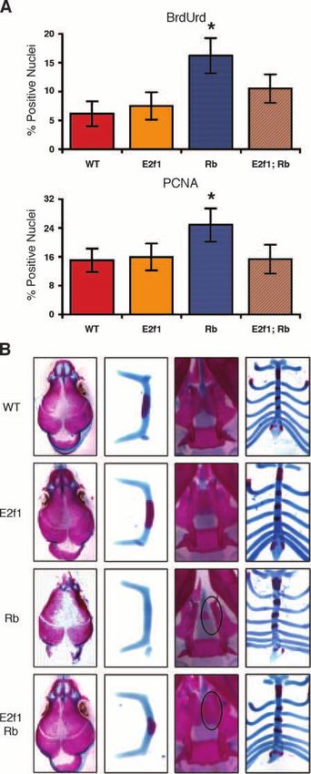
Rb Regulates Proper Bone Development           1443

and Rbc /c embryos and expanded. Two hundred and fifty                              Together, these data suggest that osteoblasts deficient for pRb
thousand cells were plated onto 3-cm tissue culture dishes and                      differentiate to a greater extent than wild-type cells in vitro, and
then induced to differentiate upon confluency. In this system,                      this correlates with the increased transcriptional levels of Runx2,
bone-like calcium deposits are secreted by fully differentiated                     OSX, and their downstream targets.
osteoblasts and can be analyzed by alizarin red staining. Based
on our in vivo data and previous in vitro differentiation studies                   Acute Ablation of pRb Promotes the Differentiation of
with fibroblasts (20), we anticipated that Rbc /c osteoblasts                       Osteoblasts In vitro
would differentiate to a lesser extent than wild-type cells.                            The wild-type and Rbc /c osteoblasts were prepared on
Contrary to this hypothesis, however, the Rbc /c osteoblasts                        embryonic day 17.5, when there was a significant difference in
secreted a greater number of calcium deposits than wild-type                        the degree of calvarial differentiation (Fig. 1A). This raised
osteoblasts based on the alizarin red staining (Fig. 3A).                           the possibility that the increased in vitro differentiation of the
   We then used quantitative real-time PCR (RT-PCR) to                              Rbc /c versus wild-type cells simply reflected the presence of
analyze the mRNA levels of several osteoblast markers during                        a larger pool of progenitor osteoblasts in the Rbc /c versus
the differentiation of these cells. Although the transcriptional                    wild-type calvaria. To address this hypothesis, we isolated
levels of Alp and Col1 were unchanged, the Rbc /c osteoblasts                       conditional Rbc/c osteoblasts. These cells were brought to
exhibited significantly greater levels of expression for several                    confluence and then infected with either a control adenovirus
other osteoblast genes compared with the wild-type cells                            containing green fluorescent protein (Adeno-GFP) or one
(Fig. 3B). Notably, Runx2 and osterix (OSX), two transcription                      expressing the Cre recombinase gene (Adeno-Cre). This
factors that are necessary to induce osteoblast differentiation
                                                                                    strategy yielded parallel populations of control and Rbc /c
(28-30), were up-regulated in the Rbc /c cells from the earliest
                                                                                    osteoblasts that had identical starting numbers of progenitors.
stages of the differentiation process (Fig. 3B). Runx2 and OSX
                                                                                    Consistent with previous studies (7), we found that the Adeno-
have been shown to induce the transcription of downstream
                                                                                    Cre was sufficient to acutely ablate pRb within 2 days of
osteoblast differentiation genes (28, 30, 31). In accordance with
                                                                                    infection (data not shown). Therefore, 2 days post-infection
these findings, we observed the increased expression of the
                                                                                    (denoted day 0 in Figs. 3C and D and Figs. 4D-F) we placed
early/mid- and late-differentiation markers, osteopontin (OPN)
                                                                                    the confluent wild-type and Rbc /c cells in differentiation
and osteocalcin (OC), respectively, in the Rbc /c osteoblasts.
                                                                                    media. The acutely ablated Rbc /c osteoblasts differentiated
                                                                                    to a greater extent than the control-infected Rbc/c cells, just as
                                                                                    we had observed with the germ line Rbc /c osteoblasts
                                                                                    (compare Fig. 3C and A). Moreover, the acutely ablated
                                                                                    Rbc /c cells expressed increased levels of Runx2, OSX, OPN,
                                                                                    and OC relative to the Adeno-GFP – infected cells in a
                                                                                    comparable manner to that observed in the germ line Rbc /c
                                                                                    osteoblasts (compare Figs. 3D and B). These data show that
                                                                                    loss of pRb acts in an intrinsic manner to increase the
                                                                                    differentiation of primary osteoblast cultures in vitro.

                                                                                    Depletion of pRb in Progenitor Osteoblasts Causes Cell
                                                                                    Cycle Exit Defects In vitro
                                                                                        We aimed to understand the molecular changes that accom-
                                                                                    panied this increased differentiation. One possibility is that
                                                                                    pRb possesses a cell cycle – independent repressive function in
                                                                                    osteoblast differentiation. In this manner, loss of pRb would
                                                                                    allow for the deregulated increase in osteoblast genes such
                                                                                    as Runx2 and OSX. We have attempted several experiments
                                                                                    to test the potential contribution of this interaction, includ-
                                                                                    ing conducting chromatin immunoprecipitations of Runx2
                                                                                    at osteoblast-specific promoters in wild-type, Rbc /c , and
                                                                                    Rbc /c ; E2f1 / calvarial preparations (data not shown).
                                                                                    These studies did not yield any evidence that Rb loss altered
                                                                                    Runx2 promoter-binding activity. Moreover, we did not detect
FIGURE 2. pRb-deficient frontal bones display decreased levels of                   any pRb binding to the Runx2 and OSX promoters. This latter,
osteoblast markers. Coronal sections of frontal bones from embryonic day
17.5 embryos were assessed by histochemical analysis of alkaline                    negative chromatin immunoprecipitation result is not particu-
phosphatase activity (left column) and in situ analysis of Collagen1a1              larly informative because pRb chromatin immunoprecipitation
mRNA (right column ). Rbc /c frontal bone sections (bottom row ) exhibit            works poorly in murine cells. However, the Runx2 and OSX
decreased levels of both markers compared with wild-type (top row ).
Original magnification, 2. The distance from the front of activity or              promoter both lack conventional E2F binding sites. Thus,
expression (arrows ) to the midline of the suture was measured in at least          although these observations do not rule out a direct, repressive
8 embryo pairs for Col1 and in 12 pairs for ALP. Columns, mean; bars, 1
SD; *, P < 0.01, statistically significant difference. WT, Rb +/c ; Mox2 +/+; Rb,
                                                                                    role for pRb in osteoblast differentiation in vitro, we have no
Rbc /c ; Mox2 +/Cre .                                                               data to support this model.

Mol Cancer Res 2008;6(9). September 2008
            Downloaded from mcr.aacrjournals.org on February 8, 2021. © 2008 American Association for Cancer
                                                      Research.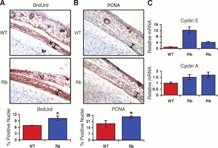
1444 Berman et al.
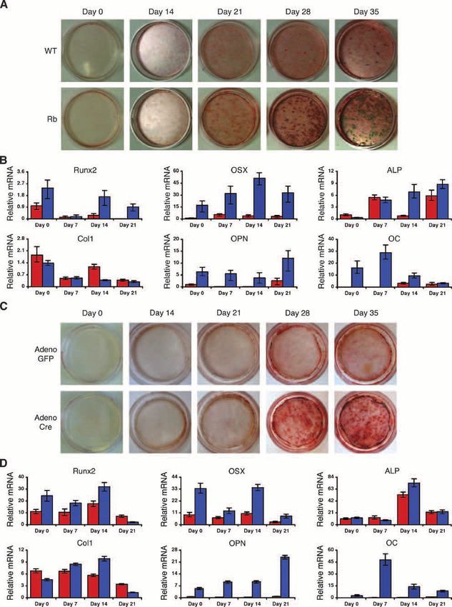
A second potential cause of the observed increase in we observed a greater number of Rbc /c osteoblast nuclei
osteoblast differentiation in vitro upon pRb loss may be related that stained positively for PCNA compared with wild-type
to cell cycle defects. Notably, the increased density of nuclei (Fig. 5B). Interestingly, at the apex of the frontal bone
osteoblast cultures is known to enhance their differentiation (the midline of the skill) where most of the osteoprogenitors
(32, 33). We hypothesized that loss of pRb may affect the were still proliferating, we did not observe a difference in
normal confluence arrest of the calvarial cells, leading to an BrdUrd or PCNA staining between the wild-type and Rbc /c
increase in proliferation and consequently, an increase in cell embryos (data not shown). This would indicate that the loss
density. Thus, we compared the proliferation of wild-type of pRb does not affect the proliferation rate of osteoproge-
versus germ line Rbc /c cells throughout the differentiation nitors but does affect their ability to properly exit the cell cycle
process. At all time points, we found that a higher proportion and to remain outside of the cell cycle. We did not observe
of the Rbc /c osteoblast nuclei incorporated 5-bromo-2- any proliferative differences between Rbc /c and wild-type
deoxyuridine (BrdUrd) compared with the wild-type controls forelimbs (data not shown), corresponding with our finding
(Fig. 4A and B). In agreement with these findings, the Rbc /c that there was no difference in the forelimbs based on alizarin
osteoblasts showed elevated levels of cyclin A and cyclin E red staining.
mRNAs (Fig. 4C). Finally, total cell counts during the initiation We also extracted RNA from the calvaria of Rbc /c and
of differentiation showed an increase in the total number wild-type embryos to examine the transcript levels of cyclin A
of cells present in Rbc /c confluent cultures compared with and cyclin E. Like PCNA, these transcripts are specifically
wild-types (Table 1). Similar results in all of these assays were induced in proliferating cells. Rbc /c calvaria typically
observed in the analyses of osteoblasts acutely ablated for pRb expressed greater mRNA levels of cyclin A and cyclin E than
(Fig. 4D-F; Table 1). Thus, we conclude that pRb loss increases wild-type skulls (Fig. 5C). Importantly, the unrestricted cell
the proliferation, and consequently, the density of confluent cycle progression in Rbc /c frontal bones was not associated
osteoblast cultures, thereby leading to an increase in primary with an apoptotic response, as determined by terminal
calvarial osteoblast differentiation in vitro. Notably, the nucleotidyl transferase – mediated nick end labeling staining
increased proliferation in Rbc /c cultures is not perpetual, as (data not shown). These data suggest that pRb deficiency
the percentage of proliferating cells does decrease to almost impairs osteoblasts from exiting the cell cycle in vivo at the
zero by day 35 (Fig. 4; data not shown). This suggests that appropriate developmental stage.
compensatory mechanisms, perhaps through the pocket proteins
p107 and p130, exist to eventually enable cell cycle exit in Deletion of E2f1 Suppresses the Cell Cycle and Ossifi-
the osteoblasts. cation Defects in Rbc /c Embryos
The cell cycle regulatory activity of pRb is known to be at
The Loss of Rb Prevents Osteoblasts from Properly least partially dependent on its ability to suppress the E2F
Exiting the Cell Cycle In vivo transcription factors and prevent the activation of genes such as
Having established a likely basis for the increased PCNA, cyclin A and cyclin E that control cell cycle progression.
differentiation of pRb-deficient osteoblasts in vitro, we wished E2F1 is an archetypal member of the E2F family. It is bound to
to determine whether a similar mechanism could explain the and inhibited by pRb in arrested cells, and it contributes to the
impaired bone development in vivo. Specifically, because activation of target genes once pRb is inactivated by either
appropriate cell cycle exit is important for the early stages of mitogenic signaling in wild-type cells or genetic lesions in
osteoblast differentiation in vivo, we hypothesized that pRb loss tumor cells. Previous work has shown that the loss of E2F1 can
might impair cell cycle exit in vivo and cause a negative effect suppress the ectopic cell cycles arising from the loss of Rb in
on bone formation. Thus, to assess cell cycle progression other tissues (34). We found that Rb and E2f1 are both
in vivo, we analyzed coronal sections of embryonic day 17.5 expressed in the calvaria (Supplemental Fig. S2). Thus, we
frontal bones for BrdUrd, which incorporates into newly crossed a mouse possessing a deletion of E2f1 into our
synthesized DNA during S phase. Embryos deficient for pRb conditional Rb model, and we then examined the compound
exhibited a significantly greater percentage of osteoblast nuclei mutant embryos to determine if E2F activity contributes to
that incorporated BrdUrd compared with the wild-type embryos the excess proliferation and ossification defects arising in the
(Fig. 5A). We also tested frontal bone sections for protein Rbc /c embryos.
expression of proliferating cell nuclear antigen (PCNA), a First, we assessed the level of cellular proliferation in the
known proliferation marker. Consistent with our BrdUrd data, embryonic osteoblasts through analysis of both BrdUrd
FIGURE 3. Rbc /c
primary osteoblasts differentiate to a greater extent than wild-type. A. Terminal differentiation of primary calvarial osteoblasts was
determined by alizarin red staining of secreted calcium deposits from 0 to 35 d. Rbc /c osteoblasts (bottom row ) secrete a greater number of calcium
deposits than wild-type osteoblasts (top row ). B. Quantitative RT-PCR results of bone marker expression levels from wild-type (red columns ) and Rbc /c
(blue columns ) osteoblasts during differentiation. Rbc /c osteoblasts express greater mRNA levels of Runx2 , osterix, osteopontin, and osteocalcin but not
alkaline phosphatase or Collagen1a1 compared with wild-type osteoblasts. Ubiquitin was used as an internal control to normalize for RNA levels within the
samples. Each time point is an average of four reactions. Columns, results from a representative littermate pair; bars, 1 SD. WT, Rb +/c ; Mox2 +/+; Rb,
Rbc /c ; Mox2 +/Cre . C. Rbc/c primary calvarial osteoblasts were infected with adenovirus expressing either the Cre recombinase enzyme or green fluorescent
protein 2 d prior to differentiation. Terminal differentiation was assessed by alizarin red staining. Rbc/c osteoblasts acutely ablated for pRb (bottom row )
secrete a greater number of calcium deposits than control-infected osteoblasts (top row ). D. Quantitative RT-PCR analysis done as described in B.
Osteoblasts acutely ablated for pRb (blue columns ) express greater mRNA levels of Runx2 , osterix, osteopontin, and osteocalcin but not alkaline
phosphatase or Collagen1a1 compared with control-infected osteoblasts (red columns ).
Mol Cancer Res 2008;6(9). September 2008
Downloaded from mcr.aacrjournals.org on February 8, 2021. © 2008 American Association for Cancer
Research.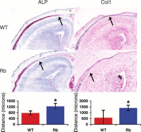
Rb Regulates Proper Bone Development   1445

Mol Cancer Res 2008;6(9). September 2008
        Downloaded from mcr.aacrjournals.org on February 8, 2021. © 2008 American Association for Cancer
                                                  Research.
1446 Berman et al.
FIGURE 4. Confluent osteoblasts in vitro exhibit excess proliferation upon loss of pRb. A. Immunofluorescence analysis of BrdUrd incorporation in
differentiating osteoblasts. Wild-type (top two rows ) and Rbc /c (bottom two rows ) osteoblasts were treated with BrdUrd (green ) for 24 h at the indicated
time points during differentiation in vitro . Nuclei are stained with 4¶,6-diamidino-2-phenylindole (blue ). Original magnification, 20. B. Quantitation of the
immunofluorescence analysis in A. A minimum of 250 cells was counted from each of three or more separate images for each sample. A greater percentage
of Rbc /c osteoblasts incorporate BrdUrd compared with wild-type cells at all time points. C. Quantitative RT-PCR analysis was done as described in Fig. 3.
Rbc /c osteoblasts (blue columns ) express greater mRNA levels of cyclin E and cyclin A relative to wild-type osteoblasts (red columns) during in vitro
differentiation. D. Nuclei of mock-infected (top two rows ) and acutely ablated (bottom two rows ) Rbc/c osteoblasts were stained for BrdUrd (green ) and 4¶,6-
diamidino-2-phenylindole (blue ). Original magnification, 20. E. Quantitation of the immunofluorescence analysis in D. A greater percentage of Rbc/c
osteoblast nuclei acutely ablated for pRb (blue columns ) stain positively for BrdUrd incorporation than control nuclei (red columns ). F. Quantitative RT-PCR
shows that acutely ablated Rbc/c osteoblasts (blue columns ) express greater mRNA levels of cyclin E and cyclin A compared with mock-infected osteoblasts
(red columns ). Bars, 1 SD. *, P < 0.05, a statistically significant difference. WT, Rb +/c ; Mox2 +/+; Rb, Rbc /c ; Mox2 +/Cre .
incorporation and PCNA expression in frontal bone sections Thus, loss of E2f1 alone seems insufficient to perturb osteoblast
from embryonic day 17.5 embryos (Fig. 6A). These two assay proliferation. Second, consistent with our prior analysis,
methods yielded highly concordant results. First, there was no proliferating osteoblasts were present at significantly higher
significant difference in the levels of either BrdUrd- or PCNA- levels in the Rbc /c frontal bone compared with the wild-type
positive nuclei in the wild-type versus the E2f1 / osteoblasts. and E2f1 / controls. Finally, the deletion of E2f1 was suf-
Mol Cancer Res 2008;6(9). September 2008
Downloaded from mcr.aacrjournals.org on February 8, 2021. © 2008 American Association for Cancer
Research.Rb Regulates Proper Bone Development 1447
ficient to almost fully suppress the excess proliferation arising Table 2. Quantitative RT-PCR Primer Pairs
in the Rbc /c embryos. The loss of E2f1 on its own or in the
Gene Primer sequence
Rbc /c background did not affect proliferation at the apex of
the frontal bone (data not shown), suggesting that the rate Alkaline phosphatase For: TCT CCA GAC CCT GCA ACC TC
of progenitor proliferation remained unaffected. Therefore, we Rev: CAT CCT GAG CAG ACC TGG TC
conclude that inappropriate activation of E2F1 contributes to Collagen 1a1 For: CGA GTC ACA CCG GAA CTT GG
Rev: GCA GGC AGG GCC AAT GTC TA
the inability of pRb-deficient osteoprogenitors to properly exit Cyclin A For: AGT TTG ATA GAT GCT GAC CC
the cell cycle in vivo. Rev: TAG GTC TGG TGA AGG TCC
We then assessed whether E2f1 inactivation modulated the Cyclin E For: TGT TTT TGC AAG ACC CAG ATG A
Rev: GGC TGA CTG CTA TCC TCG CT
Rbc /c embryonic skeletal defects observed at embryonic day Osteocalcin For: CTC TGT CTC TCT GAC CTC ACA G
17.5 (Fig. 6B). Consistent with the absence of any proliferation Rev: CAG GTC CTA AAT AGT GAT ACC G
defects, the deletion of E2f1 alone did not cause any detectable Osteopontin For: TGC TTT TGC CTG TTT GGC AT
Rev: TTC TGT GGC GCA AGG AGA TT
defects in skeletal development. As observed previously, Rb Osterix For: GCA AGG CTT CGC ATC TGA AA
deficiency caused decreased ossification in the skull and hyoid, Rev: AAC TTC TTC TCC CGG GTG TGA
Runx2 For: TGA GAT TTG TGG GCC GGA
and aberrant formation of the xiphoid process, palatine process, Rev: TCT GTG CCT TCT TGG TTC CC
and pterygoid bone. Notably, in almost all E2f1 / ; Rbc /c Ubiquitin For: TGG CTA TTA ATT ATT CGG TCT GCA T
double mutant embryos (12 of 13), the reduced ossification was Rev: GCA AGT GGC TAG AGT GCA GAG TAA
partially or completely ameliorated (Fig. 6B, first two columns).
Moreover, f40% (5 of 13) of the double mutants exhibited
normal formation of the palatine process, pterygoid bone, and activity and decreased levels of Col1 and OPN mRNA.
the xiphoid process was completely normal (Fig. 6B, latter two Previous studies have shown that deletion of the pRb-related
columns). Taken together, these data show that deletion of Rb proteins, p107 and p130, or overexpression of E2F1 affect
causes defects in embryonic skeletal development that are due, chondrocyte differentiation and development (35-37). Although
at least in part, to the inappropriate release of E2F1. our data do not rule out a role for pRb in cartilage development,
they clearly show that pRb plays a role in bone development
Discussion that is independent of chondrocytes. Specifically, Rbc /c
The RB locus is mutated or altered in >70% of all osteo- skeletons did not show any apparent defects in cartilage
sarcomas (3, 4). Moreover, several in vitro studies impli- formation, and several of the affected bones formed via
cate pRb and the pocket proteins in osteoblast differentiation intramembranous ossification, a process that does not involve
(18-21). Given these observations, we used the Mox2 +/Cre chondrocytes. Moreover, the bone defects in the Rbc /c frontal
transgene to conditionally inactivate Rb in the Rbc/c embryo bone, and presumably in other affected bones, were not the
proper, but not in the placenta, and thereby generate pRb- result of increased osteoclast activity or apoptosis. Therefore,
deficient embryos that survive until birth. This conditional our data suggest that the loss of Rb impairs osteoblast
strategy allows us to assess pRb’s role in bone development differentiation in vivo at the earliest stages of the pathway.
in vivo and primary osteoblast differentiation in vitro. Our One caveat of the in vivo studies is that they do not prove
analyses reveal a role for pRb in the promotion of osteogenesis that pRb’s requirement for osteoblast differentiation is cell
via the regulation of proper cell cycle exit. autonomous. To address this issue, we determined how the
In the developing embryo, the loss of pRb impaired bone loss of pRb affects the differentiation of primary osteoblasts
formation in a manner that caused two types of defects. Some in vitro. Given our in vivo defects and the prior observation
bones, such as the pterygoid bone, palatine process, and that pRb-deficient MEFs were impaired in their ability to
xiphoid process, developed abnormally and were misshapen, undergo osteogenesis (20), we anticipated that primary
whereas the skull and hyoid bone exhibited decreased bone osteoblasts isolated from Rbc /c embryos would display an
formation. The decreased ossification in the Rbc /c frontal impaired differentiation phenotype in vitro. However, the exact
bone was accompanied by reduced alkaline phosphatase opposite was observed: the Rbc /c osteoblasts differentiated
to a greater extent than the wild-type controls. Importantly,
we found that the acute ablation of Rb in confluent osteoblasts
Table 1. Cell Numbers at Day 0 of Differentiation was sufficient to trigger increased differentiation. These data
show that loss of pRb acts in a cell autonomous manner to
Genotype Germ line Conditional promote osteoblast differentiation in vitro.
Our study shows that two distinct molecular changes
Wild-type Rbc /c Adeno-GFP Adeno-Cre accompany the improved in vitro differentiation upon loss of
pRb. First, we observe a dramatic up-regulation of osteoblast
Cell count (1,000) 481 F16.5 656 F 14.1 483 F 24.5 579 F 17.6
genes, such as Runx2 and OSX in differentiating pRb-deficient
NOTE: Two hundred and fifty thousand cells were plated onto a 3-cm tissue osteoblasts to levels that are sometimes not reached by
culture dish and allowed to reach confluency (typically 4 days later). For ‘‘germ wild-type cells. At this time, we do not know if the extreme
line’’ cells, this confluency arrest constituted day 0 of differentiation, and the
number of cells was ascertained. For ‘‘conditional’’ cells (Rbc/c ) at confluence, up-regulation in Rbc /c cultures is due to an increased ability
adenovirus containing either green fluorescent protein or Cre recombinase was of individual cells to induce osteoblast genes, an increased
added to the medium. Two days after adenovirus addition (designated as day 0 percentage of terminally differentiated cells in the culture, or
of differentiation) cells were counted. Average cell counts from at least three
separate experiments F SD are shown. both. Interestingly, in these in vitro assays, pRb loss clearly
Mol Cancer Res 2008;6(9). September 2008
Downloaded from mcr.aacrjournals.org on February 8, 2021. © 2008 American Association for Cancer
Research.1448 Berman et al.
FIGURE 5. pRb-deficient osteoblasts do not properly exit the cell cycle in vivo . A and B. Immunohistochemical analysis of BrdUrd incorporation (A) or
PCNA protein expression (B) in coronal sections of frontal bones from embryonic day 17.5 embryos. Pregnant females were injected with BrdUrd for 2 h.
Rbc /c frontal bones (bottom ) exhibit a greater number of nuclei positively staining for BrdUrd or PCNA than wild-type littermates (top ). Original
magnification, 20. Frontal bones (bar). Columns, quantified results from four pairs of Rbc /c and wild-type frontal bone sections; bars, 1 SD; *, P < 0.05,
statistically significant difference. C. Quantitative RT-PCR analysis of cyclin E (top ) and cyclin A (bottom ) mRNA levels from Rbc /c and control littermates.
mRNA was isolated from the calvaria of embryonic day 16.5 embryos. Analysis done as described in Fig. 3. Rbc /c calvaria (blue columns ) express
increased levels of cyclin A and cyclin E relative to wild-type littermates (red columns ). Bars, 1 SD. Abbreviations: br, brain; de, dermis; fr, frontal bone. WT,
Rb +/c ; Mox2 +/+; Rb, Rbc /c ; Mox2 +/Cre .
induces some (e.g., Runx2 and OSX) but not all (ALP and the loss of E2f1 did not suppress the cell cycle defects of
Col1) osteoblast genes. The reason for this differential response osteoblasts in this in vitro setting. Thus, we have been unable
is unclear. However, we note that even prior to the induction to prove that a cell cycle exit defect can account for the
of differentiation, the ALP and Col1 mRNAs are present at increased differentiation of Rb-depleted osteoblasts in vitro.
much higher levels in the cultured osteoblasts than in the Despite this limitation, our in vivo studies provide strong
endogenous calveria. This suggests that the in vitro culture support for this model. Specifically, we find that osteoblasts of
somehow induces ALP and Col1 expression or that it selects the Rbc /c frontal bone fail to exit the cell cycle at the
for a subpopulation of the calverial cells that are committed appropriate stage of development, and we can completely sup-
to the osteoblast lineage and therefore have high ALP and press both the proliferation defect and the decreased ossifica-
Col1 expression. tion of the skull and hyoid bones through inactivation of E2f1,
The second molecular change that accompanies the a known pRb target and proliferation inducer.
improved in vitro differentiation of pRb-deficient osteoblasts If a cell cycle exit defect is the major underlying cause of
is an increase in the fraction of cells that are proliferating and both the in vitro and in vivo defects, how does this account for
the sustained presence of proliferating cells at later time points the apparently opposing effects on bone differentiation seen in
in the differentiation process. Because the density of osteoblasts the two settings? One possibility is that this is an aberrant
has been reported to correlate positively with their ability to consequence of the in vitro culture that somehow enables
differentiate in vitro (32, 33), we believe that the increased the Rb-deficient cells to overcome their differentiation defect.
proliferation of the pRb-deficient osteoblasts contributes to The alternative possibility, which we favor, is that pRb loss
their improved differentiation by increasing the density of the affects cells at early and late stages of osteoblast differentiation
confluent cells. We tried two distinct approaches to directly in a differential manner, and the in vivo and the in vitro studies
test this model. First, we attempted to maintain the Rbc/c highlight the defects in these distinct populations. Specifi-
osteoblasts in the presence of antiproliferative drugs prior to the cally, we hypothesize that pRb loss leads to ectopic proli-
ablation of pRb. However, the experiment requires several days feration that prevents early progenitors from entering osteoblast
of drug treatment to which the cells faired poorly. Second, differentiation but concomitantly enhances the differentiation
because our in vivo data indicate that deletion of E2f1 of late stage osteoblasts. In this model, the in vitro cultures
suppresses excess proliferation due to the loss of Rb, we could favor analysis of the late stage osteoblasts, thereby
analyzed the consequence of E2f1 deficiency in acutely ablated showing that pRb loss promotes osteoblast differentiation. In
and germ line – deleted Rbc /c osteoblasts. Unfortunately, contrast, the in vivo phenotype would be more complex.
Mol Cancer Res 2008;6(9). September 2008
Downloaded from mcr.aacrjournals.org on February 8, 2021. © 2008 American Association for Cancer
Research.Rb Regulates Proper Bone Development 1449
Specifically, our data clearly show ectopic proliferation of tissue was fixed in 4% paraformaldehyde and embedded
Rbc /c cells in the developing frontal bone, but we cannot in optimal cutting temperature. Frozen sections were cut at
know whether these represent uncommitted early progenitor 6 to 8 Am except for those for in situ analysis, which were cut
cells or differentiating osteoblasts that are proliferating at 10 to 12 Am. The morphology of the brain and presphenoid
inappropriately. In fact, we believe that both populations bone were used to ensure that equivalent planes of the frontal
coexist. In this event, at early time points in the bone bone were analyzed in all samples.
differentiation process, the shortage of committed osteoproge-
nitors would initially impair bone formation—exactly as we
observe in the late stage embryos. However, as the committed
osteoblasts accumulate, their increased proliferation would
eventually allow, and perhaps ultimately enhance, bone
differentiation—as we observe in the in vitro assays. Unfortu-
nately, because the Rbc /c animals die at birth, we cannot
determine whether their osteoblast density and bone deposition
ultimately exceeds that seen in wild-type animals.
There is considerable evidence to suggest that pRb plays a
direct role in regulating the transcriptional programs that
control osteoblast differentiation. Most compelling is the find-
ing that pRb can positively regulate Runx2 in vitro (20, 21).
Our findings do not discount the possibility that pRb plays
a direct role in bone differentiation through Runx2, or some
other mechanism, or that this might contribute to the bone
defects we observe in vivo. However, they argue that the
primary role of pRb in bone differentiation is to inhibit E2F1
and thereby facilitate cell cycle exit. Given that Rb inactiva-
tion is observed in a large proportion of osteosarcomas, it will
be important to develop additional models that allow a
comparison of the mechanisms by which loss of Rb affects
bone development versus osteosarcoma formation.
Materials and Methods
Animal Maintenance and Histologic Preparations
The generation of Rb c/c and Mox2-Cre mice has been
described previously (7, 27). Rbc/c and E2f1 / mice were
provided by Tyler Jacks. Mox2-Cre mice were purchased
from The Jackson Laboratory. Gestation was dated by detection
of a vaginal plug. Pregnant mice were injected with 10 AL/g
body weight of 5 mg/mL BrdUrd in PBS 2 hours prior to
tissue collection. For calcein incorporation, pregnant mice
were injected with 10 AL/g body weight of 2.5 mg/mL calcein
12 or 24 hours prior to tissue collection. Collected embryonic
FIGURE 6. Deletion of E2f1 suppresses the bone defects due to
the loss of pRb. A. Immunohistochemical analysis of BrdUrd incorpo-
ration (top ) or PCNA protein expression (bottom ) in coronal sections of
frontal bones from embryonic day 17.5 embryos, done with four to six
samples of each genotype. Deletion of E2f1 suppresses the increased
BrdUrd incorporation and PCNA expression observed in Rbc /c frontal
bone osteoblasts. B. Skeletal staining of embryonic day 17.5 embryos
as described in Fig. 1. Deletion of E2f1 suppresses the decreased
ossification found in the Rbc /c calvaria (first column ) and hyoid bone
(second column ). Deletion of E2f1 also suppresses the aberrant
formation of the palatine process and pterygoid bone (third column )
and xiphoid process (fourth column ) observed in Rbc /c skeletons. An
aberrant palatine process in the Rbc /c and a suppressed palatine
process in the double mutant are circled (third column ). Bars, 1 SD;
*, P < 0.05, statistically significant difference between Rbc /c and
wild-type, E2f1 / , or Rbc /c ; E2f1 / . WT, Rb +/c ; Mox2 +/+; E2f1 +/+;
E2f1, Rb+/c; Mox2 +/+; E2f1 / ; Rb, Rbc /c ; Mox2 +/Cre ; E2f1 +/+; RbE2f1,
Rbc /c ; Mox2 +/Cre ; E2f1 / .
Mol Cancer Res 2008;6(9). September 2008
Downloaded from mcr.aacrjournals.org on February 8, 2021. © 2008 American Association for Cancer
Research.1450 Berman et al.
Histologic Analyses 1 Ag of RNA using Superscript III reverse transcriptase
Enzymatic ALP assays were done on unfixed frozen (Invitrogen) following the instructions of the manufacturer.
sections. Briefly, 0.06 g of sodium nitrite was dissolved into Quantitative RT-PCR with 20 to 100 ng cDNA was done using
1.5 mL of water and added to 600 AL of 50 mg/mL of new SYBR Green (Applied Biosystems). Reactions were run on the
fuchsin (Sigma) in 2 mol/L of HCl. This solution was added to ABI Prism 7000 Sequence Detection System and analyzed
210 mL of Tris buffer (pH 9.0). Finally, 1.8 mL of 83.3 mg/mL using the 7000 SDS software. Primers are listed in Table 2.
naphthol AS-Bi-phosphate (Sigma) in DMF (Sigma) was
added. Sections were incubated with this overall solution for
15 min, washed in PBS and counterstained with hematoxylin. Disclosure of Potential Conflicts of Interest
Immunohistochemical analyses were done using antibodies No potential conflicts of interest were disclosed.
against BrdUrd (1:50 347580; BD Biosciences) and PCNA
(1:2,000 sc56; Santa Cruz) as previously described (38). For Acknowledgments
We are grateful to the University of Iowa Gene Transfer Vector Core for providing
Collagen1a1 in situ, digoxigenin-11-UTP – labeled single-strand the adenovirus, Tyler Jacks (Koch Institute, Massachusetts Institute of
riboprobe was prepared (probe was a gift from B. Olsen), Technology, Cambridge, MA) for the Rbc/c and E2f1 mutant strains, Bjorn
and hybridization was carried out overnight in 50% formamide Olsen (Dept. of Cell Biology, Harvard Medical School, Boston, MA) for in situ
probes, and the members of the Lees lab for helpful discussion throughout
at 55jC. Washing, detection, staining, and mounting of slides this work. We also thank Phil Iaquinta for assistance with chromatin
were carried out as described previously (39). Statistical immunoprecipitation and bioinformatics research.
significance was determined using the two-sample Student’s
t test with two-tailed distribution and unequal variance.
References
1. Weinberg RA. The retinoblastoma gene and gene product. Cancer Surv 1992;
Skeletal Staining 12:43 – 57.
Embryos were sacrificed, skinned, and eviscerated. The 2. Gurney JG, Severson RK, Davis S, Robison LL. Incidence of cancer in
remaining tissue was fixed in 95% ethanol for 4 days, children in the United States. Sex-, race-, and 1-year age-specific rates by
histologic type. Cancer 1995;75:2186 – 95.
transferred to acetone for 3 days, and subsequently transferred
3. Belchis DA, Meece CA, Benko FA, Rogan PK, Williams RA, Gocke CD.
to staining solution [final volume of 0.015% Alcian blue 8GX Loss of heterozygosity and microsatellite instability at the retinoblastoma locus in
(Sigma), 0.005% alizarin red S (Sigma), and 5% glacial acetic osteosarcomas. Diagn Mol Pathol 1996;5:214 – 9.
acid in ethanol] at 37jC for 2 days and at room temperature for 4. Feugeas O, Guriec N, Babin-Boilletot A, et al. Loss of heterozygosity of the
a 3rd day. Tissue was cleared in 1% potassium hydroxide for RB gene is a poor prognostic factor in patients with osteosarcoma. J Clin Oncol
1996;14:467 – 72. Erratum in: J Clin Oncol 1996;14:2411.
several days and ultimately stored in glycerol.
5. Lipinski MM, Jacks T. The retinoblastoma gene family in differentiation and
development. Oncogene 1999;18:7873 – 82.
Calvarial Preparations and Culture 6. Huang HJ, Yee JK, Shew JY, et al. Suppression of the neoplastic phenotype by
Calvaria from embryonic day 17.5 embryos were removed, replacement of the RB gene in human cancer cells. Science 1988;242:1563 – 6.
treated with several rounds of collagenase/trypsin digests at 7. Sage J, Miller AL, Pérez-Mancera PA, Wysocki JM, Jacks T. Acute mutation
of retinoblastoma gene function is sufficient for cell cycle re-entry. Nature 2003;
37jC, and plated onto six-well plates. Cells were grown and 424:223 – 8.
expanded in aMEM with 10% fetal bovine serum and 8. Trimarchi JM, Lees JA. Sibling rivalry in the E2F family. Nat Rev Mol Cell
penicillin/streptomycin. For differentiation, 250,000 cells were Biol 2002;3:11 – 20.
plated onto 3-cm tissue culture plates. Upon reaching 9. Clark AJ, Doyle KM, Humbert PO. Cell-intrinsic requirement for pRb in
confluence, calvarial osteoblasts were treated with medium erythropoiesis. Blood 2004;104:1324 – 6.
supplemented with 50 Ag/mL of ascorbic acid and 10 mmol/L 10. Spike BT, Dirlam A, Dibling BC, et al. The Rb tumor suppressor is required
for stress erythropoiesis. EMBO J 2004;23:4319 – 29.
of h-glycerol-phosphate. Adenovirus (University of Iowa Gene
11. Huh MS, Parker MH, Scimè A, Parks R, Rudnicki MA. Rb is required for
Transfer Vector Core) was added to the medium at 100 plaque- progression through myogenic differentiation but not maintenance of terminal
forming units per cell and washed away 24 h later. To assay for differentiation. J Cell Biol 2004;166:865 – 76.
calcium deposits, plates were stained with 1% alizarin red 12. Haigis K, Sage J, Glickman J, Shafer S, Jacks T. The related retinoblastoma
S solution (pH 5.0). (pRb) and p130 proteins cooperate to regulate homeostasis in the intestinal
epithelium. J Biol Chem 2006;281:638 – 47.
13. Yang HS, Hinds PW. pRb-mediated control of epithelial cell proliferation and
Immunofluorescence Indian hedgehog expression in mouse intestinal development. BMC Dev Biol
For BrdUrd incorporation, osteoblasts were plated onto 2007;7:6.
coverslips prior to achieving confluence. BrdUrd was added to 14. Morgenbesser SD, Williams BO, Jacks T, DePinho RA. p53-dependent
apoptosis produced by Rb-deficiency in the developing mouse lens. Nature 1994;
the medium (final concentration of 10 Amol/L) and incubated 371:72 – 4.
for 24 h prior to 4% paraformaldehyde fixation. Antigen was 15. Kim TA, Lim J, Ota S, et al. NRP/B, a novel nuclear matrix protein,
detected using antibody against BrdUrd (1:50 347580; BD associates with p110(RB) and is involved in neuronal differentiation. J Cell Biol
1998;141:553 – 66.
Biosciences) with Texas red-X goat anti-mouse secondary
16. Lee EY, Hu N, Yuan SS, et al. Dual roles of the retinoblastoma protein in cell
(1:1,000; Invitrogen). Statistical significance was determined cycle regulation and neuron differentiation. Genes Dev 1994;8:2008 – 21.
using Student’s t test. 17. Chen PL, Riley DJ, Chen Y, Lee WH. Retinoblastoma protein positively
regulates terminal adipocyte differentiation through direct interaction with C/
EBPs. Genes Dev 1996;10:2794 – 804.
Quantitative RT-PCR
18. Feuerbach D, Loetscher E, Buerki K, Sampath TK, Feyen JH. Establishment
RNA was isolated from differentiation plates using the and characterization of conditionally immortalized stromal cell lines from a
Qiagen RNeasy kit. First-strand cDNA was transcribed from temperature-sensitive T-Ag transgenic mouse. J Bone Miner Res 1997;12:179 – 90.
Mol Cancer Res 2008;6(9). September 2008
Downloaded from mcr.aacrjournals.org on February 8, 2021. © 2008 American Association for Cancer
Research.Rb Regulates Proper Bone Development 1451
19. Beck GR, Jr., Sullivan EC, Moran E, Zerler B. Relationship between alkaline transcription factor osterix is required for osteoblast differentiation and bone
phosphatase levels, osteopontin expression, and mineralization in differentiating formation. Cell 2002;108:17 – 29.
MC3T3 – 1 osteoblasts. J Cell Biochem 1998;68:269 – 80.
31. Wang X, Kua HY, Hu Y, et al. p53 functions as a negative regulator of
20. Thomas DM, Carty SA, Piscopo DM, et al. The retinoblastoma protein acts osteoblastogenesis, osteoblast-dependent osteoclastogenesis, and bone remodel-
as a transcriptional coactivator required for osteogenic differentiation. Mol Cell ing. J Cell Biol 2006;172:115 – 25.
2001;8:303 – 16.
32. Gerber I, ap Gwynn I. Influence of cell isolation, cell culture density, and cell
21. Luan Y, Yu XP, Xu K, et al. The retinoblastoma protein is an essential nutrition on differentiation of rat calvarial osteoblast-like cells in vitro . Eur Cell
mediator of osteogenesis that links the p204 protein to the Cbfa1 transcription Mater 2001;2:10 – 20.
factor thereby increasing its activity. J Biol Chem 2007;282:16860 – 70.
33. Purpura KA, Aubin JE, Zandstra PW. Sustained in vitro expansion of bone
22. Clarke AR, Maandag ER, van Roon M, et al. Requirement for a functional progenitors is cell density dependent. Stem Cells 2004;22:39 – 50.
Rb-1 gene in murine development. Nature 1992;359:328 – 30.
34. Tsai KY, Hu Y, Macleod KF, Crowley D, Yamasaki L, Jacks T. Mutation of
23. Jacks T, Fazeli A, Schmitt EM, Bronson RT, Goodell MA, Weinberg RA. E2f-1 suppresses apoptosis and inappropriate S phase entry and extends survival
Effects of an Rb mutation in the mouse. Nature 1992;359:295 – 300. of Rb-deficient mouse embryos. Mol Cell 1998;2:293 – 304.
24. Lee EY, Chang CY, Hu N, et al. Mice deficient for Rb are nonviable and 35. Cobrinik D, Lee MH, Hannon G, et al. Shared role of the pRB-related p130
show defects in neurogenesis and haematopoiesis. Nature 1992;359:288 – 94. and p107 proteins in limb development. Genes Dev 1996;10:1633 – 44.
25. Wenzel PL, Wu L, de Bruin A, et al. Rb is critical in a mammalian tissue 36. Rossi F, MacLean HE, Yuan W, et al. p107 and p130 Coordinately regulate
stem cell population. Genes Dev 2007;21:85 – 97.
proliferation, Cbfa1 expression, and hypertrophic differentiation during endo-
26. Wu L, de Bruin A, Saavedra HI, et al. Extra-embryonic function of Rb is chondral bone development. Dev Biol 2002;247:271 – 85.
essential for embryonic development and viability. Nature 2003;421:942 – 7.
37. Scheijen B, Bronk M, van der Meer T, Bernards R. Constitutive E2F1
27. Tallquist MD, Soriano P. Epiblast-restricted Cre expression in MORE mice: a tool overexpression delays endochondral bone formation by inhibiting chondrocyte
to distinguish embryonic vs. extra-embryonic gene function. Genesis 2000;26:113 – 5. differentiation. Mol Cell Biol 2003;23:3656 – 68.
28. Ducy P, Zhang R, Geoffroy V, Ridall AL, Karsenty G. Osf2/Cbfa1: a 38. Danielian PS, Bender Kim CF, Caron AM, Vasile E, Bronson RT, Lees JA.
transcriptional activator of osteoblast differentiation. Cell 1997;89:747 – 54. E2f4 is required for normal development of the airway epithelium. Dev Biol
29. Otto F, Thornell AP, Crompton T, et al. Cbfa1, a candidate gene for 2007;305:564 – 76.
cleidocranial dysplasia syndrome, is essential for osteoblast differentiation and 39. Böhme K, Li Y, Oh PS, Olsen BR. Primary structure of the long and short
bone development. Cell 1997;89:765 – 71. splice variants of mouse collagen XII and their tissue-specific expression during
30. Nakashima K, Zhou X, Kunkel G, et al. The novel zinc finger-containing embryonic development. Dev Dyn 1995;204:432 – 45.
Mol Cancer Res 2008;6(9). September 2008
Downloaded from mcr.aacrjournals.org on February 8, 2021. © 2008 American Association for Cancer
Research.You can also read