Engaging a polylactide copolymer in oral tissue regeneration: first validation of Suprathel for guided epithelial and osseous healing
←
→
Page content transcription
If your browser does not render page correctly, please read the page content below

JOURNAL of MEDICINE and LIFE

JML | ORIGINAL ARTICLE

Engaging a polylactide copolymer in oral tissue
regeneration: first validation of Suprathel®
for guided epithelial and osseous healing
Sergiu Vacaras 1, Grigore Baciut 1, Dan Gheban 2, Simion Bran 1, Horatiu Colosi 3 *, Septimiu Toader 4, Daiana Opris 1,
Winfried Kretschmer 5, Avram Manea 1, Gabriel Armencea 1, Mihaela Baciut 1, Horia Opris 1,
Ileana Mitre 1, Mihaela Hedesiu 6, Cristian Dinu 1

Author Affiliations:                                                                                    * Corresponding Author:
1. Department of Maxillofacial Surgery and Radiology, Division of Maxillofacial Surgery                 Horatiu Alexandru Colosi,
   and Implantology, Iuliu Hatieganu University of Medicine and Pharmacy,                               Associate Professor, DMD,
   Cluj-Napoca, Romania                                                                                 MS, PhD, Department of
2. Department of Morphological Sciences, Division of Pathoanatomy,                                      Medical Education, Division
   Iuliu Hatieganu University of Medicine and Pharmacy, Cluj-Napoca, Romania                            of Medical Informatics and
                                                                                                        Biostatistics, Faculty of
3. Department of Medical Education, Division of Medical Informatics and Biostatistics,
                                                                                                        Medicine, Iuliu Hatieganu
   Iuliu Hatieganu University of Medicine and Pharmacy, Cluj-Napoca, Romania
                                                                                                        University of Medicine and
4. Center for Experimental Surgery, Iuliu Hatieganu University of Medicine and Pharmacy,                Pharmacy, 6 Louis Pasteur
   Cluj-Napoca, Romania                                                                                 Street, 400349,
5. Klinik fur Mund-, Kiefer- und Plastische Gesichtschirurgie, Alb Fils Kliniken GmbH,                  Cluj-Napoca, Romania.
   Goppingen, Baden-Wurttemberg, Germany                                                                Phone: +40264597256
6. Department of Maxillofacial Surgery and Radiology, Division of Maxillofacial Radiology,              Fax: +40264893547
   Iuliu Hatieganu University of Medicine and Pharmacy, Cluj-Napoca, Romania                            E-mail: hcolosi@umfcluj.ro

                                                                                                        DOI
                                                                                                        10.25122/jml-2021-0083

                                                                                                        Dates
ABSTRACT
                                                                                                        Received: 11 February 2021
The present study investigated the capacity of Suprathel® (a copolymer membrane, so                     Accepted: 31 March 2021
far validated for skin regeneration) to also regenerate oral tissue – mucosa and bone, by
comparing this biomaterial, in a split-mouth rabbit model, to Mucoderm®, a xenogeneic
collagen matrix certified for keratinized oral mucosa healing. The clinical reason behind
this experimental animal model was to determine whether the benefits of this advanced
skin regeneration product (Suprathel®) could be conveyed for future evaluation in clinical
trials of oral tissue regeneration in humans. The outcomes of this study validated the use
of Suprathel®, a terpolymer of polylactide with trimethylene carbonate and ε-caprolac-
tone, for stimulation of oral epithelium and alveolar bone regeneration in rabbits. Both
Suprathel® and Mucoderm® exhibited comparable results and the null hypothesis stating
a comparable regenerating effect of these two materials could not be rejected.

KEYWORDS: Suprathel®, guided tissue regeneration, oral mucosa, socket preservation,
bone healing.

ABBREVIATIONS: CPB – porcine bone; FDBA – frozen-dried bone allograft; GBR –Guided
bone regeneration; PCL – polycaprolactone; PGA – poly-glycolic acid; PLA – poly-lactic
acid; PLGA – poly-lactide-co-glycolide; PVA – polyvinyl alcohol; rhBMP-2 – recombinant
human morphogenetic protein-2.

INTRODUCTION

Oral tissue regeneration is a sophisticated blend of intricated processes occurring continuously due to age, pathologic deterioration of
gingiva and bone, trauma, and teeth loss. Considering the functional and anatomic significance of the involved regions, the deficiency
can range from minor to significant or even life-altering.

© 2021 JOURNAL of MEDICINE and LIFE. VOL: 14 ISSUE: 2 MARCH-APRIL 2021
                                                                                                                                           181
JOURNAL of MEDICINE and LIFE

      Teeth loss is a process that determines resorption of the alveolar bone, which is clinically and radiologically evident by the loss of
      bone height and width [1]. The current state of the art of treating edentulous ridges is by using implant-supported prostheses [2].
      The prosthetically-driven planning involves the correct three-dimensional (3D) positioning of dental implants, representing the key to
      therapeutic success [3].

      Bone resorption occurs in time, but most of the bone is lost during the first 4 weeks after the tooth is lost, averaging 3–5 mm in width
      after 6 months [4]. Bone regeneration and other surgical augmentation procedures have been used with a high degree of success to
      restore the alveolar process and prepare the tissues for dental implant therapy with correct 3D positioning [5]. Consequently, after
      tooth extraction, it is highly beneficial to preserve the remaining bone and stop the bone resorption process [6]. Socket preservation
      procedures have long been studied and implemented, but a consensus regarding the best method of preserving the bundle bone, using
      various types of bone substitute material, with or without barrier membranes, is still lacking [5, 7].

      Tissue regeneration with an ideal barrier membrane

      A guided bone regeneration membrane needs to create and maintain a secluded space, protecting an environment favorable to the
      bone-forming cells, sheltered from the epithelial cells [8]. The ideal characteristics of the barrier membranes used in such cases need to
      be biocompatible, provide cell occlusion, integration by the host, clinical manageability, and space-making abilities [9]. Although sev-
      eral advantages of using resorbable membranes exist, such as no need for a second surgery, improved cost-effectiveness, and simplified
      surgical protocol, non-resorbable membranes still serve as the gold standard of bone regeneration [10].

      Gingival recession/lack of keratinized tissue

      Morphological characteristics of the oral mucosa are considered an important factor in the stability and integrity of the periodontium
      [11]. The non-keratinized mucosa facilitates the penetration of bacteria into the crevice, causing an inflammatory response with subse-
      quent attachment loss [12]. It has long been suggested that an adequate zone of keratinized tissue was critical for preserving the stability
      of periodontal tissues [13]. Recent studies have suggested that a wider band of keratinized gingiva may determine significant differences
      concerning the lowering of the plaque index, the values of the modified gingival index, the mucosal recession, and attachment loss [14].
      Lack of attached keratinized mucosa was linked to hygiene discomfort and lower vestibular depth [15, 16].

      Purpose of the study – Suprathel®

      The purpose of the current investigation was to assess the effectiveness of a polylactide copolymer (Suprathel®) in oral tissue regenera-
      tion and stimulate innovative research to promote oral tissue regeneration. Before this study, the split-mouth design used to compare the
      proposed material to a long-time validated product (Mucoderm®) has been perfected using a preliminary pilot study on only two rabbits,
      that has been implemented by the same research team [17], in order to optimize the assessment criteria of tissue regeneration, to yield
      reproducible outcome data and to allow an objective statistical appraisal of the experimental results. Perfecting this split-mouth design
      and its assessment criteria was also aimed at a possible translation of this preclinical experience to future clinical research.

      Connective tissue graft Mucoderm®

      The use of a connective tissue graft in enhancing the width of the keratinized tissue is considered the standard of care [18]. Also, the use
      of the graft enhances the marginal tissue thickness to improve the long-term stability of the periodontium [19]. The downsides are that
      it complicates the surgical procedure, increases morbidity, and raises the rates of wound failure during early healing. To try to balance
      off the latter, alternatives have been studied, such as human dermis and collagen matrices [20]. Xenogeneic matrices, like Mucoderm®,
      have several advantages, such as no need for human donors, no risks of transmitting diseases, and no limit to the size of the surgical
      site size [21]. Recent developments have indicated that xenogeneic collagen matrices can provide soft tissue enhancement similar to
      connective tissue grafts and also have an improved patient-reported outcome [22].

      In search for consolidating the clinical applicability of a proposed approach, the role and benefit of the method and its effect have to be
      thoroughly inspected, starting on animal models.

      MATERIAL AND METHODS

      Study design
      Twenty New Zealand rabbits (n=20) from the Biobase of the Iuliu Hațieganu University of Medicine and Pharmacy in Cluj-Napoca,
      Romania, were used in this study. All rabbits were 2 years-old males and weighed between 3–4 kilograms.

                                                             © 2021 JOURNAL of MEDICINE and LIFE. VOL: 14 ISSUE: 2 MARCH-APRIL 2021
182
JOURNAL of MEDICINE and LIFE

The sample size was chosen in line with accepted guide-
lines for sample size calculation in animal studies, accord-
ing to which the degree of freedom of an analysis of vari-
ance, E, should lie between 10 and 20, where E = total
number of animals − total number of groups [23]. All
animals that showed signs of infection or died during the
course of the experiment were excluded.

Randomization and blinding

The study aimed to evaluate both epithelial and bone
regeneration. For this reason, it was structured based on
two experimental arms that were created and followed in
all 20 rabbits, using a split-mouth pattern according to
a blinding protocol designed to compare epithelial and
bone regeneration of the two investigated materials.

The arm of the study investigating the epithelial regener-
ation potential of the two materials involved the creation
of 5x5 mm mucosal defects in the upper anterior vestibule
bilaterally in the oral cavity of all 20 rabbits (Figure 1).         Figure 1. Creation of 5x5 mm mucosal defects in the upper anterior
                                                                     vestibule (bilaterally).
The defects were covered using one of the materials on
each side (Figure 2). The second arm of the study assessed
bone healing. To this end, the interventions involved bilat-
eral extractions of two maxillary premolars on each side, as well as of one molar on one of the sides (Figures 3, 4), followed by alveolar
coverage with the investigated materials, except for the molar socket, which was left uncovered, as a control socket with spontaneous
healing (Figures 5 and 6).

All surgical interventions took place under general anesthesia, which was performed using 10% ketamine (Rotexmedica GmbH
Arzneimittelwerk, Trittau, Germany) and 2% xylazine (Bioveta, Ivanovice na Hane, Czech Republic) in a ratio of 2:1. All the involved
procedures were in accordance with the Animal Research: Reporting of In Vivo Experiments (ARRIVE) guidelines and the ethical
standards comprised in the Institutional and National Guide for the care and use of laboratory animals.

The two materials used for coverage were: a specially fabricated, double thickness (250 microns thick) Suprathel® membrane patch
(Polymedics Innovations, Denkendorf, Germany) and a commercially available Mucoderm® membrane patch (Botiss Biomaterials
GmbH, Zossen, Germany).

Neither the investigators who performed the surgical in-
terventions nor those who collected or later analyzed the
clinical and histological data knew the assignment of each
material for mucosal defect and alveoli coverage, respec-
tively. This way, the blinding protocol was followed in both
arms of the study. One of the authors (M.B.) was strictly
in charge of controlling the preservation of blinding and
the use of each material for every operated site.

The harvesting of specimens was performed three months
later, after euthanasia of the animal using Vetased (SC
Pasteur Filiala Filipești, Filipeştii de Pădure, Romania) in
overdose.

The maxillae with the mucosal and alveolar regeneration
sites were evaluated clinically for the absence of patho-
logical signs and were resected, after euthanasia, in order
to perform a histological evaluation of the mucosal heal-
ing process and the quality and quantity of alveolar bone
healing (Figures 7 and 8 a, b, c).

Histological staining was performed for all operation
sites with hematoxylin-eosin and according to Masson’s               Figure 2. Coverage of mucosal defects with either one of the com-
                                                                     pared materials on each side.
trichrome technique. All sections were examined by a his-

© 2021 JOURNAL of MEDICINE and LIFE. VOL: 14 ISSUE: 2 MARCH-APRIL 2021
                                                                                                                                             183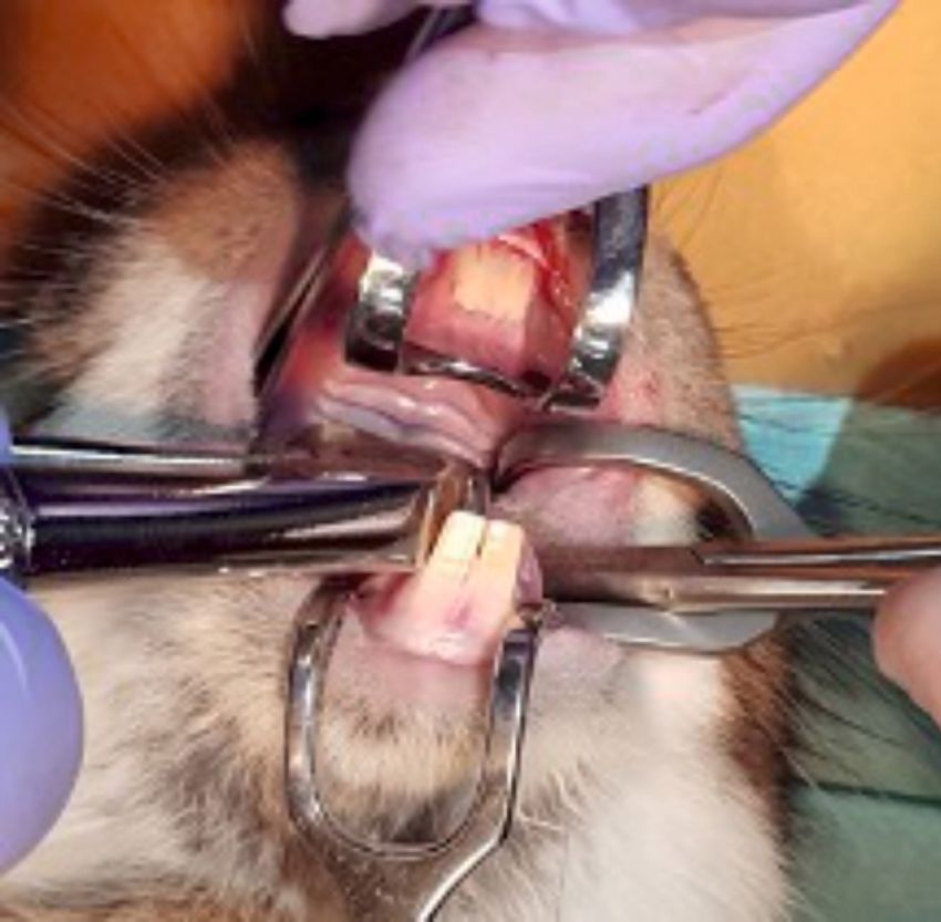
JOURNAL of MEDICINE and LIFE
Rabbit's Dental System topathologist with 25 years of experience
in assessing guided oral tissue regenera-
Vestibuloplasty - Material 1 Vestibuloplasty - Material 2 tion (G.D.) and by three other investiga-
tors (A.M; C.D.; H.O.). The histological
Right Incisors Incisors Left
regeneration of the epithelial defects and
Upper Upper bone healing were independently as-
sessed by each of these four investigators.
Extraction + Material 1 Extraction + Material 2 The evaluation process was planned
based on criteria established in a previous
Premolars Premolars pilot study, from which the array of the
most useful aspects for characterization
of the newly formed tissue emerged [17].
Molars Molars Comparison with spontaneous healing
of the mucosal defect has not been per-
formed because of the cicatricial spon-
Extraction - Control alveolus (socket) taneous healing with retractile connec-
tive scarring of the tissue, without any
Molars possibility for a meaningful comparative
Molars
evaluation to guided tissue regeneration
(GTR).
Premolars Premolars
Data for histologic evaluation of the ep-
ithelial healing comprised the following
parameters: the maximum and minimum
thickness of the superficial layer (in mi-
Incisor Incisor crometer), the total number of pathologic
Right Left
foci, presence of normal, hyper- and hy-
Lower Lower poplastic (with reduced spinous layer) epi-
Figure 3. Anatomic scheme of a rabbit’s dental system. The green circles mark the thelium, presence of inflammation, simple
paired sites of vestibuloplasty. The red circles mark the sites of dental extractions. granuloma, as well as foreign body granu-
loma in distinct microscopic fields.
As for the bone healing evaluation crite-
ria, the following aspects were recorded
and analyzed after being quantified on
a 5-points Likert scale: normal bone tra-
beculae, bone formation (filling of alve-
olus with bone), vascularization (neofor-
mation vessels), mature bone, structural
integrity at the membrane surface, pres-
ence of inflammation/granulation tissue,
material persistence, presence of ab-
scesses in the post-extraction site, newly
formed cartilaginous tissue, newly formed
fibrous tissue.
The 5-points Likert scale enabled the es-
timation of 5 categories, ranging from the
absence of the criterion to the extreme-
ly intense/clear/generalized presence of
the criterion.
Data analysis
Descriptive statistics have been computed
and reported as the median and inter-
quartile range (IQR), given the skewed
distribution of the studied dataset. Mean
and standard deviation have also been re-
ported for contrasting and an improved
Figure 4. Extraction of maxillary teeth.
overview of these descriptive results.
© 2021 JOURNAL of MEDICINE and LIFE. VOL: 14 ISSUE: 2 MARCH-APRIL 2021
184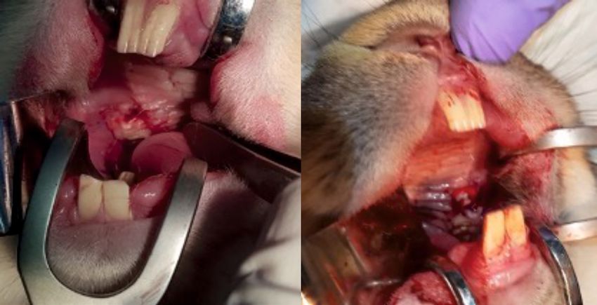
JOURNAL of MEDICINE and LIFE

Figure 5. Coverage of the paired alveoli after tooth extractions on each side – the split-mouth pattern.

Inferential statistics has investigated three main categories of questions, pertaining to the epithelial (mucosal) healing study arm and the
bone healing study arm.

Firstly, the current study tested the null hypotheses that no differences appeared during the mucosal healing process between the two
groups (Mucoderm® vs. Suprathel®) concerning the frequency of the following histological characteristics:

Figure 6. Complete coverage of all reconstruction sites: buccal mucosa and alveolar sockets bilaterally.

© 2021 JOURNAL of MEDICINE and LIFE. VOL: 14 ISSUE: 2 MARCH-APRIL 2021
                                                                                                                                               185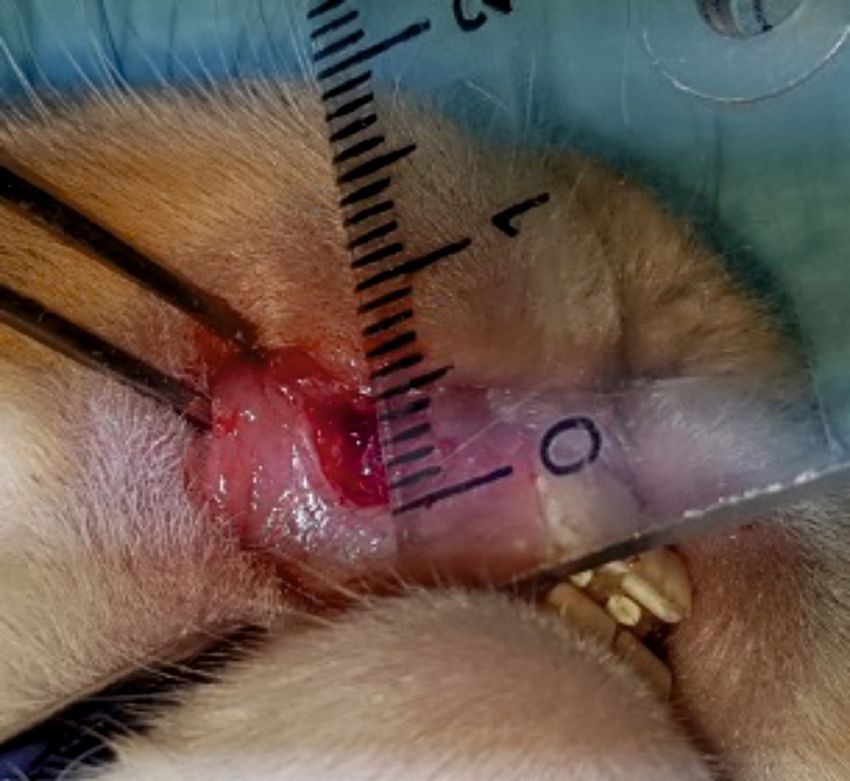
JOURNAL of MEDICINE and LIFE

  •    Presence of superficial epithelialization;
  •    Presence of profound epithelialization;
  •    Presence of remnant membrane material.

  The above hypotheses have been tested by applying
  McNemar’s test on the corresponding contingency ta-
  bles of these paired observations.

  Secondly, the study tested the null hypotheses that no
  differences appeared after the mucosal healing pro-
  cess between the two groups (Mucoderm® vs. Supra-
  thel®) concerning the minimum and the maximum
  thickness of the superficial epithelial layers of the
  healed regions, as well as the total number of identifi-
  able pathological foci.

  Since these measurements did not follow normal
  distributions, Wilcoxon tests for paired datasets have
  been used to test these hypotheses.

  Thirdly, the null hypotheses were tested that no differ-
  ences appeared during the healing process concern-
  ing the Likert scoring of the following histological
  characteristics in the compared groups (Mucoderm®
  vs. Suprathel®):

  •    Normal epithelium;
                                                                      Figure 7. Vestibulum site at 3 months postoperatively with clinically
  •    Hyperplasic epithelium;                                        healed mucosa, ready for the evaluation of the guided tissue regener-
  •    Hypoplasic epithelium;                                         ation (GTR).
  •    Inflammation;
  •    Simple granuloma;
  •    Foreign body granuloma.

  Once again, Wilcoxon tests for paired datasets have been used to test these hypotheses because the recorded Likert scores were not nor-
  mally distributed. Finally, the healing after extraction has been compared between the three alveoli (two alveoli covered with Mucoderm®
  and Suprathel® and a third one left for natural healing), concerning the following characteristics, quantified by Likert scores:

  •    Presence of normal bone trabeculae;
  •    Bone-filled alveoli;
  •    Bone maturity;
  •    Vascularization/vascular neo-formation;
  •    Presence of inflammatory tissue;
  •    Presence of abscesses in the post-extraction site;
  •    Newly formed cartilaginous tissue;
  •    Newly formed fibrous tissue;
  •    Structural integrity at the contact point with the membrane (only for the alveoli covered with Mucoderm® and Suprathel®);
  •    Persistence of membrane material (only for alveoli covered with Mucoderm® and Suprathel®).

  Freedman tests and post-hoc Wilcoxon tests for paired samples have been used to investigate possible differences in the above-men-
  tioned Likert scores between the investigated alveoli.The threshold for statistical significance has been chosen at a level α=0.05 for all
  tested hypotheses, with the exception of post-hoc hypotheses, for which a Bonferroni-corrected threshold α=0.0169 was used. Data
  were collected using Microsoft Excel, and analyses have been performed using IBM Statistical Package for the Social Sciences (SPSS)
  Statistics v.25 (IBM, Armonk, New York, United States of America). The collected data regarding mucosal epithelial healing and bone
  regeneration were described and analyzed under blind coding. Then, after unblinding, the obtained results were attributed to each of
  the studied materials (Suprathel® and Mucoderm®).

  RESULTS

  The animals had an uneventful recovery after the surgical interventions. One rabbit died on the 9th postoperative day, and no relation
  could be found with the experiment. An overview of mucosal epithelial healing characteristics under the influence of Mucoderm® and
  Suprathel® is presented in Table 1.

                                                             © 2021 JOURNAL of MEDICINE and LIFE. VOL: 14 ISSUE: 2 MARCH-APRIL 2021
186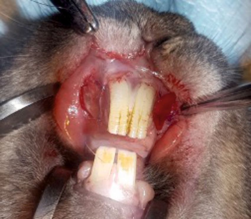
JOURNAL of MEDICINE and LIFE

    Figure 8 (A, B, C). Healed postextraction alveoli (arrows).

Results of data analysis
The null hypothesis concerning the presence of profound epithelialization has been rejected (p=0.031 – McNemar test). Profound
epithelialization was present in a significantly higher number of rabbits on the Mucoderm® side (7 rabbits) compared to the Suprathel®
side of the mouth (1 rabbit), as presented in Table 2.

None of the other null hypotheses could be rejected, as listed below:

•       Presence of remnant membrane material (p=1 – McNemar test);
•       Minimum thickness of the superficial epithelial layers (p=0.149 – Wilcoxon test);
•       Maximum thickness of the superficial epithelial layers (p=0.168 – Wilcoxon test);
•       Total number of identifiable pathological foci (p=0.206 – Wilcoxon test);
•       Likert scores regarding:

       ○     Normal epithelium (p=0.873 – Wilcoxon test);
       ○     Hyperplasic epithelium (p=0.711 – Wilcoxon test);
       ○     Hypoplasic epithelium (p=0.480 – Wilcoxon test);

© 2021 JOURNAL of MEDICINE and LIFE. VOL: 14 ISSUE: 2 MARCH-APRIL 2021
                                                                                                                                         187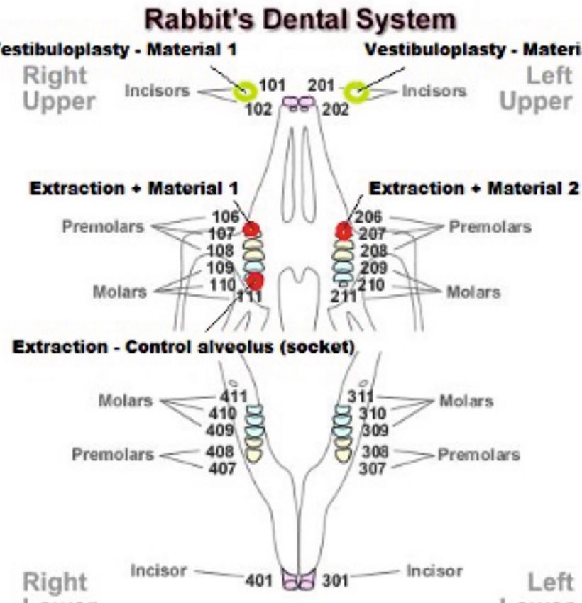
JOURNAL of MEDICINE and LIFE

       Table 1. Characteristics of epithelial healing guided by the two guided tissue regeneration – GTR materials.

                                             Mucoderm®                                        Suprathel®
                                                                                                                              P-value (Wilcoxon
                                           Std.                                            Std.                               signed ranks test)
                               Mean                   Median        IQR       Mean                    Median          IQR
                                         Deviation                                       Deviation

       Max. thickness
       of the superficial      38.42       17.799      40.00       10.00         32.11    19.099       30.00          20.00         0.168
       layer (micrometer)

       Min. thickness
       of the superficial      16.05       6.364       20.00       10.00         13.16     6.283       15.00          15.00         0.149
       layer (micrometer)

       Total number of
                               0.53        0.841       0.00         1.00         0.26      0.562        0.00          0.00          0.206
       pathologic foci

       Hyperplastic
                               2.68        1.293       2.00         2.00         2.58      1.427        3.00          2.00          0.711
       epithelium (Likert)

       Normal epithelium
                                1.16       1.302       1.00         2.00         1.05      1.353        1.00          2.00          0.873
       (Likert)

       Hypoplastic
                                0.16       0.375       0.00         0.00         0.37      0.955        0.00          0.00          0.480
       epithelium (Likert)

       Inflammation
                               4.00        0.000       4.00         0.00         3.79      0.918        4.00          0.00          0.317
       (Likert)

       Simple granuloma
                               3.89        0.459       4.00         0.00         3.95      0.229        4.00          0.00          0.655
       (Likert)

       Foreign body
                               3.89        0.459       4.00         0.00         4.00     0.000         4.00          0.00          0.317
       granuloma (Likert)

          ○    Inflammation (p=0.317 – Wilcoxon test);
          ○    Simple granuloma (p=0.655 – Wilcoxon test);
          ○    Foreign body granuloma (p=0.317 – Wilcoxon test).

      No significant differences were found between Mucoderm® and Suprathel® concerning any of the alveolar healing characteristics quan-
      tified by Likert scores in this study. Figures 9 and 10 illustrate two of these highly comparable characteristics: newly formed capillaries
      and newly formed fibrous tissue. Nevertheless, both Mucoderm® and Suprathel® exhibited significantly higher Likert scores compared
      to the naturally healing alveolus in respect to the following three characteristics (Figures 11–13):

          ○    Presence of normal bone trabeculae (p=0.004/p=0.003 – post hoc Wilcoxon test);
          ○    Bone-filled alveoli (p=0.004/p=0.003 – post hoc Wilcoxon test);
          ○    Bone maturity (p=0.006/p=0.002 – post hoc Wilcoxon test).

      Histological aspects of mucosal healing (vestibuloplasty)

      With regard to the histological results, epithelialization occurred in
      all cases. Inflammatory hyperplasia with a tendency of hyperkera-
      tosis with orthokeratosis was found more expressed for Mucoderm®
      and less for Suprathel® (Figures 14, 15). In most cases, Suprathel® was             Table 2. Contingency table of profound epithelializa-
      characterized by local epithelial hyperplasia without any other changes             tion depending on the intervention side.
      (Figure 15).
                                                                                          Profound               Profound epithelialization
                                                                                          epithelialization             Suprathel®
      Histological aspects of bone healing (extraction socket)                            Mucoderm®              Absent             Present

      Both materials induced epithelialization on the contour of the dental               Absent                      12               0
      alveolus and fibrosis (with incorporated particles of the biomaterial in
                                                                                          Present                     6                1
      the medullary space of the alveolar bone (in the case of Mucoderm®)

                                                               © 2021 JOURNAL of MEDICINE and LIFE. VOL: 14 ISSUE: 2 MARCH-APRIL 2021
188
JOURNAL of MEDICINE and LIFE

Figure 9. Vascularization and newly formed cap-

                                                       Likert Scale
illaries in the healing of the three compared al-
veoli. Bolded lines mark the median Likert score;
upper/lower whiskers mark the limits of the larg-
est/smallest values that are not greater/smaller
than the third/first quartile plus/minus 1.5 times
the interquartile range.

                                                                                                        Suprathel.            Spontaneous alveolus.
                                                                             Mucoderm.
                                                                                                   Vascularization, newly     Vascularization, newly
                                                                            Vascularization
                                                                                                     formed capillaries         formed capillaries
                                                       Likert Scale

Figure 10. Newly formed fibrous tissue in the
healing of the three compared alveoli. Bolded
lines mark the median Likert score; upper/lower
whiskers mark the limits of the largest/small-
est values that are not greater/smaller than the
third/first quartile plus/minus 1.5 times the in-
terquartile range.

                                                                                                                              Spontaneous alveolus.
                                                                      Mucoderm. Newly formed      Suprathel. Newly formed
                                                                                                                                 Newly formed
                                                                           fibrous tissue              fibrous tissue
                                                                                                                                  fibrous tissue

Figure 11. Normal trabecular bone structure in
                                                       Likert Scale

the healing of the three compared alveoli. Bolded
lines mark the median Likert score; upper/lower
whiskers mark the limits of the largest/small-
est values that are not greater/smaller than the
third/first quartile plus/minus 1.5 times the inter-
quartile range.

                                                                                                                               Spontaneous alveolus.
                                                                         Mucoderm. Normal            Suprathel. Normal
                                                                                                                                Normal trabecular
                                                                      trabecular bone structure   trabecular bone structure
                                                                                                                                  bone structure

© 2021 JOURNAL of MEDICINE and LIFE. VOL: 14 ISSUE: 2 MARCH-APRIL 2021
                                                                                                                                                       189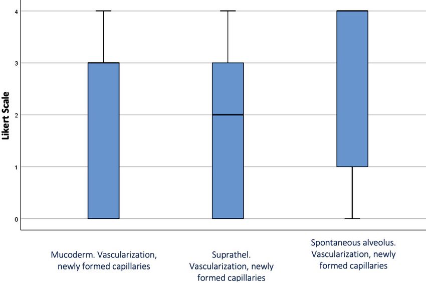
JOURNAL of MEDICINE and LIFE

                                                                                                     Figure 12. Bone formation (socket filling) in the
                                                                                                     healing of the three compared alveoli. Bolded
  Likert Scale

                                                                                                     lines mark the median Likert score; upper/lower
                                                                                                     whiskers mark the limits of the largest/small-
                                                                                                     est values that are not greater/smaller than the
                                                                                                     third/first quartile plus/minus 1.5 times the in-
                                                                                                     terquartile range.

                                                                           Spontaneous alveoulus.
                    Mucoderm. Bone                Suprathel. Bone
                                                                              Bone formation
                 formation (socket filling)   formation (socket filling)
                                                                               (socket filling)

    (Figure 16). Regarding Suprathel®, epithelialization occurred in the alveolus with nonkeratinized hyperplasic epithelium, while the
    biomaterial itself was no longer visible (Figure 17).

    DISCUSSION

    The aim of the present study has been reached by investigating whether the benefits of an advanced skin regeneration biomaterial
    (Suprathel®) may also be conveyed to oral tissue regeneration. The animal experiment that we conducted comprised both an analysis
    of mucosal defect healing (after vestibuloplasty) and bone healing (postextraction alveoli healing).

    After investigating the capacity of Suprathel® to regenerate oral tissue – mucosa and bone, by juxtaposing it in a split-mouth animal
    model to Mucoderm®, a certified biomaterial for keratinized oral mucosa healing, no significant differences between these two bioma-
    terials could be found in respect of all but one (profound epithelialization) of the investigated characteristics of guided tissue healing.

    Principles of guided tissue regeneration

    There are numerous factors that influence the success of a regeneration procedure. They include factors linked to the properties of
    the regeneration membrane (barrier), such as barrier occlusion and stability, the size of the barrier perforations, peripheral sealing,
    adequate blood supply, access to tissue-forming cells (epithelium, bone and others) [24–27].

                                                                                                     Figure 13. Bone maturity in the healing of the
  Likert Scale

                                                                                                     three compared alveoli. Bolded lines mark the
                                                                                                     median Likert score; upper/lower whiskers mark
                                                                                                     the limits of the largest/smallest values that are
                                                                                                     not greater/smaller than the third/first quartile
                                                                                                     plus/minus 1.5 times the interquartile range.

                        Mucoderm.                    Suprathel.              Spontaneous alveolus.
                        Mature bone                 Mature bone                 Mature bone

                                                                   © 2021 JOURNAL of MEDICINE and LIFE. VOL: 14 ISSUE: 2 MARCH-APRIL 2021
190
JOURNAL of MEDICINE and LIFE
Figure 14. Red arrows indicate towards the rests of Mucoderm® included in the fibrous tissue. Blue arrows point to the new hyperplasic
and hyperkeratotic epithelium. The yellow arrow shows the original normal gingival epithelium, with a reduced amount of lamellar ker-
atin at the surface.
Resorbable membranes belong to the groups of natural or synthetic polymers. Collagen and aliphatic polyesters, such as polyglycolide
or polylactide, are the most frequently used ones [28]. Collagen can be obtained from different sources and can be treated in various
ways for membrane production. Polyglycolide or polylactide can be easily produced in different forms with multivalent physical, chem-
ical and mechanical characteristics [29].
Alloplastic materials
The most frequently used resorbable synthetic polymers in tissue engineering are poly-lactic acid (PLA), poly-glycolic acid (PGA), po-
ly-lactide-co-glycolide (PLGA), polyvinyl alcohol (PVA), and polycaprolactone (PCL). There are several designs that stimulate uniform
cell distribution, vascular supply, and cell differentiation used for tissue engineering: meshes, fibers, sponges, films, scaffolds and foams
[30]. Among the multiple advantages of using these alloplastic materials, one can consider good biocompatibility, chemical versatility,
favorable mechanical properties, low immunogenic reactions, and no risk of disease transmission [31]. They are degraded and eliminat-
ed through metabolic pathways [32]. The challenge is to design a structure that promotes cell differentiation, determining normal-tissue
anatomy and function [33].
Xenogeneic collagen matrix (Mucoderm®)
Guided tissue regeneration can also be consistently and predictably sustained with the help of collagen barriers of animal origin [34].
Angiogenesis is enhanced by collagen, which promotes healing and enhances fibroblast expression. It is resorbable, and there is no
© 2021 JOURNAL of MEDICINE and LIFE. VOL: 14 ISSUE: 2 MARCH-APRIL 2021
191JOURNAL of MEDICINE and LIFE
Figure 15. Suprathel® produced the same type of hyperplasic epithelium but less hyperkeratotic (blue arrow) compared to the normal
epithelium (yellow arrow). Epithelial extensions surrounded by dense fibrous tissue were seen in the chorion (red arrow).
need for a second surgery [35]. The xenogeneic collagen matrix has in its composition collagen type I and III. It is a frequently used
augmentation biomaterial to enhance soft peri-implant soft tissue, and it is used as a replacement for autogenous grafts [20, 36, 37].
Mucoderm® (Xenogeneic collagen membrane) has a proven clinical use for increasing the keratinized mucosa width, including around
dental implants, although it does not increase its thickness like the connective tissue graft [38].
Suprathel®
The gold standard for regeneration of the deep dermal and full-thickness skin defects (burns, ulcers) is still considered the autologous
spit-thickness skin graft [39, 40]. Nevertheless, several issues have been reported with the current use of skin grafts, and due to this, a
variety of wound dressings have been developed [41]. Synthetic polymers, which the body degrades using enzymatic processes, have
been introduced in many medical areas in recent years [42, 43].
Suprathel® is a synthetic terpolymer mainly based on DL-lactide with trimethylene carbonate and ε-caprolactone. The final product is
an interconnected porous membrane clinically successfully certified for skin healing. It shows large plasticity and adapts to the wound
surface. It is water permeable, and it favors reepithelialization of wounds [44]. All of the components of Suprathel® have valuable
properties as regeneration biomaterials.
Lactic acid, lactate (anion), and lactide (di-ester derived from lactic acid) are known for their favorable properties in promoting tissue
healing in general and skin formation in particular. The responsible mechanisms are believed to be multiple. It elicits angiogenesis via
stimulation of growth factors, migration of endothelial cells, induces homing of stem cells, triggers diverse activation loops, and even
© 2021 JOURNAL of MEDICINE and LIFE. VOL: 14 ISSUE: 2 MARCH-APRIL 2021
192JOURNAL of MEDICINE and LIFE
Figure 16. The blue arrow indicates the extraction socket covered with Mucoderm®, epithelialized with keratinized hyperplasic epitheli-
um (similar hyperplasic character as in mucosal healing). The red arrow points to a fibrous nodule containing the included biomaterial
formed in the alveolar bone.
modulates gene expression in human mesenchymal stem cells [45]. It accelerates the healing and has an antiseptic effect, whereas lac-
tate-mediated oxidants stimulate fibroblasts, thus contributing to the formation of the dermis [46].
Various combinations of Suprathel®’s components have gained attention (e.g., polylactide-co-ε-caprolactone – PLLC blends with
collagen) and were subjected to research in the physiologically relevant environment in which they would be utilized, particularly as
natural biopolymers, sensitive to water content. Their properties might vary with hydration – e.g., blend elasticity increases with hy-
dration [47, 48].
Although displaying longer healing times when compared to a skin graft, Suprathel® demonstrated comparable early scar formation
[49]. This material can serve as treatment for patients with extensive burns to cover the deep dermal burn wounds, saving donor sites
© 2021 JOURNAL of MEDICINE and LIFE. VOL: 14 ISSUE: 2 MARCH-APRIL 2021
193JOURNAL of MEDICINE and LIFE
Figure 16. Epithelialized alveolus after coverage with Suprathel®. Nonkeratinized hyperplasic epithelium.
for split-thickness skin grafts to be used in full-thickness burned areas [50]. Also, this membrane significantly reduces pain, is easy to
handle, no allergic reactions are reported, and the material is easy and pain-free to remove after complete healing of the burns [50].
Mucosal healing – epithelialization
Overall, our study showed no significantly different outcomes for the healing of mucosal defects covered with Suprathel® and Mucoderm®,
except for profound epithelialization, which occurred more often on the side where tissue regeneration was guided by Mucoderm®. Both
materials favored the formation of a normal epithelium to similar degrees, whereas inflammation, simple granuloma, as well as foreign
body granuloma, were present in comparable rates.
Several studies have compared tissue healing after free gingival graft vestibuloplasty, which is considered the gold standard, to porcine
collagen matrix (Mucoderm®) with comparable results regarding the clinical and histological outcome. The main advantages of using
collagen matrix to augment keratinized tissues surrounding dental implants are that they do not require a harvesting procedure, surgery
time is reduced, and the regenerated tissues appear more esthetic. Tissue shrinkage has also been cited for both methods, with a higher
degree in the collagen matrix group [51–53].
Socket preservation – bone formation
After an extraction, the alveolar bone remodels, and the resorption process begins. Socket preservation diminishes this effect, and it
usually involves placing biomaterials into the socket to support the remodeling process and coverage with a protective membrane in or-
der to preserve bone tissues and minimize bone loss after teeth extraction [54]. Preservation has been known to produce less resorption
when compared to normal healing after tooth extraction [55].
Guided bone regeneration (GBR) is a procedure based on the protected space theory in which a barrier membrane is used for space
maintenance over a bone defect or the tooth alveolus specifically after extraction, promoting the ingrowth of osteogenic cells and pre-
venting migration of undesired cells from the overlying soft tissues into the alveolus [56]. Resorbable and non-resorbable membranes
have been used with good results in reducing bone resorption [57]. For non-resorbable membranes, a frequent complication is mem-
brane exposure, with subsequently reduced rates of new bone formation [58]. Thus, resorbable membranes offer a viable alternative
for this problem.
The need for the delayed placement of dental implants is an indication of socket preservation. It is generally performed with bone
autografts, allografts (frozen-dried bone allograft, FDBA), xenografts (collagenated porcine bone, CPB), or alloplasts (hydroxyapatite,
magnesium-enriched hydroxyapatite, calcium sulfate). FDBA [59] and CPB [60] have been shown to have a positive effect on height
and width when compared to natural healing. The xenograft provides stability to the site due to its density and low resorption rate [61].
Another promising new approach is using concentrated growth factors (recombinant human bone morphogenetic protein – rhBMP-2)
that can preserve the alveolar bone with almost no height change from baseline to 4 months [62].
An alternative to socket preservation therapy is implant placement with simultaneous contour augmentation using GBR. This tech-
nique described by Buser et al. involves extraction without flap elevation, debridement, followed by healing of the soft tissues for 4–8
© 2021 JOURNAL of MEDICINE and LIFE. VOL: 14 ISSUE: 2 MARCH-APRIL 2021
194JOURNAL of MEDICINE and LIFE
Table 3. Non-resorbable vs. resorbable membranes for alveolus bone regeneration.
Type of membrane Advantages Disadvantages Commercial products
• May include the use of titanium;
• Maintains shape until removal; • Need of an additional intervention
• Can be easily fixed with titanium for removal;
or bone tacks; • Increased patient morbidity;
• ePTFE membranes;
Non-resorbable • Regenerates more bone if • Exposure leads to the
• Titanium reinforced membranes.
complication-free; necessity of removal;
• No inflammatory response unless • Experience is required for
the membrane is exposed; optimal handling.
• Numerous studies available.
• Variable resorption rate;
• Uncertain barrier to
• No need of removal (cost effective); specific tissues;
• Low morbidity; • Difficult to handle;
• Good soft tissue healing; • Less bone regenerated • Bovine collagen matrix ;
Resorbable • Membrane exposure does not than with non-resorbable • Porcine collagen matrix ;
mandate immediate removal; membranes; • Cross-linked collagen barrier;
• Numerous studies demonstrate • Resorption with various
its validity. inflammatory response
which may interfere
with healing and GBR.
weeks, implant placement with simultaneous contour augmentation on the buccal aspect with GBR using resorbable membranes com-
bined with autogenous chips and a low resorption bone filler, as well as tension-free primary wound closure [63].
The advantages and disadvantages of the two major types of membranes used in guided bone regeneration procedures, including
socket preservation, are shown in Table 3 (non-resorbable vs. resorbable membranes).
Suprathel® was investigated in this study concerning its capacity to act as a resorbable membrane for GBR. The present study found
comparable bone healing characteristics in those alveoli that regenerated under Suprathel® membranes compared to their correspond-
ing split-mouth pairs for which healing occurred under Mucoderm® membranes.
Both these biomaterials exhibited significantly improved healing and GBR characteristics regarding their socket-filling ability and
their capability to foster the regeneration of normal bone trabeculae, with higher degrees of bone maturity, compared to the naturally
healing control alveoli.
Future developments
Research on tissue regeneration in the oral cavity is confronted with the multifaceted challenges of the complex oral environment – humidity,
microbial flora, constant mechanical impact – conditions which render it highly demanding. As the processing of materials also plays an
essential role in modulating their properties, extensive development of complex blends of polymers with natural tissue components and
healing-promoters (collagen, fibrin, growth factors) in a hydrated environment has been undertaken in the attempt to obtain materials
with improved and quantifiable physical and mechanical properties.
Electrospinning has recently gained attention as a source of an easy, cost-effective method for producing materials for medical purposes
in a homogenous solution [47]. The electrospun scaffolds made for tissue engineering applications can be seeded with cells to recondi-
tion or replace tissue. By adding drugs into the electrospinning solution, drug delivery systems, such as implants, transdermal patches,
oral forms, can be prepared. These scaffolds fulfill a similar purpose as the extracellular matrix in natural tissue. Polycaprolactone, a
resorbable polymer, component of Suprathel®, is typically used for this purpose. The resulting compounds may then be coated with
collagen to promote cell attachment, as collagen has already been spun into membranes with good results [64].
CONCLUSION
This animal study achieved the validation of Suprathel® (a resorbable material, already successfully used for skin regeneration) as a
versatile and effective oral epithelium and bone regeneration material. The results of this study pave the way for future clinical trials that
could further investigate the use in humans of this highly promising material for guided oral tissue and bone regeneration.
© 2021 JOURNAL of MEDICINE and LIFE. VOL: 14 ISSUE: 2 MARCH-APRIL 2021
195JOURNAL of MEDICINE and LIFE
ACKNOWLEDGMENTS
Ethical approval
The approval for this study was obtained from the Ethics Committee of the Iuliu Hatieganu University of Medicine and Pharmacy,
Cluj-Napoca, Romania (approval no. 211/May 4th, 2018).
Conflict of interest
The authors declare that there is no conflict of interest.
REFERENCES study. Journal of Clinical Periodontology. 2016; Blackwell
Munksgaard. 43(3):305–10.
31. Nasalapure, A. V., Chalannavar, R.K., Gani, R.S.,
Malabadi, R.B. and Kasai, D.R. (2017) Tissue engineering of
skin: A review. Trends Biomater. Artif. Organs.
16. Souza, A.B., Tormena, M., Matarazzo, F. and Araújo,
M.G. The influence of peri-implant keratinized mucosa on 32. Vacanti, C.A., Kim, W., Upton, J., Mooney, D. and
1. Pinho, M.N., Roriz, V.M., Novaes, A.B., Taba, M., Grisi, brushing discomfort and peri-implant tissue health. Clinical Vacanti, J.P. The Efficacy of Periosteal Cells Compared to
M.F.M., de Souza, S.L.S. et al. Titanium Membranes in Oral Implants Research. 2016; 27(6):650–5. Chondrocytes in the Tissue Engineered Repair of Bone
Prevention of Alveolar Collapse After Tooth Extraction. Defects. Tissue Engineering. 1995; 1(3):301–8.
Implant Dentistry. 2006; 15(1):53–61. 17. Vacaraş, S., Băciuţ, G., Gheban, D., Bran, S., Colosi, H.,
Berce, C. et al. First – in-animal experimental pilot study of 33. Ru, C., Wang, F., Pang, M., Sun, L., Chen, R. and Sun, Y.
2. Gotfredsen, K., Carlsson, G.E., Jokstad, A., Arvidson Suprathel® for oral mucosa and bone regeneration. HVM Suspended, Shrinkage-Free, Electrospun PLGA Nanofibrous
Fyrberg, K., Berge, M., Bergendal, B. et al. Implants and/or Bioflux. 2021; 13(2):56–66. Scaffold for Skin Tissue Engineering. ACS Applied Materials
teeth: consensus statements and recommendations. Journal of & Interfaces. 2015; 7(20):10872–7.
Oral Rehabilitation. 2008; John Wiley & Sons, Ltd. 18. Cordioli, G., Mortarino, C., Chierico, A., Grusovin,
35 Suppl 1(s1):2–8. M.G. and Majzoub, Z. Comparison of 2 Techniques of 34. Lorenz, J., Blume, M., Barbeck, M., Teiler, A.,
Subepithelial Connective Tissue Graft in the Treatment of Kirkpatrick, C.J., Sader, R.A. et al. Expansion of the
3. Buser, D., Martin, W. and Belser, U.C. Optimizing esthetics Gingival Recessions. Journal of Periodontology. 2001; Wiley. peri-implant attached gingiva with a three-dimensional
for implant restorations in the anterior maxilla: anatomic and 72(11):1470–6. collagen matrix in head and neck cancer patients-results from
surgical considerations. The International Journal of Oral & a prospective clinical and histological study. Clinical Oral
Maxillofacial Implants. 2004; 19 Suppl:43–61. 19. Tonetti, M.S., Cortellini, P., Pellegrini, G., Nieri, M., Investigations. 2017; 21(4):1103–11.
Bonaccini, D., Allegri, M. et al. Xenogenic collagen matrix
4. Nevins, M., Camelo, M., De, P.S., Friedland, B., Schenk, or autologous connective tissue graft as adjunct to coronally 35. Armstrong, D.G. and Jude, E.B. The role of matrix
R. and Parma-Benfenati, S. A study of the fate of the buccal advanced flaps for coverage of multiple adjacent gingival metalloproteinases in wound healing. Journal of the
wall of extraction sockets of teeth with prominent recession: Randomized trial assessing non-inferiority in root American Podiatric Medical Association. 2002; Allen Press.
roots. Primary Dental Care. coverage and superiority in oral health-related. Journal of 92(1):12–8.
2006; SAGE Publications. os13(3):90–90. Clinical Periodontology. 2018; 45(1):78–88.
36. Sanz, M., Lorenzo, R., Aranda, J.J., Martin, C. and
5. Ten Heggeler, J.M.A.G., Slot, D.E. and Van der Weijden, 20. Cairo, F., Nieri, M. and Pagliaro, U. Efficacy of Orsini, M. Clinical evaluation of a new collagen matrix
G.A. Effect of socket preservation therapies following tooth periodontal plastic surgery procedures in the treatment (Mucograft prototype) to enhance the width of keratinized
extraction in non-molar regions in humans: a systematic of localized facial gingival recessions. A systematic review. tissue in patients with fixed prosthetic restorations: a
review. Clinical Oral Implants Research. 2011; Clin Oral Journal of Clinical Periodontology. 2014; 41:S44–62. randomized prospective clinical trial. Journal of Clinical
Implants Res. 22(8):779–88. Periodontology. 2009; Wiley Online Library. 36(10):868–76.
21. McGuire, M.K. and Scheyer, E.T. Xenogeneic Collagen
6. Tarnow, D.P., Eskow, R.N. and Zamzok, J. Aesthetics and Matrix With Coronally Advanced Flap Compared to 37. Lorenzo, R., García, V., Orsini, M., Martin, C. and
implant dentistry. Periodontology 2000. 1996; Blackwell Connective Tissue With Coronally Advanced Flap for the Sanz, M. Clinical efficacy of a xenogeneic collagen matrix
Munksgaard. 11(1):85–94. Treatment of Dehiscence-Type Recession Defects. Journal of in augmenting keratinized mucosa around implants: a
Periodontology. 2010; 81(8):1108–17. randomized controlled prospective clinical trial. Clinical Oral
7. Opris, H., Bran, S., Dinu, C., Baciut, M., Prodan, Implants Research. 2012; 23(3):316–24.
D.A.D.A., Mester, A. et al. Clinical applications of avian 22. Stefanini, M., Jepsen, K., de Sanctis, M., Baldini, N.,
eggshell-derived hydroxyapatite. Bosnian Journal of Basic Greven, B., Heinz, B. et al. Patient-reported outcomes and 38. Moraschini, V., Guimarães, H.B., Cavalcante, I.C. and
Medical Sciences. 2020; 20(4):430–7. aesthetic evaluation of root coverage procedures: a 12-month Calasans-Maia, M.D. Clinical efficacy of xenogeneic collagen
follow-up of a randomized controlled clinical trial. Journal of matrix in augmenting keratinized mucosa round dental
8. Linde, A., Thorén, C., Dahlin, C. and Sandberg, E. Clinical Periodontology. 2016; 43(12):1132–41. implants: a systematic review and meta-analysis. Clinical Oral
Creation of new bone by an osteopromotive membrane Investigations. 2020; 24(7):2163–74.
technique: An experimental study in rats. Journal of Oral 23. Charan, J. and Kantharia, N. How to calculate sample
and Maxillofacial Surgery. 1993; J Oral Maxillofac Surg. 39. Giessler, G.A., Deb, R., Germann, G. and Sauerbier,
size in animal studies? Journal of Pharmacology and
51(8):892–7. M. [Primary treatment of burn patients]. Der Chirurg;
Pharmacotherapeutics. 2013; 4(4):303–6.
Zeitschrift Fur Alle Gebiete Der Operativen Medizen.
9. Karring, T., Nyman, S., Gottlow, J. and Laurell, L. 24. Kostopoulos, L. and Karring, T. Augmentation of the 2004; 75(6):560–7.
Development of the biological concept of guided tissue rat mandible using guided tissue regeneration. Clinical Oral
regeneration--animal and human studies. Periodontology 40. Qaryoute, S., Mirdad, I. and Hamail, A.A. Usage of
Implants Research. 1994; 5(2):75–82.
2000. 1993; 1(1):26–35. autograft and allograft skin in treatment of burns in children.
25. Lundgren, D., Lundgren, A.K., Sennerby, L. and Nyman, Burns. 2001; 27(6):599–602.
10. Hämmerle, C.H.F. and Jung, R.E. Bone augmentation by S. Augmentation of intramembraneous bone beyond the
41. Eaglstein, W.H. and Falanga, V. Tissue engineering and
means of barrier membranes. Periodontology 2000. skeletal envelope using an occlusive titanium barrier. An
the development of Apligraf, a human skin equivalent. Cutis.
2003; Periodontol 2000. 33(1):36–53. experimental study in the rabbit. Clinical Oral Implants
1998; 62(1 Suppl):1–8.
Research. 1995; 6(2):67–72.
11. Bartold, P. Turnover in periodontal connective
42. Amecke, B., Bendix, D. and Entenmann, G. Resorbable
tissues: dynamic homeostasis of cells, collagen and ground 26. Lundgren, A.K., Sennerby, L. and Lundgren, D. Guided polyesters: composition, properties, applications. Clinical
substances. Oral Diseases. 2008; 1(4):238–53. jaw-bone regeneration using an experimental rabbit model. Materials. 1992; 10(1–2):47–50.
International Journal of Oral and Maxillofacial Surgery.
12. Page, R.C., Offenbacher, S., Schroeder, H.E., Seymour, 1998; 27(2):135–40. 43. Hayashi, T. Biodegradable polymers for biomedical uses.
G.J. and Kornman, K.S. Advances in the pathogenesis of Progress in Polymer Science. 1994; 19(4):663–702.
periodontitis: summary of developments, clinical implications 27. Lundgren, A., Lundgren, D. and Taylor, A. Influence
and future directions. Periodontology 2000. of barrier occlusiveness on guided bone augmentation. 44. Hildebrandt, D., Ziegler, K. and Wollina, U. EIectrical
1997; 14(1):216–48. An experimental study in the rat. Clinical Oral Implants impedance and transepidermal water loss of healthy
Research. 1998; 9(4):251–60. human skin under different conditions. Skin Research and
13. Agudio, G., Cortellini, P., Buti, J. and Pini Prato, G. Technology. 1998; 4(3):130–4.
Periodontal Conditions of Sites Treated With Gingival 28. Hutmacher, D., Hürzeler, M.B. and Schliephake,
Augmentation Surgery Compared With Untreated H. A review of material properties of biodegradable 45. Zieker, D., Schäfer, R., Glatzle, J., Nieselt, K., Coerper,
Contralateral Homologous Sites: An 18- to 35-Year and bioresorbable polymers and devices for GTR and S., Northoff, H. et al. Lactate modulates gene expression in
Long-Term Study. Journal of Periodontology. GBR applications. The International Journal of Oral & human mesenchymal stem cells. Langenbeck’s Archives of
2016; 87(12):1371–8. Maxillofacial Implants. 1996; 11(5):667–78. Surgery. 2008; Springer Verlag. 393(3):297–301.
14. Lin, G.-H., Chan, H.-L. and Wang, H.-L. The 29. Fields, T. (2001) Guided bone regeneration: focus on 46. Wagner, S., Hussain, M.Z., Hunt, T.K., Bacic, B. and
Significance of Keratinized Mucosa on Implant resorbable membranes. Baylor Oral Surgery Thursday Becker, H.D. Stimulation of fibroblast proliferation by
Health: A Systematic Review. Journal of Periodontology. Morning Conference. 2001. lactate-mediated oxidants. Wound Repair and Regeneration.
2013; 84(12):1755–67. 2004; Wound Repair Regen. 12(3):368–73.
30. Dhandayuthapani, B., Yoshida, Y., Maekawa, T. and
15. Halperin-Sternfeld, M., Zigdon-Giladi, H. and Machtei, Kumar, D.S. Polymeric Scaffolds in Tissue Engineering 47. McCullen, S.D., Ramaswamy, S., Clarke, L.I. and
E.E. The association between shallow vestibular depth and Application: A Review. International Journal of Polymer Gorga, R.E. Nanofibrous composites for tissue engineering
peri-implant parameters: A retrospective 6 years longitudinal Science. 2011; 2011:1–19. applications. Wiley Interdisciplinary Reviews: Nanomedicine
© 2021 JOURNAL of MEDICINE and LIFE. VOL: 14 ISSUE: 2 MARCH-APRIL 2021
196JOURNAL of MEDICINE and LIFE
and Nanobiotechnology. 2009; Wiley Interdiscip Rev Preservation Using a (Dense) PTFE Barrier with or without 60. Barone, A., Aldini, N.N., Fini, M., Giardino, R., Calvo
Nanomed Nanobiotechnol. 1(4):369–90. Xenograft Material: A Randomized Clinical Trial. Materials. Guirado, J.L. and Covani, U. Xenograft Versus Extraction
2019; 12(18):2902. Alone for Ridge Preservation After Tooth
48. Kwon, I.K. and Matsuda, T. Co-electrospun nanofiber Removal: A Clinical and Histomorphometric Study. Journal
fabrics of poly(L-lactide-co-ε-caprolactone) with type I 55. Horowitz, R., Holtzclaw, D. and Rosen, P.S. A Review of Periodontology. 2008; 79(8):1370–7.
collagen or heparin. Biomacromolecules. 2005; American on Alveolar Ridge Preservation Following Tooth Extraction.
Chemical Society. 6(4):2096–105. Journal of Evidence Based Dental Practice. 61. Garg, A.K. Bone biology, harvesting, grafting for denal
2012; 12(3):149–60. implants: rationale and clinical applications, 2004, Chicago,
49. Keck, M., Selig, H.F., Lumenta, D.B., Kamolz, L.P., Quintessence Publ.
Mittlböck, M. and Frey, M. The use of Suprathel® in deep 56. Gottlow, J., Nyman, S., Lindhe, J., Karring, T. and
dermal burns: First results of a prospective study. Burns. Wennström, J. New attachment formation in the human 62. Fiorellini, J.P., Howell, T.H., Cochran, D., Malmquist, J.,
2012; 38(3):388–95. periodontium by guided tissue regeneration. Case reports. Lilly, L.C., Spagnoli, D. et al. Randomized study evaluating
Journal of Clinical Periodontology. 1986; 13(6):604–16. recombinant human bone morphogenetic protein-2 for
50. Uhlig, C., Rapp, M., Hartmann, B., Hierlemann, H., extraction socket augmentation. Journal of Periodontology.
Planck, H. and Dittel, K.-K. Suprathel®-An innovative, 57. Lekovic, V., Kenney, E.B., Weinlaender, M., Han, T., 2005; 76(4):605–13.
resorbable skin substitute for the treatment of burn victims. Klokkevold, P., Nedic, M. et al. A bone regenerative approach
Burns. 2007; 33(2):221–9. to alveolar ridge maintenance following tooth extraction. 63. Buser, D., Chen, S.T., Weber, H.P. and Belser, U.C. Early
Report of 10 cases. Journal of Periodontology. implant placement following single-tooth extraction in the
51. Schmitt, C.M., Moest, T., Lutz, R., Wehrhan, F., 1997; 68(6):563–70. esthetic zone: biologic rationale and surgical procedures. The
Neukam, F.W. and Schlegel, K.A. Long-term outcomes after International Journal of Periodontics & Restorative Dentistry.
vestibuloplasty with a porcine collagen matrix (Mucograft®) 58. Simion, M., Baldoni, M., Rossi, P. and Zaffe, D. A 2008; 28(5):441–51.
versus the free gingival graft: a comparative prospective comparative study of the effectiveness of e-PTFE membranes
clinical trial. Clinical Oral Implants Research. with and without early exposure during the healing period. 64. Sill, T.J. and von Recum, H.A.
2016; 27(11):e125–33. The International Journal of Periodontics & Restorative Electrospinning: Applications in drug delivery and tissue
Dentistry. 1994; 14(2):166–80. engineering. Biomaterials. 2008; 29(13):1989–2006.
52. Schmitt, C.M., Tudor, C., Kiener, K., Wehrhan, F.,
Schmitt, J., Eitner, S. et al. Vestibuloplasty: Porcine Collagen 59. Iasella, J.M., Greenwell, H., Miller, R.L., Hill, M., Drisko,
Matrix Versus Free Gingival Graft: A Clinical and Histologic C., Bohra, A.A. et al. Ridge preservation with freeze-dried
Study. Journal of Periodontology. 2013; 84(7):914–23. bone allograft and a collagen membrane compared to
extraction alone for implant site development: a clinical and
53. Menceva, Z., Dimitrovski, O., Popovska, M., Spasovski, histologic study in humans. Journal of Periodontology.
S., Spirov, V. and Petrusevska, G. Free Gingival Graft 2003; 74(7):990–9.
versus Mucograft: Histological Evaluation. Open Access
Macedonian Journal of Medical Sciences. 2018; 6(4):675–9.
54. de Carvalho Formiga, M., Dayube, U.R.C., Chiapetti,
C.K., de Rossi Figueiredo, D. and Shibli, J.A. Socket
© 2021 JOURNAL of MEDICINE and LIFE. VOL: 14 ISSUE: 2 MARCH-APRIL 2021
197You can also read