Isolation and characterization of a ranavirus from koi, Cyprinus carpio L., experiencing mass mortalities in India
←
→
Page content transcription
If your browser does not render page correctly, please read the page content below
Journal of Fish Diseases 2015, 38, 389–403                                                      doi:10.1111/jfd.12246

                        Isolation and characterization of a ranavirus from koi,
                        Cyprinus carpio L., experiencing mass mortalities in India

                        M R George, K R John, M M Mansoor, R Saravanakumar, P Sundar and V Pradeep
                        Department of Aquaculture, Fisheries College and Research Institute, Tuticorin, India

                                                                                        cultivated fish species with a current aquaculture
                        Abstract
                                                                                        production of 3.44 million tonnes (FAO year
                        We investigated mass mortalities of koi, Cyprinus               book 2010). The koi is a coloured variety of com-
                        carpio Linnaeus, 1758, experienced in South Indian              mon carp and is extensively used as an ornamental
                        fish farms by virus isolation, electron microscopy,             fish in large display aquaria and backyard ponds
                        PCR detection, sequencing of capsid protein gene                as personal hobby. The hobby has now been
                        and transmission studies. Samples of moribund koi               transformed into an art and science in Japan, and
                        brought to the laboratory suffered continuous mor-              then subsequently spread worldwide (Balon
                        tality exhibiting swimming abnormalities, intermit-             1995). Presently, koi is one of the most transcon-
                        tent surfacing and skin darkening. Irido-like virus             tinentally traded fishes among the ornamental
                        was isolated from the infected fish in the indigenous           fishes. Both common carp and koi culture suffered
                        snakehead kidney cell line (SNKD2a). Icosahedral                mass mortalities as last two decades in many
                        virus particles of 100 to 120 nm were observed in               countries, and the causative agent was identified
                        the infected cell cultures, budding from the cell               to be koi herpesvirus. However, mortalities due to
                        membrane. Virus transmission and pathogenicity                  iridoviruses have not been reported from koi.
                        studies revealed that horizontal transmission                   Iridovirus infections can cause varied clinical signs
                        occurred associated with mortality. PCR analysis of             ranging from death to no observable signs
                        infected fish and cell cultures confirmed the presence          depending on the species infected (Langdon et al.
                        of Ranavirus capsid protein sequences. Sequence                 1986; Ahne et al. 1989a; Hedrick et al. 1992;
                        analysis of the major capsid protein gene showed an             Pozet et al. 1992; Hedrick & McDowell 1995).
                        identity of 99.9% to that of largemouth bass virus                 Iridoviruses are double-stranded DNA viruses
                        isolated from North America. Detection and                      having icosahedral capsid with a size range of
                        successful isolation of this viral agent becomes the            about 120–200 nm and a genome size ranging
                        first record of isolation of a virus resembling Santee–         from 102 to 210 kbp (Jancovich et al. 2012).
                        Cooper Ranavirus from a koi and from India. We                  Iridoviruses have been found to infect both verte-
                        propose the name koi ranavirus to this agent.                   brate and invertebrate hosts including fish,
                                                                                        amphibians, reptiles, crustaceans, molluscs and
                        Keywords: fish cell line, India, koi, ranavirus,
                                                                                        insects (Williams 1996; Chinchar et al. 2009).
                        snakehead.
                                                                                        The family Iridoviridae is subdivided into five
                                                                                        genera, the Iridovirus and Chloriridovirus genera,
                                                                                        which infect insects; the Lymphocystivirus and
                        Introduction                                                    Megalocytivirus genera, which infect fish species;
                                                                                        and Ranavirus, which contains viruses that are
                        Common carp, Cyprinus carpio Linnaeus, 1758,
                                                                                        more genetically diverse and associated with mor-
                        enjoys worldwide distribution and is a widely
                                                                                        tality in amphibians, fish and reptiles (Chinchar
                                                                                        2002). Genome sizes of ranaviruses vary from 105
                        Correspondence K Riji John, Department of Aquaculture, Fish-
                        eries College and Research Institute, Tuticorin 628008, India
                                                                                        to 140 kbp with number of putative ORFs rang-
                        (e-mail: rijijohn@gmail.com)                                    ing between 92 and 139 (Chinchar, Yu &

Ó 2014
John Wiley & Sons Ltd   389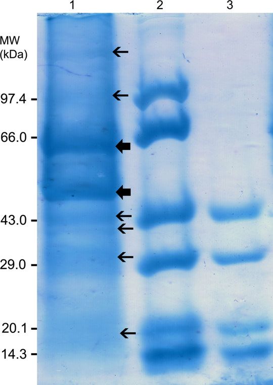
Journal of Fish Diseases 2015, 38, 389–403                          M Rosalind George et al. Koi ranavirus in India

                        Jancovich 2011). Ranaviruses can cause acute, sys-       John & George 2006) was used for isolation, multi-
                        temic disease in fish with increasing severity result-   plication and infectivity assays of this virus. Cell
                        ing from necrosis of kidney and spleen and               lines developed in the laboratory from seabass, Lates
                        haemorrhages on the skin and internal organs             calcarifer Bloch, 1790, caudal peduncle (SBCP2),
                        (Chinchar 2002; Williams, Barbosa-Solomieu &             seabass kidney (SBKD; John & George 2006) and
                        Chinchar 2005). Viruses of the genera Ranavirus          different tissues from clownfish, Amphiprion sebae
                        are of growing concern to aquaculture owing to           Bleeker, 1853, such as fin (CFFN), brain (CFBR),
                        their ability to cause large-scale mortality in a        spleen (CFSP2; John & George 2011), bluegill,
                        wide variety of host species (Ahne et al. 1997;          Lepomis macrochirus Rafinesque, 1819, fry (BF2;
                        Mao, Hedrick & Chinchar 1997; Qin et al. 2003;           Provided by Dr. Milind Patole, National Centre
                        Williams et al. 2005).                                   for Cell Sciences, Pune), Epithelioma papulosum
                           Within the genus Ranavirus, there are viruses that    cyprini (EPC; Provided by Dr. Espen Rimstad,
                        appear to be different in several respects from the      Norwegian School of Veterinary Science, Oslo) and
                        type species FV3. Two tropical ranavirus isolates,       Brown bullhead, Ictalurus nebulosis (Lesueur, 1819)
                        guppy virus 6 (GV6) and doctor fish virus (DFV),         cells (BB; Provided by Dr. Somkiat Kanchanakhan,
                        were found to be different from European and Aus-        AAHRI, Bangkok) were also used for testing the
                        tralian ranavirus isolates based on the nucleotide       susceptibility of the viral agent. Cell lines were
                        sequences of major capsid protein (MCP), DNA             grown and maintained in Lebovitz (L-15) medium
                        polymerase and neurofilament triplet H1-like             (Gibco Invitrogen) supplemented with 10% foetal
                        (NF-H1) protein gene (Holopainen et al. 2009).           bovine serum (FBS; Gibco Invitrogen) and 1%
                        However, these two viruses are found to be very          antibiotic–antimycotic mix (Gibco Life Technolo-
                        similar but not identical with the North American        gies) in 25 or 75 cm2 tissue culture flasks (Greiner
                        Santee–Cooper ranavirus isolated from largemouth         Bio-One) at 28 °C.
                        bass (Mao et al. 1999). Some authors have sug-
                        gested that the Santee–Cooper ranavirus and related
                                                                                 Fish
                        viruses such as doctor fish virus and guppy virus
                        may not belong to the genus (Hyatt et al. 2000;          Mass mortalities were observed in ornamental fish
                        Whittington, Becker & Dennis 2010).                      farms of South India where the koi were bred and
                           Ranaviruses infect multiple coldblooded verte-        reared. The koi after breeding in cement cisterns
                        brates and have been found undergone several             were transferred to open freshwater earthen ponds
                        host shifts suggesting the possibility of these          for nursery rearing. The ponds received tube well
                        viruses crossing the poikilothermic species barriers     water with water hardness ranging between 400
                        leading eventually to potentially devastating dis-       and 600 ppm as CaCO3. The juvenile koi stock
                        eases in new hosts (Jancovich et al. 2010). Some         was later transferred to a set of cement cisterns
                        strains of iridoviruses such as EHNV have also           when the mortalities were observed following clin-
                        been isolated from fishes not showing clinical dis-      ical signs such as skin darkening, loss of scales,
                        ease indicating their likely role as the carriers of     vertical hanging, uncoordinated swimming, turn-
                        the virus. Experimental inoculation resulting in         ing upside down, lateral rotation, intermittent sur-
                        sero-conversion but without clinical signs has been      facing, settling at the bottom laterally and death.
                        reported from EHNV in Australian frogs or the            The fish, which showed clinical signs, always suc-
                        cane toad Bufo marinus (Zupanovic et al. 1998).          cumbed to death despite antibiotic treatment by
                        In the present study, we have investigated infected      the farmers. In many cisterns, the mortality often
                        juvenile koi suffering from mass mortality in an         reached 100%. A sample of 25 juvenile koi of
                        ornamental fish farm with apparently no effect up        about 11.7 g average weight from one of the
                        on treatment with antibiotics.                           cement cisterns experiencing such large-scale mor-
                                                                                 talities in an ornamental fish farm was brought to
                                                                                 the laboratory in live condition for investigation.
                        Materials and methods                                    The fish were maintained in freshwater aquarium
                                                                                 of 100 L capacity with 60 L water where they
                        Cell cultures
                                                                                 exhibited clinical signs such as uncoordinated
                        Snakehead kidney cell line (SNKD2a) derived from         swimming, rolling over, intermittent surfacing and
                        striped snakehead Channa striata (Bloch, 1793;           skin darkening and continued to suffer from

Ó 2014
John Wiley & Sons Ltd   390
Journal of Fish Diseases 2015, 38, 389–403 M Rosalind George et al. Koi ranavirus in India
mortality. The fish were sampled for virus isola- having tenfold serially diluted virus suspension in
tion, detection and used for virus transmission quadruplicate. The plate was incubated at 28 °C,
studies. and development of CPE was observed for a period
of 10 days. TCID 50 mL1 was calculated using
Spearman and Karber formula (Karber 1931). Cell
Virus isolation and propagation
line susceptibility studies were carried out using cell
Pooled tissue extracts from kidney and spleen of the lines such as BB, BF2 and EPC and other indige-
infected koicarp were aseptically prepared in a nous cell lines developed in the laboratory such as
tissue homogenizer at 1/10 dilution in L15 medium SBKD, SBCP2a, CFFN, CFBR and CFSP2. All
containing 2% FBS and 19 antibiotic–antimycotic the cell lines were inoculated with the virus by
mix. The homogenate was centrifuged at 3000 g simultaneous inoculation method. The cell lines
for 10 min at 10 °C, and clarified supernatant was were subcultured for simultaneous inoculation at a
filtered through 0.22-lm syringe membrane filter rate already standardized for formation of mono-
(Millipore). The filtered homogenate (0.7 mL) was layer at the end of 24-h incubation for each cell
added to freshly prepared snakehead kidney cells line. The cell lines after subculturing were simulta-
(SNKD2a) in a 25-cm2-flask. The inoculated cells neously inoculated with 0.7 mL of the virus prepa-
along with control were incubated at 28 °C and ration of 108.5 TCID 50 mL1 in 25-cm2 flasks
observed daily for the development of CPE. Once and incubated at 28 °C along with control. The
the CPE was complete, the supernatant was flasks were observed daily for the onset and devel-
collected, clarified and filtered through 0.22-lm opment of CPE.
syringe membrane filter, and 0.7 mL was inocu-
lated to new SNKD2a cells in a 25-cm2 flask for
Transmission electron microscopy
confirmation of viral agent, and CPE was observed.
The cell culture supernatant with full blown CPE The virus was grown on SNKD2a cells (24–
at the end of first passage of virus was clarified at 48 h), fixed in 3% glutaraldehyde, washed in
3000 g for 15 min and stored in aliquots at 0.1 M cacodylate buffer, and the cell pellets were
50 °C for further use. The isolated viral agent held overnight at 4 °C. Following post-fixing in
was investigated for the presence of an envelope by 1% osmium tetroxide and washing in buffer, the
treating the virus suspension with chloroform and cell pellets were processed for electron microscopy
checking for the retention of infectivity (Feldman (John et al. 2001). Ultrathin sections (80 nm) of
& Wang 1961). The virus was also tested for its cell pellets were cut using an Ultracut microtome
ability to withstand heat treatment at 56 °C for 2 h (Leica ultracut UCT) and stained with uranyl ace-
and pH treatment at pH 3 and 9 for 30 min. tate and Reynold’s solution. The sections were
Acidic (pH 3.0) and alkaline (pH 9.0) solutions examined and photographed using a Philips 201C
were prepared by adding 0.1 N HCl/NaOH to the transmission electron microscope (the Nether-
cell culture medium without FBS. After adding lands) at 80 kV.
1 mL virus to 9.0 mL acidic or alkaline medium at
the respective pH for ½ h, an aliquot 100 lL each
Virus purification and DNA extraction
was used for titration by serial dilution to find out
the tissue culture infective dose (TCID 50 mL1) The virus was concentrated by ultracentrifugation
along with control. following propagation of the virus in EPC cells
grown in 175-cm2 flasks (Greiner Bio-one). When
CPE was extensive, culture fluid was harvested
Virus titration and cell line susceptibility
and clarified by centrifugation at 2000 g for
studies
15 min to remove the cellular debris. Supernatant
To find out the TCID 50 mL1 of the virus prep- and pelleted cells were then processed separately.
aration, the virus suspension was titrated in Pelleted cells were resuspended in 2-mL TNE buf-
SNKD2a cells in a 96-well-microtitre plate. fer and subjected 3 times to freeze-thawing in
Actively growing cells were trypsinized, and the cell liquid nitrogen. After centrifugation at 2000 g for
suspension diluted using L-15 medium supple- 10 min, the supernatant was pooled with the cell
mented with 10% FBS and antibiotics. The cells culture clarified supernatant. Approximately
were added in simultaneous mode to each well 100 mL of collected supernatant was pelleted in a
Ó 2014
John Wiley & Sons Ltd 391Journal of Fish Diseases 2015, 38, 389–403 M Rosalind George et al. Koi ranavirus in India
Beckman L 80 ultracentrifuge (Beckman) at (Eppendorf). PCRs were conducted in 50-lL
100 000 g for 90 min in an SW-41 Ti rotor reaction mixture with Smart Prime Master mix of
(Beckman) over a 50% sucrose cushion in TNE Ampliqon, Denmark. The amplification condi-
buffer (0.01 M Tris–HCl, 0.1 M-NaCl, 0.001 M- tions were as follows for the first set of primers
EDTA, pH 7.5). The virus pellets were pooled (M151 and M152): 94 °C/4 min, one cycle;
and resuspended in 1-mL TNE buffer. DNA from 94 °C/30 s, 60 °C/30 s, 72 °C/40 s, 35 cycles
purified virus preparation was extracted using with a final extension of 72 °C/5 min. Slight
DNA Extraction Solution (Merck Millipore) as change in cycling conditions was used for the sec-
per the manufacturer’s instructions. DNA quality ond set of primers (M153 and M154) as follows:
was assessed by electrophoresis using 0.4% agarose 94 °C/4 min, one cycle; 94 °C/30 s, 55 °C/30 s,
gel and ethidium bromide staining. 72 °C/60 s, 35 cycles with a final extension of
72 °C/5 min. A third set of amplification was car-
ried out with M151 and M154 primers with
Analysis of structural proteins
cycling conditions of 94 °C/4 min, one cycle;
Structural proteins of the purified virus preparation 94 °C/30 s, 58 °C/30 s, 72 °C/60 s, 35 cycles
were analysed by SDS–polyacrylamide gel electro- with a final extension of 72 °C/5 min. The
phoresis (PAGE) using a Genei Mini Gel System amplification products along with molecular
(GeNei, Merck). The proteins of the virus were markers were visualized in 1.2% agarose gels (Ge-
resolved by 12% discontinuous polyacrylamide– Nei, Merck Millipore), stained by ethidium bro-
SDS slab gels (Laemmli 1970) by electrophoresis at mide and recorded in the gel documentation
90 V for 45–55 min along with medium and low system. DNA from uninfected cell cultures and
range molecular weight markers (GeNei, Merck), fish were used as negative controls.
and the gels were stained in 0.1% Coomassie bril-
liant blue. Molecular weights of the virion proteins
Sequencing of the major capsid protein gene of
were determined by UVIDOC software in a gel
viral agent
documentation system (UVI Tec).
The PCR products obtained by two primer sets
directed for the EHNV major capsid protein
PCR detection of viral agent
(MCP) gene were purified by gel extraction kits
The dead and moribund fish from the pathogenic- (Qiagen) and sequenced by outsourcing. The
ity study, infected cell culture (EPC) pellet and sequences obtained were analysed using CLC
purified virus preparation were used for detection Main Workbench (CLC Bio) for multiple align-
of ranavirus DNA by PCR. DNA from kidney of ments and phylogenetic analysis with MCP gene
the fish was extracted using DNA Extraction Solu- of largemouth bass virus, LMBV (FR682503),
tion (GeNei, Merck Millipore) as per the manu- guppy virus, GV6 (FR677325), doctor fish virus,
facturer’s instructions. The DNA at the end of DFV (FR677324), epizootic haematopoietic
extraction was dissolved in sterile deionized water necrosis virus, EHNV (FJ433873) and frog virus
(Biocel Millipore) and subjected to amplification 3, FV3 (AY548484).
using several primer sets including the primers
(Table 1) and slightly modified protocol of Marsh
Virus transmission and pathogenicity
et al. (2002) for the detection of EHNV described
in the Manual of Diagnostic Tests for Aquatic Virus transmission and pathogenicity study were
Animals (OIE 2012) in a Master Cycler Gradient conducted using healthy juvenile koi (average
Table 1 Primers used for the successful amplification of Ranavirus DNA from the infected tissues and cell culture supernatants
Primer Sequence Target gene Product size, bp
M151 (EHNMCP1FW) AACCCGGCTTTCGGGCAGCA EHNV Major capsid protein gene 321
M152 (EHNMCP1RE) CGGGGCGGGGTTGATGAGAT
M153 (EHNMCP2FW) ATGACCGTCGCCCTCATCAC EHNV Major capsid protein gene 625
M154 (EHNMCP2RE) CCATCGAGCCGTTCATGATG
M151 (EHNMCP1FW) AACCCGGCTTTCGGGCAGCA EHNV Major capsid protein gene 1201
M154 (EHNMCP2RE) CCATCGAGCCGTTCATGATG
Ó 2014
John Wiley & Sons Ltd 392Journal of Fish Diseases 2015, 38, 389–403 M Rosalind George et al. Koi ranavirus in India
weight – 9.3 g) obtained from a local fish farm. laboratory also suffered from progressive mortality,
The fish were acclimatized to laboratory condi- and the entire sample of 25 fishes except 3 were
tions for 7 days in well-aerated glass aquarium dead in 14 days. The fishes had clinical signs such
tanks of 100-L capacity and fed with commercial as erratic and uncoordinated swimming, loss of
pelleted feed twice daily. Following acclimatiza- balance, vertical hanging, turning upside down,
tion, four fish each was assigned to five groups lateral rotation, intermittent surfacing, settling at
for four different treatments. Three groups the bottom and death. Externally, fishes had skin
received intraperitoneal injection with 50-lL tis- darkening, loss of scales, discolouration, swollen
sue extracts prepared from brain, gill and pooled and pale gills and emaciation.
samples of spleen and kidney from infected koi,
respectively. Tissue extracts were prepared by
Isolation of virus in cell culture and
homogenizing pooled individual tissues from
susceptibility of cell lines to the virus
infected fish, and 0.5 g tissues each was mixed
with 4.5 mL of L15 medium. Tissue extracts Virus isolation studies conducted with pooled tis-
were clarified by low-speed centrifugation and sues of kidney and spleen from infected koi in
membrane filtration using 0.22-lm syringe mem- SNKD2a cells showed that the viral agent was
brane filter. The fourth group was maintained in capable of growing in snakehead kidney cell line
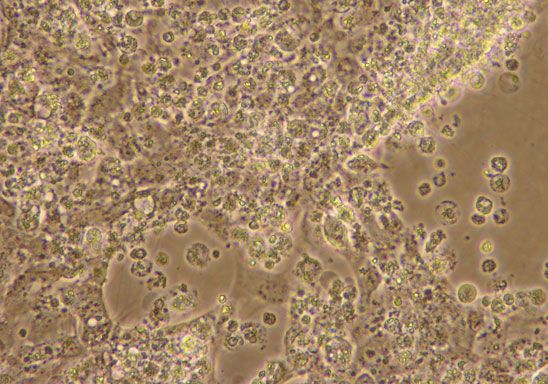
aquarium tank with 40-L freshwater mixed with at 28 °C. The CPE was characterized by focal
1/10th water from the infected fish tank. A destruction of cells discernible on day 1 and fur-
group of four fishes intraperitoneally injected ther progression over 3-day duration till the com-
with 50 lL L15 medium served as control. The plete destruction of the monolayer (Fig. 1). CPE
fishes were maintained for 35 days for observa- started with several small foci of rounded cells
tion in well-aerated 100-L glass tanks with 40 L appearing in the cell monolayer followed by
water and fed ad libitum with commercial pel- aggregation of the round cells at the periphery of
leted feed. Uneaten food and faecal matter were the foci, which became more and more enlarged
removed daily and disinfected before discharge all in size. The rounded cells got detached from the
through the duration of the experiment. Recovery monolayer, underwent lysis and CPE-progressed
of the virus was also attempted from dead and till the monolayer is completely destroyed. The
live fish by inoculating tissue homogenates of virus on first passage through the same cell line
pooled samples (n = 3) of kidney and spleen on was found to induce complete CPE in 2 day
to SNKD2a cells. post-infection (dpi). Cell line susceptibility studies
A similar study was conducted in koi juveniles demonstrated that the virus could grow on a vari-
using cell culture grown virus by intraperitoneal ety of cell lines tested viz. CFFN, CFBR, CFSP2,
injection. Koi juveniles had an average weight of SBKD, SBCP2, BF2, EPC and BB cells
7.4 g, and five fish each were maintained in dupli- (Table 2). KIRV however grew very slowly on BB
cate in 100-L glass tanks as above. Virus grown in cells. Quantitation of virus by titration experi-
EPC cells was diluted using cell culture medium, ments in SNKD2a cell line revealed that the stock
and 50 lL virus preparation containing 106 virus had a titre of 108.5 +/ 0.94 TCID 50 mL1.
TCID50 was injected to each fish intraperitone- Chloroform treatment of the virus suspension
ally. Two sets of five fish each were used as con- resulted in the loss of infectivity of the virus indi-
trol and were injected with clarified cell culture cating the likely presence of an envelope. While
supernatant without virus. Fish were maintained treatment at pH 9 did not reduce the infectivity
for a period of 4 weeks for clinical signs and of the virus, treatment of the virus at pH 3
mortality. reduced the virus infectivity by 3.5 logs and heat
treatment at 56 °C by 2.33 logs.
Results
Electron microscopy of infected cell cultures
Clinical pathology
Transmission electron microscopic analysis of the
The koi in the fish farm were experiencing contin- infected SNKD2a cell cultures revealed the pres-
uous mortalities leading to large-scale loss of ence of several icosahedral virus particles of 100–
fishes. The live fish samples brought to the 120 nm size (n = 14) scattered in the cytoplasm
Ó 2014
John Wiley & Sons Ltd 393Journal of Fish Diseases 2015, 38, 389–403 M Rosalind George et al. Koi ranavirus in India
(a) Table 2 Details of cell lines investigated for the susceptibility
to KIRV indicating onset of CPE and the complete destruction
of the monolayer
Cell line Onset of CPE (days) Completion of CPE (days)
SNKD2a 1 3
CFFN 1 3
CFBR 2 3
CFSP2 1 3
SBKD 3 5
SBCP2 2 3
BF2 2 3
EPC 1 3
BB 3 10a
a
Incomplete CPE.
(b)
ranged from 18 to 151 kDa. Two major proteins
of the virus had molecular weight of 50 and
63 kDa.
PCR detection of the Ranavirus
PCR analysis of the infected fish tissues showed
that the viral DNA is present in the infected tis-
sues such as spleen and kidney of the koi. While
the first primer set targeting the MCP amplified a
(c) characteristic product of 321 bp, second set of
primers did not amplify the expected 625-bp
amplicon. However, when the forward primer of
the set I and reverse primer of the set II were
used, an amplicon of about 1200 bp was obtained
indicating the expected target sequence amplifica-
tion. Similar results were also obtained when puri-
fied virus preparation was subjected to PCR
amplification (Fig. 5a–c). No amplicons were
obtained from uninfected healthy koi tissues and
control cell cultures used in PCR.
Figure 1 Cytopathic effect caused by KIRV in SNKD2a cell Sequence analysis of major capsid protein gene
line (a) Control uninfected cells (2009) and (b and c) infected
cells showing the induced CPE (200 and 1009, respectively). Sequences of the two gel purified amplified prod-
ucts from the PCR were multiple-aligned with
MCP genes of LMBV, GV6, DFV, FV-3 and
of the cells (Fig. 2). Large numbers of virus parti- EHNV. The analysis showed 99.91% similarity
cles were found in the periphery of the cytoplasm with LMBV with single change of one nucleotide
of the infected cells. Budding of the virus particles at position 291 of the sequence generated, which
could also be noticed from the surface of the resulted in the change of one amino acid G
membranes (Fig. 3). (Glycine) to D (Aspartic acid; Fig. 6). The rana-
virus isolated in this study showed 78.46% iden-
tity with MCP gene of EHNV and 78.38% with
Analysis of structural proteins
FV3, the type species of Ranavirus. Although,
Structural proteins of the virus resolved into two KIRV carried several nucleotide changes with
major and six minor proteins in the SDS–PAGE EHNV and FV3 in the sequenced fragment, it
analysis (Fig. 4). Molecular mass of the 8 proteins had same nucleotide at position 291 (relative to
Ó 2014
John Wiley & Sons Ltd 394Journal of Fish Diseases 2015, 38, 389–403 M Rosalind George et al. Koi ranavirus in India
Figure 2 Transmission electron micrograph of KIRV grown
in SNKD2a cells showing icosahedral particle of 100–120 nm
size. Virus particles at the end of virus morphogenesis
(bar = 200 nm).
Figure 4 SDS–PAGE analysis of structural proteins of KIRV
in 12% acrylamide gel stained with 0.1% Coomassie brilliant
blue. Lane 1: KIRV. Two major structural proteins of 60 and
53 kDa (thick arrow) and 6 minor proteins (thin arrow) are
indicated; Lane 2: Medium range molecular mass markers
(GeNei); lane 3: Low range molecular mass markers (GeNei).
uncoordinated swimming, rolling over and vertical
hanging before death. Half of the fish in the tank
having 1/10th water from the infected tank also
died during the experiment. However, the fishes,
which received brain and gill extracts by intraperi-
Figure 3 Transmission electron micrographs of KIRV particles toneal injection, did not show any mortality
distributed in the infected cell and virions seen budding from
(Fig. 8). Fish also had no clinical signs in these
the cell membrane (bar = 200 nm).
two tanks. No mortality was observed in the con-
trol fishes, which received only cell culture media
by intraperitoneal injection. The virus was recov-
start in Fig. 6) and the amino acid remained as ered from the spleen and kidney of the dead fishes
glycine similar to that of GV6, DFV, EHNV from all the treatment tanks using SNKD2a cell
and FV3 unlike that of LMBV. A phylogenetic line but not from live fish samples collected from
tree constructed with the six sequences by neigh- control tank. No mortality was observed in second
bour-joining method is in Fig. 7. set of fishes injected with virus grown in EPC cell
line.
Pathogenicity of KIRV to koi juveniles
Discussion
All the fish injected with pooled extracts of kidney
and spleen were dead by the end of the experi- Koi mortalities were found to be causing havoc in
ment. These fish had similar clinical signs such as an ornamental fish farm of Southeast coast of
Ó 2014
John Wiley & Sons Ltd 395Journal of Fish Diseases 2015, 38, 389–403 M Rosalind George et al. Koi ranavirus in India
(a) (b) (c)
Figure 5 Agarose gel electrophoretic analysis of the PCR products obtained from KIRV infected cell culture pellet and purified
virus preparation with primers targeting major capsid protein gene of EHNV (a) with M151 & M152 primers. Lane 1: KIRV
infected EPC cell culture pellet, Lane 2: uninfected EPC cell culture pellet, lane 3: Purified preparation of KIRV, Lane 4: PCR neg-
ative control, lane 5: 1 kb molecular marker (GeneRuler, Fermentas, Thermo Scientific). (b) with M151 & M154 primers. Lane 1:
KIRV infected EPC cell culture pellet, Lane 2: uninfected EPC cell culture pellet, lane 3: Purified preparation of KIRV, Lane 4:
PCR negative control, lane 5: 1 kb molecular marker. (c) PCR amplification of infected koi kidney using M151 and M152 primers.
Lane 1: 1 kb Molecular marker, Lane 2: Infected koi kidney.
India. To ascertain the aetiology of the large-scale lines. Many of the ranaviruses have wide host
mortality in the koi farm, we investigated the inci- range with EHNV infecting as many as 13 spe-
dence through virological analysis of infected cies of fishes (Whittington et al. 2010). The sus-
fishes. The fish samples brought to the laboratory ceptibility of a panel of freshwater and marine
were a collection of healthy and moribund ani- fish cell lines to the KIRV obtained in this
mals, which showed varying clinical signs such as study indicates the potential of the virus to
loss of scales, uncoordinated swimming, vertical multiply in different fish species leading to clini-
hanging, lateral rotation turning upside down and cal or subclinical infection. Further investigation
intermittent surfacing before settling at the bottom may be required to prove the susceptibility of
and death. All but three fishes died within 14 days these fish species by in vivo pathogenicity stud-
of the arrival of the fish to the laboratory. Similar ies. The virus in SNKD2a cells had grown to a
clinical manifestations such as erratic swimming or titre of 108.5 +/ 0.9428 TCID 50 mL1 similar
hyperbuoyancy associated with swim bladder over- to the titres obtained for Australian EHNV iso-
inflation were found associated with infections of lates grown in BF2 cells (Gould et al. 1995;
wild largemouth bass with Santee–Cooper ranavi- Ariel et al. 2009) and largemouth bass virus in
rus (Grizzle & Brunner 2003). Natural wild infec- EPC cells (Deng et al. 2011). Like ranaviruses
tions with variable clinical manifestations ranging in general, the KIRV also lost infectivity by low
from inapparent infection to sporadic epizootics of pH (3.0) and heat treatment at 56 °C (Janco-
mortality also reported in largemouth bass (Gold- vich et al. 2012).
berg et al. 2003). The virus grown in cell culture examined under
The virus isolate resembling Santee–Cooper the transmission electron microscope had icosahe-
ranavirus obtained in the present investigation dral particles of 100–120 nm size. The virions of
was found to grow on a variety of cell lines vertebrate iridoviruses are reported to range in size
developed in the laboratory from marine (clown from 100 to 300 nm (Hyatt & Chinchar 2008).
fish), brackish water (seabass) and freshwater The virus particles were seen at the periphery of
(snakehead) fishes and also on EPC, BF2 and the cell membrane and were also found budding
BB cell lines. In BB cell line however, KIRV out of the cell membrane. Virions of ranaviruses
did not cause full scale CPE unlike other cell are reported to accumulate in the cytoplasm
Ó 2014
John Wiley & Sons Ltd 396Journal of Fish Diseases 2015, 38, 389–403 M Rosalind George et al. Koi ranavirus in India
C AGG T CGGGC GA C T A T G T GC T C A A C T C T T G GC T GG T C C T C A AGA C C C C T C AGA T T A A A C T GC T GGCGGC C A A C C AG T T T A
. . . . . . . . . . . . . . . . . . . . . . . . . . . . . . . . . . . . . . . . . . . . . . . . . . . . . . . . . . . . . . . . . . . . . . . . . . . . . . . .
. . . . . . . . . . . . . . . . . . . . . . . . . . . . . . . . . . . . . . . . . . . . . . . . . . . . . . . . . . . . . . . . . . . . . . . . . . . . . . . .
. . . . . . . . . . . . . . . . . . . . . . . . . . . . . . . . . . . . . . . . . . . . . . . . . . . . . . . . . . . . . . . . . . . . . . . . . . . . . . . .
. . . . . . . . .g . . t . . ca . c . . . . . .g. c . . . t . . . .g. . . . . . . . . . . cg . .g. c . .g. . c . . . . . c . . a . . . . . . c . gg
. . . . . . . . .g . . t . . ca . c . . . . . .g. c . . . t . . . .g. . . . . . . . . . . cg . . g . cg . g . . c . . . . . t . . a . . . . . . c . gg
A CGC A A A CGG T A C C A T C AGA T GGA C C A A A A A T C T C A T GC A C A A CG T T G T G GAGC A CGC CG C A C T C T CG T T C A A CGAGA T T
. . . . . . . . . . . . . . . . . . . . . . . . . . . . . . . . . . . . . . . . . . . . . . . . . . . . . . . . . . . . . . . . . . . . . . . . . . . . . . . .
. . aa tg . . . . . . . . . . . . . . . . . . . . . . . . . . . . . . . . . . . . . . . . . . . . . . . . . . . . . . . . . . . . . . . . . . . . . . . . . .
. . aa tg . . . . . . . . . . . . . . . . . . . . . . . . . . . . . . . . . . . . . . . . . . . . . . . . . . . . . . . . . . . . . . . . . . . . . . . . . .
ga . ac . . . . . c . . a . . . . .g . . . . . a . .g. . c . c . . . . . . . . . . a . . . . . . . . a . . . t . a ac . . . . . a . . . . . . . . c . . c
ga . ac . . t . . c . . . . . . . .g . . . . . a . .g. . c . c . . . . . . . . . . a . . . . . . . . ag . . t . a . c . . . . . a . . . . . . . . c . . c
C AGGC C C AGC AG T T T A A C A C T GC T T T C C T G GA T GC C T GGA A CGAG T A C A C C A T GC C CGAG GC C A AGCGC A T CGGG T A T T A
. . . . . . . . . . . . . . . . . . . . . . . . . . . . . . . . . . . . . . . . . . . . . . . . . . . . . . . . . . . . . . . . . . . . . . . . . . . . . . . .
. . . . . . . . . . . . . . . . . t . . . . . . . . . . . . . . c . . . . . . . . . . . . . . . . . . . . . . . . . . . . . . . . . . . . . . . . . . . . c . .
. . . . . . . . . . . . . . . . . t . . . . . . . . . . . . . . c . . . . . . . . . . . . . . . . . . . . . . . . . . . . . . . . . . . . . . . . . . . . c . .
agc . . . . . . t cc . . . . . . . . g. . a . a . . . . . . c . . . . . . . g. . . . . . . . . . . . . . . a . . . . . . . . . . . . . . a . . c . . c . .
agc . . . . . . t cc . . . . . . . . g. . a . a . . . . . . c . . . . . . . g. . . . . . . . . . . . . . . a . . . . . . . . . . . . . ca . . c . . c . .
C A A C A T GA T T GGC A A C A C T A GCGA T C T CG T C A A T C C CGC C C C CGC C A C CG G T C A AGC AGG AGC T AGGG T C C T GC C CGC C A
. . . . . . . . . . . . . . . . . . . . . . . . . . . . . . . . . . . . . . . . . . . . . . . . . . a . . . . . . . . . . . . . . . . . . . . . . . . . . . . .
. . . . . . . . . . . . . . . . . . . . . . . . . . . . . . . . . . . . . . . . . . . . . . . . . . . a . . . . . . . . . . . . . . . . . . . . . . . . . . . .
. . . . . . . . . . . . . . . . . . . . . . . . . . . . . . . . . . . . . . . . . . . . . . . . . . . a . . . . . . . . . . . . . . . . . . . . . . . . . . . .
t . . . . . . . . a . . . . . . . . c . . . . . . . . . a . . . . c . . . . . . . .g. . . . . a . . c . . gaac . . . . . c . . . . . . . . c . .g. . . .
t . . . . . . . . a . . . . . . . . c . . . . . . . . . a . . . . c . . . . . . . .g. . . . . a . . c . . g . ac . . . . . c . . . . . . . . c . .g. . . .
AAAACC T TGT CC T T CC T C T C CCC T T C T T T T T CGGC AGAGA C AGCGGC C T G GC C C T GC C T A C AG T C A C C C T GC C T T A C A A C
. . . . . . . . . . . . . . . . . . . . . . . . . . . . . . . . . . . . . . . . . . . . . . . . . . . . . . . . . . . . . . . . . . . . . . . . . . . . . . . .
. . . . . . . . . . . . . . . . . . . . . . . . . . . . . . . . . . . . . . . . . . . . . . . . . . . . . . . . . . . . . . . . . . . . . . . . . . . . . . . .
. . . . . . . . . . . . . . . . . . . . . . . . . . . . . . . . . . . . . . . . . . . . . . . . . . . . . . . . . . . . . . . . . . . . . . . . . . . . . . . .
.g. . . . .g. . t . . . . . c . . . . . a . . . . . c . . . tc . . . . . . . . . . . . . . . . . . . . . . . . ag tc . . . t . . . . c . . c . . . . . .
.g. . . . .g. . t . . . . . c . . . . . a . . . . . c . . . tc . . . . . . . . . . . . . . . . . . . . . . . . ag tc . . . t . . . . c . . c . . . . . .
Figure 6 Partial MCP sequence alignment of KIRV, LMBV (GenBank Accession No FR682503, nt 403–1525), GV6
(FR677325), DFV (FR677324), EHNV (FJ433873, nt 21008–19886) and FV3 (AY548484, nt 97557–98679) were compared.
Amino acid translation with reference to full coding sequence of the MCP gene is indicated. Identical nucleotides are represented by
dots.
within large paracrystalline arrays and are released envelope for KIRV was also confirmed by the loss
by budding from the plasma membrane acquiring of the viral infectivity up on treatment with the
an envelope (Chinchar 2002). The presence of the organic solvent.
Ó 2014
John Wiley & Sons Ltd 397Journal of Fish Diseases 2015, 38, 389–403 M Rosalind George et al. Koi ranavirus in India
GA A A T T AGA A T C A C C A T C AG C C T GAGA T C C A T T C AGGA T C T C C T GA T T C T T C AGC A C A AG A CGA C CGGAG A AG T C A AGC C
. . . . . . . . . . . . . . . . . . . . . . . . . . . . . . . . . . . . . . . . . . . . . . . . . . . . . . . . . . . . . . . . . . . . . . . . . . . . . . . .
. . . . . . . . . . . . . . . . . . . . . . . . . . . . . . . . . . . . . . . . . . . . . . . . . . . . . . . . . . . . . . . . . . . . . . . . . . . . . . . .
. . . . . . . . . . . . . . . . . . . . . . . . . . . . . . . . . . . . . . . . . . . . . . . . . . . . . . . . . . . . . . . . . . . . . . . . . . . . . . . .
. .g. . c . .g. . a . . ag . . . a g . . . . . gg . . . . c . . . . . c . . . . . . . . c . . c . . . . . . . . c . . c . . a . .g. c . a . . . gc . .
. .g. . c . .g. . a . . ag . . . a g . . . . . gg . . . . c . . c . . c . . . . . . . . c . . c . . . . . . . . c . . c . . a . .g. c . a . . . gc . .
A A T CG T GGC C A C AGA T C T GG A AGGAGG T C T C C C AGA C A CG G T AGAGGC T C A CG T C T A C A T GA C T G T GGG T C T GG T GA C T G
. . . . . . . . . . . . . . . . . . . . . . . . . . . . . . . . . . . . . . . . . . . . . . . . . . . . . . . . . . . . . . . . . . . . . . . . . . . . . . . .
. . . . . . . . . . . . . . . . . . . . . . . . . . . . . . . . . . . . . . . . . . . . . . . . . . . . . . . . . . . . . . . . . . . . . . . . . . . . . . . .
. . . . . . . . . . . . . . . . . . . . . . . . . . . . . . . . . . . . . . . . . . . . . . . . . . . . . . . . . . . . . . . . . . . . . . . . . . . . . . . .
c . . . . . . . . . g. c . . c . . c . .g. . . . . . . . . . . c . . . . . c . . c . . . . . ca . . . . . . . . . . . . . c . . c . cc . . ca . c . . c .
c . . . . . . . . . t . c . . c . . t . cg . . . . . . . . . . . c . . . . . c . . c . . . . . ca . . . . . . . . . . . . . c . . c . cc . . ca . c . . c .
C CGC CGAGCG T C AGGC T A T G AGC AGC T C AG T C AGGGA C A T GG T GG T GGAG C AGA T GC AGA T GGC T C C AG T C C A C A T GG T C
. . . . . . . . . . . . . . . . . . . . . . . . . . . . . . . . . . . . . . . . . . . . . . . . . . . . . . . . . . . . . . . . . . . . . . . . . . . . . . . .
. . . . . . . . . . . . . . . . . . . . . . . . . . . . . . . . . . . . . . . . . . . . . . . . . . . . . . . . . . . . . . . . . . . . . . . . . . . . . . . .
. . . . . . . . . . . . . . . . . . . . . . . . . . . . . . . . . . . . . . . . . . . . . . . . . . . . . . . . . . . . . . . . . . . . . . . . . . . . . . . .
gg . a . . . . a . g. . . . . c . . . . . . . . . a . . . . . . . . . . . . . . . . c . . . . . . . . .g. . . . .g cc . . c . . . . . . . . . . . . . . .
gg . a . . . . a . a . . . . . c . . . . . . . . . a . . . . . . . . . . . . . . . . t . . . . . . . . .g. . . . .g cc . . c . . . . . . . . . . . . . . .
A A C C C C A AGA A CGC C A C CG T C T T T C A CGC A GA C C T GCGC T T T T C C C A CGC CG T C A A AGCG C T C A T G T T T A T GG T GC A A A A
. . . . . . . . . . . . . . . . . . . . . . . . . . . . . . . . . . . . . . . . . . . . . . . . . . . . . . . . . . . . . . . . . . . . . . . . . . . . . . . .
. . . . . . . . . . . . . . . . . . . . . . . . . . . . . c . . . . . . . . . . . . . . . . . . . . . . . . . . . . . . . . . . . . . . . . . . . . . . . . . .
. . . . . . . . . . . . . . . . . . . . . . . . . . . . . c . . . . . . . . . . . . . . . . . . . . . . . . . . . . . . . . . . . . . . . . . . . . . . . . . .
. . . . . . .g. . . . . . gg . . ac . . . c . . . a . c . . . a . . . .g. . c . . a . . . . . a . . . . .g. . c . .g. . . . . . . . . . . . . .g. .
. . . . . . .g. . . . . . g . . . ac . . . c . . . a . c . . . a . . . .g. . c . . a . . . . . a . . . . .g. . c t .g. . . . . . . . . . . . . .g. .
CG T C A C T C A C A AG T C T G T GG G T T C C A A C T A C A C T T GCG T C A C T C C T G T T G T T GGAGCGGG T A A C A C CG T C C T GGAGC C CG
. . . . . . . . . . . . . . . . . . . . . . . . . . . . . . . . . . . . . . . . . . . . . . . . . . . . . . . . . . . . . . . . . . . . . . . . . . . . . . . .
. . . . . . . . . . . . . . . . . . . . . . . . . . . . . . . . . . . . . . . . . . . . . . . . . . . . . . . . . . . . . . . . . . . . . . . . . . . . . . . .
. . . . . . . . . . . . . . . . . . . . . . . . . . . . . . . . . . . . . . . . . . . . . . . . . . . . . . . . . . . . . . . . . . . . . . . . . . . . . . . .
. . . . . . a . . . cc t . . c . . c . . c . . . . . t . . . . . c . . . . c . . . . . . c . . c . .g. . . . tc . a c . . . . .g. . . . . . . . . . . a .
. . . . . . a . . . cc t . . c . . c . . c . . . . . t . . . . . c . . . . . . . . . . . c . . c . .g. . . . tc . . c . . . . .g. . . . . . . . . . . a .
Figure 6 Continued.
The SDS–PAGE analysis of the structural pro- formed the major proteins of the virus. The major
teins of the virus indicated the presence of major capsid protein of the EHNV (Jancovich et al.
and minor viral proteins in the purified virus prepa- 2010), FV3 (Mao et al. 1996) and GIV (Murali
ration having molecular weights ranging from 18 to et al. 2002) also has similar molecular weight of
151 kDa. Two proteins of the size 50 and 63 kDa about 50 kDa. The major capsid proteins of the size
Ó 2014
John Wiley & Sons Ltd 398Journal of Fish Diseases 2015, 38, 389–403 M Rosalind George et al. Koi ranavirus in India
C C C T GGC CG T CGA T C CGG T C A AGAGCGC C A G T C T GG T G T A CGA A A A C A C T A C C AGGC T T C C AGA C A T GAG CG T AGAG T A C
. . . . . . . . . . . . . . . . . . . . . . . . . . . . . . . . . . . . . . . . . . . . . . . . . . . . . . . . . . . . . . . . . . . . . . . . . . . . . . . .
. . . . . . . . . . . . . . . . . . . . . . . . . . . . . . . . . . . . . . . . . . . . . . . . . . . . . . . . . . . . . . . . . . . . . . . . . . . . . . . .
. . . . . . . . . . . . . . . . . . . . . . . . . . . . . . . . . . . . . . . . . . . . . . . . . . . . . . . . . . . . . . . . . . . . . . . . . . . . . . . .
. . . . . . .g. . g. . . . . c . . . . . . . . . . . . . . c . . . . . . . . . . . . . . . . . c . . a . . . . . c . . c . . . c . .g. a . . c . . . . . .
. . . . t . .g. . a . . . . . c . . . . . . . . . . . . . . c . . . . . . . . . . . . . . . . . c . . a . . . . . c . . c . . . . . .g. a . . c . . . . . .
T A T T C C C T GG T GC AGC C C T G G T A C T A CGC A C C CGC C A T T C C C A T C AGC A C T GGC C A C C A C C T C T A C T C T T A CGC C C T GAG
. . . . . . . . . . . . . . . . . . . . . . . . . . . . . . . . . . . . . . . . . . . . . . . . . . . . . . . . . . . . . . . . . . . . . . . . . . . . . . . .
. . . . . . . . . . . . . . . . . . . . . . . . . . . . . . . . . . . . . . . . . . . . . . . . . . . . . . . . . . . . . . . . . . . . . . . . . . . . . . . .
. . . . . . . . . . . . . . . . . . . . . . . . . . . . . . . . . . . . . . . . . . . . . . . . . . . . . . . . . . . . . . . . . . . . . . . . . . . . . . . .
. . c . .g. . . . . . . . . . . . . . . . . . . . t . . c a . . t . . . . c . . ag . . . . . . . c . .g. . . . . . . . . . . . . . . . . t . . . . . c . .
. . c . .g. . . . . .g. . . . . . . . . . . . . t . . c a . . t . . . . c . . ag . . . . . . . c . .g. . . . . . . . . . . . . . . . . t . . . . . c . .
C C T C A A CGA T CC T CACCC T T C AGGG T C T A C C A A T T T CGG T CGC C T GA C C A A CGC A AGC A T C A A CG T G T C T C T G T C T GC CG
. . . . . . . . . . . . . . . . . . . . . . . . . . . . . . . . . . . . . . . . . . . . . . . . . . . . . . . . . . . . . . . . . . . . . . . . . . . . . . . .
. . . . . . . . . . . . . . . . . . . . . . . . . . . . . . . . . . . . . . . . . . . . . . . . . . . . . . . . . . . . . . . . . . . . . . . . . . . . . . . .
. . . . . . . . . . . . . . . . . . . . . . . . . . . . . . . . . . . . . . . . . . . . . . . . . . . . . . . . . . . . . . . . . . . . . . . . . . . . . . . .
. . . gc . g . . c . . c . . . . . a . . c . . a . . c . . . . . . . a . . . c a . a . . . . . . . . . . . c . . . c . t . . . . . ca . c . . . . . c . . t .
. . . gc . g . . c . . c . . . . . a . . c . . a . . c . . . . . . . a . . . . a . a . . . . . . . . . . . c . . . c . t . . . . . ca . c . . . . . c . . t .
AGGC CGGA A C T GC CGC CGGA GGAGGAGGGG C AGA C A A C T C T GGC T A C A A A A A C C C T C AGA A A T A CGC C C T GG T GG T C A T G
. . . . . . . . . . . . . . . . . . . . . . . . . . . . . . . . . . . . . . . . . . . . . . . . . . . . . . . . . . . . . . . . . . . . . . . . . . . . . . . .
. . . . . . . . . . . . . . . . . . . . . . . . . . . . . . . . . . . . . . . . . . . . . . . . . . . . . . . . . . . . . . . . . . . . . . . . . . . . . . . .
. . . . . . . . . . . . . . . . . . . . . . . . . . . . . . . . . . . . . . . . . . . . . . . . . . . . . . . . . . . . . . . . . . . . . . . . . . . . . . . .
. . . . . acc . . g. . . t . . . c . . . . . . c . . a . gc . . . . . . . . . . . g . . . . cc . c .g. c . . a . .g. . . . . . . . ca . c . . t c . .
. . . . . acc . . g. . . . . . . c . . . . . . t . . a . gta . . . . . . . . . . g . . . . cc . c .g. c . . a . .g. . . . . . . . ca . c . . t c . .
GC C
. . .
. . .
. . .
. . .
. . .
Figure 6 Continued.
range 48–55 kDa form about 40% of the proteins expected 321 fragment, the second set of primers
of the family Iridoviridae (Jancovich et al. 2012). did not amplify the expected 625 fragment indi-
Two sets of primers targeting the MCP of cating a sequence difference from EHNV. How-
EHNV were used to amplify the MCP gene of ever, an approximately 1200-bp fragment was
the KIRV isolate. While the first set amplified the amplified when the forward primer of the first set
Ó 2014
John Wiley & Sons Ltd 399Journal of Fish Diseases 2015, 38, 389–403 M Rosalind George et al. Koi ranavirus in India
Figure 7 Phylogenetic tree constructed using partial MCP gene sequences of six similar ranaviruses LMBV (FR682503), GV6
(FR677325), DFV (FR677324), EHNV (FJ433873) and FV3 (AY548484) by neighbour-joining method using CLC Main Work-
bench indicating the relationship of KIRV with other similar ranaviruses including the type virus of the genus, FV3.
120
Brain
Gill
Spleen & Kidney
100 Infected water
Control
CumulaƟve mortaility (%)
80
60
40
20
0
0 5 10 15 20 25 30 35 40
Days post-challenge
Figure 8 Cumulative mortality of koi juveniles experimentally challenged by intraperitoneal injection with 0.22-lm membrane fil-
tered pooled tissue extracts of infected koi along with control and koi challenged with 1/10th infected tank water.
and reverse primer of the second set were used. by blast searches of the full MCP gene. With
Although the OIE primer sequences used were other major group of ranaviruses comprising
based on EHNV MCP sequence, they were not EHNV and FV3, which shows 97.7% homogene-
present in LMBV sequence. However, the analysis ity of MCP gene between them, the ranavirus iso-
of sequences showed that the primers shared some lated in the present study showed an identity of
identity with LMBV MCP sequence FR682503. only 78.46% with EHNV and 78.38% with FV3
As the EHNV primer sets could amplify both for the fragment of 1123 bp. The present analysis,
321- and 1200-bp fragments, we proceeded however, showed an interesting change in the
through sequencing of the amplicons for identify- amino acid sequence in the LMBV and FV3,
ing the pathogen instead of further redesigning the type species of the Ranavirus group. While the
primers based on LMBV sequences. amino acid at position 168 of the FV3 and
The sequence analysis of the partial coding EHNV MCP gene is a glycine, it is an aspartic
region of the MCP gene amplified in the present acid for LMBV. However, the present ranavirus
virus spanning 1123 bp demonstrated that the isolate from India compares favourably with the
isolate is similar to the Santee–Cooper ranavirus type species FV3 in this regard by retaining the
(LMBV) with 99.91% sequence homogeneity. amino acid glycine at this position.
LMBV MCP sequence shows 99.21% identity Early epizootic ranavirus EHNV was isolated in
with DFV and GV6 MCP sequences, which 1985 from Australia (Langdon et al. 1986), later
within themselves are identical (Ohlemeyer et al. European sheatfish virus (ESV) and European cat-
2011). MCP gene sequences have been used as fish virus (ECV) from Europe (Ahne, Schlotfeldt &
main identifying character of the ranaviruses Thomsen 1989b; Pozet et al. 1992). The vertebrate
(Whittington et al. 2010), which shows over 84% ranavirus in North America was isolated in 1995
percentage similarity between the ranavirus groups from an epizootic affecting largemouth bass
Ó 2014
John Wiley & Sons Ltd 400Journal of Fish Diseases 2015, 38, 389–403 M Rosalind George et al. Koi ranavirus in India
Micropterus salmoides (Lacepe0 de) in Santee–Cooper experimental infection by causing 50% mortality in
reservoir in SC, USA (Plumb et al. 1996). Two koi. EHNV has been reported to be an indiscrimi-
virus isolates similar to Santee–Cooper virus were nate pathogen of several fish with high variability in
reported later from ornamental fishes imported susceptibility of host species. While redfin perch are
from South-East Asia to the United States, the highly susceptible, rainbow trout were resistant to
guppy virus (GV6) and doctor fish virus (DFV), bath exposure in 102.2 TCID 50 mL1 and suc-
which had the partial MCP nucleotide sequence cumbed only after intraperitoneal infection (Whit-
almost identical to that the MCP gene of Santee– tington & Reddacliff 1995).
Cooper ranavirus (Mao et al. 1999). The present The experimental fish after exhibiting clinical
isolate KIRV is more homologous to Santee–Coo- signs, died in 2–3 days. Samples of moribund and
per ranavirus at 99.91% than to GV6 and DFV, dead fish were processed for virus isolation, and the
which is showing only 99.2% homology for the results indicated the presence of a viral agent capa-
same region of 1123 bp, thus forming a separate ble of multiplying in snakehead kidney cells. The
isolate from these two Asian ranaviruses. Singapore virus caused cytopathic effect in snakehead cell cul-
grouper iridovirus (SGIV), another Asian ranavirus tures in 2 days characterized by the presence refrac-
isolate, shows only 71.2% identity with Santee– tile detached cells causing damaged areas in the cell
Cooper ranavirus isolate over the full MCP gene monolayer. The confirmatory diagnosis for the
sequence and hence has substantial difference presence of ranavirus DNA in the infected fish tis-
between the Indian ranavirus isolate obtained in sues was obtained by the PCR amplification of
the present study as well. DNA in the infected fish tissues. The recovery of
Pathogenicity study was conducted for the virus the virus from the dead fishes in the laboratory
isolate using filtered infected tissue homogenate experiment inoculated with the virus isolate indi-
administration and by cell culture grown virus with cated that the virus isolated from the dying fishes
two groups of koi. While all the fish injected with tis- was associated with the mortality of koi in the farm.
sue homogenates of spleen and kidney developed The role of koi herpesvirus (KHV) in inducing the
similar clinical signs seen in the farm-infected fishes mortality was not investigated as the virus isolate
and died at the end of the experiment, there was no obtained was a ranavirus like agent. As the cell cul-
mortality in the fish injected with brain or gill homo- ture grown ranavirus could not induce mortality,
genates indicating the importance of kidney as an the possibility of original infection carrying other
ideal organ for isolation of ranaviruses from infected viral agents such as KHV could not be ruled out.
fishes (Ariel et al. 2009). Present study also indicated We have thus isolated and characterized the
that the virus could be transmitted through contami- viral agent from infected koi experiencing large-
nated water. However, the cell culture grown virus scale mortality in the koi farms of South India.
did not induce mortality in koi injected with virus The physicochemical properties of the viral agent
intraperitoneally. Similar instances were also isolated, size of major capsid protein, sequence
reported in experimental infections of largemouth identify of the MCP gene with the Santee–Cooper
bass, where different populations have demonstrated virus (LMBV) coupled with the clinical pathology
profound variability in susceptibility to Santee–Coo- of infection prove that virus belongs to the genus
per ranavirus (Goldberg et al. 2005). Experimental Ranavirus of the family Iridoviridae. The disease
infections with no clinical signs or mortality was also caused by Ranavirus in fish is a problem in many
observed in 1-year-old wild largemouth bass injected countries but was hither to unreported from
with Santee–Cooper ranavirus although viraemia India. The detection of a ranavirus isolate from
was established (Plumb et al. 1996). At the same infected koi in India in the present investigation
time, mortality up to 100% in 5 days was observed becomes the first record of isolation of a virus
in largemouth bass infected via injection of Santee– resembling Santee–Cooper ranavirus infection of
Cooper ranavirus (Plumb & Zilberg 1999). koi and in the country. The name koi ranavirus
Although it is reported that there are only lower lev- (KIRV) is proposed for this agent.
els of environmental virus shedding through cutane-
ous mucus (Plumb et al. 1996; Woodland et al.
Acknowledgements
2002), in the present study, the infection was trans-
mitted through water taken from the infected tank The financial assistance from the Department
when used as a source of the virus to induce of Biotechnology, New Delhi is gratefully
Ó 2014
John Wiley & Sons Ltd 401Journal of Fish Diseases 2015, 38, 389–403 M Rosalind George et al. Koi ranavirus in India
acknowledged. We thank Dr Espen Rimstad, Goldberg T.L., Coleman D.A., Inendino K.R., Grant E.C. &
Norwegian Veterinary School, Oslo, Dr Somkiat Philipp D.P. (2003) Strain variation in an emerging
iridovirus of warm water fishes. Journal of Virology 77,
Kanachanakhan, AAHRI, Thailand and Dr M Pa- 8812–8818.
tole NCCS Pune for providing cell cultures. We
Goldberg T.L., Grant E.C., Inendino K.R., Kassler T.W.,
acknowledge the services of the Department of Claussen J.E. & Philipp D.P. (2005) Increased infectious
Gastroenterology, Christian Medical College, Vel- disease susceptibility resulting from outbreeding depression.
lore for the electron microscopy. Conservation Biology 19, 455–462.
Gould A.R., Hyatt A.D., Hengstberger S.H., Whittington R.J.
Publication History & Coupar B.E.H. (1995) A polymerase chain reaction
(PCR) to detect epizootic haematopoietic necrosis virus and
Received: 10 December 2013 Bohle iridovirus. Diseases of Aquatic Organisms 22, 211–215.
Revision received: 24 January 2014 Grizzle J.M. & Brunner C.J. (2003) Review of largemouth
Accepted: 10 February 2014 bass virus. Fisheries 28, 10–13.
This paper was edited and accepted under the Editorship of Hedrick R.P. & McDowell T.S. (1995) Properties of iridoviruses
Professor Ron Roberts. from ornamental fish. Veterinary Research 26, 423–427.
Hedrick R.P., McDowell T.S., Ahne W., Torhy C. & De
Kinkelin P. (1992) Properties of three iridovirus-like agents
References associated with systemic infections of fish. Diseases of Aquatic
Organisms 13, 203–209.
Ahne W., Bearzotti M., Bremont M. & Essbauer S. (1989a)
Comparison of European systemic piscine and amphibian Holopainen R., Ohlemeyer S., Sch€utze H., Bergmann S.M. &
iridoviruses with epizootic haematopoietic necrosis virus and Tapiovaara H. (2009) Ranavirus phylogeny and
frog virus 3. Journal of Veterinary Medicine Series B 45, differentiation based on major capsid protein., DNA
373–383. polymerase and neurofilament triplet H1-like protein genes.
Diseases of Aquatic Organisms 85, 81–91.
Ahne W., Schlotfeldt H.J. & Thomsen I. (1989b) Fish viruses,
isolation of an icosahedral cytoplasmic deoxyribovirus from Hyatt A.D. & Chinchar V.G. (2008) Iridoviruses of
sheatfish (Silurus glanis). Journal of Veterinary Medicine Series vertebrates. In: Encyclopaedia of Virology. 3rd edn (ed. by
B 36, 333–336. B.W.J. Mahy & M.H.V. Van Regenmortel), pp. 161–167.
Elsevier, Oxford.
Ahne W., Bremont M., Hedrick R.P., Hyatt A.D. &
Whittington R.J. (1997) Iridoviruses associated with epizootic Hyatt A.D., Gould A.R., Zupanovic Z., Cunningham A.A.,
haematopoietic necrosis (EHN) in aquaculture. World Journal Hengstberger S., Whittington R.J., Kattenbelt J. &
of Microbiology and Biotechnology 13, 367–373. Coupar B.E.H. (2000) Comparative studies of piscine
and amphibian iridoviruses. Archives of Virology 145,
Ariel E., Nicolajsen N., Christophersen M.B., Holopainen R., 301–331.
Tapiovaara H. & Jensen B. (2009) Propagation and
isolation of ranaviruses in cell culture. Aquaculture 294, Jancovich J.K., Bremont M., Touchman J.W. & Jacobs B.L.
159–164. (2010) Evidence for multiple recent host species shifts
among the Ranaviruses (family Iridoviridae). Journal of
Balon E.K. (1995) Origin and domestication of the wild carp., Virology 84, 2636–2647.
Cyprinus carpio, from Roman gourmets to the swimming
flowers. Aquaculture 129, 3–48. Jancovich J.K., Chinchar V.G., Hyatt A., Miyazaki T.,
Williams T. & Zhang Q.Y. (2012) Family Iridoviridae. In:
Chinchar V.G. (2002) Ranaviruses (family Iridoviridae) Virus Taxonomy, Ninth Report of the International
emerging coldblooded killers. Archives of Virology 147, Committee on Taxonomy of Viruses (ed. by A.M.Q. King,
447–470. M.J. Adams, E.B. Carstens & E.J. Lefkowitz), pp. 193–
Chinchar V.G., Hyatt A., Miyazaki T. & Williams T. (2009) 210. Elsevier, San Diego.
Family Iridoviridae, poor viral relations no longer. Current John K.R. & George M.R. (2006) Development and
Topics in Microbiology and Immunology 328, 123–170. characterisation of fish cell lines from warm water fishes.
Chinchar V.G., Yu K.H. & Jancovich J.K. (2011) The Completion report of the ICAR Project, Department of
molecular biology of frog virus 3 and other iridoviruses Aquaculture, Fisheries College and Research Institute,
infecting cold-blooded vertebrates. Viruses 3, 1959–1985. Tuticorin, India.
Deng G., Li S., Xie J., Bai J., Chen K., Ma D., Jiang X., Lao John K.R. & George M.R. (2011) Development and
H. & Yu L. (2011) Characterization of a ranavirus isolated characterization of cell lines from clownfish (Amphiprion sp.)
from cultured largemouth bass (Micropterus salmoides) in and their applications, Completion Report of the DBT
China. Aquaculture 312, 198–204. Project, Department of Aquaculture, Fisheries College and
FAO year book (2010) Fishery and Aquaculture Statistics, FAO, Research Institute, Tuticorin, India.
Rome, pp 78. John K.R., George M.R., Richards R.H. & Frerichs G.N.
Feldman H.A. & Wang S.S. (1961) Sensitivity of various (2001) Characteristics of a new reovirus isolated from
viruses to chloroform. Proceedings of the Society for epizootic ulcerative syndrome infected snakehead fish.
Experimental Biology and Medicine 106, 736–738. Diseases of Aquatic Organisms 46, 83–92.
Ó 2014
John Wiley & Sons Ltd 402Journal of Fish Diseases 2015, 38, 389–403 M Rosalind George et al. Koi ranavirus in India
Karber G. (1931) Beitrag zur kollektiven behandlung Plumb J.A. & Zilberg D. (1999) The lethal dose of
pharmakologischer Reihenversuche. Archives of Experimental largemouth bass virus in juvenile largemouth bass and the
Pathology and Pharmacology 162, 480–483. comparative susceptibility of striped bass. Journal of Aquatic
Laemmli U.K. (1970) Cleavage of structural proteins during Animal Health 11, 246–252.
assembly of the head of bacteriophage T4. Nature 227, Plumb J.A., Grizzle J.M., Young H.E., Noyes A.D. &
680–685. Lamprecht S. (1996) An iridovirus isolated from wild
Langdon J.S., Humphrey J.D., Williams L.M., Hyatt A.D. & largemouth bass. Journal of Aquatic Animal Health 8,
Westbury H.A. (1986) First virus isolation from Australian 265–270.
fish, an iridovirus-like pathogen from redfin perch, Perca Pozet F., Morand M., Moussa A., Torhy C. & De Kinkelin P.
fluviatilis L. Journal of Fish Diseases 9, 263–268. (1992) Isolation and preliminary characterization of a
Mao J., Tham T.N., Gentry G.A., Aubertin A. & Chinchar pathogenic icosahedral deoxyribovirus from the catfish
V.G. (1996) Cloning, sequence analysis, and expression of Ictalurus melas. Diseases of Aquatic Organisms 14, 35–42.
the major capsid protein of the iridovirus frog virus 3. Qin Q.W., Chang S.F., Ngoh-Lim G.H., Gibson-Kueh S., Shi
Virology 216, 431–436. C. & Lam T.J. (2003) Characterization of a novel ranavirus
Mao J.H., Hedrick R.P. & Chinchar V.G. (1997) Molecular isolated from grouper Epinephelus tauvina. Diseases of
characterization, sequence analysis, and taxonomic position Aquatic Organisms 53, 1–9.
of newly isolated fish iridoviruses. Virology 229, 212–220. Whittington R.J. & Reddacliff G.L. (1995) Influence of
Mao J., Wang J., Chinchar G.D. & Chinchar V.G. (1999) environmental temperature on experimental infection of
Molecular characterization of a ranavirus isolated from redfin perch (Perca fluviatilis) and rainbow trout
largemouth bass Micropterus salmoides. Diseases of Aquatic (Oncorhynchus mykiss) with epizootic haematopoietic necrosis
Organisms 37, 107–114. virus, an Australian iridovirus. Australian Veterinary Journal
72, 421–424.
Marsh I.B., Whittington R.J., O’Rourke B., Hyatt A.D. &
Chrisholm O. (2002) Rapid differentiation of Australian., Whittington R.J., Becker J.A. & Dennis M.M. (2010)
European and American ranaviruses based on variation in Iridovirus infections in finfish—Critical review with
major capsid protein gene sequence. Molecular and Cellular emphasis on ranaviruses. Journal of Fish Diseases 33, 95–
Probes 16, 137–151. 122.
Murali S., Wu M.F., Gou I.C., Chen S.C., Yang H.W. & Williams T. (1996) The iridoviruses. Advances in Virus
Chang C.Y. (2002) Molecular characterization and Research 46, 345–412.
pathogenicity of a grouper iridovirus (GIV) isolated from Williams T., Barbosa-Solomieu V. & Chinchar V.G. (2005) A
yellow grouper, Epinephelus awoara (Temminick and decade of advances in iridovirus research. Advances in Virus
Schlegel). Journal of Fish Diseases 25, 91–100. Research 65, 173–248.
Ohlemeyer S., Holopainen R., Tapiovaara H., Bergmann Woodland J.E., Brunner C.J., Noyes A.D. & Grizzle J.M.
S.M. & Sch€ utze H. (2011) Major capsid protein gene (2002) Experimental oral transmission of largemouth bass
sequence analysis of the Santee-Cooper ranaviruses DFV, virus. Journal of Fish Diseases 25, 669–672.
GV6, and LMBV. Diseases of Aquatic Organisms 96, 195– Zupanovic Z., Musso C., Lopez G., Louriero C.L., Hyatt
207. A.D., Hengstberger S. & Robinson A.J. (1998) Isolation
OIE (2012) Chapter 2.3.1. Epizootic heameopoitic Necroisis. and characterization of iridoviruses from the giant toad
In: Manual of Diagnostic Tests for Aquatic Animals pp. 256– Bufo marinus in Venezuela. Diseases of Aquatic Organisms
276. Office International des Epizooties, Paris. 33, 1–9.
Ó 2014
John Wiley & Sons Ltd 403You can also read