Pan cancer analysis of the prognostic and immunological role of PSMB8
←
→
Page content transcription
If your browser does not render page correctly, please read the page content below
www.nature.com/scientificreports
OPEN Pan‑cancer analysis
of the prognostic
and immunological role of PSMB8
Danxiang Chen1,2, Cong Jin1,2, Xubin Dong1,2, Jialiang Wen1, Erjie Xia1, Qingxuan Wang1* &
Ouchen Wang1*
Recently some evidence has demonstrated the significance of PSMB8 in various malignancies.
Nevertheless, PSMB8 (proteasome subunit beta 8), more familiar in the field of immunology
contributing to the process of antigen presentation, is indeterminate in the role as a survival
predictor of human pan-cancer. Besides, how PSMB8 interacts with immune cell infiltration in the
tumor microenvironment requires further research. We then penetrated into the analysis of the
PSMB8 expression profile among 33 types of cancer in the TCGA database. The results show that
overexpression of PSMB8 was associated with poor clinical outcomes in overall survival (Sartorius
et al. in Oncogene 35(22):2881–2892, 2016), disease-specific survival (DSS), disease-free interval
(DFI), and progression-free interval (PFI) in most cancer varieties. In addition, there existed distinctly
positive correlations between PSMB8 and immunity, reflected straightforwardly in the form of
immune scores, tumor-infiltrating immune cells (TIICs) abundance, microsatellite instability, tumor
mutation burden, and neoantigen level. Notably, specific markers of dendrite cells exhibited the
tightest association with PSMB8 expression in terms of tumor-related immune infiltration patterns.
Moreover, gene enrichment analysis showed that elevated PSMB8 expression was related to multiple
immune-related pathways. We finally validated the PSMB8 expression in our local breast samples
via quantitative PCR assays and concluded that PSMB8 appeared to perform well in predicting
the survival outcome of BRCA patients. These findings elucidate the pivotal role of the antigen
presentation-related gene PSMB8, which could potentially serve as a robust biomarker for prognosis
determination in multiple cancers.
Abbreviations
APCs Antigen-presenting cells
APG Antigen-presenting gene
ACC Adrenocortical carcinoma
BLCA Bladder urothelial carcinoma
BRCA Breast invasive carcinoma; ovarian serous cystadenocarcinoma
CCLE The Broad Institute Cancer Cell Line Encyclopedia
CESC Cervical squamous cell carcinoma and endocervical adenocarcinoma
CHOL Cholangiocarcinoma
CI Confidence intervals
COAD Colon adenocarcinoma
CTLs Cytotoxic T lymphocytes
DCs Dendritic cells
DFI Disease-free interval
DLBC Lymphoid neoplasm diffuse large B-cell Lymphoma
DSS Disease-specific survival
ESCA Esophageal carcinoma
GBM Glioblastoma multiforme
GSEA Gene Set Enrichment Analysis
GTEx The Genotype-Tissue Expression
1
Department of Breast Surgery, The First Affiliated Hospital of Wenzhou Medical University, Nan Bai Xiang Street,
Wenzhou 325006, Zhejiang, China. 2These authors contributed equally: Danxiang Chen, Cong Jin and Xubin Dong.
*
email: wangqingxuan1991@163.com; woc@wmu.edu.cn
Scientific Reports | (2021) 11:20492 | https://doi.org/10.1038/s41598-021-99724-9 1
Vol.:(0123456789)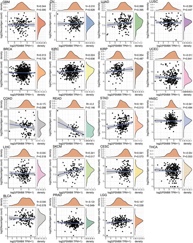
www.nature.com/scientificreports/
HNSC Head and neck squamous cell carcinoma
HRs Hazard ratios
KICH Kidney chromophobe;
KIRC Kidney renal clear cell carcinoma
KIRP Kidney renal papillary cell carcinoma
LAML Acute myeloid leukemia
LGG Lower grade glioma
LUAD Lung adenocarcinoma
LUSC Lung squamous cell carcinoma
MESO Mesothelioma
MHC Major Histocompatibility Complex
MSI Microsatellite instability
NES Net enrichment score
NIK/NF-kB Nuclear factor kappa-light-chain-enhancer of activated B cells;
OS Overall survival
OV Ovarian serous cystadenocarcinoma
PAAD Pancreatic adenocarcinoma
PCPG Pheochromocytoma and paraganglioma
PFI Progression-free interval
PRAD Prostate adenocarcinoma
READ Rectum adenocarcinoma
SARC Sarcoma
SKCM Skin cutaneous melanoma
STAD Stomach adenocarcinoma
TCGA The Cancer Genome Atlas
TGCT Testicular Germ Cell Tumors
THCA Thyroid carcinoma
THYM Thymoma
TIC Tumor-infiltrating immune cell
TMB Tumor mutation burden
TME Tumor microenvironment
UCEC Uterine corpus endometrial carcinoma
UCS Uterine carcinosarcoma
UVM Uveal melanoma
Currently, several checkpoint-blocking drugs such as anti-CTLA4 and anti-PD-L1 have provided superior per-
formance compared to conventional cytotoxic drugs, which has fueled the field of immune-related therapeutic
targets in oncology. Considering the recent advances in immunotherapy, it is impossible to ignore the substantial
influence of the tumor microenvironment (TME), which is comprised of resident stromal cells and recruited
immune cells. The antigen-presenting system, in concert with immune effector cells, orchestrate sustained input
to the TME-tumor interaction via the enhancement of tumor immunogenicity and the modulation of an anti-
tumor microenvironment.
The PSMB8 gene encodes an essential subunit of a specialized immunoproteasome c omplex1. And the
generated peptides has higher affinity with major histocompatibility complex (MHC) I molecules and in turn
enhanced antigenicity to CD8+ T c ells2–4. Meanwhile, PSMB8 mutations have been observed to contribute to
auto-inflammation and lipodystrophy in humans1, which show the pleiotropic functions involved in the mainte-
nance of dynamic equilibrium. Previous studies have revealed the context-specific role of PSMB8, which varies
in diverse cancers. Recent studies have recognized a neovascularization-suppressive role exerted by PSMB8 in
glioma, via modulation of the ERK1/2 and PI3K/AKT signaling pathways5,6. In the orthotopic mouse model,
inducible knock-down of PSMB8 dampened the expression of vascular endothelial growth factor (VEGF) and
CD31; and thus, favored invasive capacity in glioblastoma. In another functional analysis, the role of PSMB8
was recapitulated in mucinous ovarian carcinoma pathogenesis, which identified PSMB8 as a mediator between
antigen presentation of exogenous antigen via MHC class I molecules and the noncanonical nuclear factor
kappa-light-chain-enhancer of activated B cells (NIK/NF-kB) pathway7. Furthermore, PSMB8 meditated PI3K/
AKT pathway activation in acute myeloid leukemia (LAML) was established and was suggested as a promotor
of tumorigenesis8. In addition to the previously mentioned tumors, a large-scale public database analysis and
laboratory investigations have successively confirmed the role of PSMB8 in the evolution of cutaneous squamous
cell carcinoma, papillary thyroid carcinoma, and prostate adenocarcinoma9–11. The association of aberrant expres-
sion of PSMB8 have been asynchronous with regard to tumor prognosis. In contrast, there is also a growing body
of research lending support to a shielding role for PSMB8 via the promotion of immune cell infiltration. In T
cell-mediated anti-tumor immunity12, the overexpression of PSMB8 was reported to reduce colony formation
after radiation with a significant increase in expression of apoptosis-inducing molecules, such as cleaved PARP
and cleaved caspase-313. To date, the mechanisms that underline the tumorigenesis capacity of PSMB8 are not
fully understood, and the immunological and prognostic roles of PSMB8 in the pan-cancer background remain
to be elucidated.
Previously published studies conducting the cancer-associated analysis involving PSMB8 have been limited
to specific cancer type. Herein, we present a comprehensive evaluation of the immune-related prognostic land-
scape of PSMB8 in the pan-cancer field pooling information from publicly available databases. Our study was
Scientific Reports | (2021) 11:20492 | https://doi.org/10.1038/s41598-021-99724-9 2
Vol:.(1234567890)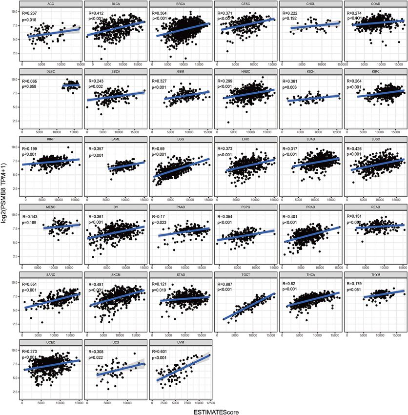
www.nature.com/scientificreports/
exploratory and interpretative in nature, and required a longitudinal analysis. In detail, we first compared the
expression of PSMB8 in normal tissues, tumor cell lines, and pan-cancer. Transcriptome-sequencing patterns
were conventionally followed by survival analysis. Subsequently we evaluated the prognostic value of PSMB8 in
pan-cancer using datasets from The Cancer Genome Atlas (TCGA) database. Next, we analyzed the association
between PSMB8 expression and the degree of immune cell infiltration, immune checkpoint expression, and
mutational burden. To this end, we have shifted the research focus of PSMB8 toward a multi-dimensional analysis
of clinical relevance, with an unique immuno-correlation study based on pan-cancer analysis. This study sheds
light on the potential role of PSMB8 as a prognostic-indicator in different cancers.
Materials and methods
Data acquisition and processing. TCGA, a landmark cancer genomic database containing vast informa-
tion on cancer samples spanning 33 cancer types, was exploited to extract expression profile data from matched
tumor and adjacent normal samples, as well as information detailing the corresponding clinicopathological
traits. Another comprehensive public resource, GTEx, was applied to enrich tissue-specific normal samples
alongside those obtained from TCGA. The broad institute CCLE was interrogated for PSMB8 mRNA expression
in human cancer cell lines for a multi-dimensional inspection of PSMB8 expression.
Prior to commencing the study, the transcript data were checked for their robustness and normalization.
Subsequently, the RNA sequencing data were adjusted to eliminate missing and duplicated results, and were
transformed by a log2(TPM + 1) normalization using the R package of "rma" in an R environment (R version:
3.6.1). Cases in pan-cancer were acquired having a thorough follow-up for survival analysis and their correspond-
ing outcomes in terms of overall s urvival14, disease-specific survival (DSS), and in the disease-free interval (DFI)
and progression-free interval (PFI).
PSMB8 gene expression analysis and correlation with malignancy prognosis. The recruited
samples from TCGA in the section of the survival analysis were preliminarily screened for their data integrity
in terms of both PSMB8 expression and follow-up information. Consequently, a prognosis-correlated analysis
of the respective outcome events of OS, DSS, DFI, PFI was conducted. The optimal PSMB8 cut-off values for
Kaplan–Meier curves were calculated using the function "survcutpoint" of "survminer" package in R (R version:
3.6.1). Kaplan–Meier curves and log-rank tests were used to estimate the prognosis-predictive value of PSMB8.
Hazard ratios (HRs) with 95% confidence intervals and log-rank Ps were calculated. HRs with a value less than
1 were detrimental to survival, while a value greater than 1 were beneficial to prognosis.
TIMER analysis and immune microenvironment correlation analysis. To investigate the interplay
between tumor and the TME, an immune correlation analysis was carried out. First, the TIMER (Tumor Immune
Estimation Resource) web server (https://cistrome.shinyapps.io/timer/) was used to visualize the immune infil-
tration-associated expression pattern of PSMB8 in diverse cancer types. In the gene module of the TIMER data-
base, we investigated the levels of PSMB8 expression and the abundance of tumor-infiltrating immune cells
(TIICs), including CD4+ T cells, CD8+ T cells, B cells, neutrophils, dendritic cells (DCs), and macrophages.
The scatter plots displayed purity-corrected partial Spearman’s rho values and statistical significances evaluated
by the Wilcoxon test. In addition, the ESTIMATE algorithm was exploited to infer the ratio of immune and
stromal components from PSMB8 expression data in pan-cancer, whose results were presented in the form of
and immune score, stromal score, and ESTIMATE score with the relative correlation coefficients. Following
the application of these immune algorithms, immune-stimulatory and immunoinhibitory gene markers were
gathered for additional correlation analysis in pan-cancer, which comprised BLTA, CD200, TNFRSF14, NRP1,
LAIR1, TNFSF4, CD244, LAG3, ICOS, CD40LG, CTLA4, CD48, CD28, CD200R1, HAVCR2, ADORA2A,
CD276, KIR3DL1, CD80, PDCD1, LGALS9, CD160, TNFSF14, IDO2, ICOSLG, TMIGD2, VTCN1, IDO1,
PDCD1LG2, HHLA2, TNFSF18, BTNL2, CD70, TNFSF9, TNFRSF8, CD27, TNFRSF25, VSIR, TNFRSF4,
CD40, TNFRSF18, TNFSF15, TIGIT, CD274, CD86, CD44, and TNFRSF9. To further evaluate the immune-
predictor value of PSMB8 in pan-cancer, we evaluated two emerging parameters—tumor mutation burden
(TMB) and microsatellite instability (MSI)—in the context of immunotherapy. We proposed that TMB and MSI-
associated PSMB8 expression would provide evidence that a higher degree of genomic instability, as defined by
the TMB and MSI, could extrapolate to more immune-surveillance opportunities. Furthermore, the relationship
between different immune checkpoint genes and PSMB8 expression were analyzed via correlation coefficients.
Gene set enrichment analysis. To reflect the underlying biological function of PSMB8, we subsequently
anatomized the biological traits by applying GSEA. Genes enriched in the predefined gene sets in GSEA (http://
software.broadinstitute.org) served as the reference to assess the overall coupling of aberrantly-expressed PSMB8
enriched in the KEGG and HALLMARK collections, respectively. The significantly enriched pathways were
finally identified based on the calculated net enrichment score (NES) with a false discovery rate (FDR) < 0.05 as
the cut-off criterion.
Association analysis of PSMB8 With DNA mismatch repair (MMR) genes and methyltrans-
ferases. DNA mismatch repair is an essential intracellular repair mechanism, which leads to the risk reduc-
tion of genomic instability. DNA methylation is a form of epigenetic modification that does not alter the DNA
sequence. The correlation of five MMRs genes (MLH1, MSH2, MSH6, PMS2, EPCAM) as well as four methyl-
transferases with PSMB8 expression was assessed in TCGA.
Scientific Reports | (2021) 11:20492 | https://doi.org/10.1038/s41598-021-99724-9 3
Vol.:(0123456789)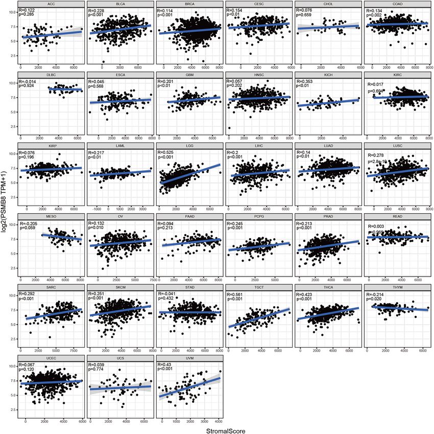
www.nature.com/scientificreports/
Figure 1. Flowchart illustrating the pan-cancer analysis.
Quantitative real‑time PCR assays (qRT‑PCR). qRT-PCR was used to test the relative expression of
PSMB8 from BRAC cells. RNA was extracted by Trizol reagent and then exploited for the process of reverse
transcription via a reverse transcription reagent kit (Toyobo, Osaka, Japan). Finally, qRT-PCR was conducted
using THUNDERBIRD SYBR qPCR Mix (Toyobo, Osaka, Japan) with the machine of Applied Biosystems 7500.
Each sample was conducted at least triplicate and the relative expression of PSMB8 was measured by the 2 −ΔΔCt
calculation fomula compared to GAPDH expression. The sequences of the primers used for cDNA amplification
were listed as followed: PSMB8 Forward: 5′-GCTGCCTTCAACATAACATCA-3′, and Reverse: 5′-CTGCCA
CCACCACCATTA -3′; GADPH Forward: 5′-GTCTCCTCTGACTTCAACAGCG-3′ and Reverse: 5′-ACCACC
CTGTTGCTGTAGCCAA-3′.
Epigenomic deconvolution. In the complicated background of tumor cell-type heterogeneity, we utilized
the epigenomic deconvolution (EDec), a reference-free algorithm, to infer cell-type proportions from DNA
methylation and clarify the immunocyte-specific expression pattern of P SMB815. The simulations was based on
an open source software EDec(http://genboree.org/theCommons/projects/edec) and the output file containing
the mean and standard error estimates of cell type and subtype-specific gene expression profiles based on the
TCGA BRCA dataset was directly utilized for the presentation(http://genboree.org/theCommons/documents/
571).
Statistical analysis. Gene expression data from the TCGA, GTEx, and CCLE databases were analyzed
using Student’s t-test. The Kruskal Wallis test was used to evaluate PSMB8 expression in pan-cancer, and the
Wilcoxon test was used to evaluate gene expression differences between normal and tumor tissues. OS was
calculated using the Kaplan–Meier method, and survival curves were compared using log-rank tests. Pearson
analysis was performed to evaluate the correlation between PSMB8 expression levels with checkpoint-related
genes. All statistical analysis was conducted using R software (version 3.6.1). A P-value < 0.05 was considered
statistically significant.
Results
Pan‑cancer expression profile of PSMB8. To start with, our research procedure towards PSMB8 in
the pan-cancer was shown in Fig. 1. Previous studies investigating PSMB8 have alluded to two contradictory
notions, one of which considered that elevated PSMB8 expression was adversely associated with tumor progres-
sion, such as in PTC, LAML, and GBM, while the other regarded PSMB8 as an immune-stimulatory factor with
protective characteristics. Thus, we first performed a comprehensive evaluation of PSMB8 mRNA expression in
normal tissues in the GTEx database and in cell lines in CCLE database. The results revealed that PSMB8 was
expressed in relatively lower levels in the bone marrow, muscle, and testis, while expression was much higher in
the spleen, bladder, and lung (Kruskal–Wallis test P < 0.05) (Fig. 2A). As shown in Fig. 2B, the PSMB8 expres-
Scientific Reports | (2021) 11:20492 | https://doi.org/10.1038/s41598-021-99724-9 4
Vol:.(1234567890)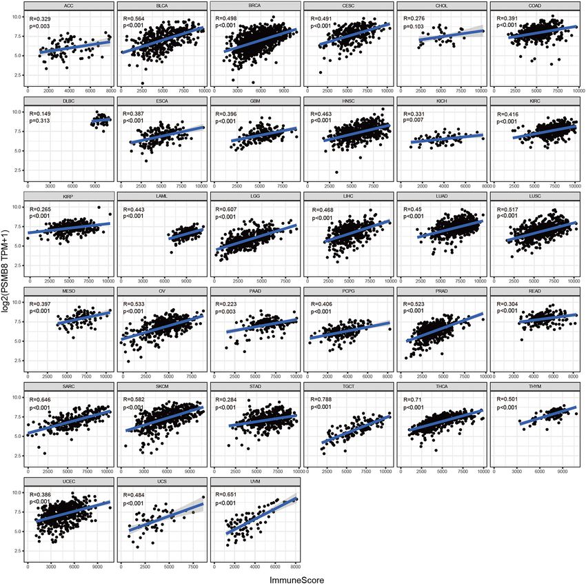
www.nature.com/scientificreports/

                                  Figure 2.  Expression of PSMB8 in TCGA cohorts, GTEx datasets, and CCLE cell lines. (A,B) It Significant
                                  variation of PSMB8 expression can be observed across different normal tissues and normal cell lines. (C,D)
                                  Differential expression between normal and tumor tissues in TCGA and GTEx databases. T, tumor; N, normal.
                                  X axis represented the number of tumor and normal samples. Y axis represented transcripts per million
                                  [log2(TPM + 1)]. *P < 0.05, **P < 0.01, and ***P < 0.001.

                                  sion in 22 normal cell lines extracted from the CCLE database was also observed with substantially significant
                                  differences (Kruskal–Wallis test: P = 3.9e−47).
                                      Further, to distinguish between the two potential roles of PSMB8, we conducted a pan-cancer expression
                                  profile analysis using datasets from TCGA and the GTEx databases to compare differences in expression between
                                  tumor and normal tissue. The results obtained from the preliminary expression analysis of PSMB8 are presented
                                  in Fig. 2C,D. The analysis of differentially expressed PSMB8 in TCGA (Fig. 2C) revealed significant differences
                                  except for kidney chromophobe (KICH), pancreatic adenocarcinoma (PAAD), and prostate adenocarcinoma
                                  (PRAD). Subsequently, we combined gene expression from normal samples in the GTEx with those from TCGA
                                  and generated a new plot. As shown in Fig. 2D, PSMB8 was overexpressed in tumor tissues compared to normal
                                  tissues on average, except for lung squamous cell carcinoma (LUSC) and adrenocortical carcinoma.

                                  Prognostic significance of PSMB8 across cancers. The first part of our analysis explored the expres-
                                  sion profile of PSMB8, which raised the issue of its prognostic value. We utilized TCGA database comprising
                                  Affymetrix microarray data of 33 cancer types as well as their survival-related follow-up to investigate the impact
                                  of PSMB8 expression on OS, DSS, DFI, and PFI. Cox regression analysis revealed that PSMB8 denoted favorable
                                  OS and DSS in bladder urothelial carcinoma (BLCA), breast invasive carcinoma (BRCA), ovarian serous cyst
                                  adenocarcinoma, mesothelioma (MESO), and skin cutaneous melanoma (SKCM). Our results regarding OS,
                                  DSS, PFI showed a robust carcinogenic-predictive role of PSMB8 in brain lower grade glioma (LGG), uveal
                                  melanoma (UVM), and PAAD. Furthermore, high expression of PSMB8 was associated with poor survival in
                                  LAML and lung adenocarcinoma (LUAD). With regard to DFI, increased PSMB8 expression showed a satisfac-
                                  tory prognosis in BRCA patients, and a poor outcome in PAAD, instead. The significant results of the survival
                                  analysis are plotted in Fig. 3. All results of all cancer types at all survival times was presented in ’Supplementary
                                  Data S1’.

                                  Correlation of PSMB8 expression with immune infiltration level and components of immune
                                  cells. Previously, a variety of studies have revealed that TIICs influence the response rate of immunotherapy,

Scientific Reports |   (2021) 11:20492 |               https://doi.org/10.1038/s41598-021-99724-9                                                    5

                                                                                                                                                Vol.:(0123456789)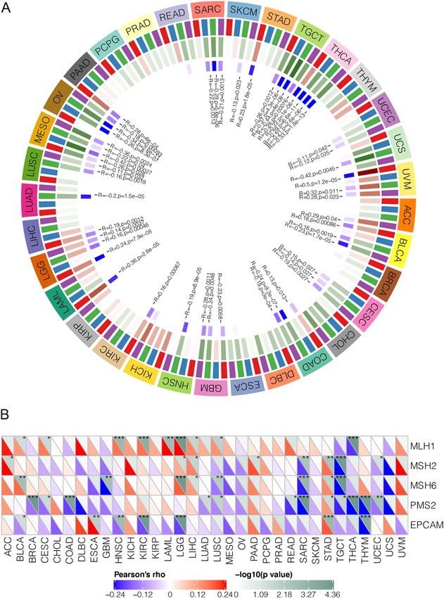
www.nature.com/scientificreports/

                                            Figure 3.  Kaplan–Meier survival curves of survival comparing high and low expression of PSMB8 in pan-
                                            cancer derived from TCGA. (A–K) Overall survival differences between groups in BLCA, BRCA, LAML, LGG,
                                            LUAD, MESO, OV PAAD, SKCM, THCA, and UVM. (L–S) Disease-specific survival differences between
                                            groups in BLCA, BRCA, LGG, MESO, OV, PAAD, SKCM, and UVM. (T–U) Disease-free interval difference
                                            between groups in BRCA and PAAD. (V–AA) Progression-free interval differences between groups in BLCA,
                                            BRCA, LGG, PAAD, SKCM and UVM.

          Scientific Reports |   (2021) 11:20492 |             https://doi.org/10.1038/s41598-021-99724-9                                            6

Vol:.(1234567890)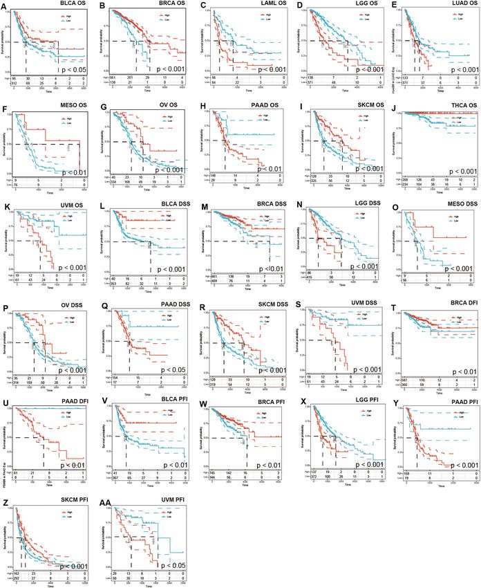
www.nature.com/scientificreports/
the efficacy of chemotherapy, and the ultimate prognosis of malignancies. Chen et al. demonstrated the seven
steps of the cancer-immunity cycle, which have become the basic framework of cancer immunotherapy research.
Portrayed as one of the constitutive proteasome genes (CP), expression of PSMB8 has been associated with levels
of MHC-I, the antigen-presenting cells, and TIILs, and consequently plays a crucial part in the above-mentioned
immunity cycle16. Albeit proteasome inhibitors, such as bortezomib, may induce proteotoxic stress and apop-
totic activity as targeted therapy, there are currently investigative areas worthy of attention involving the sup-
pression of immunoproteasome (i-proteasome) activity to induce immune evasion and metastatic progression17.
Therefore, it is necessary to evaluate the immune properties of PSMB8 in the context of pan-cancer.
The online TIMER database was exploited to calculate the expression of the immune cells in association with
a target gene. We found that all six subtypes of immune infiltration cells were associated with elevated PSMB8
expression in the following tissues: kidney renal papillary cell carcinoma (KIRP), brain lower grade glioma
(LGG), liver hepatocellular carcinoma (LIHC), lung adenocarcinoma (LUAD), lung squamous cell carcinoma
(LUSC), prostate adenocarcinoma (PRAD), sarcoma (SARC), skin cutaneous melanoma (SKCM), stomach ade-
nocarcinoma (STAD), and testicular Germ Cell Tumors (TGCT). In contrast, there seemed to be no immune
infiltration-associated expression patterns with PSMB8 in cholangiocarcinoma (CHOL) and rectum adenocarci-
noma (READ). Furthermore, the Spearman correlation coefficient determined for the immune infiltration levels
of B cells, CD8+ T cells, CD4+ T cells, macrophages, neutrophils, and dendritic cells were notably significant in
23, 23, 25, 23, 19, and 26 cancer types, which comprised a large proportion of the 32 cancer types interrogated
(Supplementary Figures S1–S8).
An additional immune algorithm, called the ESTIMATE algorithm, was applied. Three of the calculated scores
were the StromalScore, ImmuneScore, and ESTIMATEScore, which were determined to be directly proportional
to the ratio of the corresponding stromal, immune components, and their aggregates. In most cancer types,
PSMB8 emerged as a statistically significant marker of the TME status. Thus, correlations between PSMB8 expres-
sion and StromalScore, ImmuneScore, and ESTIMATEScore were determined in 20, 31, and 29 cancer types,
respectively (Figs. 4, 5, 6). The top three tumors most significantly associated with overexpression of PSMB8
based on the ESTIMATEScore were listed the following: TGCT, THCA, and UVM. Nevertheless, CHOL and
DLBC showed no statistically significant association across all three scores. The ESTIMATE outcome generated
interesting insight into THCA, which ranked second in the ESTIMATEScore as well as the ImmuneScore, and
ranked fourth in the StromalScore, and thus, hereinafter we switched our focus to the role of PSMB8 in THCA.
To determine the underlying mechanism of PSMB8 in the enhanced immunological response, we calculated
the Spearman correlations of PSMB8 expression with immune-stimulatory and immunoinhibitory factors. We
set the statistical criteria as P < 0.05. As illustrated in the heatmap, the bulk of cancer types were positively
associated with most immune elements, with the exception of CHOL, DLBC, ESCA, GBM, KICH, MESO, and
READ. These seven cancer types identified a subset in which the TME was irrelevant to PSMB8 expression, and
thus the predictive role of PSMB8 was far from optimal in this subgroup, which is frequently encountered in the
search for tumor biomarkers.
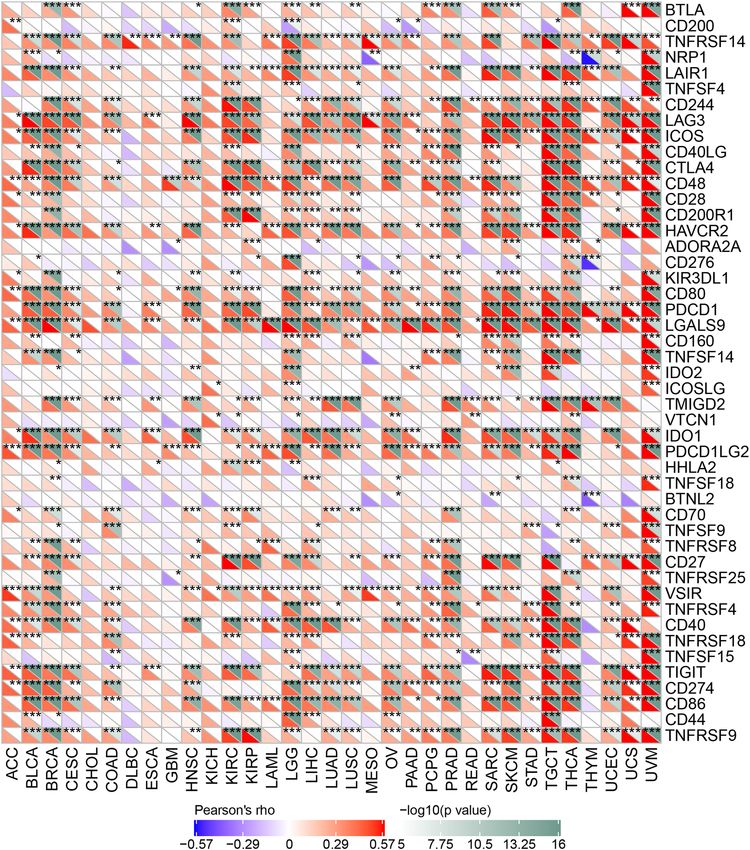
Our results revealed that a positive relationship between PSMB8 expression and the levels of immune check-
point genes in various tumors, such as TGCT, THCA and UVM (Fig. 7). Thus, these findings indicated that
PSMB8 could play a role in tumor immunity by regulating the expression of these immune checkpoint genes.
As shown in Fig. 8, we determined that the expression of PSMB8 was positively correlated with the number of
neoantigens only in SKCM tumor tissues (R = 0.241, P < 0.05). Of note only a P-value of < 0.05 and R > 0.20 may
be considered as significant and positive, respectively.
The TMB and MSI have conceptually emerged as predictors of an effective immune response, which are
promoted within the spectrum of tumor exacerbation. We further evaluated the strength of the relationship
between PSMB8 expression and the TMB or MSI in pan-cancer. The most distinct correlation coefficients in the
analysis of the TMB and MSI were ± 0.47 and ± 0.31, respectively. In detail, a significant correlation was identified
between overexpression of PSMB8 and an increased TMB in UCS, STAD, SKCM, PAAD, LGG, KIRC, ESCA,
COAD, and BRCA tissues, while an opposite association was observed in THYM and PRAD tumor tissues
(Fig. 9A). Similarly, increased PSMB8 expression was significantly associated with an increased MSI in THCA,
KIRC, DLBC, and COAD tissues and was inversely correlated with in UCEC, TGCT, PRAD, OV, MESO, LUSC,
LUAD, and CESC (Fig. 9B).
Association analysis of PSMB8 with DNA mismatch repair genes and methyltransferases. As
shown in Fig. 10A, a significant correlation between PSMB8 and four methyltransferase expression levels could
be observed in most cancer types. In addition, DNA mismatch repair gene expressions were almost inversely
correlated with the PSMB8 expression in pan-cancer, whereas MLH1 was of positive correlation (Fig. 10B). Our
analysis revealed that PSMB8 could regulate epigenetic status in pan-cancer.
Gene‑annotation and pathway enrichment analysis. To uncover the potential signaling pathways
and immunocompetences associated with the involvement of PSMB8 in tumorigenesis, we applied GSEA using
KEGG and HALLMARK terms. Only enriched gene sets with the absolute value of normalized enrichment
score (NES) > 1, nominal (NOM) P < 0.05, and an FDR q-value < 0.25 were considered statistically significant. As
shown in Fig. 11A,C, KEGG and HALLMARK enriched terms showed that overexpression of PSMB8 was mainly
associated with immunological processes, including antigen processing and presentation (NES = − 2.6, NOM
P < 0.05), natural killer cell-mediated cytotoxicity (NES = − 2.3, NOM P < 0.05), allograft rejection (NES = − 2.6,
NOM P < 0.05), the interferon (IFN)-gamma response (NES = − 2.7, NOM P < 0.05), and the IFN-alpha response
(NES = − 2.5, NOM P < 0.05). Furthermore, high expression of PSMB8 was associated with metabolic syndromes,
Scientific Reports | (2021) 11:20492 | https://doi.org/10.1038/s41598-021-99724-9 7
Vol.:(0123456789)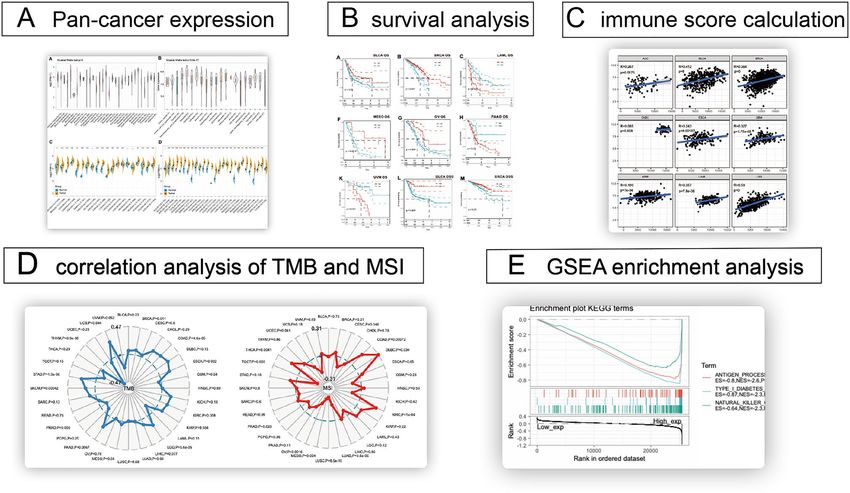
www.nature.com/scientificreports/

                                            Figure 4.  Correlation analysis between PSMB8 expression in 33 types of cancers and stromal score.

                                            such as type I diabetes mellitus (NES = − 2.3, NOM P < 0.05). However, there was no significant enrichment in
                                            the analysis based on low expression of PSMB8 (Fig. 11B,D).

                                            Native validation of PSMB8 expression in the BRCA subtypes. To further validation, we chose 60
                                            pairs BRCA tissues and their corresponding adjacent normal tissues to perform qRT-PCR. Our local specimens
                                            were all sampled from the operation room in the First Affiliated Hospital of Wenzhou Medical University, which
                                            were made up of luminal (HR+, HER2−), TNBC (HR−, HER2−), and HER2+ (HR−, HER2+) subgroups. And
                                            Fig. 12 directly show the disparity tendency of PSMB8 expression between tumor and normal tissues with sig-
                                            nificantly statistical difference (*P < 0.05, **P < 0.01, and ***P < 0.001).

                                            Immunocyte‑specific expression pattern of psmb8 in brca by epigenomic deconvolu-
                                            tion. Tumor heterogeneity partially reflected in the proportion of the various cell-types in a tumor (com-
                                            posed of immunocyte, stromal cell, tumor cell and normal cell), aroused the desire to micro dissect and then
                                            infer cell-type composition. By applying EDec to TCGA BRCA queue, the expression profile was delineated
                                            precisely based on the subtype and cell type. The mean and standard error estimates of immunoproteasome
                                            subunit encoding gene expression was filed in Supplementary Data S2. And the comparative histogram reveal-

          Scientific Reports |   (2021) 11:20492 |              https://doi.org/10.1038/s41598-021-99724-9                                              8

Vol:.(1234567890)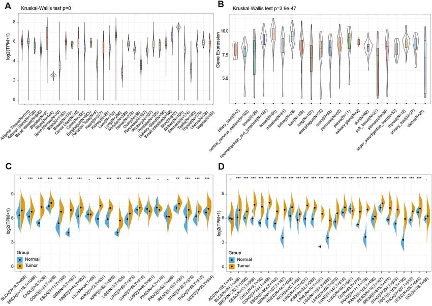
www.nature.com/scientificreports/
Figure 5. Correlation analysis between PSMB8 expression in 33 types of cancers and immune score.
ing the expression pattern of 10 catalytic subunit validated the immunocyte-specific profile of PSMB8 (shown
in Fig. 13).
Discussion
The awareness of remodeling an efficient anti-tumor TME improved, along with the widely-spread application of
immune checkpoint blockade tumor therapy. With the exception of immunologically-hot cancer types benefiting
much from blocking immune checkpoints, the overall objective response rates of other tumors merely ranged
from 15 to 25%, which was far from satisfactory18. Recently, the third generation of combined immunotherapy
strategies, integrating an immunomodifier with immune checkpoint inhibition (ICI), has achieved synergistic
effects in the maintenance of a dominant and enhanced immunological response19, which fully clarified a posi-
tive anti-tumor immune modification was of significance when ICI didn’t work.
PSMB8, a catalytic subunit of immunoproteasomes, plays a critical role in the process of proteolysis to
generate the antigenic epitopes, which are in turn transmitted to MHC class I molecules for further antigen-
presentation. Thus, the nature of antigen presentation, to some extent, significantly enhanced the pool of MHC-I
compatible peptides. The increased release of IFN-gamma by the TIIL in the TME trigger the incorporation
of immuno-subunits into the catalytic core of the proteasome and transform the excessive proteasomes into
Scientific Reports | (2021) 11:20492 | https://doi.org/10.1038/s41598-021-99724-9 9
Vol.:(0123456789)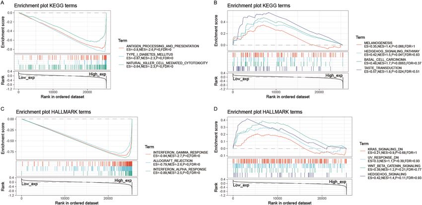
www.nature.com/scientificreports/
Figure 6. Correlation analysis between PSMB8 expression in 33 types of cancers and the estimated immune
score.
immunoproteasomes. The IFN-gamma inducible genes (PSMB8/β5i, PSMB9/β1i, PSMB10/β2i), together with
their chaperones have command of the tumor vulnerability to antigen-dependent killer c ells20. Thus, essential
subunits (more explicitly referred to PSMB units) were fundamentally engaged in the immunoproteasome trans-
formation, acting against the construction of anti-tumor immunity.
On the other hand, malignancies are usually subjected to proteotoxic stress, under which circumstances
tumorigenic proteins induced by genomic aberrations are assembled at the expense of proteasome-promoted
activities regulating proteostasis21. Accordingly, a variety of tumors addictively resort to immunoproteasome
activities. An experimental study determined that down-regulated incorporation of PSMB8 into immunoprotea-
some could attenuate its formation, laying the foundation of the key part of PSMB8 in the proteolytic activity and
protein homeostasis in maintenance of tumor development. In a previous study, the immunoproteasome (β1i-
β2i-β5i) and the two intermediate proteasomes (β1-β2-β5i and β1i-β2-β5i), except for the standard proteasome
Scientific Reports | (2021) 11:20492 | https://doi.org/10.1038/s41598-021-99724-9 10
Vol:.(1234567890)www.nature.com/scientificreports/
Figure 7. Each cancer was conducted the correlation analysis between 47 immune checkpoint genes and the
gene expression of PSMB8, which was presented in the heatmap. The spearman correlation coefficients were
filled in accordance with the corresponding colors at the bottom of chart (*P < 0.05, **P < 0.01, and *** P < 0.001).
(β1-β2-β5), exhibited the comparative efficiency of degrading ubiquitinated or oxidized proteins, indicating the
more significant role of β5i/PSMB8 than β1i/PSMB9 and β2i/PSMB10.
The above contradictory arguments underlined a chaotic picture of the functional orientation of PSMB8 in
neoplasms. To sum up, PSMB8, the intrinsic APG, capable of enlarging the pool of MHC-I compatible peptides,
credibly reinforced the capacity of antigen presentation to the immune system and in turn antigen-dependent
cytotoxicity. On the reverse side, more in-depth mechanism studies discovered its interaction with oncogenic
signaling pathway and the essential function in oncoprotein homeostasis.
As far as we are concerned, in consideration of the immunological relevance, it would be better to analyze the
candidate gene, PSMB8, in the circumstance of specific TIME. Szekely et al. noticed a coordinated down regula-
tion of PSMB8-10(β5i, β1i, β2i) in the metastases than the primary breast tumor, together with the TIL counts and
Scientific Reports | (2021) 11:20492 | https://doi.org/10.1038/s41598-021-99724-9 11
Vol.:(0123456789)www.nature.com/scientificreports/
Figure 8. Correlation analysis between the level of neoantigens and PSMB8 expression in pan-cancer.
Scientific Reports | (2021) 11:20492 | https://doi.org/10.1038/s41598-021-99724-9 12
Vol:.(1234567890)www.nature.com/scientificreports/
Figure 9. Correlation of PSMB8 expression with tumor mutation burden (TMB) and microsatellite instability
(MSI) in multiple cancer types. (A) Correlation between TMB and PSMB8 expression. (B) Correlation between
MSI and PSMB8 expression. Spearman’s correlation coefficients are shown above the bar graphs. (Spearman
Correlation test, P < 0.05 was considered statistically significant).
immunomodulator signatures. We boldly speculate that PSMB8 alone could not make sense to the prognosis of
immunologically-inert metastases and the other cancer types likewise. And just like the circumstantial impression
made by PD-(L)1, only in the immune activation background, could the antigen-presentation effect be stimulated
sufficiently; Otherwise the PSMB8 orientate its function towards the ontogenetic downstream signaling, such as
PSMB8-mediated PI3K/PTEN/AKT /MTOR pathway activation, which was verified to facilitate tumorigenesis.
This gene has not yet been fully exploited, particularly in the immuno-regulation level. Hence, our study
which investigated the general applicability of the antigen-presentation regulator PSMB8 as a prognostic bio-
marker in pan-cancer from an immuno-oncological perspective, could provide a rational and theoretical foun-
dation for future mechanism studies.
As high-throughput sequencing techniques become relatively easier to perform, large-scale public databases
have flourished. Independent datasets with pan-cancer expression profiles were acquired from TCGA, GTEx, and
CCLE, which include normal tissues, tumor tissues, and cancer cell lines. We extracted the expression of PSMB8
and visually rendered the comparison in the pan-cancer. Using GTEx and TCGA, we found that PSMB8 expres-
sion was generally lower in normal tissues, while exhibited varied expression in tumor cell lines of CCLE. Next,
we explored differences in expression between tumor and normal tissues using TCGA datasets and determined
that PSMB8 was generally prone to be over-expressed in the most tumor tissues compared to normal tissues,
except for LUSC, PAAD, PRAD, and KICH tumors. When data from normal tissues in the GTEx was added
to the TCGA, PSMB8 expression was higher in PAAD, PRAD, and KICH than adjacent normal tissues. Thus,
only PSMB8 expression in LUSC was inconsistent and finally drew a conclusion that overexpression of PSMB8
appeared more frequently in the tumor tissues than in their normal counterparts.
We next attempted to identify the prognosis-predictor value for PSMB8. Our findings showed that high
expression of PSMB8 in BLCA, BRCA, MESO, OV, and SKCM was successful in predicting better OS and DSS.
In view of these survival outcomes, the function of PSMB8 was orientated more like a protective role. Conversely,
increased PSMB8 expression revealed poor overall survival for LAML, LGG, LUAD, PAAD, and UVM based
on Kaplan–Meier analysis. In a word, our results sustained coherence with prior studies, as well as revealed the
functional pleiotropy of PSMB8, albeit with unknown molecular mechanisms.
Given the overpowering energy of the immune microenvironment implicated in the process of tumorigenesis,
a significant correlation-analysis in pan-cancer was conducted. More than two-thirds of cancer types exhibited
a significant association between PSMB8 and the immune infiltrating cells. Since the TME are constantly in a
state of flux, tumor progression along a relatively unmanageable pathway may demand a metabolic reprogram-
ming obtained from the TIICs, which leads to subsequent vicious circle of tumor differentiation. Unfortunately,
CHOL was considered an exception on account of the irrelevance between PSMB8 and the TME constituents,
including both immune and stromal elements, which could be partly in explanation of its very poor survival rates.
Proteasomes promoted the degradation of endogenous and exogenous antigens presented by the MHC
class I system, and thus were linked to antigen presentation. The maturation of dendritic cells (DC), which are
acknowledged as the most powerful APCs, orchestrate the changes in proteasomal composition. More immu-
noproteasomes populate DCs, and inducible cytotoxic T lymphocytes (CTLs) are unable to properly iden-
tify tumor cells that constitutively express these p roteasomes22. Inhibition of the catalytic subunit involved in
Scientific Reports | (2021) 11:20492 | https://doi.org/10.1038/s41598-021-99724-9 13
Vol.:(0123456789)www.nature.com/scientificreports/
Figure 10. Correlation analysis between PSMB8 expression and (A) four methyltransferases and (B) five
MMR genes in various tumor types. The color of the second outer ring represented the orderly represented Dna
methyltransferases (Dnmt1, Dnmt2, Dnmt3 and Dnmt4). The Pearson’s rho and − log10(p value) was presented
below.
Scientific Reports | (2021) 11:20492 | https://doi.org/10.1038/s41598-021-99724-9 14
Vol:.(1234567890)www.nature.com/scientificreports/
Figure 11. GSEA for samples with high PSMB8 expression and low expression. (A) The enriched gene sets
in following KEGG analysis using high PSMB8 expression samples. (B) The enriched gene sets in KEGG by
samples with low PSMB8 expression. (C) Enriched gene sets in HALLMARK collection, the immunologic
gene sets, by samples of high PSMB8 expression. (D) Enriched gene sets in HALLMARK by the low PSMB8
expression. Each line represents one gene set with a unique color, and up-regulated genes located in the left
approaching the origin of the coordinates, by contrast the down-regulated genes are indicated on the right of the
x-axis. Only gene sets with NOM P < 0.05 and FDR q-value < 0.05 were considered significant and displayed in
the plot.
immunoproteasome-transformed activities, herein referred to as PSMB8, promoted an alteration of antigenic
peptide-repertoires expressed by antigen-loaded DCs. In our ESTIMATE algorithm-based analysis, all cancers
types except for READ, CHOL, UVM, THYM, ESCA, GBM, and DLBC, presented a significant correlation
between PSMB8 expression and DC. In brief, DCs were the most closely-associated with PSMB8 levels in the
scope of pan-cancer, and both were observed to correlate with the antigen-presenting system.
Notably, one of the strongest positive associations was observed between PSMB8 expression in BRCA and
immune markers, which comprised the Estimate Score, various levels of TIIC, TMB, and MSI. Validation of the
PSMB8 in TCGA BRCA cohort through EDec algorithm, supported its immunological relationship.
To sum up, as there have not been any credible immuno-oncology evidences emerging in the previous
decades, we explored this antigen presentation gene (APG) from an immunological standpoint. As justified by
previous experimental or bioinformatic studies combined with our pan-cancer survey described herein, differ-
ent tumor types and their corresponding microenvironments did not alter the essential antigen-presentation
function of PSMB8, but definitely bent its orientation concerning tumor prognosis through the downstream
oncogenic signaling. Szekely et al. noticed a coordinated down regulation of PSMB8-10 (β5i, β1i, β2i) in the
metastases than the primary breast tumor, together with the TIL counts and immunomodulator signatures. We
boldly speculate that PSMB8 alone could not make sense to the prognosis of immunologically-inert metastases
and the other cancer types likewise. And just like the circumstantial impression made by PD-(L)1, only in the
immune activation background, could the antigen-presentation effect be stimulated sufficiently.
Nevertheless, this study extending across multiple databases presented some limitations. First, we conducted
a bioinformatic analysis of PSMB8, from which it was difficult to assess the value of clinical transformation.
Although it was clear that more mechanism analysis added to our preliminary analysis would be useful to
understand the details of interaction and the role of PSMB8 as an immunomodulator.
In addition, since the resources in all the databases were tissue-derived, these findings cannot be validated
with in vitro/in vivo models. Besides, an alternative technique to qPCR, such as RNAseq/microarray would be
more precise. In the comparative biodiversity study, the methodological factors, such as preservation buffer, tem-
plate concentration, DNA polymerase, PCR enhancer, was indeed identified to introduce the critical variability.
Finally, the polytrophic function of PSMB8 may represent an initial effect, and the underlying mechanisms
engaged in tumor activities remain still elusive. Our pan-cancer research merely embodied an immune-related
pan-cancer analysis and awaits future investigation into the mechanism involved in tumorigenesis.
Scientific Reports | (2021) 11:20492 | https://doi.org/10.1038/s41598-021-99724-9 15
Vol.:(0123456789)www.nature.com/scientificreports/
Figure 12. PSMB8 is overexpressed compared to adjacent normal tissues in local BRCA tissues by Student’s
t-test, which is composed of (A) luminal subtype, (B) TNBC subtype, and (C) HER2+ subtype. 2−ΔΔCt is used to
present the fold change in qRT-PCR experiment. (*P < 0.05, **P < 0.01, and ***P < 0.001).
Scientific Reports | (2021) 11:20492 | https://doi.org/10.1038/s41598-021-99724-9 16
Vol:.(1234567890)www.nature.com/scientificreports/
Figure 13. Cell type-based expression profile derived from TCGA BRCA in 10 catalytic proteasome subunits.
And the immunoproteasome subunit, comprising (H)PSMB8, (I)PSMB9 and (J)PSMB10, was observed in an
immunocyte-specific expression pattern.
Scientific Reports | (2021) 11:20492 | https://doi.org/10.1038/s41598-021-99724-9 17
Vol.:(0123456789)www.nature.com/scientificreports/
Data availability
The data sets and additional images supporting the conclusions of this study are included in this article. Raw
data are available on the main electronic data storage system of the First Affiliated Hospital of Wenzhou Medical
University, and access can be provided upon request to the authors. Ethical approval for this study was obtained
from the Ethics Committee of the First Affiliated Hospital of Wenzhou Medical University.
Received: 22 July 2021; Accepted: 21 September 2021
References
1. Agarwal, A. K. et al. PSMB8 encoding the beta5i proteasome subunit is mutated in joint contractures, muscle atrophy, microcytic
anemia, and panniculitis-induced lipodystrophy syndrome. Am. J. Hum. Genet. 87(6), 866–872 (2010).
2. Angeles, A., Fung, G. & Luo, H. Immune and non-immune functions of the immunoproteasome. Front. Biosci. (Landmark Ed.)
17, 1904–1916 (2012).
3. Durgeau, A. et al. Different expression levels of the TAP peptide transporter lead to recognition of different antigenic peptides by
tumor-specific CTL. J. Immunol. 187(11), 5532–5539 (2011).
4. Neefjes, J., Jongsma, M. L., Paul, P. & Bakke, O. Towards a systems understanding of MHC class I and MHC class II antigen pres-
entation. Nat. Rev. Immunol. 11(12), 823–836 (2011).
5. Chang, H. H., Cheng, Y. C., Tsai, W. C. & Chen, Y. PSMB8 inhibition decreases tumor angiogenesis in glioblastoma through
vascular endothelial growth factor A reduction. Cancer Sci. 111(11), 4142–4153 (2020).
6. Yang, B. Y. et al. PSMB8 regulates glioma cell migration, proliferation, and apoptosis through modulating ERK1/2 and PI3K/AKT
signaling pathways. Biomed. Pharmacother. 100, 205–212 (2018).
7. Liew, P. L. et al. Distinct methylation profile of mucinous ovarian carcinoma reveals susceptibility to proteasome inhibitors. Int.
J. Cancer 143(2), 355–367 (2018).
8. Lei, M. et al. LncRNA HCP5 promotes LAML progression via PSMB8-mediated PI3K/AKT pathway activation. Naunyn. Schmiede-
bergs Arch. Pharmacol. 393(6), 1025–1032 (2020).
9. Fan, X. & Zhao, Y. miR-451a inhibits cancer growth, epithelial-mesenchymal transition and induces apoptosis in papillary thyroid
cancer by targeting PSMB8. J. Cell Mol. Med. 23(12), 8067–8075 (2019).
10. Liu, Y., Yang, H. Z., Jiang, Y. J. & Xu, L. Q. miR-451a is downregulated and targets PSMB8 in prostate cancer. Kaohsiung J. Med.
Sci. 36(7), 494–500 (2020).
11. Wang, Q. et al. Genetic associations of T cell cancer immune response with tumor aggressiveness in localized prostate cancer
patients and disease reclassification in an active surveillance cohort. Oncoimmunology 8(1), e1483303 (2019).
12. Mayes, K. et al. BPTF depletion enhances T-cell-mediated antitumor immunity. Cancer Res 76(21), 6183–6192 (2016).
13. Ha, Y. J. et al. PSMB8 as a candidate marker of responsiveness to preoperative radiation therapy in rectal cancer patients. Int J
Radiat Oncol Biol Phys 98(5), 1164–1173 (2017).
14. Sartorius, C. A. et al. Estrogen promotes the brain metastatic colonization of triple negative breast cancer cells via an astrocyte-
mediated paracrine mechanism. Oncogene 35(22), 2881–2892 (2016).
15. Onuchic, V. et al. Epigenomic deconvolution of breast tumors reveals metabolic coupling between constituent cell types. Cell Rep
17(8), 2075–2086 (2016).
16. Rouette, A. et al. Expression of immunoproteasome genes is regulated by cell-intrinsic and -extrinsic factors in human cancers.
Sci Rep 6, 34019 (2016).
17. Adwal, A. et al. Tradeoff between metabolic i-proteasome addiction and immune evasion in triple-negative breast cancer. Life Sci
Alliance 3(7), e201900562 (2020).
18. Bellmunt, J. et al. Pembrolizumab as second-line therapy for advanced urothelial carcinoma. N. Engl. J. Med. 376(11), 1015–1026
(2017).
19. Tang, T. et al. Advantages of targeting the tumor immune microenvironment over blocking immune checkpoint in cancer immu-
notherapy. Signal Transduct. Target Ther. 6(1), 72 (2021).
20. Wroblewska, A. et al. Protein barcodes enable high-dimensional single-cell CRISPR screens. Cell 175(4), 1141-1155 e1116 (2018).
21. Chen, Y., Zhang, Y. & Guo, X. Proteasome dysregulation in human cancer: implications for clinical therapies. Cancer Metastasis
Rev. 36(4), 703–716 (2017).
22. Dannull, J. et al. Immunoproteasome down-modulation enhances the ability of dendritic cells to stimulate antitumor immunity.
Blood 110(13), 4341–4350 (2007).
Acknowledgements
The authors would like to thank the investigators and staff who contributed to this study.
Author contributions
C.D.X., J.C. and D.X.B. have contributed equally to this work. C.D.X. and J.C. conceptualized and designed the
study. W.J.L. and X.E.J. performed the bioinformatics analysis. All authors wrote sections of the manuscript.
W.O.C. and W.Q.X. discussed the results and participated in the critical review of the manuscript. All authors
contributed to the article and approved the submitted version.
Funding
This study was supported by Wenzhou Science and Technology Planning Project (Y20180848), Scientific Research
Incubator Project of The First Affiliated Hospital of Wenzhou Medical University (FHY2019060), the Major
Science and Technology Projects of Zhejiang Province(2015C03052).
Competing interests
The authors declare no competing interests.
Additional information
Supplementary Information The online version contains supplementary material available at https://doi.org/
10.1038/s41598-021-99724-9.
Correspondence and requests for materials should be addressed to Q.W. or O.W.
Scientific Reports | (2021) 11:20492 | https://doi.org/10.1038/s41598-021-99724-9 18
Vol:.(1234567890)www.nature.com/scientificreports/
Reprints and permissions information is available at www.nature.com/reprints.
Publisher’s note Springer Nature remains neutral with regard to jurisdictional claims in published maps and
institutional affiliations.
Open Access This article is licensed under a Creative Commons Attribution 4.0 International
License, which permits use, sharing, adaptation, distribution and reproduction in any medium or
format, as long as you give appropriate credit to the original author(s) and the source, provide a link to the
Creative Commons licence, and indicate if changes were made. The images or other third party material in this
article are included in the article’s Creative Commons licence, unless indicated otherwise in a credit line to the
material. If material is not included in the article’s Creative Commons licence and your intended use is not
permitted by statutory regulation or exceeds the permitted use, you will need to obtain permission directly from
the copyright holder. To view a copy of this licence, visit http://creativecommons.org/licenses/by/4.0/.
© The Author(s) 2021
Scientific Reports | (2021) 11:20492 | https://doi.org/10.1038/s41598-021-99724-9 19
Vol.:(0123456789)You can also read